Embed Size (px)
Citation preview

UNIQUE PRE-ROMANESQUE MURALS IN KOSTOL’ANY PODTRÍBECOM, SLOVAKIA: THE PAINTING TECHNIQUE AND
CAUSES OF DAMAGE*
D. HRADIL,1,2† J. HRADILOVÁ,2 E. KOCÍ,1 S. ŠVARCOVÁ,1,2 P. BEZDICKA1 andJ. MARÍKOVÁ-KUBKOVÁ3
1Institute of Inorganic Chemistry of the Academy of Sciences of the Czech Republic (ASCR), v.v.i., laboratory ALMA,250 68 Husinec-Rež, Czech Republic
2Academy of Fine Arts in Prague, laboratory ALMA, U Akademie 4, 170 22 Praha 7, Czech Republic3Institute of Archaelogy of the Academy of Sciences of the Czech Republic (ASCR), Prague, v.v.i., Letenská 4, 118 01 Praha 1,
Czech Republic
Pre-Romanesque murals at Kostol’any pod Tríbecom, Slovakia, have been investigated byportable X-ray fluorescence and by microanalytical methods to identify painting materials(pigments and binders), and to explain the degradation of colours. Today, missing greenand blue shades have been reconstructed according to residual concentrations of Cu, whichcorrespond to copper chlorides—products of salt corrosion of the copper carbonates azuriteand/or malachite, accelerated by micro-organisms. As confirmed by powder X-ray microdif-fraction, original minium (Pb3O4) has been transformed to brown–black plattnerite (PbO2). Inincreased humidity, even insoluble pigments are washing down from the walls and the intensityof colours further diminishes.
KEYWORDS: PRE-ROMANEQUE MURALS, PAINTING MATERIALS ANALYSIS,DEGRADATION OF PIGMENTS, PORTABLE X-RAY FLUORESCENCE, X-RAY
MICRODIFFRACTION
INTRODUCTION
The St George Church in Kostol’any pod Tríbecom, Slovakia, represents a simple single-aisleconstructution with a rectangular presbytery. This style is a very typical for the ecclesiasticalarchitecture of the early Middle Ages and is documented in a wide region including, for example,northern Italy, Switzerland, Austria, Germany and France (Ewald 1991; Brogiolio 2002; Terrier2003). In Czech and Slovak countries, there are only a few other examples, such as the church inModrá, near Velehrad (Czech Republic), or the St Marghreth Church in Kopcany (Slovakia). Thelast-mentioned one originally belonged to the settlement of Mikulcice—one of the centres of thefirst Slavic state in Central Europe, called Great Moravia (9th to early 10th century). Otherchurches of the same style can only be found in archaeological excavations in the Mikulcice area(Czech Republic), but they document clearly that this type of architecture was already wellestablished in this region and period.
The church of St George in Kostol’any is the only one in the region in which a set of uniquewall paintings from the early Middle Ages has been discovered beneath the modern wall paint.These murals were systematically documented for the first time in 1960s, as part of a programmeof preservation works (Fodor 1967) and art-historical (Krása 1967; Bakoš 1968) investigations.
*Received 28 February 2012; accepted 4 June 2012†Corresponding author: email [email protected]
bs_bs_banner
Archaeometry ••, •• (2012) ••–•• doi: 10.1111/j.1475-4754.2012.00704.x
© University of Oxford, 2012

From the viewpoint of art history, the investigation identified scenes of the central Marian cycleand dated them to the first half of the 11th century (Maríková-Kubková 2006). The research alsoincluded materials analyses which, however, failed to positively identify the painting techniqueand did not address in detail the causes of the chemical degradation of the colour layers.
Nowadays, it is known that copper pigments, for instance, and some lead pigments are unstablein conditions of increased humidity and alkalinity, as is typical for fresh lime plasters (e.g., Azeet al. 2008; Mattei et al. 2008). This fact limits their application in wall paintings, particularlywhen they are dispersed in pure or lime water and applied directly on the wet plaster—thistechnique has been known as true (buon) fresco technique since Antiquity (Vitruvius 1812 [27bc]). Knowledge about the stability of pigments, however, developed gradually as the techno-logical procedures responded to empirical experience. Moreover, the painting techniques keptchanging—unstable pigments were mixed with organic binders and applied on a dry base into thealready carbonized lime plaster, which no longer contained fresh alkaline lime (the seccotechnique rather than the true fresco technique). This method may also have been used only forcertain parts of paintings. The use of organic binders increased the colour stability of pigments.As additives, the artists used egg, yolk or glue tempera, casein or polysacharide binders (Thomp-son 1956). In 1437 Cennino Cennini, in his book Il libro del arte (Cennini 1978 [1437]), aptlywrote about orange minium, stating that ‘the paint is suitable only for work on panel paintings.Because when used on walls, it turns black and loses colour as soon as it sees air’. Still, what thelevel of expertise had been in this field 400 years earlier—that is, at the time when the paintingsat Kostol’any were created—is hard to guess, as no literary sources are available.
A frequent (but not the only) cause of degradation of inorganic pigments in all types of wallpaintings is their reaction with dissolved salts, which migrate in porous plasters and subsequentlyslowly crystallize just beneath the surface (subflorescence) or on the surface (efflorescence). Inaddition to chemical degradation, mechanical degradation also occurs due to an increasedcrystallization pressure (López-Acevedo et al. 1997). The sources of the salts may not be in themasonry itself, but they may be brought in from the soil or as a result of atmospheric humidity.Salts migrating from the subsoil are chlorides in particular (especially in close proximity to watercourses or overland roads) or nitrates (in close proximity to graveyards or industrially fertilizedfields). The polluted atmosphere of Central Europe contains mainly sulphate anions, whichresults in the development of omnipresent gypsum efflorescence (Schweigstillová and Hradil2007).
Within our background research, we have already investigated degradation processes of leadand copper pigments interacting with salt solutions in laboratory conditions (Kotulanová et al.2009; Švarcová et al. 2009). As shown by the results of these experiments, all copper pigmentsin an environment with increased humidity and chloride activity gradually transform into lightgreen alkaline copper chlorides, most frequently rhombic atacamite and triclinic paratacamite—both Cu2Cl(OH)3. The fastest transformation occurs in acetates: verdigris Cu(CH3COO)2·H2O(100% conversion as early as in 6 months), followed by carbonates; azurite Cu3(CO3)2(OH)2
(10% conversion in 20 months) and malachite Cu2(CO3)(OH)2 (2% conversion in 20 months).The example of the most stable malachite makes it possible to demonstrate the accelerating effectof oxalic acid (H2C2O4)—in its presence, almost 20% of malachite transformed in 4 weeks,mostly into atacamite. Similarly, in an environment with sulphate anions, oxalic acid acceleratesthe transformation of Cu pigments into brochantite Cu4SO4(OH)6 (Švarcová et al. 2009).
We have also experimentally verified that the degradation of lead white (hydrocerussitePb3(CO3)2(OH)2 and/or cerussite PbCO3) in a suspension with different salt solutions alwaysresults in the formation of compounds containing only divalent Pb, while no significant darkening
2 D. Hradil et al.
© University of Oxford, 2012, Archaeometry ••, •• (2012) ••–••

occurs. On the contrary, minium (Pb3O4), in a suspension with any salt solution that containsdissolved atmospheric CO2 in the form of hydrogen carbonate anions, spontaneously dispropor-tionates into a mixture of plattnerite (PbO2) and cerussite (PbCO3). In order to get the sameproducts as in the transformation of minium (i.e., plattnerite + cerussite), the transformation oflead white needs to occur in the presence of a strong oxidizing agent. However, experimentalcomparisons of the effects of hydrogen peroxide (H2O2) and the mildewcide SAVO (a 5%solution of NaClO) have shown that darkening in a short period of time occurs only in thepresence of sodium hypochlorite and not in the presence of peroxide. This means that relativelydrastic conditions are needed for the lead white to darken in wall paintings (e.g., the applicationof mildewcides). If the darkening occurs, the products in the case of lead white and minium arethe same—cerussite (PbCO3) and plattnerite (PbO2). Just as in the case of lead white, thedarkening of yellow massicot (PbO) also requires strong oxidizing agents (e.g., NaClO); if suchagents are not present, massicot carbonates gradually in a humid environment, which results inthe formation of cerussite (Kotulanová et al. 2009).
The aim of this research is to interpret the composition of original pigments in colour layers ofpre-Romanesque murals, to describe the painting technique and to reconstruct the processes thatlead to the diminishing of the colour and the evident fading of paintings. For this purpose,non-invasive screening of the elemental composition by means of portable X-ray fluorescence isused to supplement a visual inspection of murals by optical methods. A detailed materialsanalysis of the microsamples is then carried out using a combination of microanalyticalmethods, particularly scanning electron microscopy and microanalysis, and powder X-raymicrodiffraction.
METHODS
Portable X-ray fluorescence
The materials investigation of the paintings at Kostol’any was conducted in two stages. In the firststage, non-invasive X-ray fluorescence (XRF) measurements were completed in situ, using theX-MET 3000 TXR portable EDXRF spectrometer (Oxford Instruments). This method has pro-vided information about the areal distribution of chemical elements heavier than potassium (Z >19). Identification of lighter elements is not possible when measured in the air (measuringconditions: Rh anode, voltage 40 kV, detector resolution 230–250 eV, measuring time 60 s).
Microanalytical laboratory methods
We used the OLYMPUS BX-60 optical microscope, equipped with the Olympus DP 70 digitalcamera, to investigate polished cross-sections of microprobes of painting layers fixed in polyesterresin and prepared on the Kompakt 1031 grinder. The observations in visible reflected light werecomplemented with observations of the luminescence of pigments and binders in incident UVlight (Hg discharge lamp, light type UVA: 330–380 nm).
An analysis of the elemental composition of the cross-sections was made using the PhilipsXL30 CP scanning electron microscope, working under low vacuum contitions (0.5 mbar,without any need to cover the sample surface with a metallic coat) at a voltage of 25 kV, usingthe Robinson detector of backscattered electrons (RBS) and the EDAX X-ray detector (SEM–EDS: detection limit Z > 4, resolution 135 eV).
Pre-Romanesque murals in Kostol’any pod Tríbecom, Slovakia 3
© University of Oxford, 2012, Archaeometry ••, •• (2012) ••–••

Powder X-ray microdiffraction (mXRD) was used for the direct detection of crystalline phases.Selected fragments and polished samples were measured with the Philips X’Pert PRO diffrac-tometer, using a monocapillary to collimate the primary X-ray beam into an elliptical track witha constant width of ~140 mm and variable length, depending on the angle of the incident rays(usually 1.63–0.22 mm for the angle interval 10–80° 2q). The measurement conditions were asfollows: radiation Co–Ka, current 45 mA, voltage 30 kV, angle interval 4–80° 2q, measuring step0.0167°, reading time 2300 s per step, multichannel detector X’Celerator. The advantages ofusing of this method have already been reported by our team (Švarcová et al. 2010).
The basic identification of organic binders was performed by means of infrared microspec-troscopy with Fourier transformation (micro-FTIR). Fragments and their polished cross-sectionswere measured at the Polymer Institute in Brno on the infrared Continuum microscope with aNexus microspectrometer (ThermoNicolet, USA), in the reflective mode in the range 4000–650 cm-1 and using a resolution of 4 cm-1. For more accurate distinction of the protein binders,we used MALDI-TOF (i.e., Matrix Assisted Laser Desorption Ionization Time of Flight) massspectroscopy, performed by the Department of Biochemistry and Microbiology of the Institute ofChemical Technology (VŠCHT). Separated fragments were first enzymatically split with serineprotease—trypsin—and subsequently analysed on the Bruker-Daltonics Biflex IV mass spec-trometer (Kucková et al. 2007).
RESULTS OF PORTABLE XRF MEASUREMENTS
On the basis of visual inspection in the church, we can say that all the paintings were paintedfollowing one general concept. They may be roughly divided into three main sections: thenorthern wall, the southern wall and the presbytery. In the presbytery, the original paintings havebeen covered with probably Gothic paintings, on a new plaster layer, dating from the 13thcentury. There are four horizontal stripes containing individual scenes on both the southern andthe northern walls. A brief iconographic determination of the best-preserved scenes is listed inTable 1. In general, the paintings are faded today; the colours are represented only by yellow, red,white, grey to grey–blue and brown–black to black.
A portable XRF was used to measure 290 points distributed all over the painted surfaces in anirregular random network in order to determine the chemical composition of the pigments usedin the original paintings and to identify potential modern retouches and repaints. In the white andgrey parts of the paintings, only calcium has been identified. It is not surprising, because the black
Table 1 Descriptions of individual scenes of the central Marian cycle preserved in the middle stripe ofwall paintings
Southern wall (from left to right)Fragmentary (probably votive scene
addressed to church donors)Lady Day Visitation of the
Virgin MaryNativity scene
(Annunciation to theVirgin Mary)
(Birth of Jesus)
Northern wall (from left to right)Arrival of the three Magi (appearance
of the Bethlehem star)Adoration of the
three MagiEscape of the Holy
Family to EgyptFragmentary (not
definitely interpreted)
Presbytery (almost completely covered by 13th-century repaints)Maiestas Domini
4 D. Hradil et al.
© University of Oxford, 2012, Archaeometry ••, •• (2012) ••–••

pigments, either on a carbon basis (e.g., wine black or charcoal black) or as calcium phosphates(e.g., ivory black), cannot be detected by portable XRF when measuring in air rather than invacuum. The increased levels of iron in yellows and reds were again in agreement with theprevious expectations—they indicated the use of iron ochres and iron red. Still, the mostinteresting result is the areal distribution of zinc (Zn), lead (Pb) and copper (Cu); other elementswere present only sporadically. The presence of zinc (and in some cases also barium or chromiumand cadmium) indicates modern repaints, retouches and fillings with zinc white (ZnO, widelyused from 1834 onwards; see Eastaugh et al. 2004) and sometimes baryte white (BaSO4, appliedfrom c. 1810 in a natural form, and from 1830 also in a synthetic form; see Eastaugh et al. 2004),which are still evident on a fairly large area. They are present to a much greater extent on thesouthern wall (Fig. 1). According to literary sources (Fodor 1967), the wall paintings wererenovated in the 1960s, and thus the occurrence of zinc and baryte whites is most probablyconnected with those restoration works.
As a heavy metal, lead can be measured with XRF even at very low concentrations. AtKostol’any, however, some places can be found where lead dominates completely: it is in allthe black and brown–black colours; for example, in the framing of the scenes, the figurecontours and so on (Fig. 1). One may therefore justifiably expect the utilization of a leadpigment, which has turned dark. It may have been minium and/or lead white or massicot.Considering the earlier published results of our experiments, which show that the darkening oflead white is induced only by strong oxidizing agents (Kotulanová et al. 2009), it is likely thatthe original pigment in the darkened parts was mainly orange minium. It is very logical toexpect that light red colouring was original—particularly on paintings of cheeks and the con-tours of the Virgin Mary in the Nativity scene (Fig. 2). The red colour of the aureoles andalso the use of minium (now turned black) are common in other Romanesque wall paintings(Knoepfli and Emmenegger 1990; Demus 1992).
Even more interesting was the finding of copper in numerous measurements, indicating thepotential presence of copper pigments, despite the fact that there were practically no visible greenor blue shades on the paintings. However, any other sources of copper are hard to imagine. The factthat the found copper actually comes from a copper pigment can be very clearly demonstrated bythe areal distribution of copper in the individual scenes. Krása (1967) has divided each field withthe scene into three horizontal zones: top blue-grey, central red and bottom ochrous. The detectionof copper, however, has shown not only that the top zone was originally rather blue and representedthe sky, but also that the copper pigments were used also in the bottom zone, where they—incombination with the yellow ochres that are still visible even now—created a rather green orbrown–green ground instead of a yellow one (Fig. 1). On the northern wall, some residual copperpigments have survived in the clothes of the three Magi. One can retrospectively visualize that inthe first scene from the left (Arrival of the three Magi), the figures featured the following clothingcolours (from left to right): the first Magus was wearing green leggings and a light (white) coat, thesecond Magus was wearing light (white) leggings and an ochrous coat, and the third Magus waswearing ochrous leggings and a green or blue coat. In the second scene from the left (Adoration ofthe three Magi), the order of the Magi is different, as can be seen in Figure 3.
PLASTER
Apart from carbonates, the original lime plaster at Kostol’any contains chloride anions and Ca2+
and Mg2+ cations in particular, which are connected with the use of dolomitic (i.e., magnesium-rich) lime. The concentrations of ions found in the leachate have already been described by
Pre-Romanesque murals in Kostol’any pod Tríbecom, Slovakia 5
© University of Oxford, 2012, Archaeometry ••, •• (2012) ••–••

Figure 1 The results of non-invasive measurements using the portable X-ray fluorescence method. A comparison ofrelative contents of (a) zinc (Zn), (b) lead (Pb) and (c) copper (Cu), related to the total content of elements heavier thancalcium. Zinc is an indicator of modern repainting and retouches on the surface of the paintings, lead indicatesapplication of lead pigments (particularly minium in the original paintings) and copper indicates the presence of copperpigments or their remnants after degradation.
6 D. Hradil et al.
© University of Oxford, 2012, Archaeometry ••, •• (2012) ••–••

Kotulanová et al. (2009): HCO3- (978 mg kg-1), Cl- (347 mg kg-1), SO4
2– (153 mg kg-1), NO3-
(30 mg kg-1), Ca2+ (201 mg kg-1), Mg2+ (145 mg kg-1), Na+ (161 mg kg-1) and K+ (56 mg kg-1).Within this study, no petrographic analysis of plasters was performed. It was impossible to collectsufficiently representative samples, as the paintings were not being restored. In close proximityto the colour layers, samples of only several mm2 were collected.
The elemental analyses via SEM–EDS have shown that the dolomite content (CaMg(CO3)2) inthe raw material might have been ~20 wt%. The recrystallized binder is not homogeneous; onecan find even coarse grains of calcite (CaCO3) and dolomite, which may represent part ofunreacted raw material (Zeman and Ružicková 1996). According to the mXRD results, theextender used in the plaster was sand, containing quartz (SiO2) in particular, and aluminosilicates
Figure 2 The Nativity scene, southern wall: a detail of the head of the Virgin Mary, with nicely visible contours in theface painted in orange minium, and now transformed to brown–black plattnerite.
Pre-Romanesque murals in Kostol’any pod Tríbecom, Slovakia 7
© University of Oxford, 2012, Archaeometry ••, •• (2012) ••–••

such as micas and feldspars. Furthermore, the plaster contains iron-rich heterogeneities, with ahigh content of free Fe oxides (particularly hematite Fe2O3). These admixtures are often viewedas crushed bricks in the literature, to make up the extender (Thompson 1956). In our case, thepresence of bricks has not been confirmed because no calcium silicates have been found inassociation with Fe oxides by mXRD. Therefore, we can conclude that iron pigments (ochres andreds) were intentionally added to the plaster with the aim of modifying the colour.
The paintings on the plaster support at Kostol’any follow the roughness of the stonework. It isalso clearly possible to discern horizontal lines of overlapping of the fresh plaster and earlierlayers—the so-called pontate. The overlapping areas (seams) can be found 2 m apart and theyframe a working area for a person utilizing scaffolding. They provide evidence of the scaffoldingused at various stages of the work (Knoepfli and Emmenegger 1990).
PIGMENTS AND PAINTING TECHNIQUE
Preparatory lines made with tightened twisted strings outline the individual scenes horizontallyand vertically into several fields. The lines were marked in the wet plaster with ferric ochre. Theyhad been incorrectly interpreted by Fodor (1967) as brush painting. The mural paintings them-selves currently mostly consist of only one or two colour layers and the colour range seems to bereduced. Optical microscopy of micro-sections of the colour layers has shown that pigments weremixed with lime water or whitewash (i.e., a mixture of lime and chalk—calcium carbonate) and
Figure 3 The areal distribution of copper (Cu) and zinc (Zn) in the surface layers, based on spot measurements bynon-invasive X-ray fluorescence and a schematic reconstruction of the original blue and/or green colouring in selectedparts of the scenes depicting the Arrival of the three Magi (a) and the Adoration of the three Magi (b), both from thenorthern wall. Full (black) circles, Cu content >10 wt%; black crosses, Zn content >10 wt% indicating retouching,calculated as relative amounts among elements heavier than calcium (Ca). In other measurements (open circles), theseelements were not identified. In all measurements, calcium (Ca) and iron (Fe) are present in addition. Originally blueand/or green parts of paintings are indicated by grey shading. The order of the Magi is indicated using numbers.Reconstruction drawing by K. Vytejcková.
8 D. Hradil et al.
© University of Oxford, 2012, Archaeometry ••, •• (2012) ••–••

painted on the uneven plaster surface. The painting was made on the wet plaster, as has alreadybeen mentioned by Fodor (1967). North of the Alps, this type of painting is called lime wallpainting (Knoepfli and Emmenegger 1990), which has a similar technological meaning as mostfrequently used term, fresco.
The results of materials analyses of pigments provided by SEM–EDS and mXRD can besummarized as follows: the colouring of the original layers is mainly given by the presence ofyellow ochres, iron reds, blackened lead pigments (minium), whitewash—carbonized dolomiticlime—and carbon blacks; in Incarnates, dolomitic lime with ferric ochres was used. Details offaces and figure contours, and also the framing of the individual scenes, contain minium (nowblackened). We originally assumed that, taking their blue shade into consideration, the grey–blueparts of the paintings (e.g., the coat of the Virgin Mary) would contain at least some residues ofcopper pigments. However, no copper was found in those colour layers. A cross-section of themicrosample taken from this part clearly shows only the colour layer pigmented with black. It iswell known that blue coats in wall paintings with azurite or malachite were often completely orpartly underlaid with a black pigment, which a technique is called veneda by Theophillus. Thedark layer under a blue pigment enables the light dispersion on the white underlying plaster to bereduced, which increases the covering power of the blue pigment. Knoepfli and Emmenegger(1990) have reported the veneda technique; for example, in the case of early Christian wallpaintings in Cappadocia, Turkey, and also in Egypt (beneath Egyptian blue). In Romanesquepaintings in the cathedral in Norwich, United Kingdom, a layer of ground lapis lazuli (i.e., naturalultramarine) was applied on areas painted black (Howard and Gasol 1996). In the case of thepaintings at Kostol’any, however, no blue pigment was found. The blue tint is achieved here bymixing—in the same layer—carbon black of vegetable origin with iron black (magnetite Fe3O4),with a metallic bluish lustre.
SEM–EDS microanalysis of polished samples has confirmed the occurrence of residual copperpigments only in those parts of paintings where they had been previously detected by makingnon-invasive XRF measurements. However, the copper concentrations are very low in general(mostly near the detection limit of EDS analysis: 1 at%). Only locally (in two microsamples) dothey reach several atomic per cent. The highest concentration of Cu was 17 at%. Although greenor blue mineral grains were not found at all, a positive correlation can clearly be observedbetween Cu and Cl in the EDS measurements (Fig. 4). No Cu-containing phases were found bymXRD when measuring Cu-enriched fragments taken from the bottom ochrous zone of thebackground on the southern wall, or from the painting of the greenish leggings of the first Magusin the Arrival of the three Magi scene. In these parts, only yellow ochres containing clay mineralsand iron hydroxides (goethite) are dominating today (Table 2 and Fig. 5). It is not possible toindicate the provenance of these earthy pigments. The most interesting result is the presence ofsmectite group minerals, which are not very frequent in mid-European paints. They appear moreoften in Italian pigments (Hradil et al. 2003) and thus their presence could (speculatively)indicate the importation of materials from southern Europe. Also, the other well-known tradi-tional source of earths in north-east Turkey (the so-called ‘Armenian bole’) was rich in smectites(Dehn 2005).
Sporadically, dark brown grains can also be found at Kostol’any, with high Mn contents(revealed by SEM–EDS) that indicate the use of umber. In these grains, the Cu contents arehigher compared to the surrounding area. This is not surprising, because the umber from the mosttraditional source in Cyprus may eventually contain copper in very small quantities because of itsclose relation to copper deposits in ultrabasic rocks (Dill et al. 2007). Although Mn is accom-panied by some Cu (Fig. 4), it is fairly certain that umber was not the only original pigment
Pre-Romanesque murals in Kostol’any pod Tríbecom, Slovakia 9
© University of Oxford, 2012, Archaeometry ••, •• (2012) ••–••
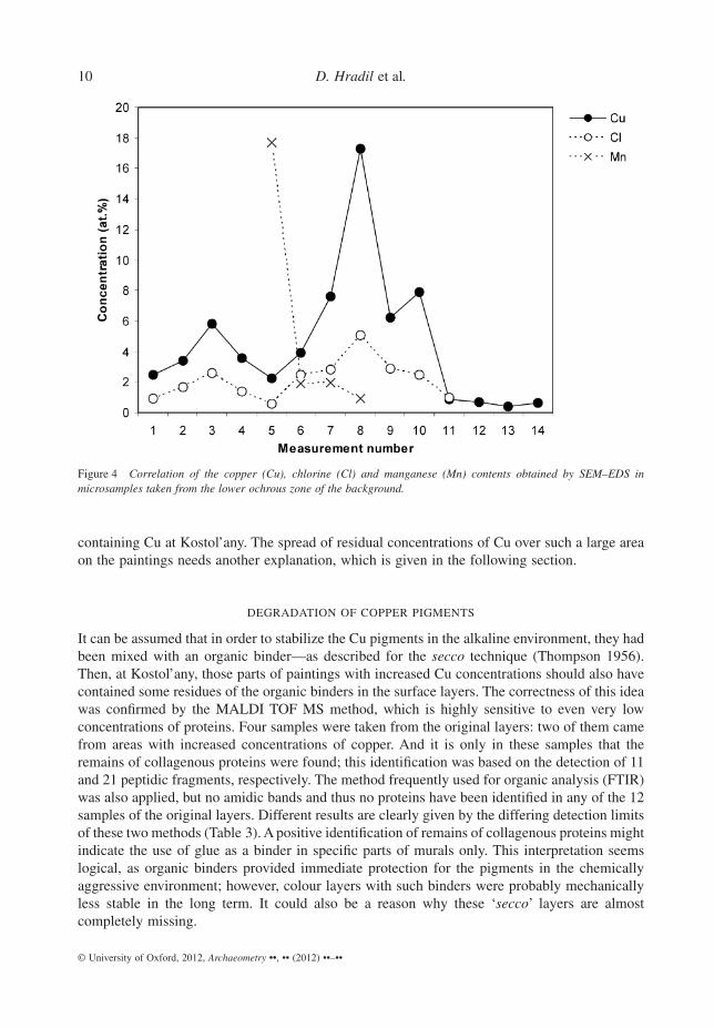
containing Cu at Kostol’any. The spread of residual concentrations of Cu over such a large areaon the paintings needs another explanation, which is given in the following section.
DEGRADATION OF COPPER PIGMENTS
It can be assumed that in order to stabilize the Cu pigments in the alkaline environment, they hadbeen mixed with an organic binder—as described for the secco technique (Thompson 1956).Then, at Kostol’any, those parts of paintings with increased Cu concentrations should also havecontained some residues of the organic binders in the surface layers. The correctness of this ideawas confirmed by the MALDI TOF MS method, which is highly sensitive to even very lowconcentrations of proteins. Four samples were taken from the original layers: two of them camefrom areas with increased concentrations of copper. And it is only in these samples that theremains of collagenous proteins were found; this identification was based on the detection of 11and 21 peptidic fragments, respectively. The method frequently used for organic analysis (FTIR)was also applied, but no amidic bands and thus no proteins have been identified in any of the 12samples of the original layers. Different results are clearly given by the differing detection limitsof these two methods (Table 3). A positive identification of remains of collagenous proteins mightindicate the use of glue as a binder in specific parts of murals only. This interpretation seemslogical, as organic binders provided immediate protection for the pigments in the chemicallyaggressive environment; however, colour layers with such binders were probably mechanicallyless stable in the long term. It could also be a reason why these ‘secco’ layers are almostcompletely missing.
Figure 4 Correlation of the copper (Cu), chlorine (Cl) and manganese (Mn) contents obtained by SEM–EDS inmicrosamples taken from the lower ochrous zone of the background.
10 D. Hradil et al.
© University of Oxford, 2012, Archaeometry ••, •• (2012) ••–••

Degradation of organic binders is accelerated particularly by the activities of micro-organisms. Their metabolic product is oxalic acid, which subsequently decomposes other sub-stances. The most stable products of such reactions are calcium oxalates (namely whewelliteCaC2O4(H2O) and/or weddellite CaC2O4 ·2H2O). As they tend to crystallize, they can be
Table 2 A list of the results of powder X-ray microdiffraction measurements on fragments and cross-sections (thenumber of samples containing the corresponding phase is expessed in parentheses, if different from 1)
Sample no. Description Major phases Minor phases
12, 25 Original plaster CaCO3 calcite (2) CaMg (CO3)2 dolomite (2)SiO2 quartzK-mica; for example, muscovite
8 Heterogeneity in theplaster (red colour)
Fe2O3 hematite CaCO3 calcite
2, 7, 11, 12, 14, 26 Original Pb pigments(blackened)
PbO2 plattnerite (6) PbCO3 cerussite (4)CaCO3 calcite (3) PbO2 scrutinyite (4)PbCO3 cerussite CaCO3 calcite (2)PbMg(CO3)2 PbMg (CO3)2 (2)
Pb3(CO3)2(OH)2 hydrocerussite (2)CaSO4·2H2O gypsum (2)PbO massicotPbSO4 anglesite
15, 16, 21, 34 Original yellows andreds
CaCO3 calcite (4) Al2Si2O5(OH)8 kaolinite (3)CaC2O4(H2O) whewellite (4) K-mica; for example, illite (3)CaSO4·2H2O gypsum (2) Expandable clay structure—
smectite group mineral (3)Al2Si2O5(OH)8 kaoliniteCaSO4·2H2O gypsumK-mica; for example, illiteSiO2 quartzSiO2 quartzNaAlSi3O8 Na-feldspar, albiteFeO(OH) goethite
18 Original Incarnate CaCO3 calcite CaC2O4(H2O) whewelliteCaSO4·2H2O gypsumPbCO3 cerussite
27 Original blue-greycolour
CaCO3 calcite CaC2O4(H2O) weddelliteAl2Si2O5(OH)8 kaoliniteSiO2 quartzCaMg(CO3)2 dolomite
30 13th-century repaints(red)
CaCO3 calcite Fe2O3 hematiteSiO2 quartz Al2Si2O5(OH)8 kaolinite
K-mica; for example, illitePb3(CO3)2(OH)2 hydrocerussite
31 13th-century repaints(yellow)
Ca5(PO4)3(OH) hydroxylapatite CaCO3 calciteCaC2O4(H2O) whewellite
10 Baroque repaint (red) CaCO3 calcite SiO2 quartzAl2Si2O5(OH)8 kaolinite K-mica; for example, illite
Pb3(CO3)2(OH)2 hydrocerussiteFeOOH lepidocrocite
20 20th-century peck ZnO zincite BaSO4 bariteCaCO3 calciteCaSO4·2H2O gypsumSiO2 cristoballiteAl2Si2O5(OH)8 kaolinite
Pre-Romanesque murals in Kostol’any pod Tríbecom, Slovakia 11
© University of Oxford, 2012, Archaeometry ••, •• (2012) ••–••

positively identified in microsamples as remnants of the above-mentioned processes, by meansof mXRD. At Kostol’any, we found them in all parts of the original paintings, and to a greaterextent in the areas with the increased Cu contents. They were absent only in the black colourlayers, since the Pb-containing phases might have been toxic for the micro-organisms (Table 3and Fig. 5). In this sense it is interesting that, apart from the decomposition of organic
Figure 5 X-ray microdiffraction patterns of the yellow background layer with increased copper (Cu) content (a) and theblackened layer with degraded lead (Pb) pigments (b), respectively.
Table 3 Organic binders and metabolic products of micro-organisms (oxalates) as identified by differentanalytical methods
Parts of the paintings Proteins Wax Calcium oxalates
Micro-FTIR MALDI-TOF Micro-FTIR mXRD
Pre-Romanesque (blackened minium) 0/2 n.a. 0/2 0/5Pre-Romanesque (containing Cu) 0/4 2/2 0/2 3/3Pre-Romanesque (other parts) 0/6 0/2 0/6 5/5Gothic 1/2 n.a. 0/2 0/1Modern-day interventions (20th century, pecks) 0/3 n.a. 3/3 0/1
Note: ratios indicate the number of positive identifications with respect to the total number of samples analysed; n.a., not analysed.
12 D. Hradil et al.
© University of Oxford, 2012, Archaeometry ••, •• (2012) ••–••

substances, micro-organisms may also accelerate decomposition of copper pigments as such, asdocumented by Švarcová et al. (2009). Copper oxalates, as unstable intermediates of the deg-radation processes, have been found in some wall paintings (Nevin et al. 2008), but unfortu-nately not at Kostol’any.
The high concentrations of chlorides in plasters, as found at Kostol’any, are no exception inhistoric buildings. These chlorides could cause the transformation of copper pigments intoatacamite Cu2Cl(OH)3; atacamite and other secondary phases have also been identified in anumber of other medieval wall paintings (e.g., Dei et al. 1998; Vandenbeele et al. 2005). AtKostol’any, however, the residual concentrations of Cu are very low, and even a detailed inves-tigation did not identify original or transformed pigment grains. The only indication of thetransformation process described above is the correlation of the residual Cu and Cl concentra-tions in colour layers at Kostol’any (Fig. 4). We can imagine that after corrosion caused by salts,probably accelerated by micro-organisms and oxalic acid produced by them, the secondaryphases (e.g., copper oxalates and atacamite) were almost completely washed out from the wall bythe physical action of water. Because we can exclude the possibility that this ‘washing out’ wasintentional—that is, resulting from any documented process of cleaning or repaints removal—itis probably connected with the truly alarming climatic conditions in the church. According tomeasurements carried out in the winter season from October 2008 to April 2009, the relative airhumidity in the interior varies in the range from 61.4 to 95.3%. The average monthly values varyfrom 75.7 to 88.1% near the entrance to the church, and from 74.6 to 81.1% in the presbytery. Thesame factor—in other words, not chemical degradation, but the physical effects of the condensedhumidity on the walls—probably resulted in the washing off and thinning of the other colourlayers and overall fading of the colouring.
DEGRADATION OF LEAD PIGMENTS
Another evident colour alteration at Kostol’any is the darkening of minium, as documented by thenon-invasive XRF measurements and also the mXRD measurements on microsamples. ThemXRD pattern in Figure 5 documents a typical mineralogical composition of a blackened colourlayer found in many places—apart from brown–black plattnerite and white cerussite (bothproducts of disproportionation of orange minium), there is also a high quantity of lead–magnesium carbonate [PbMg(CO3)2], which developed as a secondary product, and this againdocuments the use of dolomitic lime as a binder in the colour layer.
Mildewcides containing NaClO can also act as oxidizing agents to enhance the transforma-tion of lead white, but there is no existing evidence of their application at Kostol’any. Formermicrobiological investigations of the wall paintings at Kostol’any (Šujanová and Motaj 1966)have identified several types of Cladosporium mildews, particularly Cladosporium brevi-compactum. According to the authors, the mildew attacked practically the entire surface of thepaintings in spring 1966, as a result of deteriorating climatic conditions. The most affectedparts of the church were allegedly the northern wall of the nave and the presbytery. Themildew was only finally removed by ventilation, drying and heating of the walls with infraredlamps. Moreover, as documented by the restorer’s documentation from 1960s and by samplescollected at that time, the blackening of lead pigments must have occurred earlier. On the otherhand, a visual comparison of the paintings after their initial exposure and their current appear-ance clearly indicates a significant diminishing of the colour, particularly on the northern wall.This means that the degradation of colours of this unique set of paintings continues toprogress.
Pre-Romanesque murals in Kostol’any pod Tríbecom, Slovakia 13
© University of Oxford, 2012, Archaeometry ••, •• (2012) ••–••

LATER PAINTINGS
Later (13th-century) wall paintings have been found in the eastern part of the presbytery aroundthe pastoforium, made over the original paintings on the new plaster. The analysed samples weretaken from the figure’s Incarnate and from the red robe. It is very interesting that the plaster witha higher sand content, and even the paintings themselves, also contain dolomitic lime. TheIncarnate and the red drapery are pigmented with iron ochres and reds, and the colour layerscontain increased levels of phosphorus, related to the use of bone white (hydroxylapatite) andwhitewash, which were used here. In one out of every two samples, protein binder has beenidentified by FTIR (Tables 2 and 3), but a definite interpretation of the painting technique was notcarried out.
The ornamental decorations on the western wall of the chancel are also later. Dolomitic limewas used here in the lime plaster and in the lime coating, as well as in the paintings. In both ofthe above-mentioned cases, the later wall paintings have monotonous colouring and they fail tocontain any specific pigments that would allow their closer historical dating. All of the laterpaintings were left for further research.
CONCLUSION
The cycle of the oldest wall paintings in the church of St George in Kostol’any pod Tríbecom,Slovakia, was surely created for the original church and was painted shortly after its constructionin around ad 1000. According to the iconography, style and painting technique, these paintingsmost probably belong to the pre-Romanesque art of the 10th century.
Today, the colouring of these wall paintings is faded. The blue and/or green copper pigmentswere probably first altered by salt (chloride) corrosion, which was accelerated by the activities ofmicro-organisms that produce an oxalic acid as their metabolic product. Subsequently, they werewashed out by humidity condensing on the walls. In this respect, the climatic conditions in thechurch deteriorated significantly in 1960s after the floor and ceiling had been covered withconcrete. On the basis of the residual concentrations of copper in the colour layers, it is now atleast partly possible to recall the original colouring; for example, a blue zone of the sky in thebackground, the colourful clothes of the three Magi and so on. Unlike the washing out of copperpigments and the related overall thinning and fading of the colour layers, the blackening of theoriginally orange minium is a natural process. This degradation occurred much earlier in thehumid environment, where minium in the colour layer was not protected by any organic binder.The binder used in the paintings was dolomitic (magnesium-rich) lime and the paints wereapplied into the wet plaster (a fresco technique). The remains of collagenous proteins found intwo samples indicate a potential use of glue exclusively in parts with copper pigments (a seccotechnique). The present colouring of the original paintings is given by the presence of yellowochres, iron red, carbon black and blackened minium; the white is exclusively whitewash, and theuse of lead white in admixture is uncertain. A special feature is the grey–blue colour shade—forexample, on the coat of the Virgin Mary—which was not achieved by means of a blue pigment,but by an admixture of iron black (magnetite) with a metallic grey–blue lustre. The condition ofthe paintings continues to deteriorate as a result of the continuing degradation due to high andvarying humidity. This degradation has become a physical process in which probably eveninsoluble pigment particles are washed down from the walls, and the intensity of coloursdiminishes very quickly.
14 D. Hradil et al.
© University of Oxford, 2012, Archaeometry ••, •• (2012) ••–••

ACKNOWLEDGEMENTS
This research was funded by the Academy of Sciences of the Czech Republic (AV0Z40320502)and partly by the Czech Science Foundation (P103/12/2211). We hereby offer thanks, for theirprofessional co-operation, to all our colleagues from the ALMA laboratory, and to Peter Baxa(Heritage Authority of the Slovak Republic, Bratislava), Štepánka Kucková (Institute of Chemi-cal Technology—VŠCHT Prague), Zlata Vrátnícková (Polymer Institute in Brno) and graphicdesigner Katerina Vytejcková. We also thank Jan Cervenák (STP Prague) for the atmospherichumidity measurements and Daniela Cebecauerová (Heritage Authority of the Slovak Republic,Bratislava) for lending us selected archived samples for comparison purposes.
REFERENCES
Aze, S., Vallet, J., M., Detalle, V., Grauby, O., and Baronnet, A., 2008, Chromatic alterations of red lead pigments inartwork: a review, Phase Transitions, 81, 145–54.
Bakoš, J., 1968, The origin of the wall paintings at Kostol’any pod Tríbecom, Vlastivedný Casopis, 17, 178–81 (inSlovak).
Brogiolio, G. P., 2002, Oratori funerary tra VII e VIII sedilo nelle campagne transpadane, Hortus Artium Medievalium,8, 9–31.
Cennini, C., 1978 [1437], Il libro dell’ arte, the craftsman’s handbook, trans. D. V. Thompson Jr, Dover Publications,New York.
Dehn, E., 2005, Über Armenischen Bolus, Diploma work, Technical University Munich, Germany.Dei, L., Ahle, A., Baglioni, P., Dini, D., and Ferroni, E., 1998, Green degradation products of azurite in wall paintings:
identification and conservation treatment, Studies in Conservation, 43, 80–8.Demus, O., 1992, Romanische Wandmalerei, Hirmer Verlag, Munich.Dill, H. G., Fussl, M., and Botz, R., 2007, Mineralogy and (economic) geology of zeolite–carbonate mineralization in
basic igneous rocks of the Troodos Complex, Cyprus, Neues Jahrbuch fur Mineralogie–Abhandlungen, 183(3),251–68.
Eastaugh, N., Walsh, V., Chaplin, T., and Siddall, R., 2004, Pigment compendium: a dictionary of historical pigments,Elsevier Butterworth-Heinemann, Oxford.
Ewald, J., 1991, Kirchen und Kirchengrabungen im Baselbiet. Ein Beitrag zur Geschichte der Kirchen-Landschaft derNordschweiz, in Methoden und Perspektiven der Archeologie des Mittelalters (ed. J. Tauber), 57–84, Archäologieund Museum 20.
Fodor, P., 1967, Wall paintings at Kostol’any pod Tríbecom—the painting technique. Monumentorum Tutela, 2, 97–114(in Slovak).
Howard, H., and Gasol, R., 1996, The investigation of the Romanesque paintings in the Norwich cathedral, TechnologiaArtis, 17–24.
Hradil, D., Grygar, T., Hradilová, J., and Bezdicka, P., 2003, Clay and iron oxide pigments in the history of painting,Applied Clay Science, 22(5), 223–36.
Knoepfli, A., and Emmenegger, O., 1990, Wandmalerei bis zum Ende des Mittelalters, in Reclams Handbuch derkunstlerischen Technikem. Band 2—Wandmalerei Mosaik (eds. A. Knoepfli, O. Emmenegger, M. Koller and A.Meyer), 7–212, Reclam, Philipp Reklam jun. Verlag, Stuttgart.
Kotulanová, E., Bezdicka, P., Hradil, D., Hradilová, J., Švarcová, S., and Grygar, T., 2009, Degradation of lead-basedpigments by salt solutions, Journal of Cultural Heritage, 10, 367–78.
Krása, J., 1967, Wall paintings at Kostol’any pod Tríbecom, Monumentorum Tutela, 2, 115–27 (in Czech).Kucková, Š., Hynek, R., and Kodícek, M., 2007, Identification of proteinaceous binders used in artworks by MALDI-
TOF mass spectrometry, Analytical and Bioanalytical Chemistry, 388, 201–6.López-Acevedo, V., Viedma, C., González, V., and La Iglesia, A., 1997, Salt crystallization in porous construction
materials II. Mass transport and crystallization processes, Journal of Crystal Growth, 182, 103–10.Maríková-Kubková, J., 2006, Wall paintings in the church of St. George at Kostol’any pod Tríbecom, Technologia Artis,
5, 85–92 (in Czech).Mattei, E., de Vivo, G., De Santis, A., Gaetani, C., Pejosi, C., and Santamaria, U., 2008, Raman spectroscopic analysis
of azurite blackening, Journal of Raman Spectroscopy, 39, 302–6.
Pre-Romanesque murals in Kostol’any pod Tríbecom, Slovakia 15
© University of Oxford, 2012, Archaeometry ••, •• (2012) ••–••

Nevin, A., Melia, R. L., Osticioli, I., Gautier, G., and Colombini, M. P., 2008, The identification of copper oxalates in a16th century Cypriot exterior wall painting using micro FTIR, micro Raman spectroscopy and gas chromatography– mass spectrometry, Journal of Cultural Heritage, 9(2), 54–161.
Terrier, J., 2003, Approche archéologique des églises rurales édifiées au voisinage de la ville de Genève, Hortus ArtiumMedievalium, 9, 213–32.
Thompson, D. V., 1956, The materials of medieval painting, Dover Publications, New York.Schweigstillová, J., and Hradil, D., 2007, Salt formation on the Cretaceous sandstones in north and northwest Bohemia,
in Sandstone landscapes (eds. H. Hartel, V. Cílek, T. Herben, A. Jackson and R. Williams), 76–9, Academia incollaboration with Bohemian Switzerland National Park, Administration and Royal Botanic Gardens Kew, Prague.
Šujanová, O., and Motaj, S., 1966, Kostolany pod Tribecom. Stopping-down of the fungi growth, Unpublished report No.V-43/1, SÚPSOP Bratislava (in Slovak).
Švarcová, S., Hradil, D., Hradilová, J., Kocí, E., and Bezdicka, P., 2009, Microanalytical evidence of origin anddegradation of copper pigments found in Bohemian Gothic murals, Analytical and Bioanalytical Chemistry, 395,2037–50.
Švarcová, S., Kocí, E., Bezdicka, P., Hradil, D., and Hradilová, J., 2010, Evaluation of laboratory powder X-raymicro-diffraction for applications in the field of cultural heritage and forensic science, Analytical and BioanalyticalChemistry, 398, 1061–76.
Vandenbeele, P., Lambert, K., Matthys, S., Schudel, W., Bergmans, A., and Moens, L., 2005, In situ analysis of mediaevalwall paintings: a challenge for mobile Raman spectroscopy, Analytical and Bioanalytical Chemistry, 383, 707–12.
Vitruvius, 1812 [27 bc], The civil architecture of Vitruvius, trans. W. Wilkins, Thomson Davison, London.Zeman, A., and Ružicková, E., 1996, Research on the materials composition of plasters, Technologia Artis, 4, 93–9 (in
Czech).
16 D. Hradil et al.
© University of Oxford, 2012, Archaeometry ••, •• (2012) ••–••