Embed Size (px)
Citation preview
Current Concept Review
Copyright @ 2021 JPOSNA www.jposna.org
Unicameral Bone Cysts: Treatment Rationale and Approach
Soroush Baghdadi, MD1 and Alexandre Arkader, MD1,2
1Division of Orthopaedics, The Children’s Hospital of Philadelphia, Philadelphia, PA; 2Perelman School of Medicine at the University of Pennsylvania, Philadelphia, PA
Introduction Unicameral bone cyst (UBC) is a common benign, cystic lesion of the skeleton, also known as solitary or simple bone cyst. While it does not represent a true neoplasm, expansion and weakening of the bone might lead to symptoms and/or pathologic fractures. Virchow is often credited with the first report of UBC, although his
description was published in the pre-Roentgen era. Several authors have expanded our knowledge in the twentieth century, including Jaffe and Lichtenstein, who defined the pathological characteristics of the lesion, as well as the cyst lining and fluid features.
Abstract: Unicameral bone cyst (UBC) is a benign cystic lesion most commonly diagnosed in the proximal aspects of the hu-merus and femur of growing children. Medullary venous obstruction is the leading pathogenesis theory, resulting in fluid accumulation, bone resorption, and cortical thinning. Most UBCs are asymptomatic and likely go undiagnosed while the most common presentation is a pathologic fracture. Younger children tend to present with active lesions, which are uniloculated, abut the physis, and have a higher recurrence or persistence rate after treatment. Latent lesions in older children have migrated away from the growth plate and might become multiloculated. Radiographs are often diagnostic although advanced imaging is also helpful in some cases. Most UBCs do not need treatment; when indi-cated, management ranges from aspiration and steroid injection to decompression, curettage and grafting, and internal fixation. Percutaneous or open surgical approaches are acceptable and often yield good results although high recur-rence rates should be expected, especially in younger children with active lesions.
Key Concepts: • UBCs are not a true neoplasm and do not have malignant potential. However, they may cause complications such
as pathologic fractures and growth disturbance.
• Treatment should be personalized to each patient’s demographics, functional needs, desires of the family, andlesion characteristics.
• Patients/families should be aware of the high recurrence rate and the possible need for repeat intervention.
• A percutaneous or open surgical approach, with decompression, curettage of the cyst, grafting, and internal fixa-tion if necessary, is the mainstay of management.
1

JPOSNA Volume 3, Number 2, May 2021
Copyright @ 2021 JPOSNA www.jposna.org
Although UBC is currently favored in the literature over solitary bone cyst, the term “unicameral” is not always accurate. Some UBC have one cavity in the initial stages, yet many develop a multiloculated structure, especially after fractures or treatment. UBC typically arises in the metaphysis of tubular bones adjacent to the growth plate and is almost exclusively diagnosed in the immature skeleton (although it can persist into adulthood), with reported mean age of 11 years at diagnosis. UBC represents up to 3% of all reported bone lesions, but similar to other benign pathologies, most cases may go undiagnosed; hence its true incidence is likely higher.
While UBCs have no malignant potential and have a relatively favorable long-term prognosis regardless of the treatment, controversy still exists regarding the natural history, indications for treatment, and preferred technique. In this review, we focus on the different therapeutic approaches based on the available literature and institutional experience.
Etiology and Pathogenesis The exact etiology and pathogenesis of UBC remain unclear. The most favored pathogenesis theory is a blockage in the intramedullary venous drainage postulated by Cohen.1,2 In brief, an insult to the bone causes venous obstruction, which in turn leads to fluid accumulation, increased pressure, and bone resorption.1-3 Several therapeutic approaches have been established based on this mechanism, including decompression or fenestration of the cyst.
On gross pathologic examination, UBC is usually a single fluid-filled cavity. The pathognomonic feature is clear, straw-colored fluid on aspiration. However, the fluid might have a bloody tinge following a pathologic fracture. Histologic analysis of the cyst lining reveals a thin fibrous membrane with occasional multinucleated giant cells. Secondary changes, including the presence of hemosiderin, macrophages, and reactive woven bone, are also common in the presence of a fracture.
At the cellular level, osteoclasts are believed to play a role in the pathogenesis of UBCs. Osteoclasts are present in the cyst lining and fluid, and the messengers and products of the continued osteoclast-mediated bone destruction have been confirmed in the cyst,3 including prostaglandin-E2 (PGE2),4 lysosomal enzymes,5 and acid phosphatase levels.6 However, a complex signaling pathway exists between cells of different lineages, and osteoblasts are required for activation of osteoclastic bone degradation. Additionally, increased osteoblastic activity and concentration of RANKL (receptor activator of nuclear factor kappa-Β ligand) which activates osteoclasts, has been found in UBC lining and fluid.3
Clinical Presentation Similar to other benign bone lesions of childhood, most UBCs are asymptomatic and likely go undiagnosed unless there is an incidental finding or pathologic fracture. Symptomatic patients typically present with either an acute pathologic fracture or an insufficiency/stress fracture. In the latter group, the patient might present with mechanical symptoms, painful range of motion, or limping. In a systematic review, more than three-quarters of the published UBC cases were diagnosed after an acute fracture, 15% with an insufficiency fracture, and the rest as an incidental finding.7
Although UBCs may occur in any site, the metaphysis and diaphysis of long bones are the most common location. More than half of the lesions are diagnosed in the proximal humerus, followed by the proximal femur and proximal tibia.7 The calcaneus is also a relatively common site for UBC which tends to occur in the slightly older population. Boys are twice as likely to be diagnosed with a UBC, although it is unclear whether this represents a higher incidence or is the result of higher fracture risk in boys.
Imaging UBCs appear as centrally located, well-defined, lucent metaphyseal lesions on plain radiographs, with a narrow zone of transition, and often abutting the physis (active
2

JPOSNA Volume 3, Number 2, May 2021
Copyright @ 2021 JPOSNA www.jposna.org
phase). With longitudinal growth, the lesion “migrates” away from the physis and becomes diaphyseal (latent phase). Larger lesions may lead to bone expansion and result in cortical thinning. Periosteal reaction or cortical destruction are absent unless a fracture occurs. Soft tissue extension is not a feature. The “fallen fragment sign” was first described by Reynolds to help distinguish UBC from other radiolucent but solid intramedullary lesions.8,9 Following a fracture, the fluid within the UBC has no resistance to the bone fragments falling into the cavity, thus creating the distinctive intracavitary displaced bone fragment. Although the fallen fragment sign is helpful in some cases, it has a low sensitivity.10
Advanced imaging may be valuable in differentiating a UBC from similar lesions in equivocal cases. Computed tomography (CT) scan shows cortical thinning, fluid density inside the lesion, and occasionally the fallen fragment sign.11 On magnetic resonance imaging (MRI), UBC is characterized by a hypointense signal on T1-weighted images and hyperintense on T2-weighted and fluid-sensitive sequences. Septations, if present, are also visible on MRI. On post-contrast images, peripheral enhancement is appreciated. When a fracture is present, T1-weighted images will be hypersignal due to blood, while marrow edema, periosteal reaction, and surrounding edema are hyperintense on T2-weighted and fluid-sensitive sequences. Fluid-fluid levels may be present, especially in the presence of a pathologic fracture. MRI is particularly useful for the evaluation of a suspected insufficiency fracture and physeal involvement/ disruption by the cyst.
Differential Diagnosis All primarily cystic bone lesions are included in the differential diagnosis list, including aneurysmal bone cyst (ABC), non-ossifying fibroma (NOF), and fibrous dysplasia. In addition to the imaging findings, the specific demographics and pertinent clinical symptoms of each lesion are helpful in distinguishing the diagnosis.
ABC is the primary differential diagnosis of UBC due to similarities in demographics and imaging characteristics.
ABC is an expansile, lytic lesion that affects the metaphysis of long bones, similar to UBC, but in an eccentric location. ABC can be locally aggressive with cortical expansion and disruption. On MRI, multiple septations and fluid-fluid levels are present due to the bloody contents of the cavities. The signal intensity is heterogeneous, with individual lobules having different signal characteristics.12 An intact UBC is easier to distinguish from ABC while a fractured or partially healed UBC can be challenging to differentiate. During surgery, aspiration of clear straw-colored fluid is diagnostic for UBC. A bloody fluid, although not diagnostic, is more suggestive of ABC.
NOFs have a lytic, lobular appearance but are cortical based, rather than central, and are classically surrounded by a sharp sclerotic border. Mild cortical expansion and pathologic fracture can be present. MRI demonstrates the fibrous content of the lesion.
Monostotic fibrous dysplasia is in the differential diagnosis of latent, diaphyseal UBCs. The presence of a ground-glass appearance instead of a fluid-filled cavity helps differentiate the two lesions on advanced imaging. Furthermore, fibrous dysplasia lesions may undergo cystic degeneration which increases the similarities with UBC and ABC.13
Natural History The literature is inconclusive regarding the true natural history of UBCs. Most lesions are believed to heal by the time the patient reaches skeletal maturity although some studies have shown that the cyst persists, but the risk of pathologic fracture decreases. Several risk factors for complications (i.e., pathologic fracture, recurrence/persistence after treatment) have been identified. One of the earliest factors recognized is the distance of the lesion from the physis.14 Active cysts abutting the growth plate have a higher risk of progression, growth, fracture, and recurrence after treatment, while latent cysts distant from the growth plate are more likely to resolve with or without treatment. In one study, healing was achieved in cysts
3

JPOSNA Volume 3, Number 2, May 2021
Copyright @ 2021 JPOSNA www.jposna.org
that were > 2 cm away from the growth plate, but not in those closer to the physis.15 Age is also an important predictor for healing and recurrence after surgery. Patients older than 10 years have a lower rate of persistence or recurrence.16,17 The risk of pathologic fracture is related to the lesion size and location. UBCs of the lower limb (weight bearing bones) have a higher risk of fracture as do larger lesions. The exact threshold that will increase fracture risk is uncertain, although evidence suggests that lesions wider than 50-80% of the bone width are at a high risk for fracture.18,19 Overall, around 10-15% of cysts are expected to heal after a fracture,16,20-22 especially for complete or displaced fractures.
Healing of the UBCs can be assessed with the classification system developed by Neer,23,24 who defined a healed cyst as one filled by new bone formation with radiolucent areas < 1 cm in size. “Healing with defect” is a lesion with radiolucent areas < 50% of the diameter of the bone with enough cortical thickness to prevent fracture. A persistent cyst has radiolucent areas > 50% of the bone diameter with a thin cortical rim. Finally, a recurrent cyst is one that has reappeared in a previously healed area, or more commonly, a radiolucent area that has increased in size.
Management Over the years, several management strategies and surgical techniques have been introduced for UBC. Nevertheless, there is significant controversy on the optimal management strategy, indications for treatment, and the surgical procedure of choice. This controversy is, in part, rooted in the benign, nonaggressive nature of UBCs. The risk of complications, including pathologic fracture and growth arrest, should be weighed against the risk of surgical treatment and recurrence, as well as the potential for spontaneous healing over time. Treatment should be personalized based on each patient’s
demographics and unique lesion characteristics. Age, patient’s activity level, location, size of the lesion, and family preferences are important factors in tailoring a treatment plan for UBC (Figure 1).
Asymptomatic patients Most asymptomatic, incidentally found UBCs are amenable to observation alone, with the notable exception of lesions around the hip and those with very high risk of pathologic fracture, and juxta physeal lesions with a risk of potential growth disturbance. Asymptomatic upper extremity lesions can almost universally be treated conservatively. It is important that the patient and family are reassured about the benign nature of the cyst and educated about the risk of pathologic fracture as well as surgical risks and the
Figure 1. A sample algorithm for management of unicameral bone cysts. Refer to text for details.
4

JPOSNA Volume 3, Number 2, May 2021
Copyright @ 2021 JPOSNA www.jposna.org
potential for recurrence. Active patients with a high risk for fracture or symptoms may elect to undergo prophylactic treatment for large, active lesions. Large (> 50% bone width) femoral lesions, especially around the hip, have a high risk of fracture, and prophylactic treatment is most often recommended.
Symptomatic patients A thorough clinical and imaging workup is warranted to evaluate the cause of the patient’s symptoms. In general, the culprit is an impending or insufficiency/stress fracture evident by surrounding marrow edema (MRI) and cortical disruption (CT or MRI). Although a trial of conservative treatment can initially be pursued, these patients often require surgery, especially for lower extremity lesions.
Pathologic fractures Pathologic fractures may lead to decompression of the cyst and stimulate partial or complete healing in 10-15% of the cases. Therefore, conservative treatment is usually the initial preferred method, particularly for nondisplaced pathologic fractures and fractures of the upper extremities. Recurrent fractures may be an indication for surgical treatment. Proximal femoral UBC is a special case in which surgical treatment of an impending or overt fracture is indicated at presentation due to the morbidity associated with a displaced fracture around the hip.
Treatment Options
Steroid Injection The use of intralesional steroid injection, most often methylprednisolone acetate (MPA), was first attempted by Scaglietti in the 70s,25 and later adapted by several other authors.26-32 The increased concentrations of prostaglandins in the cyst fluid motivated its use. The mechanism of action of steroids is still unknown, and some believe that the cyst decompression, rather than the steroids itself, is responsible for healing. The results vary widely and the recurrence or persistence rate ranges from 15–80% after one injection. For the most part,
MPA injection alone yields unpredictable results and complete healing after the first injection is uncommon.33 In an attempt to increase healing rates, autologous bone marrow (ABM) and/or demineralized bone matrix (DBM) have been used in combination with MPA, betting on their osteogenic potential to increase the chances of healing.
The literature is inconclusive on which injection material (or combination of) leads to a higher healing rate26-
28,30,32,34 but the addition of bone marrow derivatives does not seem to add a significant benefit and may add donor-site morbidity in the case of ABM.26 Overall, one injection as a standalone procedure tends to be un-successful and most patients eventually need multiple injections. In a systematic review, MPA injection had a 71.4% healing rate, while ABM and DBM injection had a 77.9-98% healing rate,7 although many patients required multiple injections.
Surgical Treatment While indications for surgical treatment are not standardized, surgical intervention can be considered for symptomatic lesions, stress or recurrent fractures, displaced pathologic fractures of lower extremities, lesions posing risk to the physis, or lesions with a high risk for complications such as those around the hip. There is a wide array of techniques available, and the treating surgeon can personalize the treatment plan based on the several available techniques, patient’s characteristics, availability, and personal preference. The range of all the treatment modalities are described below and include aspiration and decompression, curettage, grafting, and internal fixation (Table 1).
Aspiration and Decompression UBC is a closed cavity with fluid under pressure. Therefore, drainage and decompression of the lesion may promote healing. Cyst decompression has historically been performed from an outside-in (extramedullary decompression) technique utilizing large-bore needles (e.g., Jamshidi or similar needle), curettes, or Kirshner wires. Cannulated screws and
5
JPOSNA Volume 3, Number 2, May 2021
Copyright @ 2021 JPOSNA www.jposna.org
absorbable pins have also been utilized to provide “continuous decompression” of the cyst35 although there is no evidence that the decompression is actually continuous or that this method leads to a higher healing rate. Intra-medullary decompression with flexible intramedullary nails is an alternative technique.36-42 The intramedullary hardware violates the cyst membrane and connects the UBC to the normal medullary cavity, which may provide bone marrow as an autologous bone graft. Intramedullary nailing with or without concomitant curettage and bone grafting has been reported to have a high healing rate.7,36-42 Additionally, nails may be retained to provide structural support.
Curettage Curettage of the cyst can be done percutaneously, endoscopic, or open. Curettage not only decompresses the cyst, but also connects the cyst with the adjacent normal medullary canal and removes the cyst lining which may decrease the risk of recurrence and provides pathologic confirmation of the diagnosis. Fluoroscopic guidance, especially when combined with intralesional injection of contrast material for a “cystography,”
provides visual confirmation of the nature and extent of the cyst. It also helps to determine if septations are present in the cyst. The minimally invasive technique of percutaneous curettage with the aid of fluoroscopy and bone grafting was introduced by Dormans et al. and is one of the most successful treatment strategies in UBC with excellent short-term results.22 However, a follow-up study of the same patient cohort showed a persistence/recurrence rate of 20%,18 which is similar to other techniques.
Grafting The use of bone grafting following curettage and decompression provides structural support and may also help to promote a healing response through osteoconductive/osteoinductive properties. While the bone graft and/or bone substitute easily incorporates in the UBC, recurrence or persistence may still occur. The use of autograft, allograft (cancellous and cortical strut), and synthetic bone substitutes have all been reported in the literature with similar results. There is no clear advantage to a particular graft type although bone substitute is more attractive due to their unlimited
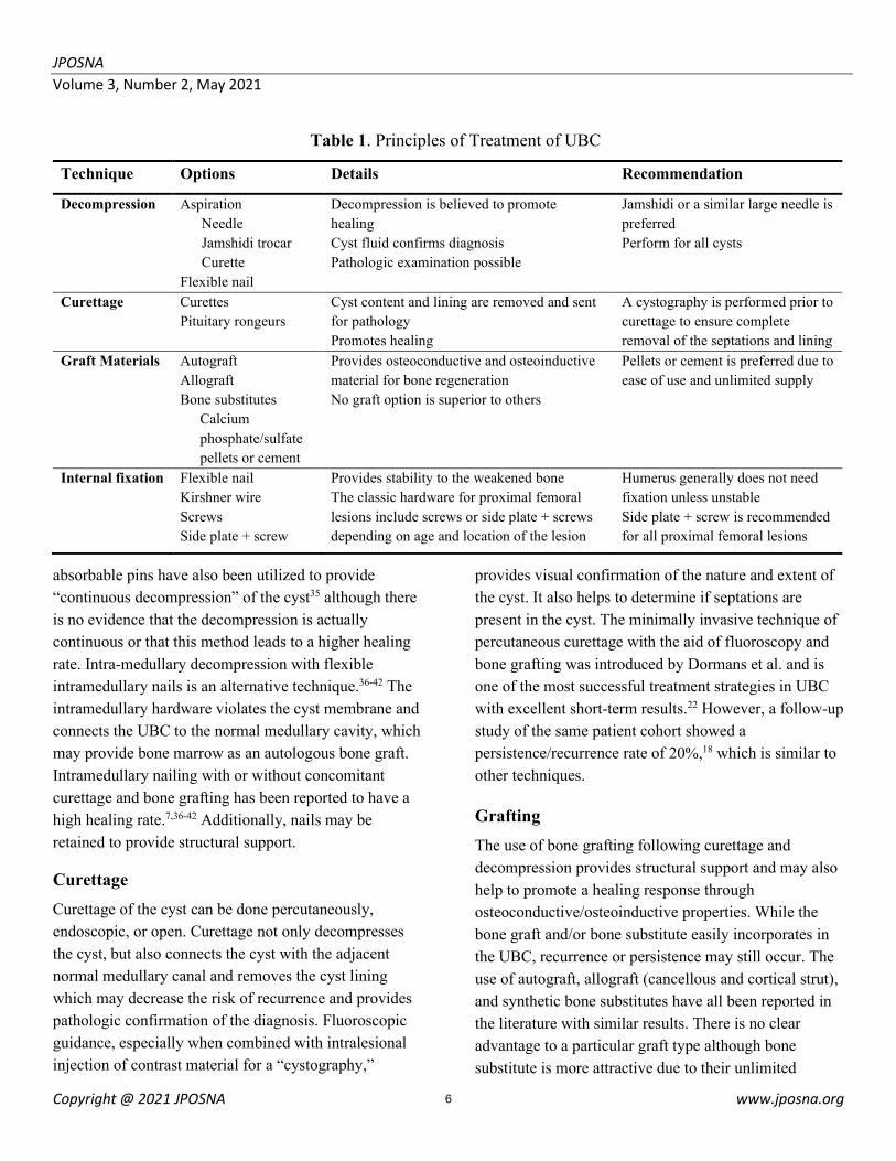
Technique Options Details Recommendation
Decompression Aspiration Needle Jamshidi trocar Curette
Flexible nail
Decompression is believed to promote healing Cyst fluid confirms diagnosis Pathologic examination possible
Jamshidi or a similar large needle is preferred Perform for all cysts
Curettage Curettes Pituitary rongeurs
Cyst content and lining are removed and sent for pathology Promotes healing
A cystography is performed prior to curettage to ensure complete removal of the septations and lining
Graft Materials Autograft Allograft Bone substitutes
Calcium phosphate/sulfate pellets or cement
Provides osteoconductive and osteoinductive material for bone regeneration No graft option is superior to others
Pellets or cement is preferred due to ease of use and unlimited supply
Internal fixation Flexible nail Kirshner wire Screws Side plate + screw
Provides stability to the weakened bone The classic hardware for proximal femoral lesions include screws or side plate + screws depending on age and location of the lesion
Humerus generally does not need fixation unless unstable Side plate + screw is recommended for all proximal femoral lesions
Table 1. Principles of Treatment of UBC
6

JPOSNA Volume 3, Number 2, May 2021
Copyright @ 2021 JPOSNA www.jposna.org
supply, absence of donor site morbidity, and no risk of disease transmission. Furthermore, several bone substitute formulations can provide immediate structural support to the lesion. The literature abounds with studies on curettage and grafting of UBCs, and although most studies are case series or otherwise low-level evidence, virtually all graft types have been successful (to a certain degree) in promoting healing of the lesion with a lower recurrence rate than decompression and curettage alone.7,18,22,28,29,36,43-49
Internal Fixation Internal fixation is not routinely required as part of the surgical management of UBCs, especially for upper extremity lesions where functional bracing is usually preferred. Intramedullary flexible nails provide sufficient
stability for most upper extremity lesions and some lower extremity cysts, but lesions around the hip often require a more stable, rigid fixation for adequate healing and early mobilization. Surgical management of UBCs does not generally require internal fixation.
Surgical Technique Lesion and patient characteristics dictate the preferred surgical approach. Upper extremity lesions, those in younger children, diaphyseal lesions, and UBCs of the tibia are generally amenable to a percutaneous approach. Most proximal femoral lesions will require a formal open approach, as well as other locations, if surgical exposure is to be performed for internal fixation.
Figure 2. Percutaneous treatment of UBC: Lesion is located and perforated (A-B), cystogram and a thorough curettage is performed (C-D), and a flexible nail is used to decompress the lesion (E-F). Bone graft (in this case calcium sulfate pellets) is packed in the cavity (G), and a functional brace is fitted (H). Figure reproduced with permission from CHOP Orthopaedics, Children’s Hospital of Philadelphia, Philadelphia, PA
7

JPOSNA Volume 3, Number 2, May 2021
Copyright @ 2021 JPOSNA www.jposna.org
Percutaneous Approach As previously described,18,22 the cyst is located with fluoroscopy, a 1 cm incision is made, and a Jamshidi or similar large-bore needle is used to perforate the cyst. The intra-lesional fluid is aspirated to confirm the clear serous or serous-sanguineous appearance. Radiopaque contrast is injected into the cyst to delineate the extent of the cyst. The cystogram also provides visual confirmation of the nature of the cyst, as UBCs lack or have minimal septations. Under fluoroscopic guidance, curettes and pituitary rongeurs can be used to remove the cyst lining and contents, which are sent to pathology. Care is taken not to miss the proximal and distal extents of the cyst. Next, a flexible nail is entered through the same incision and advanced distally (and proximally if possible) to decompress the cyst into the normal medullary canal. The nail is then removed, and a bone graft of choice is used to fill the cyst cavity. After fluoroscopic confirmation, the incision is sutured and temporary immobilization, in the form of a functional brace (e.g., Sarmiento), is recommended (Figure 2).
Open Approach Similar to the percutaneous approach, fluoroscopy is used to locate the lesion. A cortical window is made over the cyst, and the cavity is curetted thoroughly. While some authors recommend screw or wire fixation for femoral neck lesions, we recommend utilizing fixation with a lateral buttress (plates) for all cysts around the hip. Lesions that extend to the subtrochanteric region may be stabilized with load-sharing devices (intramedullary nails). If the lesion extends to the proximal femoral physis, epiphyseal fixation is recommended, especially in skeletally mature patients. In skeletally immature patients, fixation is preferably stopped short of the physis, or if possible, the physis is crossed with a nonthreaded fixation that would allow continued growth (Figure 3).
While surgical management of UBCs may have a higher success rate than observation (natural history) or injection-only, both percutaneous and open approaches
Figure 3. Open approach to UBC: Lesion is located, an oval window is created, and a thorough curettage is performed (A-B). Adjuvant cauterization is optional to achieve extended curettage (C-D). The cavity is filled with bone graft (in this case, calcium cement), and internal fixation is performed. Growth-friendly options with partially threaded screws are also available to prevent growth arrest (F-G). Figure reproduced with permission from CHOP Orthopaedics, Children’s Hospital of Philadelphia, Philadelphia, PA
8

JPOSNA Volume 3, Number 2, May 2021
Copyright @ 2021 JPOSNA www.jposna.org
still have a recurrence rate of up to 25%. The patient/family should be aware of the chance of persistence or recurrence even with the most comprehensive treatment strategies and that repeated treatment might be required.7 Younger children with active lesions (abutting the physis) have a substantially higher rate of recurrence compared to older patients with latent diaphyseal cysts. The approach to a recurrent lesion is generally similar to an untreated lesion and should be personalized to each patient, considering their function and expectations as well as lesion characteristics. However, the use of internal fixation at the initial surgery, especially in the form of screw/side plate constructs in the proximal femur, may allow the surgeon to be more conservative if a recurrence occurs.
Other Complications and Pitfalls While the most common, a fracture is not the only complication of UBCs. Recurrence is a major concern that occurs in up to 25% of cases7,21,31,50 and is generally treated similarly to the primary lesion. Active lesions abutting the physis have the potential to grow into the epiphysis through the growth plate. With the “physis at risk,” there is the potential for physeal injury and growth arrest: therefore, surgical treatment may be indicated for such lesions. Depending on the location and extent of physeal injury, angular deformity and/or longitudinal growth disturbance may occur (Figure 4).
Summary In conclusion, UBCs have a benign, likely improving or resolving natural history. Most patients will fare well with observation alone. The decision to surgically treat lesions should be made after an in-depth discussion with the family/patient. The surgical approach should sim-ilarly be individualized to the patient’s demographics, functional needs, and lesion characteristics.
A comprehensive percutaneous or open surgical treatment including decompression, curettage, and grafting, plus internal fixation in select cases, has the highest success rate, although a 20-25% recurrence rate should be expected. The current literature on the topic is generally low-level evidence, which future large, multicenter studies might be able to tackle.
Additional Links • How I do it and why? Unicameral Bone Cysts
Alexandre Arkader, MD; IPOS. http://www.posnacademy.org/media/Unicameral+Bone+Cyst/0_0dyb83i0/19139952
• Pathologic fractures of the Proximal Femur Alexandre Arkader, MD; IPOS http://www.posnacademy.org/media/Pathologic+Fractures+of+the+Proximal+Femur/1_uliu7lkk/19139952
Figure 4. Complications of UBC include pathologic fractures (A), recurrence (B), physeal injury (C), as seen on coronal fat-saturated MRI), and growth disturbance (D). Figure reproduced with permission from CHOP Orthopaedics, Children’s Hospital of Philadelphia, Philadelphia, PA
9

JPOSNA Volume 3, Number 2, May 2021
Copyright @ 2021 JPOSNA www.jposna.org
References 1. Cohen J. Simple bone cysts: studies of cyst fluid in six cases with a theory of pathogenesis. JBJS. 1960;42(4):609-16.
2. Cohen J. Etiology of Simple Bone Cyst. JBJS. 1970;52(7).
3. Aarvold A, Smith JO, Tayton ER, Edwards CJ, Fowler DJ, Gent ED, Oreffo RO. The role of osteoblast cells in the pathogenesis of unicameral bone cysts. J Child Orthop. 2012;6(4):339-46.
4. Shindell R, Huurman W, Lippiello L, Connolly J. Prostaglandin levels in unicameral bone cysts treated by intralesional steroid injection. J Pediatr Orthop. 1989;9(5):516-9.
5. Gerasimov A, Toporova S, Furtseva L, Berezhnoy A, Vilensky E, Alekseeva R. The role of lysosomes in the pathogenesis of unicameral bone cysts. Clin Orthop Relat Res. 1991 (266):53-63.
6. Markovic B, Cvijetic A, Karakasevic J. Acid and alkaline phosphatase activity in bone-cyst fluid. The Journal of bone and joint surgery British volume. 1988;70(1):27-8.
7. Kadhim M, Thacker M, Kadhim A, Holmes Jr L. Treatment of unicameral bone cyst: systematic review and meta analysis. J Child Orthop. 2014;8(2):171-91.
8. Reynolds J. The “fallen fragment sign” in the diagnosis of unicameral bone cysts. Radiology. 1969;92(5):949-53.
9. Lee JE, Reinus WR, Wilson AJ. Quantitative analysis of the plain radiographic appearance of unicameral bone cysts. Invest Radiol. 1999;34(1):28-37.
10. Alyas F, Tirabosco R, Cannon S, Saifuddin A. “Fallen fragment sign” in Langerhans' cell histiocytosis. Clin Radiol. 2008;63(1):92-6.
11. Kadhim M, Sethi S, Thacker MM. Unicameral bone cysts in the humerus: treatment outcomes. J Pediatr Orthop. 2016;36(4):7.
12. Keenan S, Bui-Mansfield LT. Musculoskeletal lesions with fluid-fluid level: a pictorial essay. J Comput Assist Tomogr. 2006 May-Jun;30(3):517-24. Epub 2006/06/17.
13. Baghdadi S, Arkader A. Fibrous Dysplasia: Recent Developments and Modern Management Alternatives. JPOSNA. 2020;2(2).
14. Jaffe HL, Lichtenstein L. Solitary unicameral bone cyst: with emphasis on the roentgen picture, the pathologic appearance and the pathogenesis. Arch Surg. 1942;44(6):1004-25.
15. Haidar SG, Culliford DJ, Gent ED, Clarke NM. Distance from the growth plate and its relation to the outcome of unicameral bone cyst treatment. J Child Orthop. 2011;5(2):151-6.
16. Dormans JP, Pill SG. Fractures through bone cysts: unicameral bone cysts, aneurysmal bone cysts, fibrous cortical defects, and nonossifying fibromas. Instr Course Lect. 2002;51:457-67.
17. Pretell-Mazzini J, Murphy RF, Kushare I, Dormans JP. Unicameral bone cysts: general characteristics and management controversies. JAAOS-Journal of the American Academy of Orthopaedic Surgeons. 2014;22(5):295-303.
18. Mik G, Arkader A, Manteghi A, Dormans JP. Results of a minimally invasive technique for treatment of unicameral bone cysts. Clinical Orthopaedics and Related Research®. 2009;467(11):2949-54.
19. Snyder BD, Hauser-Kara DA, Hipp JA, Zurakowski D, Hecht AC, Gebhardt MC. Predicting fracture through benign skeletal lesions with quantitative computed tomography. J Bone Joint Surg. 2006;88(1):55-70.
20. Garceau GJ, Gregory CF. Solitary unicameral bone cyst. JBJS. 1954;36(2):267-80.
21. Cha SM, Shin HD, Kim KC, Park JW. Does fracture affect the healing time or frequency of recurrence in a simple bone cyst of the proximal femur? Clinical Orthopaedics and Related Research®. 2014;472(10):3166-76.
10
JPOSNA Volume 3, Number 2, May 2021
Copyright @ 2021 JPOSNA www.jposna.org
22. Dormans JP, Sankar WN, Moroz L, Erol B. Percutaneous Intramedullary Decompression, Curettage, and Grafting With Medical-Grade Calcium Sulfate Pellets for Unicameral Bone Cysts in Children: A New Minimally Invasive Technique. Journal of Pediatric Orthopaedics. 2005;25(6).
23. Neer CS, Francis KC, Johnston AD, Kiernan Jr HA. Current concepts on the treatment of solitary unicameral bone cyst. Clinical Orthopaedics and Related Research®. 1973;97:40-51.
24. Neer CS, Francis KC, Marcove RC, Terz J, Carbonara PN. Treatment of unicameral bone cyst: a follow-up study of one hundred seventy-five cases. J Bone Joint Surg. 1966;48(4):731-45.
25. Scaglietti O. L’azione osteogenetica dell acetato di metilprednisolone. Bull Sci Med. 1974;146:15-7.
26. Chang C, Stanton R, Glutting J. Unicameral bone cysts treated by injection of bone marrow or methylprednisolone. The Journal of bone and joint surgery British volume. 2002;84(3):407-12.
27. Cho H-S, Oh JH, Kim H-S, Kang H, Lee S. Unicameral bone cysts: a comparison of injection of steroid and grafting with autologous bone marrow. The Journal of bone and joint surgery British volume. 2007;89(2):222-6.
28. D’Amato RD, Memeo A, Fusini F, Panuccio E, Peretti G. Treatment of simple bone cyst with bone marrow concentrate and equine-derived demineralized bone matrix injection versus methylprednisolone acetate injections: A retrospective comparative study. Acta Orthop Traumatol Turc. 2020;54(1):49.
29. Hou H-Y, Wu K, Wang C-T, Chang S-M, Lin W-H, Yang R-S. Treatment of unicameral bone cyst: a comparative study of selected techniques. JBJS. 2010;92(4):855-62.
30. Leclet H, Adamsbaum C. Intraosseous cyst injection. Radiol Clin North Am. 1998;36(3):581-7.
31. Traub F, Eberhardt O, Fernandez FF, Wirth T. Solitary bone cyst: a comparison of treatment options
with special reference to their long-term outcome. BMC Musculoskelet Disord. 2016;17(1):1-7.
32. Wright JG, Yandow S, Donaldson S, Marley L, Group SBCT. A randomized clinical trial comparing intralesional bone marrow and steroid injections for simple bone cysts. JBJS. 2008;90(4):722-30.
33. Hashemi-Nejad A, Cole W. Incomplete healing of simple bone cysts after steroid injections. The Journal of bone and joint surgery British volume. 1997;79(5):727-30.
34. Di Bella C, Dozza B, Frisoni T, Cevolani L, Donati D. Injection of demineralized bone matrix with bone marrow concentrate improves healing in unicameral bone cyst. Clinical Orthopaedics and Related Research®. 2010;468(11):3047-55.
35. Brecelj J, Suhodolcan L. Continuous decompression of unicameral bone cyst with cannulated screws: a comparative study. Journal of Pediatric Orthopaedics B. 2007;16(5):367-72.
36. Kokavec M, Fristakova M, Polan P, Bialik GM. Surgical options for the treatment of simple bone cyst in children and adolescents. The Israel Medical Association Journal: IMAJ. 2010;12(2):87-90.
37. Roposch A, Saraph V, Linhart WE. Flexible intramedullary nailing for the treatment of unicameral bone cysts in long bones. JBJS. 2000;82(10):1447.
38. de Sanctis N, Andreacchio A. Elastic stable intramedullary nailing is the best treatment of unicameral bone cysts of the long bones in children?: Prospective long-term follow-up study. Journal of Pediatric Orthopaedics. 2006;26(4):520-5.
39. Journeau P, Ciotlos D. Treatment of solitary bone cysts by intra-medullary nailing or steroid injection in children. Rev Chir Orthop Reparatrice Appar Mot. 2003;89(4):333-7.
40. Kanellopoulos AD, Mavrogenis AF, Papagelopoulos PJ, Soucacos PN. Elastic intramedullary nailing and DBM-bone marrow injection for the treatment of simple bone cysts. World J Surg Oncol. 2007;5(1):1-8.
11

JPOSNA Volume 3, Number 2, May 2021
Copyright @ 2021 JPOSNA www.jposna.org
41. Zhang P, Zhu N, Du L, Zheng J, Hu S, Xu B. Treatment of simple bone cysts of the humerus by intramedullary nailing and steroid injection. BMC Musculoskelet Disord. 2020;21(1):70.
42. Zhou J, Ning S, Su Y, Liu C. Elastic intramedullary nailing combined with methylprednisolone acetate injection for treatment of unicameral bone cysts in children: a retrospective study. J Child Orthop. 2020;15(0):1-8.
43. Aiba H, Kobayashi M, Waguri-Nagaya Y, Goto H, Mizutani J, Yamada S, Okamoto H, Nozaki M, Mitsui H, Miwa S. Treatment of simple bone cysts using endoscopic curettage: a case series analysis. J Orthop Surg Res. 2018;13(1):1-9.
44. Cho HS, Seo SH, Park SH, Park JH, Shin DS, Park IH. Minimal invasive surgery for unicameral bone cyst using demineralized bone matrix: a case series. BMC Musculoskelet Disord. 2012;13(1):1-7.
45. Dong C, Klimek P, Abächerli C, De Rosa V, Krieg AH. Percutaneous cyst aspiration with injection of two
different bioresorbable bone cements in treatment of simple bone cyst. J Child Orthop. 2020;14(1):76-84.
46. Gentile JV, Weinert CR, Schlechter JA. Treatment of unicameral bone cysts in pediatric patients with an injectable regenerative graft: a preliminary report. Journal of Pediatric Orthopaedics. 2013;33(3):254-61.
47. Mavčič B, Saraph V, Gilg MM, Bergovec M, Brecelj J, Leithner A. Comparison of three surgical treatment options for unicameral bone cysts in humerus. Journal of Pediatric Orthopaedics B. 2019;28(1):51-6.
48. Nunziato C, Williams J, Williams R. Synthetic Bone Graft Substitute for Treatment of Unicameral Bone Cysts. Journal of Pediatric Orthopaedics. 2021;41(1):e60-e6.
49. Sung AD, Anderson ME, Zurakowski D, Hornicek FJ, Gebhardt MC. Unicameral bone cyst: a retrospective study of three surgical treatments. Clin Orthop Relat Res. 2008;466(10):2519-26.
50. Wilkins RM. Unicameral bone cysts. JAAOS-Journal of the American Academy of Orthopaedic Surgeons. 2000;8(4):217-24.
12