Embed Size (px)
Citation preview
INVESTIGATION
Unexpected Function of the GlucanosyltransferaseGas1 in the DNA Damage Response Linked to
Histone H3 Acetyltransferases inSaccharomyces cerevisiae
Moriah Eustice* and Lorraine Pillus*,†,1
*Section of Molecular Biology, Division of Biological Sciences, and †UC San Diego Moores Cancer Center, University of California,San Diego, La Jolla, California 92093-0347
ABSTRACT Chromatin organization and structure are crucial for transcriptional regulation, DNA replication, and damage repair.Although initially characterized in remodeling cell wall glucans, the b-1,3-glucanosyltransferase Gas1 was recently discovered toregulate transcriptional silencing in a manner separable from its activity at the cell wall. However, the function of Gas1 in modulatingchromatin remains largely unexplored. Our genetic characterization revealed that GAS1 had critical interactions with genes encodingthe histone H3 lysine acetyltransferases Gcn5 and Sas3. Specifically, whereas the gas1 gcn5 double mutant was synthetically lethal,deletion of both GAS1 and SAS3 restored silencing in Saccharomyces cerevisiae. The loss of GAS1 also led to broad DNA damagesensitivity with reduced Rad53 phosphorylation and defective cell cycle checkpoint activation following exposure to select genotoxins.Deletion of SAS3 in the gas1 background restored both Rad53 phosphorylation and checkpoint activation following exposure togenotoxins that trigger the DNA replication checkpoint. Our analysis thus uncovers previously unsuspected functions for both Gas1 andSas3 in DNA damage response and cell cycle regulation.
CHROMATIN packages DNA in the nucleus and regulatesaccessibility to the underlying genome. Tightly com-
pacted chromatin, or heterochromatin, impedes nuclearprocesses including transcription, DNA replication, andDNA damage repair (reviewed in Li and Reinberg 2011;Papamichos-Chronakis and Peterson 2013). Thus, genesfound within heterochromatic regions are repressed orsilenced (reviewed in Rusche et al. 2003). However, thedegree of chromatin compaction is highly dynamic, ascells must continuously alter transcriptional programs inresponse to environmental or metabolic demands whilepromoting replication and repair processes.
The basic unit of chromatin is the nucleosome, consistingof DNA wrapped around an octamer of conserved corehistone proteins (Kornberg and Lorch 1999). Post-translational
modification (PTM) of histones is a prime means for alteringchromatin structure. These modifications are dynamic andtightly controlled as they regulate higher order chromatinstructure and DNA accessibility by altering the interactionbetween DNA and histones in addition to recruiting chro-matin-modifying enzymes (reviewed in Kouzarides 2007;Campos and Reinberg 2009). The localization of chromatinwithin the nucleus also plays a fundamental role in chroma-tin dynamics, such that localization to the nuclear peripheryregulates processes including silencing and the DNA damageresponse (DDR) (reviewed in Bermejo et al. 2012; Taddeiand Gasser 2012).
The b-1,3-glucanosyltransferase Gas1, a member of theGas family of proteins, was initially characterized at the cellwall where it remodels chains of b-1,3-glucan (Ragni et al.2007). However, a pool of Gas1 also localizes to the nuclearperiphery (Huh et al. 2003) and genome-wide studies haveidentified genetic and physical interactions between Gas1and diverse components of the chromatin modifying ma-chinery (www.thebiogrid.org). Reflecting these findings, de-letion of GAS1 was recently discovered to lead to a uniqueconstellation of silencing defects in yeast. Specifically, loss of
Copyright © 2014 by the Genetics Society of Americadoi: 10.1534/genetics.113.158824Manuscript received October 17, 2013; accepted for publication January 29, 2014;published Early Online February 13, 2014.Supporting information is available online at http://www.genetics.org/lookup/suppl/doi:10.1534/genetics.113.158824/-/DC1.1Corresponding author: Section of Molecular Biology, University of Calfornia, SanDiego, 9500 Gilman Dr., La Jolla, CA 92093-0347. E-mail: [email protected]
Genetics, Vol. 196, 1029–1039 April 2014 1029
Gas1 catalytic activity increases rDNA silencing anddecreases telomeric silencing, yet has no observable changeat the HM cryptic mating-type loci. These alterations in silenc-ing are not remediated by the osmoregulator sorbitol (Kochand Pillus 2009), which rescues the cell wall-associateddefects of gas1 and other cell wall mutants (Turchini et al.2000; Levin 2005). Combined, these data support a functionfor Gas1 in chromatin-mediated processes that is separablefrom its role at the cell wall.
A genome-wide screen reported that GAS1 has a negativegenetic interaction with GCN5 (Costanzo et al. 2010), whichencodes a prominent lysine acetyltransferase (KAT). Gcn5-catalyzed acetylation of histone and nonhistone substratesaffects numerous chromatin-dependent processes (reviewedin Lee and Workman 2007; Koutelou et al. 2010). Gcn5functions in several important complexes including SAGA,ADA, and SLIK/SALSA (Grant et al. 1997; Pray-Grant et al.2002) to acetylate nucleosomal substrates on histone H3,with lysine 14 (K14) as a predominant target (Kuo andAndrews 2013). Gcn5 acts as a coactivator, with H3K14acetylation correlating with active transcription (Pokholoket al. 2005) and Gcn5 is enriched at the promoters of activegenes (Robert et al. 2004).
Gcn5 functionally overlaps with another KAT, Sas3. Gcn5and Sas3 share nucleosomal H3 targets (reviewed in Lafonet al. 2007) and deletion of both GCN5 and SAS3 is synthet-ically lethal (Howe et al. 2001). Further, both Gcn5 and Sas3are recruited to similar genomic regions (Rosaleny et al.2007). Whereas Gcn5 has been studied extensively, less isknown about Sas3, due in part to the functional overlapswith Gcn5 as well as the limited independent phenotypesdefined for SAS3 mutants. Deletion of SAS3 leads to a mod-est increase in silencing at the HM loci (Reifsnyder et al.1996) and Sas3 localizes at the boundary of the HM loci,blocking the spread of silent chromatin (Tackett et al. 2005).Sas3 physically associates with the N terminus of Spt16,a subunit of the FACT elongation complex (John et al.2000), which is essential for recovery from replication stress(O’Donnell et al. 2004) and boundary formation (Tackettet al. 2005).
In addition to functions in transcriptional regulation andsilencing, Gcn5 and other histone modifying enzymes alsohave crucial roles in the DDR. One of the earliest marksassociated with DDR activation in yeast is the phosphoryla-tion of H2A at serine 129 (S129), which serves as a scaffoldthat amplifies the DNA damage signal in part by recruitingthe repair machinery (reviewed in Rossetto et al. 2010).Subsequently, phosphorylation of other mediators, promi-nently including the Rad53 kinase, triggers a cascade thatleads to changes in transcription and activation of cell cyclecheckpoints, which foster the repair of damaged DNA(reviewed in Branzei and Foiani 2006; Sirbu and Cortez2013).
Deletion of GCN5 renders cells sensitive to DNA damag-ing agents such as the topoisomerase I inhibitor camptothe-cin (CPT), the radiomimietic drug methyl methanesulfonate
(MMS) and the replication inhibitor hydroxyurea (HU)(Choy and Kron 2002; Burgess et al. 2010). Indeed, Gcn5-catalzyed acetylation of both histone and nonhistone sub-strates features prominently at numerous stages of the DNAdamage response (Burgess et al. 2010; Lee et al. 2010;Charles et al. 2011).
There is also some evidence that Sas3 may play a role inthe DDR. For example, Sas3 has a reported physical inter-action with the DNA damage checkpoint effector kinaseChk1 (Liu et al. 2000), although the functional significanceof this interaction has not been established. Further, mutantsof H3K14 and H3K23, nucleosomal substrates of Gcn5and Sas3, are sensitive to DNA-damaging agents (Qin andParthun 2002; Tamburini and Tyler 2005). However, whatrole, if any, Sas3 may play in DNA damage has not beendefined.
Here we report that GAS1 has strong genetic interactionswith the histone H3 lysine acetyltransferases encoded byboth GCN5 and SAS3. The gas1 gcn5 combination was syn-thetically lethal. In contrast, the gas1 sas3 double mutantwas viable and, moreover, displayed selective mutual sup-pression of each individual mutant’s phenotypes. We alsodiscovered that gas1 has broad DNA damage sensitivity fol-lowing exposure to the genotoxins MMS, HU, and CPT.Sensing and initial activation of the DNA damage responsewas intact in gas1 strains, as evidenced by phosphorylationof histone H2A. However, the MMS and HU sensitivity ofgas1 reflects failure to trigger the DNA damage cell cyclecheckpoint as demonstrated by loss of both the cell cycledelay and Rad53 phosphorylation. The deletion of SAS3 inthe gas1 background specifically suppressed both MMS andHU sensitivity, leading to restoration of cell cycle delay andRad53 phosphorylation. These findings define a role for Gas1in the DNA damage response that is separable from its cellwall function. We have also identified a specific role for Sas3in antagonizing the replication checkpoint, which is uniqueand opposite to the role previously identified for Gcn5.
Materials and Methods
Yeast strains and plasmids
Strains are listed in Supporting Information, Table S1, plas-mids in Table S2, and oligonucleotides in Table S3. Allmutations are deletions, unless otherwise noted, and wereconstructed using standard techniques (Amberg et al. 2005).
Growth, silencing, and DNA damage assays
Plate assays are fivefold serial dilutions adjusted to an A600
of 1.0 after growth to saturation in synthetic complete (SC)medium. Dilution assays were incubated at 30�, exceptwhere noted. Telomeric silencing assays were performedwith the TELVR::URA3 reporter strain grown in SC mediumand plated on SC as growth control or SC supplementedwith 0.1% 5-fluoroorotic acid (5-FOA) to assay silencing(Renauld et al. 1993; Van Leeuwen and Gottschling
1030 M. Eustice and L. Pillus

2002). Silent mating-type analysis was performed with thehml::TRP1 reporter (Le et al. 1997). Silencing of the rDNAwas assayed using the RDN::Ty-1-mURA3 construct (Smithand Boeke 1997). Strains were plated on SC as a growthcontrol and SC 2Ura for rDNA silencing. HU sensitivity wasanalyzed with 0.2 M HU. MMS sensitivity was analyzed with0.015% MMS. CPT sensitivity was analyzed using 20 mg/mlCPT dissolved in DMSO added to plates buffered with 100mM potassium phosphate (pH 7.5) to maintain CPT activity(Nitiss and Wang 1988) with growth control plates at thesame concentration of DMSO. DMSO is shown as a controlwith all CPT images as gas1 is mildly sensitive to DMSO. Forultraviolet light (UV) sensitivity, strains were plated at A600
of 1.0 and exposed to 60 J/m2. Where indicated, plates weresupplemented with 1 M sorbitol.
Protein immunoblots
Strains for analysis of H2AS129 and Rad53 phosphorylationfollowing genotoxin exposure were incubated at 30� to anA600 of 0.4. Cultures were then treated with either indicatedgenotoxin or untreated as a control. The concentrations ofHU, MMS, and CPT were the same as in dilution assays.Cells were incubated with genotoxin for 2 hr at 30� withshaking. Cell extracts were prepared by bead beating(Clarke et al. 1999). Proteins were separated on 18%(H2A) or 8% (Rad53) SDS-polyacrylamide gels and trans-ferred to nitrocellulose. H2AS129 phosphorylation levelswere analyzed with the primary antiserum anti-H2A phos-pho S129 (1:5000, Abcam) and blots were imaged usingECL Plus (GE Healthcare Amersham) with anti-H2A(1:5000, Abcam) used as a probe for protein loading. Foranalysis of Rad53 phosphorylation, the primary antiserumwas anti-Rad53 (1:5000 dilution; Pike et al. 2003, a giftfrom J. Heierhorst). Antitubulin (1:10000; Bond et al.1986) used as a probe for protein loading.
Flow cytometry
Cells were grown in SC with genotoxin conditions as usedfor immunoblots, fixed with ethanol overnight, and thentreated with RNase A (Clarke et al. 1999). Cells were stainedwith propidium iodide for 2 days at 4�, sonicated, and ana-lyzed with Accuri (BD).
Results
The synthetic lethality of GAS1 with GCN5 is separablefrom cell wall functions
The function of Gas1 at the cell wall has been studied ex-tensively (reviewed in Popolo and Vai 1999; Orlean 2012),but less is known about the pool of Gas1 that is contiguouswith the nuclear periphery (Huh et al. 2003). Genome-widestudies report .50 interactions of GAS1 with genes encod-ing nuclear proteins, many of which are active in chromatindynamics and/or the DDR (www.thebiogrid.org). However,few of these interactions have been independently vali-dated. Based on the silencing defects of gas1 and its
reported interactions, we chose to further define thechromatin-based functions of Gas1 by analyzing interactionswith genes encoding nuclear factors. We selected thesebased on previous genome-wide analysis of synthetic inter-actions, such as the synthetic lethality for gas1 and orc2-1(Suter et al. 2004) or based on independent observationsfrom our laboratory. The initial analysis included genesencoding the Orc2 subunit of the DNA replication originrecognition complex, the histone lysine deacetylase Rpd3,and the ATPase Swr1. The double mutants gas1 rpd3 andgas1 orc2-1 were synthetically lethal; however, these inter-actions were at least partially rescued by the osmoregulatorsorbitol (Figure S1, A and B), which rescues phenotypes ofcell wall-defective mutants, including gas1 (Turchini et al.2000; Levin 2005). Conversely, deletion of SWR1 rescuedboth gas1 temperature and calcofluor white (CFW) sensitiv-ity (Figure S1C), which disrupts the cell wall by inhibitingchitin synthesis (Roncero and Duran 1985). Although theseresults do not eliminate the possibility that the proteinsencoded by these genes may also be significant for Gas1-related chromatin functions, we directed our focus to otherchromatin modifying enzymes as a means to define the rolesof Gas1 in chromatin dynamics that are separable from itscell wall function.
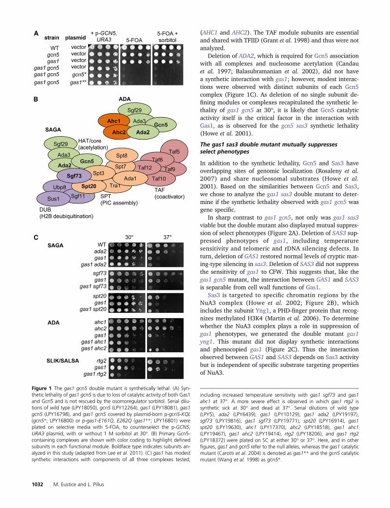
A recent genome-wide study indicated that GAS1 andGCN5 have a negative genetic interaction (Costanzo et al.2010). We found that the gas1 gcn5 heterozygous doublemutant failed to sporulate unless covered by a plasmidencoding either GAS1 or GCN5. When dissected, the result-ing haploid double mutants were not viable without thecovering plasmid as demonstrated in two ways: first bythe inferred genotype of dead spores and second by inabilityto grow on 5-FOA, which selects against the URA3-markedcovering plasmids. The catalytic activity of both Gas1 andGcn5 is required for viability, as neither of the previouslydefined catalytically inactive mutants, gcn5-KQL (gcn5*;Wang et al. 1998) or gas1-E161Q, E262Q (gas1**; Carottiet al. 2004), rescued the lethality of the double mutant inplasmid-shuffle tests. Additionally, the osomoregulator sor-bitol did not rescue the synthetic lethality of gas1 gcn5 (Fig-ure 1A). Thus, the synthetic lethality of gas1 gcn5 is due toloss of the catalytic activities of Gas1 and Gcn5 and is sep-arable from cell wall-associated functions.
The substrate specificity of Gcn5 is largely defined by themacromolecular complexes in which it is found, includingSAGA, ADA, and SLIK/SALSA (Grant et al. 1999; Lee et al.2011; Figure 1B). To determine whether the synthetic le-thality observed for gas1 gcn5 was specifically mediatedthrough one complex or functional module, double mutantswere generated with gas1 to include genes encoding com-ponents of the SAGA modules and unique subunits for bothSLIK/SALSA and ADA. These included genes encoding a cen-tral component of the HAT module (ADA2), key structural orfunctional components of other SAGA modules includingDUB (SGF73) and SPT (SPT20), in addition to genes encod-ing unique components of SLIK/SALSA (RTG2) and ADA
Gas1 and Sas3 Function in DDR 1031
(AHC1 and AHC2). The TAF module subunits are essentialand shared with TFIID (Grant et al. 1998) and thus were notanalyzed.
Deletion of ADA2, which is required for Gcn5 associationwith all complexes and nucleosome acetylation (Candauet al. 1997; Balasubramanian et al. 2002), did not havea synthetic interaction with gas1; however, modest interac-tions were observed with distinct subunits of each Gcn5complex (Figure 1C). As deletion of no single subunit de-fining modules or complexes recapitulated the synthetic le-thality of gas1 gcn5 at 30�, it is likely that Gcn5 catalyticactivity itself is the critical factor in the interaction withGas1, as is observed for the gcn5 sas3 synthetic lethality(Howe et al. 2001).
The gas1 sas3 double mutant mutually suppressesselect phenotypes
In addition to the synthetic lethality, Gcn5 and Sas3 haveoverlapping sites of genomic localization (Rosaleny et al.2007) and share nucleosomal substrates (Howe et al.2001). Based on the similarities between Gcn5 and Sas3,we chose to analyze the gas1 sas3 double mutant to deter-mine if the synthetic lethality observed with gas1 gcn5 wasgene specific.
In sharp contrast to gas1 gcn5, not only was gas1 sas3viable but the double mutant also displayed mutual suppres-sion of select phenotypes (Figure 2A). Deletion of SAS3 sup-pressed phenotypes of gas1, including temperaturesensitivity and telomeric and rDNA silencing defects. Inturn, deletion of GAS1 restored normal levels of cryptic mat-ing-type silencing in sas3. Deletion of SAS3 did not suppressthe sensitivity of gas1 to CFW. This suggests that, like thegas1 gcn5 mutant, the interaction between GAS1 and SAS3is separable from cell wall functions of Gas1.
Sas3 is targeted to specific chromatin regions by theNuA3 complex (Howe et al. 2002; Figure 2B), whichincludes the subunit Yng1, a PHD-finger protein that recog-nizes methylated H3K4 (Martin et al. 2006). To determinewhether the NuA3 complex plays a role in suppression ofgas1 phenotypes, we generated the double mutant gas1yng1. This mutant did not display synthetic interactionsand phenocopied gas1 (Figure 2C). Thus the interactionobserved between GAS1 and SAS3 depends on Sas3 activitybut is independent of specific substrate targeting propertiesof NuA3.
Figure 1 The gas1 gcn5 double mutant is synthetically lethal. (A) Syn-thetic lethality of gas1 gcn5 is due to loss of catalytic activity of both Gas1and Gcn5 and is not rescued by the osomoregulator sorbitol. Serial dilu-tions of wild type (LPY18050), gcn5 (LPY12264), gas1 (LPY18081), gas1gcn5 (LPY16798), and gas1 gcn5 covered by plasmid-born p-gcn5-KQL(gcn5*; LPY16800) or p-gas1-E161Q, E262Q (gas1**; LPY16801) wereplated on selective media with 5-FOA, to counterselect the p-GCN5,URA3 plasmid, with or without 1 M sorbitol at 30�. (B) Primary Gcn5-containing complexes are shown with color coding to highlight definedsubunits in each functional module. Boldface type indicates subunits an-alyzed in this study (adapted from Lee et al. 2011). (C) gas1 has modestsynthetic interactions with components of all three complexes tested,
including increased temperature sensitivity with gas1 sgf73 and gas1ahc1 at 37�. A more severe effect is observed in which gas1 rtg2 issynthetic sick at 30� and dead at 37�. Serial dilutions of wild type(LPY5), ada2 (LPY6439), gas1 (LPY10129), gas1 ada2 (LPY19197),sgf73 (LPY19816), gas1 sgf73 (LPY19771), spt20 (LPY16914), gas1spt20 (LPY19630), ahc1 (LPY17370), ahc2 (LPY18518), gas1 ahc1(LPY19467), gas1 ahc2 (LPY19414), rtg2 (LPY18206), and gas1 rtg2(LPY18372) were plated on SC at either 30� or 37�. Here, and in otherfigures, gas1 and gcn5 refer to the null alleles, whereas the gas1 catalyticmutant (Carotti et al. 2004) is denoted as gas1** and the gcn5 catalyticmutant (Wang et al. 1998) as gcn5*.
1032 M. Eustice and L. Pillus

Based on the mutual suppression observed in the gas1sas3 double mutant, we next tested whether deletion ofSAS3 suppressed the gas1 gcn5 synthetic lethality. The triplemutant gas1 gcn5 sas3 was not viable (Figure 2D). Theseresults suggest that the interactions between GAS1 andGCN5 or SAS3 are of distinct and opposite outcomes.
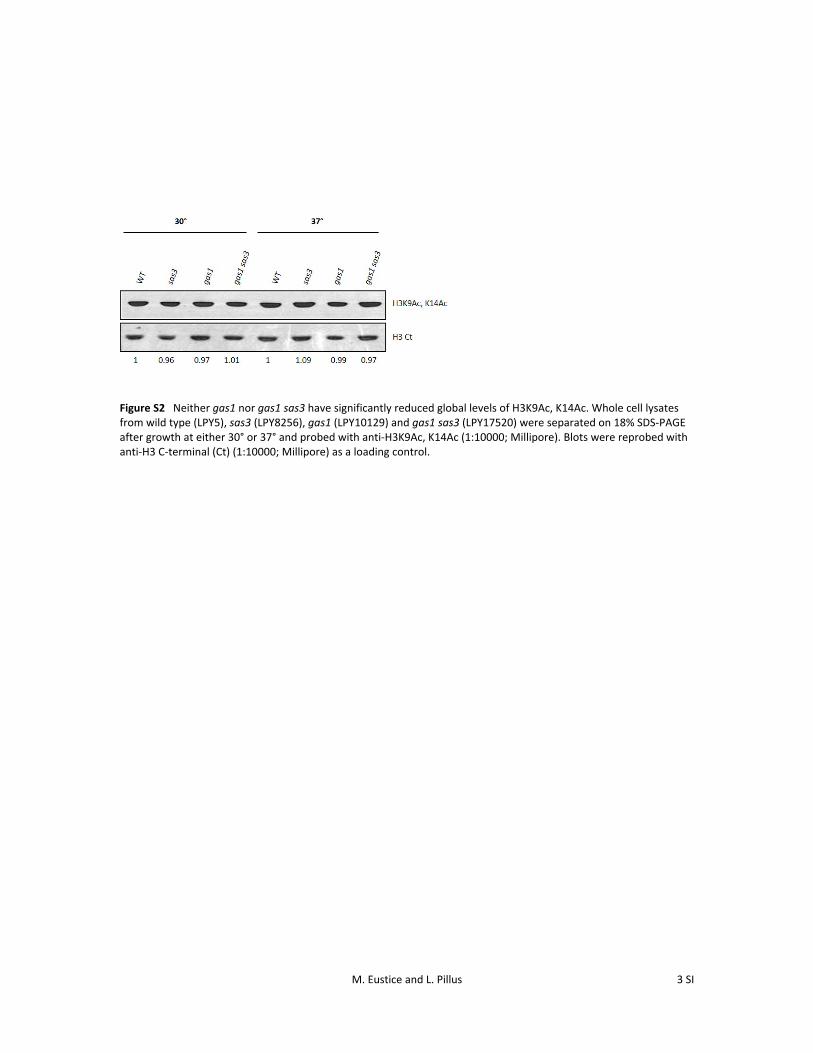
Due to the strength of the genetic interactions with H3KATs, we analyzed H3 acetylation (H3Ac) levels undersuppressing conditions. As previously reported, deletionof GAS1 did not alter levels of H3K9Ac, H3K14Ac at 30�(Koch and Pillus 2009), which are targets of both Gcn5and Sas3 (reviewed in Lafon et al. 2007). At 37�, a condi-tion under which deletion of SAS3 suppresses gas1 tem-perature sensitivity, neither the gas1 strain nor gas1 sas3had altered global levels of H3K9Ac, H3K14Ac (FigureS2). This suggests that the suppression phenotypes ofgas1 sas3 are not mediated through changes in globalH3K9Ac, H3K14Ac levels, which are largely intact in sas3strains due to Gcn5.
Deletion of GAS1 leads to broad DNA damagesensitivity with specific suppression in the absenceof SAS3
Several studies have demonstrated a role for Gcn5-basedacetylation of histone and nonhistone substrates in the DDR(Choy and Kron 2002; Qin and Parthun 2002; Tamburiniand Tyler 2005; Liang et al. 2007; Burgess et al. 2010; Wanget al. 2012). GAS1, SAS3, and GCN5 also all have numerousgenetic and physical interactions with key components ofthe DDR, as defined from previous genome-wide screens(www.thebiogrid.org). Based on these connections, weasked whether the chromatin functions of GAS1 may alsoinfluence DDR.
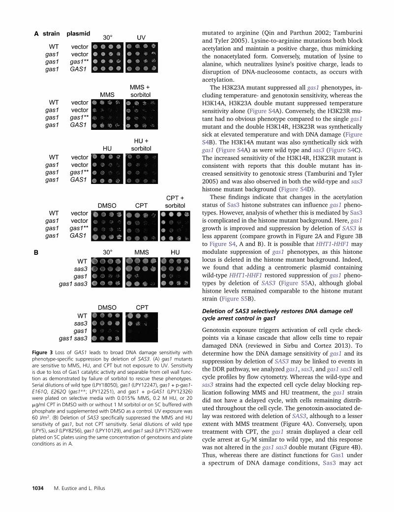
We evaluated the sensitivity of gas1 to a spectrum of DNAdamaging agents including MMS, HU, CPT, and UV irradia-tion, which generates bulky DNA adducts (Sertic et al.2012). Deletion of GAS1 led to sensitivity to all chemicalagents tested, but not to UV. The genotoxin sensitivity wasdue to loss of the b-1,3-glucanosyltransferase activity ofGas1 and was not rescued by sorbitol (Figure 3A). DNAdamage sensitivity was not shared with other members ofthe GAS family, nor other components of the cell wall ma-chinery tested (Figure S3), demonstrating that the sensitiv-ity was not a general phenotype of mutants with cell walldefects.
As deletion of SAS3 suppressed specific phenotypes ofgas1, we analyzed the gas1 sas3 double mutant upon DNAdamage. Deletion of SAS3 suppressed both the MMS andHU sensitivity of gas1 but did not rescue the CPT sensitivity(Figure 3B). These results indicated that, whereas Gas1 hasa broad role in the DDR, Sas3 has a more specific, andantagonistic, function.
Based on the DNA damage phenotypes, we performedgenetic analysis of nucleosomal targets of Sas3 that havebeen implicated in DDR. The residues, H3K14 and H3K23,have increased sensitivity to DNA damaging agents when
Figure 2 Mutual suppression of phenotypes in the gas1 sas3 double mutant.(A) Deletion of SAS3 rescues gas1 temperature sensitivity and silencing defectsat the telomere and rDNA array but not CFW sensitivity. In turn, deletion ofGAS1 restores HM silencing in sas3 to wild-type levels. The sir2 mutant isincluded as a positive control for disruption of silencing. Top panel: Serialdilutions of wild type (LPY4924), sir2 (LPY5035), sas3 (LPY19731), gas1(LPY19773), and gas1 sas3 (LPY16444) were plated on SC at 30� and 37�,SC with 5-FOA (TELVR::URA3) or SC 2Trp (hml::TRP1). Middle panel: Serialdilutions of wild type (LPY2444), sir2 (LPY2447), sas3 (LPY17686), gas1(LPY10074), and gas1 sas3 (LPY17685) were plated on SC or SC 2Ura(RDN::Ty-1-mURA3) at 30�. Bottom panel: wild type (LPY5), sas3 (LPY8256),gas1 (LPY10129), and gas1 sas3 (LPY17520) were plated on either SC or SCwith 10 mg/ml CFW. (B) NuA3 complex with subunits analyzed herein shadedgreen (adapted from Lafon et al. 2007). (C) Deletion of YNG1 does not havesynthetic interactions with gas1. Serial dilutions of wild type (LPY6285), yng1(LPY5526), gas1 (LPY9820), and gas1 yng1 (LPY16997) were plated on SC ateither 30� or 37�. (D) Analysis of GAS1, GCN5, and SAS3 reveals distinct andopposing outcomes for synthetic interactions. Serial dilutions of wild type(LPY5), gcn5 (LPY8242), sas3 (LPY16039), gas1 (LPY10129), gas1 gcn5 + p-GCN5, URA3 (LPY16736), gas1 sas3 (LPY19823), and gas1 gcn5 sas3 + p-GCN5, URA3 (LPY19101) were plated on SC or SC with 5-FOA, to selectagainst p-GCN5, URA3, at 30� with and without 1 M sorbitol.
Gas1 and Sas3 Function in DDR 1033
mutated to arginine (Qin and Parthun 2002; Tamburiniand Tyler 2005). Lysine-to-arginine mutations both blockacetylation and maintain a positive charge, thus mimickingthe nonacetylated form. Conversely, mutation of lysine toalanine, which neutralizes lysine’s positive charge, leads todisruption of DNA-nucleosome contacts, as occurs withacetylation.
The H3K23A mutant suppressed all gas1 phenotypes, in-cluding temperature- and genotoxin sensitivity, whereas theH3K14A, H3K23A double mutant suppressed temperaturesensitivity alone (Figure S4A). Conversely, the H3K23R mu-tant had no obvious phenotype compared to the single gas1mutant and the double H3K14R, H3K23R was syntheticallysick at elevated temperature and with DNA damage (FigureS4B). The H3K14A mutant was also synthetically sick withgas1 (Figure S4A) as were wild type and sas3 (Figure S4C).The increased sensitivity of the H3K14R, H3K23R mutant isconsistent with reports that this double mutant has in-creased sensitivity to genotoxic stress (Tamburini and Tyler2005) and was also observed in both the wild-type and sas3histone mutant background (Figure S4D).
These findings indicate that changes in the acetylationstatus of Sas3 histone substrates can influence gas1 pheno-types. However, analysis of whether this is mediated by Sas3is complicated in the histone mutant background. Here, gas1growth is improved and suppression by deletion of SAS3 isless apparent (compare growth in Figure 2A and Figure 3Bto Figure S4, A and B). It is possible that HHT1-HHF1 maymodulate suppression of gas1 phenotypes, as this histonelocus is deleted in the histone mutant background. Indeed,we found that adding a centromeric plasmid containingwild-type HHT1-HHF1 restored suppression of gas1 pheno-types by deletion of SAS3 (Figure S5A), although globalhistone levels remained comparable to the histone mutantstrain (Figure S5B).
Deletion of SAS3 selectively restores DNA damage cellcycle arrest control in gas1
Genotoxin exposure triggers activation of cell cycle check-points via a kinase cascade that allow cells time to repairdamaged DNA (reviewed in Sirbu and Cortez 2013). Todetermine how the DNA damage sensitivity of gas1 and itssuppression by deletion of SAS3 may be linked to events inthe DDR pathway, we analyzed gas1, sas3, and gas1 sas3 cellcycle profiles by flow cytometry. Whereas the wild-type andsas3 strains had the expected cell cycle delay blocking rep-lication following MMS and HU treatment, the gas1 straindid not have a delayed cycle, with cells remaining distrib-uted throughout the cell cycle. The genotoxin-associated de-lay was restored with deletion of SAS3, although to a lesserextent with MMS treatment (Figure 4A). Conversely, upontreatment with CPT, the gas1 strain displayed a clear cellcycle arrest at G2/M similar to wild type, and this responsewas not altered in the gas1 sas3 double mutant (Figure 4B).Thus, whereas there are distinct functions for Gas1 undera spectrum of DNA damage conditions, Sas3 may act
Figure 3 Loss of GAS1 leads to broad DNA damage sensitivity withphenotype-specific suppression by deletion of SAS3. (A) gas1 mutantsare sensitive to MMS, HU, and CPT but not exposure to UV. Sensitivityis due to loss of Gas1 catalytic activity and separable from cell wall func-tion as demonstrated by failure of sorbitol to rescue these phenotypes.Serial dilutions of wild type (LPY18050), gas1 (LPY12247), gas1 + p-gas1-E161Q, E262Q (gas1**; LPY12251), and gas1 + p-GAS1 (LPY12326)were plated on selective media with 0.015% MMS, 0.2 M HU, or 20mg/ml CPT in DMSO with or without 1 M sorbitol or on SC buffered withphosphate and supplemented with DMSO as a control. UV exposure was60 J/m2. (B) Deletion of SAS3 specifically suppressed the MMS and HUsensitivity of gas1, but not CPT sensitivity. Serial dilutions of wild type(LPY5), sas3 (LPY8256), gas1 (LPY10129), and gas1 sas3 (LPY17520) wereplated on SC plates using the same concentration of genotoxins and plateconditions as in A.
1034 M. Eustice and L. Pillus
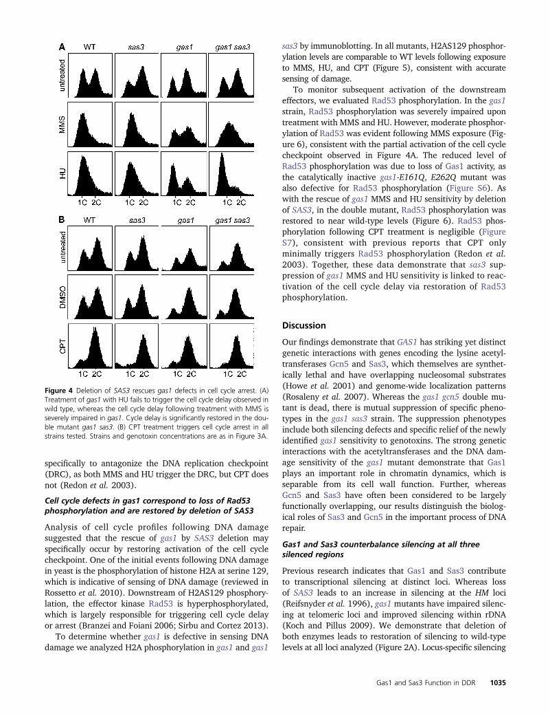
specifically to antagonize the DNA replication checkpoint(DRC), as both MMS and HU trigger the DRC, but CPT doesnot (Redon et al. 2003).
Cell cycle defects in gas1 correspond to loss of Rad53phosphorylation and are restored by deletion of SAS3
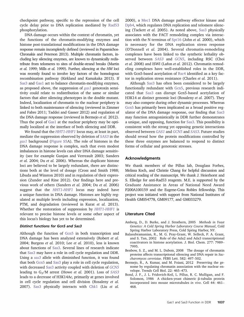
Analysis of cell cycle profiles following DNA damagesuggested that the rescue of gas1 by SAS3 deletion mayspecifically occur by restoring activation of the cell cyclecheckpoint. One of the initial events following DNA damagein yeast is the phosphorylation of histone H2A at serine 129,which is indicative of sensing of DNA damage (reviewed inRossetto et al. 2010). Downstream of H2AS129 phosphory-lation, the effector kinase Rad53 is hyperphosphorylated,which is largely responsible for triggering cell cycle delayor arrest (Branzei and Foiani 2006; Sirbu and Cortez 2013).
To determine whether gas1 is defective in sensing DNAdamage we analyzed H2A phosphorylation in gas1 and gas1
sas3 by immunoblotting. In all mutants, H2AS129 phosphor-ylation levels are comparable to WT levels following exposureto MMS, HU, and CPT (Figure 5), consistent with accuratesensing of damage.
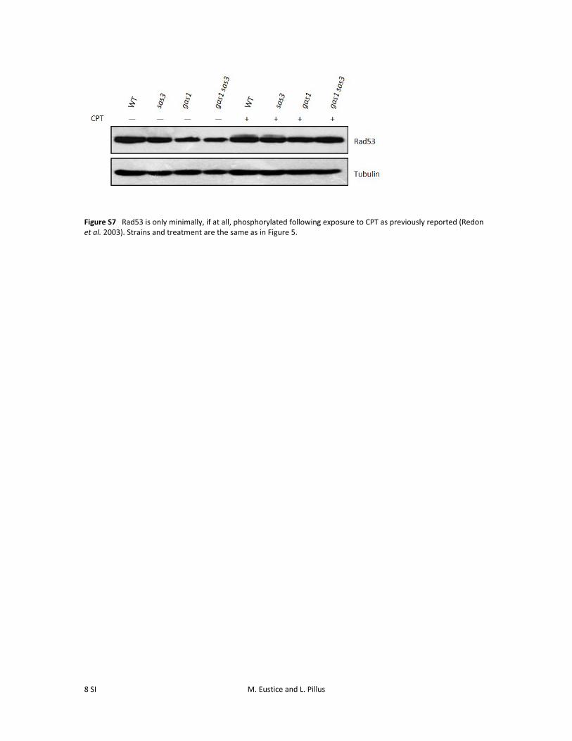
To monitor subsequent activation of the downstreameffectors, we evaluated Rad53 phosphorylation. In the gas1strain, Rad53 phosphorylation was severely impaired upontreatment with MMS and HU. However, moderate phosphor-ylation of Rad53 was evident following MMS exposure (Fig-ure 6), consistent with the partial activation of the cell cyclecheckpoint observed in Figure 4A. The reduced level ofRad53 phosphorylation was due to loss of Gas1 activity, asthe catalytically inactive gas1-E161Q, E262Q mutant wasalso defective for Rad53 phosphorylation (Figure S6). Aswith the rescue of gas1 MMS and HU sensitivity by deletionof SAS3, in the double mutant, Rad53 phosphorylation wasrestored to near wild-type levels (Figure 6). Rad53 phos-phorylation following CPT treatment is negligible (FigureS7), consistent with previous reports that CPT onlyminimally triggers Rad53 phosphorylation (Redon et al.2003). Together, these data demonstrate that sas3 sup-pression of gas1 MMS and HU sensitivity is linked to reac-tivation of the cell cycle delay via restoration of Rad53phosphorylation.
Discussion
Our findings demonstrate that GAS1 has striking yet distinctgenetic interactions with genes encoding the lysine acetyl-transferases Gcn5 and Sas3, which themselves are synthet-ically lethal and have overlapping nucleosomal substrates(Howe et al. 2001) and genome-wide localization patterns(Rosaleny et al. 2007). Whereas the gas1 gcn5 double mu-tant is dead, there is mutual suppression of specific pheno-types in the gas1 sas3 strain. The suppression phenotypesinclude both silencing defects and specific relief of the newlyidentified gas1 sensitivity to genotoxins. The strong geneticinteractions with the acetyltransferases and the DNA dam-age sensitivity of the gas1 mutant demonstrate that Gas1plays an important role in chromatin dynamics, which isseparable from its cell wall function. Further, whereasGcn5 and Sas3 have often been considered to be largelyfunctionally overlapping, our results distinguish the biolog-ical roles of Sas3 and Gcn5 in the important process of DNArepair.
Gas1 and Sas3 counterbalance silencing at all threesilenced regions
Previous research indicates that Gas1 and Sas3 contributeto transcriptional silencing at distinct loci. Whereas lossof SAS3 leads to an increase in silencing at the HM loci(Reifsnyder et al. 1996), gas1 mutants have impaired silenc-ing at telomeric loci and improved silencing within rDNA(Koch and Pillus 2009). We demonstrate that deletion ofboth enzymes leads to restoration of silencing to wild-typelevels at all loci analyzed (Figure 2A). Locus-specific silencing
Figure 4 Deletion of SAS3 rescues gas1 defects in cell cycle arrest. (A)Treatment of gas1 with HU fails to trigger the cell cycle delay observed inwild type, whereas the cell cycle delay following treatment with MMS isseverely impaired in gas1. Cycle delay is significantly restored in the dou-ble mutant gas1 sas3. (B) CPT treatment triggers cell cycle arrest in allstrains tested. Strains and genotoxin concentrations are as in Figure 3A.
Gas1 and Sas3 Function in DDR 1035

relies on a balance of silencing proteins and other chromatinfactors, some of which are limiting (Smith et al. 1998;Benbow and Dubois 2008). Altering the distribution of thesefactors can lead to changes in the strength of silencing be-tween loci (Lustig et al. 1996). As silencing is both strength-ened and/or disrupted at specific loci in the mutants understudy, one potential explanation for the mutual suppressionobserved in the gas1 sas3 strain is that localization of limit-ing silencing factors is normalized. In this case, Sas3 andGas1 counteract the influence of each other, such that inthe absence of both enzymes balance is restored. This ideais in agreement with our previous observation of a physicalinteraction between Gas1 and the deacetyltransferase Sir2(Koch and Pillus 2009), a limiting factor essential for estab-lishment and maintenance of silencing (Rusche et al. 2003).
Analysis of DNA damage sensitivity in gas1 cells revealsthat Sas3 antagonizes the DNA replication checkpoint
In addition to previously defined silencing defects (Koch andPillus 2009) we found that deletion of GAS1 led to DNAdamage sensitivity. Strains lacking Gas1, or with defectivecatalytic activity, were sensitive to the genotoxins MMS, HU,and CPT but not UV (Figure 3A). Thus, although Gas1 playsa broad role in DNA damage, there are distinctions for par-ticular types of damage or repair pathways.
Whereas H2AS129 phosphorylation, indicating sensingand initial DDR activation, was intact in all strains analyzed,the levels of Rad53 phosphorylation were significantly re-duced in gas1 and restored by deletion of SAS3. Impairmentof the MMS or HU DNA damage-associated cell cycle delayand Rad53 phosphorylation levels in gas1 strains (Figure 4
and Figure 5) indicates that Gas1 may function in triggeringhyperphosphorylation of Rad53 and the subsequent cell cy-cle checkpoint. Although GAS1 mutants failed to arrest inresponse to MMS and HU they did undergo CPT-inducedG2/M arrest. These observations strengthen the idea thatGas1 is broadly relevant to DDR, yet its contributions appearto depend on the type of lesion.
Distinct mechanistic roles for Gas1 in DNA damage arefurther supported by the suppression seen with deletion ofSAS3, which rescued MMS and HU sensitivity but not CPTsensitivity (Figure 3B). MMS and HU elicit a largely over-lapping transcriptional response, which is primarily depen-dent on Rad53 phosphorylation of substrates. By contrast,CPT leads to induction of a markedly different set of genes(Travesa et al. 2012; Travesa and Wittenberg 2012). BothMMS and HU trigger the replication checkpoint via forkarrest or by slowing fork progression by reducing dNTPpools, respectively (reviewed in Branzei and Foiani 2007).Conversely, CPT is considered to be “checkpoint blind” asexposure leads to only modest induction of Rad53 phos-phorylation and does not trigger the replication checkpoint(Redon et al. 2003; Tourriere and Pasero 2007).
The primary checkpoints activated by DNA damageinclude delay of the G1/S transition and block of the G2/Mtransition and the S-phase checkpoints. Although there areoverlaps in the proteins mediating these checkpoints thereare also distinctions that depend on the phase of the cellcycle, type of DNA damage, and repair pathway choice(reviewed in Warmerdam and Kanaar 2010; Symingtonand Gautier 2011; Gobbini et al. 2013). Cell cycle check-points and DNA damage repair require both positive andnegative regulation to ensure proper spatiotemporal dynam-ics and maintenance of genomic integrity (reviewed inPanier and Durocher 2013). Thus, Sas3 may be particularlyrelevant in antagonizing activation of the replication
Figure 5 H2AS129 is phosphorylated following genotoxin exposure in allstrains. Levels of H2AS129 phosphorylation following exposure to MMS(top), HU (middle), and CPT (bottom) are comparable to wild type in allstrains analyzed. Strains and genotoxin concentrations are as in Figure3A.
Figure 6 Rad53 phosphorylation is significantly reduced in gas1 and re-stored in gas1 sas3 following exposure to MMS (top) and HU (bottom).Note that overall levels of Rad53 are diminished in gas1. Strains andgenotoxin concentrations are as in Figure 3A.
1036 M. Eustice and L. Pillus
checkpoint pathway, specific to the repression of the cellcycle delay prior to DNA replication mediated by Rad53phosphorylation.
DNA damage occurs within the context of chromatin, yetthe functions of the chromatin-modifying enzymes andhistone post-translational modifications in the DNA damageresponse remain incompletely defined (reviewed in Papamichos-Chronakis and Peterson 2013). Multiple chromatin factors, in-cluding key silencing enzymes, are known to dynamically redis-tribute from telomeres to sites of double-strand breaks (Martinet al. 1999; Mills et al. 1999). Further, silencing at the HM lociwas recently found to involve key factors of the homologousrecombination pathway (Kirkland and Kamakaka 2013). IfSas3 and Gas1 act to balance chromatin-modifying enzymes,as proposed above, the suppression of gas1 genotoxin sensi-tivity could relate to redistribution of the same or similarfactors that alter silencing phenotypes in the double mutant.Indeed, localization of chromatin to the nuclear periphery islinked to both maintenance of silencing (reviewed in Zimmerand Fabre 2011; Taddei and Gasser 2012) and regulation ofthe DNA damage response (reviewed in Bermejo et al. 2012).Thus the pool of Gas1 at the nuclear periphery may be opti-mally localized at the interface of both silencing and DDR.
We found that the HHT1-HHF1 locus may, at least in part,mediate the suppression observed by deletion of SAS3 in thegas1 background (Figure S5A). The role of histones in theDNA damage response is complex, such that even modestimbalances in histone levels can alter DNA damage sensitiv-ity (see for example Gunjan and Verreault 2003; Sanderset al. 2004; Du et al. 2006). Whereas the duplicate histoneloci are believed to be largely redundant, there are distinc-tions both at the level of dosage (Cross and Smith 1988;Libuda and Winston 2010) and in regulation of their expres-sion (Zunder and Rine 2012). Our findings here and pre-vious work of others (Sanders et al. 2004; Du et al. 2006)suggest that the HHT1-HHF1 locus may indeed havea unique function in DNA damage. Histones are highly reg-ulated at multiple levels including expression, localization,PTM, and degradation (reviewed in Kurat et al. 2013).Whether the restoration of suppression by HHT1-HHF1 isrelevant to precise histone levels or some other aspect ofthis locus’s biology has yet to be determined.
Distinct functions for Gcn5 and Sas3
Although the function of Gcn5 in both transcription andDNA damage has been analyzed extensively (Robert et al.2004; Burgess et al. 2010; Lee et al. 2010), less is knownabout functions of Sas3. Several lines of research indicatethat Sas3 may have a role in cell cycle regulation and DDR.Using a sas3 allele with diminished function, it was foundthat both Gcn5 and Sas3 play a role in cell cycle regulation,with decreased Sas3 activity coupled with deletion of GCN5leading to G2/M arrest (Howe et al. 2001). Loss of SAS3leads to a decrease of H3K14Ac, primarily at genes involvedin cell cycle regulation and cell division (Rosaleny et al.2007). Sas3 physically interacts with Chk1 (Liu et al.
2000), a Mec1 DNA damage pathway effector kinase andDpb4, which regulates DNA replication and telomere silenc-ing (Tackett et al. 2005). As noted above, Sas3 physicallyassociates with the FACT remodeling complex via interac-tion with the N terminus of Spt16 (John et al. 2000), whichis necessary for the DNA replication stress response(O’Donnell et al. 2004). Several chromatin-remodelingcomplexes have been linked to the synthetic lethality ob-served between SAS3 and GCN5, including RSC (Choiet al. 2008) and ISWI (Lafon et al. 2012). Chromatin remod-eling complexes have well-established roles in the DDR,with Gcn5-based acetylation of Rsc4 identified as a key fac-tor in replication stress resistance (Charles et al. 2011).
Although Sas3 has often been considered to be largelyfunctionally redundant with Gcn5, previous research indi-cated that Sas3 can disrupt Gcn5-based acetylation ofH3K14 at distinct genomic loci (Rosaleny et al. 2007). Theymay also compete during other dynamic processes. WhereasGcn5 has primarily been implicated as a broad positive reg-ulator of the DNA damage response, our finding that Sas3may function antagonistically in DDR further demonstratesa unique, and opposing, function for Sas3. This possibility isconsistent with the strong yet opposing genetic interactionsobserved between GAS1 and GCN5 and SAS3. Future studiesshould reveal how the protein modifications controlled bythese three enzymes are balanced to respond to distinctforms of cellular and genotoxic stresses.
Acknowledgments
We thank members of the Pillus lab, Douglass Forbes,Melissa Koch, and Christie Chang for helpful discussion andcritical reading of the manuscript. We thank J. Heierhorst andS. Elledge for anti-Rad53 reagents. M.E. is supported by theGraduate Assistance in Areas of National Need AwardP200A100159 and the Eugene-Cota Robles fellowship. Thisproject was initiated with support from National Institutes ofHealth GM054778, GM09177, and GM033279.
Literature Cited
Amberg, D., D. Burke, and J. Strathern, 2005 Methods in YeastGenetics: A Cold Spring Harbor Laboratory Course Manual, ColdSpring Harbor Laboratory Press, Cold Spring Harbor, NY.
Balasubramanian, R., M. G. Pray-Grant, W. Selleck, P. A. Grant,and S. Tan, 2002 Role of the Ada2 and Ada3 transcriptionalcoactivators in histone acetylation. J. Biol. Chem. 277: 7989–7995.
Benbow, S. Z., and M. L. Dubois, 2008 The dosage of chromatinproteins affects transcriptional silencing and DNA repair in Sac-charomyces cerevisiae. FEBS Lett. 582: 497–502.
Bermejo, R., A. Kumar, and M. Foiani, 2012 Preserving the ge-nome by regulating chromatin association with the nuclear en-velope. Trends Cell Biol. 22: 465–473.
Bond, J. F., J. L. Fridovich-Keil, L. Pillus, R. C. Mulligan, and F.Solomon, 1986 A chicken-yeast chimeric b-tubulin proteinincorporated into mouse microtubules in vivo. Cell 44: 461–468.
Gas1 and Sas3 Function in DDR 1037
Branzei, D., and M. Foiani, 2006 The Rad53 signal transductionpathway: replication fork stabilization, DNA repair, and adapta-tion. Exp. Cell Res. 312: 2654–2659.
Branzei, D., and M. Foiani, 2007 Interplay of replication check-points and repair proteins at stalled replication forks. DNA Re-pair (Amst.) 6: 994–1003.
Burgess, R. J., H. Zhou, J. Han, and Z. Zhang, 2010 Gcn5 inreplication-coupled nucleosome assembly. Mol. Cell 37: 469–480.
Campos, E. I., and D. Reinberg, 2009 Histones: annotating chro-matin. Annu. Rev. Genet. 43: 559–599.
Candau, R., J. X. Zhou, C. D. Allis, and S. L. Berger, 1997 Histoneacetyltransferase activity and interaction with ADA2 are criticalfor GCN5 function in vivo. EMBO J. 16: 555–565.
Carotti, C., E. Ragni, O. Palomares, T. Fontaine, G. Tedeschi et al.,2004 Characterization of recombinant forms of the yeast Gas1protein and identification of residues essential for glucanosyl-transferase activity and folding. Eur. J. Biochem. 271: 3635–3645.
Charles, G. M., C. Chen, S. C. Shih, S. R. Collins, P. Beltrao et al.,2011 Site-specific acetylation mark on an essential chromatin-remodeling complex promotes resistance to replication stress.Proc. Natl. Acad. Sci. USA 108: 10620–10625.
Choi, J. K., D. E. Grimes, K. M. Rowe, and L. Howe,2008 Acetylation of Rsc4p by Gcn5p is essential in the absenceof histone H3 acetylation. Mol. Cell. Biol. 28: 6967–6972.
Choy, J. S., and S. J. Kron, 2002 NuA4 subunit Yng2 function inintra-S-phase DNA damage response. Mol. Cell. Biol. 22: 8215–8225.
Clarke, A. S., J. E. Lowell, S. J. Jacobson, and L. Pillus,1999 Esa1p is an essential histone acetyltransferase requiredfor cell cycle progression. Mol. Cell. Biol. 19: 2515–2526.
Costanzo, M., A. Baryshnikova, J. Bellay, Y. Kim, E. D. Spear et al.,2010 The genetic landscape of the cell. Science 327: 425–431.
Cross, S. L., and M. M. Smith, 1988 Comparison of the structureand cell cycle expression of mRNAs encoding two histone H3–H4 loci in Saccharomyces cerevisiae. Mol. Cell. Biol. 8: 945–954.
Du, L.-L., T. M. Nakamura, and P. Russell, 2006 Histone modifi-cation-dependent and independent pathways for recruitment ofcheckpoint protein Crb2 to double-strand breaks. Genes Dev. 20:1583–1596.
Gobbini, E., D. Cesena, A. Galbiati, A. Lockhart, and M. P.Longhese, 2013 Interplays between ATM/Tel1 and ATR/Mec1 in sensing and signaling DNA double-strand breaks.DNA Repair (Amst.) 12: 791–799.
Grant, P. A., L. Duggan, J. Côté, S. M. Roberts, J. E. Brownell et al.,1997 Yeast Gcn5 functions in two multisubunit complexes toacetylate nucleosomal histones: characterization of an Ada com-plex and the SAGA (Spt/Ada) complex. Genes Dev. 11: 1640–1650.
Grant, P. A., D. Schieltz, M. G. Pray-Grant, D. J. Steger, J. C. Reeseet al., 1998 A subset of TAF(II)s are integral components ofthe SAGA complex required for nucleosome acetylation andtranscriptional stimulation. Cell 94: 45–53.
Grant, P. A., A. Eberharter, S. John, R. G. Cook, B. M. Turner et al.,1999 Expanded lysine acetylation specificity of Gcn5 in nativecomplexes. J. Biol. Chem. 274: 5895–5900.
Gunjan, A., and A. Verreault, 2003 A Rad53 kinase-dependentsurveillance mechanism that regulates histone protein levels inS. cerevisiae. Cell 115: 537–549.
Howe, L., D. Auston, P. Grant, S. John, R. G. Cook et al.,2001 Histone H3 specific acetyltransferases are essential forcell cycle progression. Genes Dev. 15: 3144–3154.
Howe, L., T. Kusch, N. Muster, R. Chaterji, J. R. Yates, 3rd et al.,2002 Yng1p modulates the activity of Sas3p as a component ofthe yeast NuA3 histone acetyltransferase complex. Mol. Cell.Biol. 22: 5047–5053.
Huh, W. K., J. V. Falvo, L. C. Gerke, A. S. Carroll, R. W. Howsonet al., 2003 Global analysis of protein localization in buddingyeast. Nature 425: 686–691.
John, S., L. Howe, S. T. Tafrov, P. A. Grant, R. Sternglanz et al.,2000 The something about silencing protein, Sas3, is the cat-alytic subunit of NuA3, a yTAF(II)30-containing HAT complexthat interacts with the Spt16 subunit of the yeast CP (Cdc68/Pob3)-FACT complex. Genes Dev. 14: 1196–1208.
Kirkland, J. G., and R. T. Kamakaka, 2013 Long-range hetero-chromatin association is mediated by silencing and double-strand DNA break repair proteins. J. Cell Biol. 201: 809–826.
Koch, M. R., and L. Pillus, 2009 The glucanosyltransferase Gas1functions in transcriptional silencing. Proc. Natl. Acad. Sci. USA106: 11224–11229.
Kornberg, R. D., and Y. Lorch, 1999 Twenty-five years of thenucleosome, fundamental particle of the eukaryote chromo-some. Cell 98: 285–294.
Koutelou, E., C. L. Hirsch, and S. Y. Dent, 2010 Multiple faces ofthe SAGA complex. Curr. Opin. Cell Biol. 22: 374–382.
Kouzarides, T., 2007 Chromatin modifications and their function.Cell 128: 693–705.
Kuo, Y. M., and A. J. Andrews, 2013 Quantitating the specificityand selectivity of Gcn5-mediated acetylation of histone H3.PLoS ONE 8: e54896.
Kurat, C. F., J. Recht, E. Radovani, T. Durbic, B. Andrews et al.,2013 Regulation of histone gene transcription in yeast. Cell.Mol. Life Sci. 71: 599–613.
Lafon, A., C. S. Chang, E. M. Scott, S. J. Jacobson, and L. Pillus,2007 MYST opportunities for growth control: yeast genes illu-minate human cancer gene functions. Oncogene 26: 5373–5384.
Lafon, A., E. Petty, and L. Pillus, 2012 Functional antagonismbetween Sas3 and Gcn5 acetyltransferases and ISWI chromatinremodelers. PLoS Genet. 8: e1002994.
Le, S., C. Davis, J. B. Konopka, and R. Sternglanz, 1997 Two newS-phase-specific genes from Saccharomyces cerevisiae. Yeast 13:1029–1042.
Lee, H. S., J. H. Park, S. J. Kim, S. J. Kwon, and J. Kwon, 2010 Acooperative activation loop among SWI/SNF, gamma-H2AX andH3 acetylation for DNA double-strand break repair. EMBO J. 29:1434–1445.
Lee, K. K., M. E. Sardiu, S. K. Swanson, J. M. Gilmore, M. Toroket al., 2011 Combinatorial depletion analysis to assemble thenetwork architecture of the SAGA and ADA chromatin remodel-ing complexes. Mol. Syst. Biol. 7: 503.
Lee, K. K., and J. Workman, 2007 Histone acetyltransferase com-plexes: one size doesn’t fit all. Nat. Rev. Mol. Cell Biol. 8: 284–295.
Levin, D. E., 2005 Cell wall integrity signaling in Saccharomycescerevisiae. Microbiol. Mol. Biol. Rev. 69: 262–291.
Li, G., and D. Reinberg, 2011 Chromatin higher-order struc-tures and gene regulation. Curr. Opin. Genet. Dev. 21: 175–186.
Liang, B., J. Qiu, K. Ratnakumar, and B. C. Laurent, 2007 RSCfunctions as an early double-strand-break sensor in the cell’sresponse to DNA damage. Curr. Biol. 17: 1432–1437.
Libuda, D. E., and F. Winston, 2010 Alterations in DNA replica-tion and histone levels promote histone gene amplificaition inSaccharomyces cerevisiae. Genetics 184: 985–997.
Liu, Y., G. Vidanes, Y. C. Lin, S. Mori, and W. Siede,2000 Characterization of a Saccharomyces cerevisiae homo-logue of Schizosaccharomyces pombe Chk1 involved in DNA-damage-induced M-phase arrest. Mol. Gen. Genet. 262:1132–1146.
Lustig, A. J., C. Liu, C. Zhang, and J. P. Hanish, 1996 TetheredSir3p nucleates silencing at telomeres and internal loci in Sac-charomyces cerevisiae. Mol. Cell. Biol. 16: 2483–2495.
1038 M. Eustice and L. Pillus
Martin, D. G., K. Baetz, X. Shi, K. L. Walter, V. E. MacDonald et al.,2006 The Yng1p plant homeodomain finger is a methyl-histonebinding module that recognizes lysine 4-methylated histone H3.Mol. Cell. Biol. 26: 7871–7879.
Martin, S. G., T. Laroche, N. Suka, M. Grunstein, and S. M. Gasser,1999 Relocalization of telomeric Ku and SIR proteins in re-sponse to DNA strand breaks in yeast. Cell 97: 621–633.
Mills, K. D., D. A. Sinclair, and L. Guarente, 1999 MEC1-depen-dent redistribution of the Sir3 silencing protein from telomeresto DNA double-strand breaks. Cell 97: 609–620.
Nitiss, J., and J. C. Wang, 1988 DNA topoisomerase-targetingantitumor drugs can be studied in yeast. Proc. Natl. Acad. Sci.USA 85: 7501–7505.
O’Donnell, A. F., N. K. Brewster, J. Kurniawan, L. V. Minard, G. C.Johnston et al., 2004 Domain organization of the yeast histonechaperone FACT: the conserved N-terminal domain of FACTsubunit Spt16 mediates recovery from replication stress. NucleicAcids Res. 32: 5894–5906.
Orlean, P., 2012 Architecture and biosynthesis of the Saccharo-myces cerevisiae cell wall. Genetics 192: 775–818.
Panier, S., and D. Durocher, 2013 Push back to respond better:regulatory inhibition of the DNA double-strand break response.Nat. Rev. Cancer 13: 661–672.
Papamichos-Chronakis, M., and C. L. Peterson, 2013 Chromatinand the genome integrity network. Nat. Rev. Genet. 14: 62–75.
Pike, B. L., S. Yongkiettrakul, M. D. Tsai, and J. Heierhorst,2003 Diverse but overlapping functions of the two forkhead-associated (FHA) domains in Rad53 checkpoint kinase activa-tion. J. Biol. Chem. 278: 30421–30424.
Pokholok, D. K., C. T. Harbison, S. Levine, M. Cole, N. M. Hannettet al., 2005 Genome-wide map of nucleosome acetylation andmethylation in yeast. Cell 122: 517–527.
Popolo, L., and M. Vai, 1999 The Gas1 glycoprotein, a putative wallpolymer cross-linker. Biochim. Biophys. Acta 1426: 385–400.
Pray-Grant, M. G., D. Schieltz, S. J. McMahon, J. M. Wood, E. L.Kennedy et al., 2002 The novel SLIK histone acetyltransferasecomplex functions in the yeast retrograde response pathway.Mol. Cell. Biol. 22: 8774–8786.
Qin, S., and M. R. Parthun, 2002 Histone H3 and the histoneacetyltransferase Hat1p contribute to DNA double-strand breakrepair. Mol. Cell. Biol. 22: 8353–8365.
Ragni, E., T. Fontaine, C. Gissi, J. P. Latge, and L. Popolo,2007 The Gas family of proteins of Saccharomyces cerevisiae:characterization and evolutionary analysis. Yeast 24: 297–308.
Redon, C., D. R. Pilch, E. P. Rogakou, A. H. Orr, N. F. Lowndes et al.,2003 Yeast histone 2A serine 129 is essential for the efficientrepair of checkpoint-blind DNA damage. EMBO Rep. 4: 678–684.
Reifsnyder, C., J. Lowell, A. Clarke, and L. Pillus, 1996 Yeast SASsilencing genes and human genes associated with AML and HIV-1 Tat interactions are homologous with acetyltransferases. Nat.Genet. 14: 42–49.
Renauld, H., O. M. Aparicio, P. D. Zierath, B. L. Billington, S. K.Chhablani et al., 1993 Silent domains are assembled continu-ously from the telomere and are defined by promoter distanceand strength, and by SIR3 dosage. Genes Dev. 7: 1133–1145.
Robert, F., D. K. Pokholok, N. M. Hannett, N. J. Rinaldi, M. Chandyet al., 2004 Global position and recruitment of HATs andHDACs in the yeast genome. Mol. Cell 16: 199–209.
Roncero, C., and A. Duran, 1985 Effect of Calcofluor white andCongo red on fungal cell wall morphogenesis: in vivo activationof chitin polymerization. J. Bacteriol. 163: 1180–1185.
Rosaleny, L. E., A. B. Ruiz-Garcia, J. Garcia-Martinez, J. E. Perez-Ortin,and V. Tordera, 2007 The Sas3p and Gcn5p histone acetyltrans-ferases are recruited to similar genes. Genome Biol. 8: R119.
Rossetto, D., A. W. Truman, S. J. Kron, and J. Côté,2010 Epigenetic modifications in double-strand break DNAdamage signaling and repair. Clin. Cancer Res. 16: 4543–4552.
Rusche, L. N., A. L. Kirchmaier, and J. Rine, 2003 The establish-ment, inheritance, and funciton of silenced chromatin in Saccha-romyces cerevisiae. Annu. Rev. Biochem. 72: 481–516.
Sanders, S. L., M. Portoso, J. Mata, J. Bahler, R. C. Allshire et al.,2004 Methylation of histone H4 lysine 20 controls recruitmentof Crb2 to sites of DNA damage. Cell 119: 603–614.
Sertic, S., S. Pizzi, F. Lazzaro, P. Plevani, and M. Muzi-Falconi,2012 NER and DDR: classical music with new instruments.Cell Cycle 11: 668–674.
Sirbu, B. M., and D. Cortez, 2013 DNA damage response: threelevels of DNA repair regulation. Cold Spring Harb. Perspect.Biol. 5: a012724.
Smith, J. S., and J. D. Boeke, 1997 An unusual form of transcrip-tional silencing in yeast ribosomal DNA. Genes Dev. 11: 241–254.
Smith, J. S., C. B. Brachmann, L. Pillus, and J. D. Boeke,1998 Distribution of a limited Sir2 protein pool regulates thestrength of yeast rDNA silencing and is modulated by Sir4p.Genetics 149: 1205–1219.
Suter, B., A. Tong, M. Chang, L. Yu, G. W. Brown et al., 2004 Theorigin recognition complex links replication, sister chromatidcohesion and transcriptional silencing in Saccharomyces cerevi-siae. Genetics 167: 579–591.
Symington, L. S., and J. Gautier, 2011 Double-strand break end re-section and repair pathway choice. Annu. Rev. Genet. 45: 247–271.
Tackett, A. J., D. J. Dilworth, M. J. Davey, M. O’Donnell, J. D.Aitchison et al., 2005 Proteomic and genomic characterizationof chromatin complexes at a boundary. J. Cell Biol. 169: 35–47.
Taddei, A., and S. M. Gasser, 2012 Structure and function in thebudding yeast nucleus. Genetics 192: 107–129.
Tamburini, B. A., and J. K. Tyler, 2005 Localized histone acetyla-tion and deacetylation triggered by the homologous recombina-tion pathway of double-strand DNA repair. Mol. Cell. Biol. 25:4903–4913.
Tourriere, H., and P. Pasero, 2007 Maintenance of fork integrityat damaged DNA and natural pause sites. DNA Repair (Amst.) 6:900–913.
Travesa, A., D. Kuo, R. A. de Bruin, T. I. Kalashnikova, M. Guaderramaet al., 2012 DNA replication stress differentially regulates G1/Sgenes via Rad53-dependent inactivation of Nrm1. EMBO J. 31:1811–1822.
Travesa, A., and C. Wittenberg, 2012 Turned on by genotoxicstress. Cell Cycle 11: 3145–3146.
Turchini, A., L. Ferrario, and L. Popolo, 2000 Increase of externalosmolarity reduces morphogenetic defects and accumulaiton ofchitin in a gas1 mutant of Saccharomyces cerevisiae. J. Bacteriol.182: 1167–1171.
van Leeuwen, F., and D. E. Gottschling, 2002 Assays for genesilencing in yeast. Methods Enzymol. 350: 165–186.
Wang, L., L. Liu, and S. L. Berger, 1998 Critical residues for his-tone acetylation by Gcn5, functioning in Ada and SAGA com-plexes, are also required for transcriptional function in vivo.Genes Dev. 12: 640–653.
Wang, Y., S. P. Kallgren, B. D. Reddy, K. Kuntz, L. Lopez-Mauryet al., 2012 Histone H3 lysine 14 acetylation is required foractivation of a DNA damage checkpoint in fission yeast. J. Biol.Chem. 287: 4386–4393.
Warmerdam, D. O., and R. Kanaar, 2010 Dealing with DNA dam-age: relationships between checkpoint and repair pathways.Mutat. Res. 704: 2–11.
Zimmer, C., and E. Fabre, 2011 Principles of chromosomal orga-nization: lessons from yeast. J. Cell Biol. 192: 723–733.
Zunder, R. M., and J. Rine, 2012 Direct interplay among histones,histone chaperones, and a chromatin boundary protein in the con-trol of histone gene expression. Mol. Cell. Biol. 32: 4337–4349.
Communicating editor: M. Hampsey
Gas1 and Sas3 Function in DDR 1039

GENETICSSupporting Information
http://www.genetics.org/lookup/suppl/doi:10.1534/genetics.113.158824/-/DC1
Unexpected Function of the GlucanosyltransferaseGas1 in the DNA Damage Response Linked to
Histone H3 Acetyltransferases inSaccharomyces cerevisiae
Moriah Eustice and Lorraine Pillus
Copyright © 2014 by the Genetics Society of AmericaDOI: 10.1534/genetics.113.158824

2 SI M. Eustice and L. Pillus
Figure S1 The synthetic lethality of gas1 with orc2‐1 or rpd3 is at least partially rescued by sorbitol, whereas deletion of SWR1 rescued both gas1 temperature and CFW sensitivities. (A) Wild type (LPY10266), orc2‐1 (LPY10267), gas1 (LPY10271) and gas1 orc2‐1 covered by p‐GAS1 (LPY10270) were plated on SC or SC with 5‐FOA, to counterselect p‐GAS1, URA3, with or without 1M sorbitol at 25°. (B) Wild type (LPY4196), rpd3 (LPY14355), gas1 (LPY19200), gas1 rpd3 covered by p‐GAS1, URA3 (LPY15695) were plated at 30° on SC and SC with 5‐FOA, to counterselect p‐GAS1, URA3, with or without 1M sorbitol. (C) Wild type (LPY5), swr1 (LPY16104), gas1 (LPY10129) and gas1 swr1 (LPY17161) were plated on SC at 30°, 37°, and SC with CFW at 30°.
M. Eustice and L. Pillus 3 SI
Figure S2 Neither gas1 nor gas1 sas3 have significantly reduced global levels of H3K9Ac, K14Ac. Whole cell lysates from wild type (LPY5), sas3 (LPY8256), gas1 (LPY10129) and gas1 sas3 (LPY17520) were separated on 18% SDS‐PAGE after growth at either 30° or 37° and probed with anti‐H3K9Ac, K14Ac (1:10000; Millipore). Blots were reprobed with anti‐H3 C‐terminal (Ct) (1:10000; Millipore) as a loading control.

4 SI M. Eustice and L. Pillus
Figure S3 Genotoxin sensitivity is not a common feature of the GAS family or cell wall disruption. Wild type (LPY5), gas1 (LPY10129), gas2 (LPY10047), gas3 (LPY10051), gas5 (LPY11544) and bgl2 (LPY13102) were plated on SC or SC with HU, MMS or CPT, with DMSO as a control, and incubated at 30°. Among the five‐membered GAS family, GAS2, like GAS4 (not shown) is expressed meiotically, whereas GAS1, GAS3, and GAS5 are vegetatively expressed (Ragni et al. 2007). BGL2 encodes a cell wall endo‐β‐1,3‐glucanase (Mrsa et al. 1993).

M. Eustice and L. Pillus 5 SI
Figure S4 H3K23A mutants suppress gas1 temperature and DNA damage sensitivity phenotypes. (A) H3K23A mutant in gas1 rescues temperature, HU and MMS sensitivity. This suppression is decreased in the absence of SAS3 as well as in the double mutant H3K14A, K23A. (B) Mutation of the same single residues to arginine does not alter phenotypes of either gas1 or gas1 sas3 yet, as in A, the double mutant exacerbates the phenotypes. (C/D) Wild type and sas3 controls analyzed as in A and B. Although phenotypes are similar to wild type, sas3 decreased growth at elevated temperature. For these experiments gas1 (LPY18343), gas1 sas3 (LPY19878), wild type (LPY12242) and sas3 (LPY16432) were freshly transformed with indicated histone mutants and struck out on 5‐FOA to select against the covering wild type plasmid (pJH33; Ahn et al. 2005). Transformations were performed with plasmids containing wild type H3‐H4 (HHT2‐HHF2; pLP1775), H3K14A (pLP1777), H3K23A (pLP3086), H3K14A, K23A (pLP3078), H3K14R (pLP3018), H3K23R (pLP3050) and H3K14R, K23R (pLP3064). Mutants were generated with site‐directed mutagenesis with oligonucleotides listed in Table S3.

6 SI M. Eustice and L. Pillus
Figure S5 Suppression of gas1 phenotypes by deletion of SAS3 is at least partially dependent on the presence of HHT1‐HHF1. (A) Diminished suppression by deletion of SAS3 is observed in the histone mutant background deleted for HHT1‐HHF1. Suppression is restored when the HHT1‐HHF1 locus is provided on a CEN plasmid in the gas1 sas3 double mutant. (B) However, this is not due to global changes in histone levels. Genotoxin and growth conditions are the same as in Figure S4. Strains are as in Figure S4, except those carrying the p‐HHT1‐HHF1 (pLP3145), which also have HHT2‐HHF2 (pLP1775). Strains plated in (A) were subsequently used for analysis in (B). The immunoblot was probed with anti‐H3‐Ct (1:10000; Millipore), anti‐H4 (1:10000; Millipore), anti‐H2A (1:5000; Abcam) and anti‐tubulin (1:10000; Bond et al. 1986) as a loading control.

M. Eustice and L. Pillus 7 SI
Figure S6 Reduction of Rad53 protein levels and phosphorylation isoforms is dependent on the β‐1,3‐glucanosyltransferase activity of Gas1. Wild type (LPY5), gas1 (LPY10129), gas1 + p‐gas1** (LPY12251) and gas1 + p‐GAS1 (LPY122326) were treated with HU or MMS. Whole cell lysates were separated on 8% SDS‐PAGE and probed with anti‐Rad53 followed by anti‐tubulin as loading control, as done for Figure 5.
8 SI M. Eustice and L. Pillus
Figure S7 Rad53 is only minimally, if at all, phosphorylated following exposure to CPT as previously reported (Redon et al. 2003). Strains and treatment are the same as in Figure 5.

M. Eustice and L. Pillus 9 SI
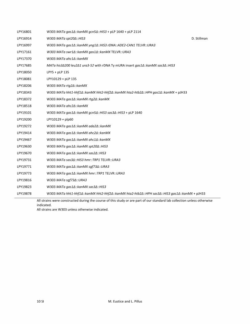
Table S1 Yeast strains used in this study
Strain Genotype Source
LPY5 (W303‐1a) MATa ade2‐1 can1‐100 his3‐11,15 leu2‐3,112 trp1‐1 ura3‐1 R. Rothstein
LPY1597 W303 MATa sas2Δ::TRP1
LPY2444 MATα his3Δ200 leu2Δ1 ura3‐52 with rDNA Ty mURA insert J.S. Smith
LPY2447 MATα his3Δ200 leu2Δ1 ura3‐52 with rDNA Ty mURA insert sir2Δ2::HIS3 J.S. Smith
LPY4196 LPY5 + pLP60
LPY4924 W303 MATa hmr::TRP1 TELVR::URA3
LPY5035 W303 MATa sir2Δ::HIS3 hmr::TRP1 TELVR::URA3
LPY5526 W303 MATa yng1Δ::HIS3 rDNA::ADE2‐CAN1 TELVR::URA3
LPY6285 W303 MATa rDNA::ADE2‐CAN1 TELVR::URA3 K. Runge
LPY6439 W303 MATa ada2Δ::kanMX R. Rothstein
LPY8242 W303 MATa gcn5Δ::HIS3
LPY8256 W303 MATa sas3Δ::HIS3
LPY9820 W303 MATa gas1Δ::kanMX rDNA::ADE2‐CAN1 TELVR::URA3
LPY10047 W303 MATa rDNA::ADE2‐CAN1 hmr::TRP1 gas2Δ::kanMX
LPY10051 W303 MATa rDNA::ADE2‐CAN1 hmr::TRP1 gas3Δ::kanMX
LPY10074 MATα his3Δ200 leu2Δ1 ura3‐52 with rDNA Ty mURA insert gas1Δ::kanMX
LPY10129 MATa ade2‐1 can1‐100 his3‐11,15 leu2‐3,112 trp1‐1 ura3‐1 gas1Δ::kanMX
LPY10266 W303 MATα rDNA::CAN:ADE2 + pLP1823
LPY10267 W303 MATα orc2‐1 rDNA::CAN:ADE2 + pLP1823
LPY10270 W303 MATα gas1Δ::kanMX orc2‐1 rDNA::CAN:ADE2 + pLP1823
LPY10271 W303 MATα gas1Δ::kanMX orc2‐1 rDNA::CAN:ADE2 + pLP1823
LPY11544 W303 MATa gas3Δ::kanMX rDNA::CAN:ADE2 hmr::TRP1
LPY12232 W303 MATa hht1‐hhf1Δ::kanMX hht2‐hhf2Δ::kanMX hta2‐htb2Δ::HPH + pJH33 M.M. Smith
LPY12247 LPY10129 + pLP135
LPY12251 LPY10129 + pLP2114
LPY12264 W303 MATa rDNA::CAN:ADE2 hmr::TRP1 gcn5Δ::NatMX
LPY12326 LPY10129 + pLP1951
LPY13102 W303 MATa rDNA::CAN:ADE2 hmr::TRP1 bgl2Δ::kanMX
LPY14355 W303 MATa rpd3Δ::kanMX + pLP60
LPY15695 W303 MATa gas1Δ::kanMX rpd3Δ::kanMX + pLP60 + pLP1823
LPY16039 W303 MATa sas3Δ::HIS3
LPY16104 W303 MATa swr1Δ::kanMX
LPY16432 W303 MATa hht1‐hhf1Δ::kanMX hht2‐hhf2Δ::kanMX hta2‐htb2Δ::HPH sas3Δ::HIS3 + pJH33
LPY16444 W303 MATa sas3Δ::HIS3 hmr::TRP1 TELVR::URA3
LPY16736
LPY16798
W303 MATa gas1Δ::kanMX gcn5Δ::HIS3 + pLP1640
W303 MATa gas1Δ::kanMX gcn5Δ::HIS3 + pLP 1640 + pLP 135
LPY16800 W303 MATa gas1Δ::kanMX gcn5Δ::HIS3 + pLP 1640 + pLP 1950
10 SI M. Eustice and L. Pillus
LPY16801 W303 MATa gas1Δ::kanMX gcn5Δ::HIS3 + pLP 1640 + pLP 2114
LPY16914 W303 MATa spt20Δ::HIS3 D. Stillman
LPY16997 W303 MATa gas1Δ::kanMX yng1Δ::HIS3 rDNA::ADE2‐CAN1 TELVR::URA3
LPY17161 W303 MATa swr1Δ::kanMX gas1Δ::kanMX TELVR::URA3
LPY17370 W303 MATa ahc1Δ::kanMX
LPY17685 MATa his3Δ200 leu2Δ1 ura3‐52 with rDNA Ty mURA insert gas1Δ::kanMX sas3Δ::HIS3
LPY18050 LPY5 + pLP 135
LPY18081 LPY10129 + pLP 135
LPY18206 W303 MATa rtg2Δ::kanMX
LPY18343 W303 MATa hht1‐hhf1Δ::kanMX hht2‐hhf2Δ::kanMX hta2‐htb2Δ::HPH gas1Δ::kanMX + pJH33
LPY18372 W303 MATa gas1Δ::kanMX rtg2Δ::kanMX
LPY18518 W303 MATa ahc2Δ::kanMX
LPY19101 W303 MATa gas1Δ::kanMX gcn5Δ::HIS3 sas3Δ::HIS3 + pLP 1640
LPY19200 LPY10129 + plp60
LPY19272 W303 MATa gas1Δ::kanMX ada2Δ::kanMX
LPY19414 W303 MATa gas1Δ::kanMX ahc2Δ::kanMX
LPY19467 W303 MATa gas1Δ::kanMX ahc1Δ::kanMX
LPY19630 W303 MATa gas1Δ::kanMX spt20Δ::HIS3
LPY19670 W303 MATa gas1Δ::kanMX sas2Δ::HIS3
LPY19731 W303 MATa sas3Δ::HIS3 hmr::TRP1 TELVR::URA3
LPY19771 W303 MATa gas1Δ::kanMX sgf73Δ::URA3
LPY19773 W303 MATa gas1Δ::kanMX hmr::TRP1 TELVR::URA3
LPY19816 W303 MATa sgf73Δ::URA3
LPY19823 W303 MATa gas1Δ::kanMX sas3Δ::HIS3
LPY19878 W303 MATa hht1‐hhf1Δ::kanMX hht2‐hhf2Δ::kanMX hta2‐htb2Δ::HPH sas3Δ::HIS3 gas1Δ::kanMX + pJH33
All strains were constructed during the course of this study or are part of our standard lab collection unless otherwise indicated. All strains are W303 unless otherwise indicated.
M. Eustice and L. Pillus 11 SI
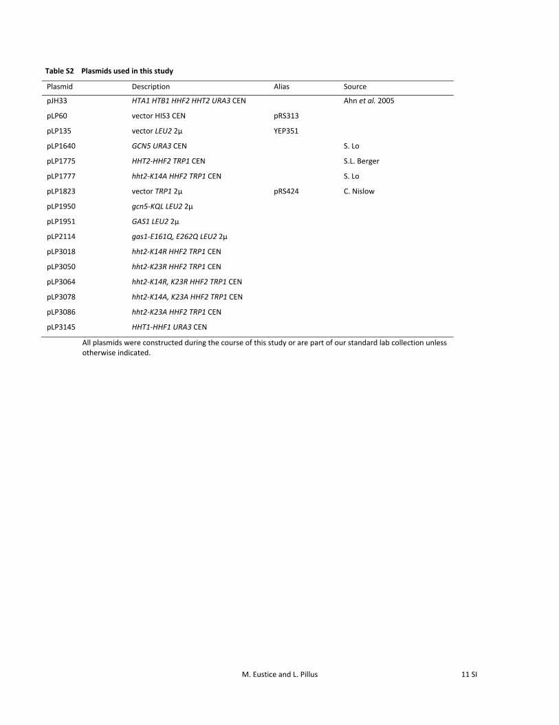
Table S2 Plasmids used in this study
All plasmids were constructed during the course of this study or are part of our standard lab collection unless otherwise indicated.
Plasmid Description Alias Source
pJH33 HTA1 HTB1 HHF2 HHT2 URA3 CEN Ahn et al. 2005
pLP60 vector HIS3 CEN pRS313
pLP135 vector LEU2 2μ YEP351
pLP1640 GCN5 URA3 CEN S. Lo
pLP1775 HHT2‐HHF2 TRP1 CEN S.L. Berger
pLP1777 hht2‐K14A HHF2 TRP1 CEN S. Lo
pLP1823 vector TRP1 2μ pRS424 C. Nislow
pLP1950 gcn5‐KQL LEU2 2μ
pLP1951 GAS1 LEU2 2μ
pLP2114 gas1‐E161Q, E262Q LEU2 2μ
pLP3018 hht2‐K14R HHF2 TRP1 CEN
pLP3050 hht2‐K23R HHF2 TRP1 CEN
pLP3064 hht2‐K14R, K23R HHF2 TRP1 CEN
pLP3078 hht2‐K14A, K23A HHF2 TRP1 CEN
pLP3086
pLP3145
hht2‐K23A HHF2 TRP1 CEN
HHT1‐HHF1 URA3 CEN
12 SI M. Eustice and L. Pillus
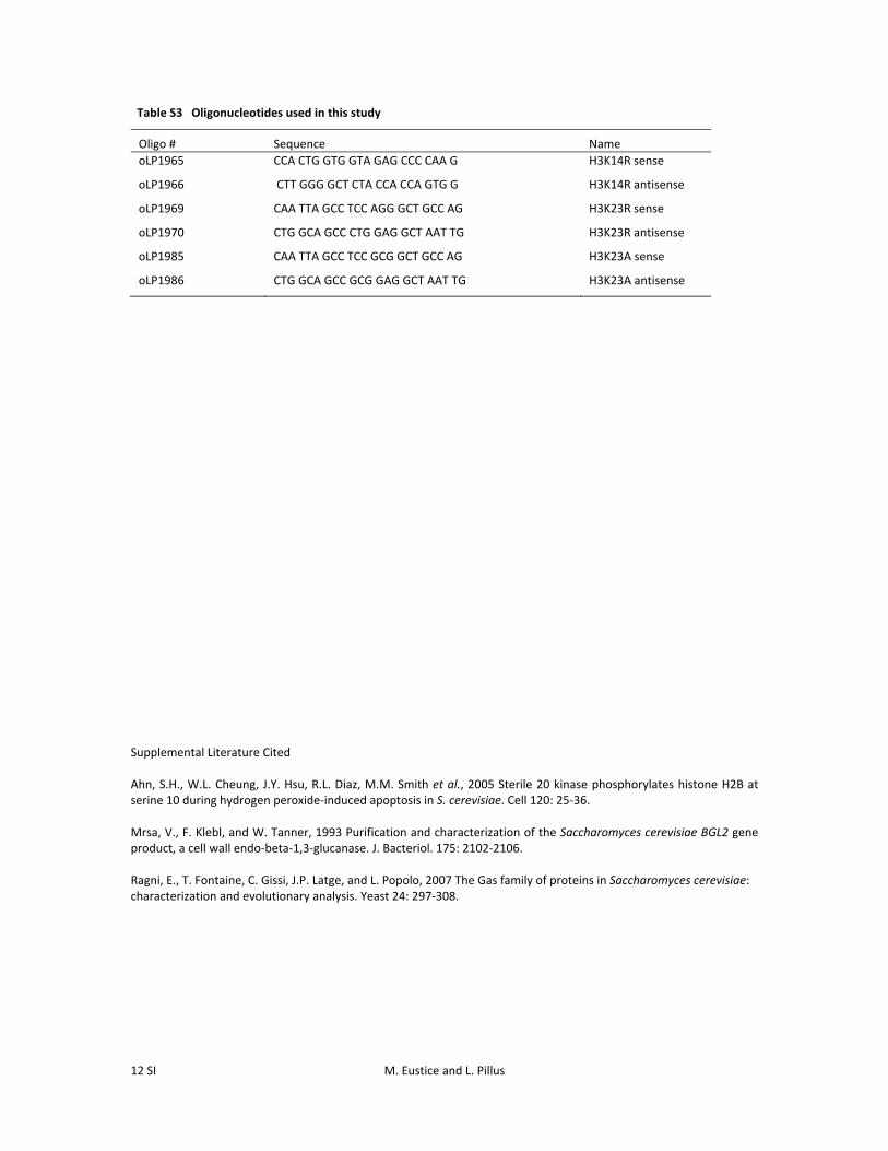
Table S3 Oligonucleotides used in this study
Oligo # Sequence Name
oLP1965 CCA CTG GTG GTA GAG CCC CAA G H3K14R sense
oLP1966 CTT GGG GCT CTA CCA CCA GTG G H3K14R antisense
oLP1969 CAA TTA GCC TCC AGG GCT GCC AG H3K23R sense
oLP1970 CTG GCA GCC CTG GAG GCT AAT TG H3K23R antisense
oLP1985 CAA TTA GCC TCC GCG GCT GCC AG H3K23A sense
oLP1986 CTG GCA GCC GCG GAG GCT AAT TG H3K23A antisense
Supplemental Literature Cited Ahn, S.H., W.L. Cheung, J.Y. Hsu, R.L. Diaz, M.M. Smith et al., 2005 Sterile 20 kinase phosphorylates histone H2B at serine 10 during hydrogen peroxide‐induced apoptosis in S. cerevisiae. Cell 120: 25‐36. Mrsa, V., F. Klebl, and W. Tanner, 1993 Purification and characterization of the Saccharomyces cerevisiae BGL2 gene product, a cell wall endo‐beta‐1,3‐glucanase. J. Bacteriol. 175: 2102‐2106. Ragni, E., T. Fontaine, C. Gissi, J.P. Latge, and L. Popolo, 2007 The Gas family of proteins in Saccharomyces cerevisiae: characterization and evolutionary analysis. Yeast 24: 297‐308.