Embed Size (px)
Citation preview

565
Review
www.expert-reviews.com ISSN 1478-9450© 2010 Expert Reviews Ltd10.1586/EPR.10.40
Parkinson’s disease (PD) is a very common highly debilitating neurodegenerative disease, which affects all human populations [1,2]. It is age related, generally becoming first apparent in the sixth decade of life, affecting approximately 2% of the population over 60 years of age, and rising to 5% over 80 years of age. The major symptoms include tremor, akinesia, postural instability and bradykinesia. Although many neuronal types are affected to some degree, the problems with body movement essentially result from a loss of dopaminergic neurons in the sub-stantia nigra pars compacta [3]. It is estimated
that approximately 70% of dopaminergic cells have to be lost before clinical symptoms are observed. This suggests that the number with diagnosed disease reflects only the tip of the ice-berg, and a high percentage with asymptomatic disease in the general population. At present, the only available treatments are those to amel-iorate the symptoms of the disease; there are no treatments targeting prevention or slowing its progression.
The major pathological feature associated with sporadic PD is the presence of Lewy bodies in surviving dopaminergic neurons [4]. There is
Philip A RobinsonSection of Ophthalmology and Neuroscience, Wellcome Trust Brenner Building, Leeds Institute for Molecular Medicine, St James’s University Hospital, Leeds LS9 7TF, UK Tel.: +44 113 343 8419 Fax: +44 113 343 8603 [email protected]
These are really exciting times in the field of Parkinson’s disease research. Although the etiology of sporadic disease still remains a mystery, many of the proteins associated with hereditary disease (5–10% of all disease) have now been identified. Only time will tell whether proteins associated with hereditary disease are involved in the development of sporadic disease. The most valuable proteomic studies performed to date are, and continue to be, those aimed at identifying endogenous binding partners, substrates, post-translational modifications and cellular pathways affected by these proteins. Similar to global proteomic approaches, even these approaches have surprisingly often been characterized by the production of very long lists of proteins. Consequently, the parallel development of more refined protein–protein interactions maps has aided the chance of identifying those protein complexes and/or cellular pathways, which, when disrupted, lead to the development of disease. The knowledge gained from these studies is essential, as targeting the activities of these proteins, or the pathways they operate in, currently offers the best opportunity to develop new therapeutic strategies to treat the disease. They may include agents to modulators of kinase activities (e.g., PINK1 and LRRK2), modulators of the activity of the ubiquitin–protein ligase, Parkin, proteostasis agents to block a-synuclein filament assembly and toxicity, or promote the refolding of mutant proteins, modulators of a-synuclein transfer between cells, reagents to regulate cargo dynamics along axonal microtubule networks, stimulators of autophagy and/or modulators of cellular stress pathways. The second major challenge will be to identify biomarkers to enable population screening to identify those with asymptomatic early-stage disease. Whether the analysis of blood or urine samples will yield such a marker, remains to be determined. Success or failure will be highly dependent on adopting strict standard operating procedures for the collection, processing and storage of samples, combined with the need for the identification of the most robust methods of prefractionation of samples to remove the most abundant proteins prior to proteomic screening.
Keywords: a-synuclein • DJ-1• HtrA2 • LRRK2 • Parkin • Parkinson’s disease • PINK1
Understanding the molecular basis of Parkinson’s disease, identification of biomarkers and routes to therapyExpert Rev. Proteomics 7(4), 565–578 (2010)
For reprint orders, please contact [email protected]

Expert Rev. Proteomics 7(4), (2010)566
Review Robinson
still controversy whether it is the physical presence of Lewy bod-ies, per se, which ultimately results in cell death, or whether fila-mentous or oligomeric structures of its major protein constituent, a-synuclein, are the toxic entities [5]. From proteomic analyses, the constituent make-up of Lewy bodies is very complex, with more than 250 proteins being described [6–8]. Loss of activity of such a large number of components from the cell as they integrate into the inclusion makes it difficult to determine which, if any, are associated with disease onset.
Although the majority of PD is apparently sporadic, 5–10% is inherited [1]. The genes associated with many of these inher-ited autosomal dominant and recessive forms of the disease have now been identified (Table 1), although there are also a few chromosomal loci where the nature of genes still need to be described (e.g., PARK3, 10, 11 and 12) [1,9]. The list of pro-teins includes kinases (PINK1 and LRRK2), proteins regulat-ing protein folding and disposal (Parkin, ATP13A2, HtrA2 and DJ-1), anti oxidants (DJ-1), and those whose function are prov-ing difficult to determine (a-synuclein). The deubiquitylating enzyme, UCH-L1, was also reported to be a disease-susceptibil-ity gene [10,11]. However, a subsequent genetic analysis brought this conclusion into question [12]. The protein is, therefore, not included in Table 1. Many different proteomic approaches have been used to identify binding partners and substrates [13]. The cellular processes that they may regulate are numerous (Table 1). Interestingly, recent genome-wide association studies on spo-radic PD patients have identified a few susceptibility loci, which include a-synuclein, LRRK2 and tau (plus two others) [14,15]. The former two are of obvious interest in relation to hereditary disease, but the latter is also interesting in light of its relationship to the development of Alzheimer’s disease. Combined with the knowledge that aggregated a-synuclein, in the form of Lewy bodies, is also observed in other neuronal cell types in different neurodegenerative diseases – the ‘a-synucleinopathies’ [16] – these data hint at a potential common mechanism of disease onset, at least for some of the neurodegenerative diseases [17,18].
We do not yet know what triggers the initiation of sporadic dis-ease, how the disease progresses or what drives the progression. However, recent studies demonstrating that (aggregating) a-synu-clein can be internalized by cells suggests a mechanism based on cell–cell transmission [19–22]. The neurons initially affected could depend purely on chance exposure to an environmental trigger, the response to which depends on the ability of the cells, at that time, to deal with the challenge. The response to this initiating event could then be determined by a combination of other complex genetic factors that regulate, for instance, response to toxic chemicals, or reflects a neuron’s underlying ability to deal with oxidative stress.
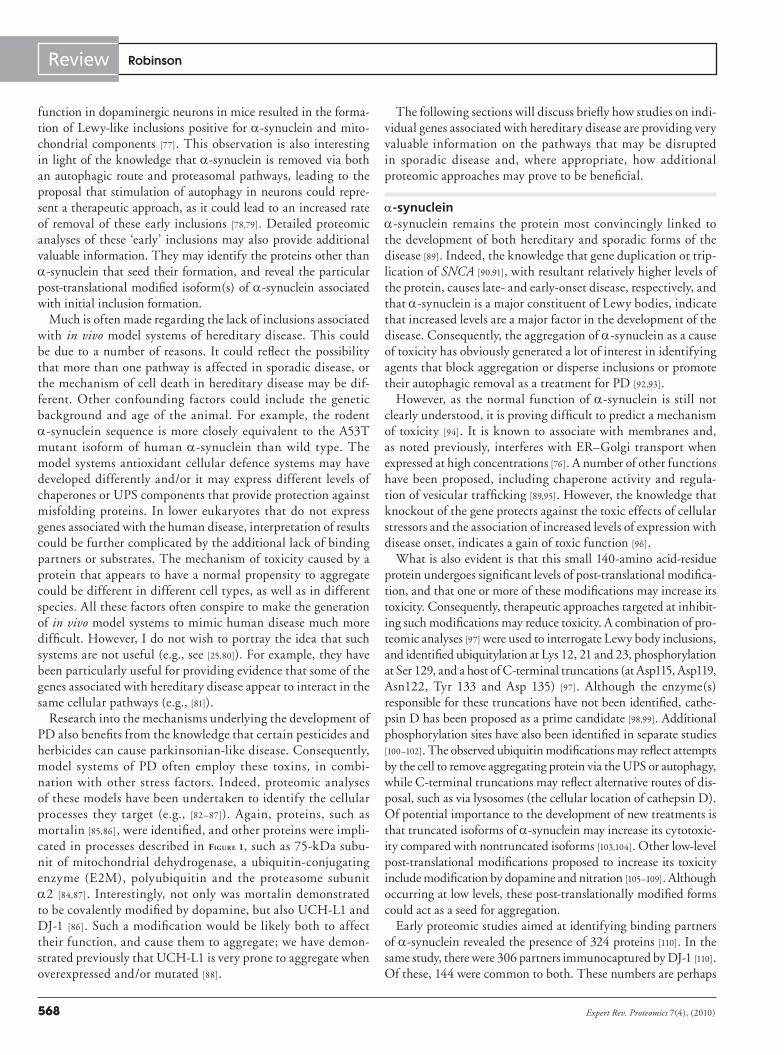
However, a general over-riding scheme to explain the develop-ment of sporadic disease appears to revolve around problems with mitochondrial dynamics, resulting in oxidative stress [23–33], calcium flux [34–36], and protein folding, transport and/or degradation [37–40] (Figure 1; see also [41]). The problems may be exacerbated by an age-related (or genetic) inability to deal efficiently with cellular stress caused by external agents and/or dopamine [42]. How mitochondrial activity and protein processing affect one another is the subject of intense research activity. For example, the ubiquitin–proteasome system (UPS) controls mitochondrial fission and fusion [43–45], and fission followed by selective fusion is required for the removal of defective mitochondria [46,47]. Similarly, increased output of reac-tive oxygen species (ROS) from mitochondria associated with the development of disease will produce protein damage, and potentially decreased ATP output from mitochondria, thereby affecting the efficiency of the UPS and chaperone activity. Furthermore, as ROS can also act as a signaling molecule, they may activate transcription of proteins to deal with this increased stress analogous to activa-tion of the unfolded-protein response by proteins misfolding in the endoplasmic reticulum (ER) or in mitochondria [48–51].
It is essential that the schematic shown in Figure 1 should be viewed in the context of a neuron. What is not clear is which arms of the model initially drive disease progression. Damage to mitochondria or decreased proteasomal activity [37] could lead to increased levels of ROS, and activation of ROS-regulated signaling pathways [50],
and localized oxidative damage to proteins in the vicinity of the mitochondria. The size and shape of a neuron requires that sub-cellular organelles, such as mitochondria and other cargos, such as proteins, have to be transported over relatively huge dis-tances along axons [52,53]. Synaptic regions of dopaminergic cells may be particularly sensitive, as they are rich in mitochondria, a-synuclein and dopamine, an inherently unstable chemical [54–56]. a-synuclein aggre-gation could lead to reduced proteasomal activity, interference with mitochondrial dynamics and calcium flux [35], eventu-ally setting off a chain of events that would require constant retrograde transport of both aggregating a-synuclein and also ‘damaged’ or depolarized mitochondria to the cell body for macroautophagy [52,57,58].
Table 1. Proteins associated with hereditary forms of Parkinson’s disease.
Protein Mitochondrial association
Cytoskeletal association
Reported activities
a-synuclein Y† Y Chaperone/vesicular trafficking
Parkin Y† Y Ubiquitin–protein ligase
PINK1 Y Y Kinase
DJ-1 Y Y Chaperone/antioxidant
LRRK2 Y† Y Kinase
ATP13A2 N N P-type ATPase
HTRA2 Y Y Protease
Proteins demonstrating a mitochondrial association (Y), may have a mitochondrial location if overexpressed, or as a result of cellular stress (Y†), indicated in the second column. Interaction with or modulation of the cytoskeleton or cytoskeletal proteins are indicated in the third column (Y). The reported activities of each protein are indicated in the fourth column. This does not preclude the assignment of additional functions in the future (e.g., for a-synuclein). ATP13A2 is a lysosomal protein. N: No; Y: Yes.
www.expert-reviews.com 567
ReviewProteomics & Parkinson’s disease
For efficient transport, microtubule tracks in neurons must be kept free. If transport is hindered, then the constant supply of cargos destined for the synapses could be inhibited, possibly resulting in a loss of synaptic cell–cell contact and a retraction of the axon.
A number of global proteomic approaches have been employed to identify disease-related changes in brain tissue or substantia nigra [59–61]. Major changes were consistent with the model described pre-viously, as they included increases in mitochondrial complex III and ATP synthase D chain (mitochondrial dysfunction) and L-type calcium channel d-subunit (calcium flux), and a decrease in levels of mortalin expression (protein folding) [59,60]. Interestingly, they also suggest modifications to the cellular cytoskeleton as decreased levels of the intermediate filament (NF) proteins, NF-L and NF-M [59], and an increase in profilin, an actin-binding protein, were also observed. We also recently observed increased interaction of cytoskeletal elements with mitochondria in a cellular model, which was created to overexpress a-synuclein [62]. In future screens, focus-ing specifically on changes in the expression profile of components of the cytoskeleton and/or changes to the interaction of proteins under study with components of the cytoskeleton in regions of neurons, such as synaptic extracts or axons, may prove to be par-ticularly informative, whether using in vitro or in vivo cell models.
The late presentation of sporadic disease presents its own unique problems when try-ing to develop animal models to mimic its development, as we are always presented with end-stage disease [63]. We do not know what initiates the disease process, why it is pro-gressive, whether there are complex genetic factors involved and/or if there are disease-promoting factors in the cellular microenvi-ronment. Furthermore, the animal models themselves may have a genetic background that causes a different response to environ-mental agents compared with humans, or lack interacting partners of suspect proteins. Some model systems (and particularly lower eukaryotes) may lack the orthologue of the genes associated with hereditary forms of human disease under study, such as a-synu-clein or their interaction partners (e.g., [64]). The criticism is then often raised regarding the relevance of models to sporadic disease. Any protein (whether wild type or mutant) prone to aggregate could be potentially toxic if expressed at high enough levels. In addi-tion, over expression of an ubiquitin–protein ligase or protein kinase may promote inter-action with, and subsequent modification of, low-affinity binding partners, which are not normal physiological substrates. Only rigor-ous follow-up studies including comparative proteomic profiling would provide evidence for involvement in disease development.
With some of the models based on hereditary forms of the dis-ease, phenotypes are observed [65–67]. The severity of the pheno-types are often enhanced when two or more genes associated with hereditary forms of the disease are overexpressed, knocked out and/or mutated [68–70], or additional stress factors, such as chemi-cal toxins, are included in the model systems (e.g., [71,72]). These models can also demonstrate the protective effect of some of the proteins associated with PD, counter acting the toxicity of another (e.g., Parkin and G2019S LRRK2, respectively) [73]. These model systems have often been interrogated using proteo mics technolo-gies to identify pathways that are affected [74,75]. Even if a model system, such as those based on genes associated with hereditary disease, does not accurately reproduce the phenotype of PD observed in sporadic disease, they can often provide valuable information. For example, a model based on a high-throughput screen to block the toxicity of a-synuclein in yeast demonstrated how a-synuclein may interfere with the ER-to-Golgi transport in humans when expressed at high levels [76].
In the long term, whether Lewy bodies, per se, are the toxic entity in PD, or not, eventually, the model must be able to explain their presence. In this regard, targeted loss of 26S proteasomal
Figure 1. Etiology of Parkinson’s disease. Central to the onset of Parkinson’s disease are decreased levels of UPS activity, altered mitochondrial dynamics and output of ROS. Problems with mitochondrial dynamics as a result of a cellular stress, for example, may result in an increased output of ROS, leading to increased protein damage if not dealt with by antioxidant defences, such as SOD or glutathione. In time, increased protein damage may result in inclusion/Lewy body formation. Their physical presence may, ultimately, lead to cell death. Prior attempts by the cell to remove aggregating protein may involve both autophagy and the UPS. Released aggregated a-synuclein may be taken up by adjoining cells, or engulfed by cells of the immune system. Initial problems with mitochondria may result from problems of mitochondrial fission and fusion resulting from lower levels of UPS activity (which may be age related or disease related). Defective, depolarized mitochondria may be removed from cells via mitophagy promoted by Parkin (and PINK1/DJ-1), and be dependent on binding to microtubules and transport along axons to the cell body. The stress response ensuing from any of these activities may activate kinases, such as LRRK2 or PINK1. GSH: Glutathione; ROS: Reactive oxygen species; SOD: Superoxide dismutase; UPS: Ubiquitin–proteasome system.
Inclusions(Aggresomes)
Proteindamage
(Localized)
(Syn)n+
Lewybodies
Glialcells
Endocytosis
Chemicals,parasites
Autophagy
ATP13A2 Cell death
Fission
Fusion
Parkin/PINK1/DJ1
SODGSH
ROS(H2O2, O2
-•,OH•, NO•)
(Oxidativestress)
UPS proteinprocessing
Environmentalstress
Mitochondrialactivity/dynamics
Expert Rev. Proteomics 7(4), (2010)568
Review Robinson
function in dopaminergic neurons in mice resulted in the forma-tion of Lewy-like inclusions positive for a-synuclein and mito-chondrial components [77]. This observation is also interesting in light of the knowledge that a-synuclein is removed via both an autophagic route and proteasomal pathways, leading to the proposal that stimulation of autophagy in neurons could repre-sent a therapeutic approach, as it could lead to an increased rate of removal of these early inclusions [78,79]. Detailed proteomic analyses of these ‘early’ inclusions may also provide additional valuable information. They may identify the proteins other than a-synuclein that seed their formation, and reveal the particular post-translational modified isoform(s) of a-synuclein associated with initial inclusion formation.
Much is often made regarding the lack of inclusions associated with in vivo model systems of hereditary disease. This could be due to a number of reasons. It could reflect the possibility that more than one pathway is affected in sporadic disease, or the mechanism of cell death in hereditary disease may be dif-ferent. Other confounding factors could include the genetic background and age of the animal. For example, the rodent a-synuclein sequence is more closely equivalent to the A53T mutant isoform of human a-synuclein than wild type. The model systems antioxidant cellular defence systems may have developed differently and/or it may express different levels of chaperones or UPS components that provide protection against misfolding proteins. In lower eukaryotes that do not express genes associated with the human disease, interpretation of results could be further complicated by the additional lack of binding partners or substrates. The mechanism of toxicity caused by a protein that appears to have a normal propensity to aggregate could be different in different cell types, as well as in different species. All these factors often conspire to make the generation of in vivo model systems to mimic human disease much more difficult. However, I do not wish to portray the idea that such systems are not useful (e.g., see [25,80]). For example, they have been particularly useful for providing evidence that some of the genes associated with hereditary disease appear to interact in the same cellular pathways (e.g., [81]).
Research into the mechanisms underlying the development of PD also benefits from the knowledge that certain pesticides and herbicides can cause parkinsonian-like disease. Consequently, model systems of PD often employ these toxins, in combi-nation with other stress factors. Indeed, proteomic analyses of these models have been undertaken to identify the cellular processes they target (e.g., [82–87]). Again, proteins, such as mortalin [85,86], were identified, and other proteins were impli-cated in processes described in Figure 1, such as 75-kDa subu-nit of mitochondrial dehydrogenase, a ubiquitin-conjugating enzyme (E2M), polyubiquitin and the proteasome subunit a2 [84,87]. Interestingly, not only was mortalin demonstrated to be covalently modified by dopamine, but also UCH-L1 and DJ-1 [86]. Such a modification would be likely both to affect their function, and cause them to aggregate; we have demon-strated previously that UCH-L1 is very prone to aggregate when overexpressed and/or mutated [88].
The following sections will discuss briefly how studies on indi-vidual genes associated with hereditary disease are providing very valuable information on the pathways that may be disrupted in sporadic disease and, where appropriate, how additional proteomic approaches may prove to be beneficial.
a-synuclein a-synuclein remains the protein most convincingly linked to the development of both hereditary and sporadic forms of the disease [89]. Indeed, the knowledge that gene duplication or trip-lication of SNCA [90,91], with resultant relatively higher levels of the protein, causes late- and early-onset disease, respectively, and that a-synuclein is a major constituent of Lewy bodies, indicate that increased levels are a major factor in the development of the disease. Consequently, the aggregation of a-synuclein as a cause of toxicity has obviously generated a lot of interest in identifying agents that block aggregation or disperse inclusions or promote their autophagic removal as a treatment for PD [92,93].
However, as the normal function of a-synuclein is still not clearly understood, it is proving difficult to predict a mechanism of toxicity [94]. It is known to associate with membranes and, as noted previously, interferes with ER–Golgi transport when expressed at high concentrations [76]. A number of other functions have been proposed, including chaperone activity and regula-tion of vesicular trafficking [89,95]. However, the knowledge that knockout of the gene protects against the toxic effects of cellular stressors and the association of increased levels of expression with disease onset, indicates a gain of toxic function [96].
What is also evident is that this small 140-amino acid-residue protein undergoes significant levels of post-translational modifica-tion, and that one or more of these modifications may increase its toxicity. Consequently, therapeutic approaches targeted at inhibit-ing such modifications may reduce toxicity. A combination of pro-teomic analyses [97] were used to interrogate Lewy body inclusions, and identified ubiquitylation at Lys 12, 21 and 23, phosphorylation at Ser 129, and a host of C-terminal truncations (at Asp115, Asp119, Asn122, Tyr 133 and Asp 135) [97]. Although the enzyme(s) responsible for these truncations have not been identified, cathe-psin D has been proposed as a prime candidate [98,99]. Additional phosphorylation sites have also been identified in separate studies [100–102]. The observed ubiquitin modifications may reflect attempts by the cell to remove aggregating protein via the UPS or autophagy, while C-terminal truncations may reflect alternative routes of dis-posal, such as via lysosomes (the cellular location of cathepsin D). Of potential importance to the development of new treatments is that truncated isoforms of a-synuclein may increase its cytotoxic-ity compared with non truncated isoforms [103,104]. Other low-level post-translational modifications proposed to increase its toxicity include modification by dopamine and nitration [105–109]. Although occurring at low levels, these post-translationally modified forms could act as a seed for aggregation.
Early proteomic studies aimed at identifying binding partners of a-synuclein revealed the presence of 324 proteins [110]. In the same study, there were 306 partners immunocaptured by DJ-1 [110]. Of these, 144 were common to both. These numbers are perhaps

www.expert-reviews.com 569
ReviewProteomics & Parkinson’s disease
greater than was expected, but could reflect the capture of a number of large multiprotein complexes and/or reflect the knowledge that both proteins display chaperone-like activities [95,111]. A separate study identified more than 250 proteins [112]. Proteins included were those associated with ribosomes, RNA binding and protein synthesis, the cytoskeleton and chaperones. Interestingly, the same group performed a study 2 years previously, where they looked at the expression profiles of mitochondrial proteins extracted from the substantia nigra of brains of mice treated with 1-methyl-4-phenyl-1,2,3,6-tetrahydropyridine (MPTP), a potent mitochon-drial toxin that produces parkinsonian-like disease in humans [113]. They identified DJ-1, and noted its colocalization with a-synuclein in substantia nigra and to cytoplasmic inclusions.
The post-translational modification of a-synuclein at ser129 has been the most intensely studied to date, owing to its relatively high abundance in Lewy bodies. This phosphorylation modifica-tion appears to be cell specific, as a-synuclein is not phosphory-lated in platelets in PD [114]. Its presence in Lewy bodies has led to the suggestion that this modification may promote aggregation. By contrast, a recent study indicates that phosphorylation at S87 may protect against oligomerization [102]. As such, the kinase that promotes this activity could also represent a therapeutic target.
Although the functional consequence of ser129 phosphoryla-tion is not known, we, and others, have found that this is post-translational modification occurs at low levels in the absence of large protein aggregates [35,115]. Pull-down assays, followed by mass spectrometry analysis using phosphorylated and nonphos-phorylated peptides of this region of a-synuclein, suggested that the former interacts with mitochondrial electron transport chain components, but the unmodified peptide did not [115]. Interactants of the phosphorylated peptide also included cytoskeletal proteins and vesicular trafficking proteins. It is interesting from nonbiased proteomic analyses of a previous study, and our recent studies on the interaction of a-synuclein with mitochondria [62], that both phosphorylation of a-synuclein and overexpression of a-synuclein identified not only mitochondrial components, but also cyto-skeletal components [116]. This supports the proposal put forward in a previous article in this journal, by Fasano and Lopiano [117], that the inter action of a-synuclein with cyto skeletal components may be an important modulator of its toxicity. It could also sup-port the idea that a-synuclein could disrupt transport along axons or neurites, which is dependent on cytoskeletal interactions, as has been proposed for other neurodegenerative diseases [52]. We also noted that overexpression of a-synuclein caused a reduction in the levels of two components of OXPHOS complex 1, and a small but significant reduction in OXPHOS 1 activity [118]. How many oth-ers of the 46 component OXPHOS complex are affected by high levels of a-synuclein was not determined but could be the focus of future studies. Not only could changes to the levels of individual units be monitored, but also their post-translational modification status, using approaches such as stable isotope labeling by amino acids in cell culture combined with immunocapture.
The observation that a-synuclein is toxic in lower eukaryo-tes [76] has led to their use for various high-throughput screens and proteomic analyses; the assumption is made that the method
of toxicity is the same as in humans. Furthermore, as the A53T mutant in humans is reported to aggregate more readily, this iso-form is often used in place of wild-type a-synuclein to study toxic function. However, we have recently observed that the wild-type isoform appears to produce more changes to the mitochondrial proteome than the A53T mutant isoform, despite both being phosphorylated [62]. This may indicate that, although high con-centrations of a-synuclein affects mitochondrial function, this may not be the cause of its cytotoxicity. The gain of toxic function may be multifactorial, and properties, such as its interference with ER–Golgi transport, are more likely to be involved [76].
In vitro, we found that mammalian cells can proliferate when a-synuclein is expressed at relatively high levels. Toxicity is not observed until cells are cultured under conditions that produce further cell stress, or expressed at relatively high levels in eukaryo-tic cells not normally expressing the protein [76], or perhaps lack sufficient levels of the chaperones normally preventing such aggre-gation. The choice of cell type may be very important as, similar to the group of Pamela McLean [119], we found that the H4 neuro-glioma cell line produces sodium dodecyl sulfate (SDS)-insoluble aggregates when a-synuclein is overexpressed, but not in HEK293 or SH-SY5Y cells, for example.
Parkin/PINK1/DJ-1 (& ATP13A2)Loss of Parkin activity, associated with hereditary disease, can occur through loss of enzymic activity or misfolding leading to decreased solubility [120]. It appears that phosphorylation, both by casein kinase I and cyclin-dependent kinase 5 (cdk5), can also result in loss of activity as the consequence of reducing its solubility, and inhibition of these kinase activities can partially block aggregation of disease-related mutants [121].
A number of key observations over the last few years have aided our understanding of the mechanism of action of Parkin based on the establishment of links between it, PINK1 and DJ-1. Perhaps the first key observation for a role of Parkin in mitochondrial function came from a Drosophila model system, demonstrating morphologi-cal defects in Parkin-null animals [27]. This was followed up by the observation that overexpression of Parkin in Drosophila could rescue the phenotypic defects caused by PINK1 mutants [81,122]. More recently, Parkin was demonstrated to target depolarized mitochondria for autophagic removal (mitophagy) from cells [123], and PINK1 is required for the regulation of mitophagy [124,125]. Subsequent studies have indicated roles for Beclin 1, p62, mitofusin and VDAC1 in Parkin/PINK1-mediated mitophagy [47,126,127].
Parkin expression and activity is not restricted to one type of cell. It is widely distributed. Why it affects dopaminergic cells in particular in hereditary PD is not understood. It may be that these cells are normally subject to higher levels of cellular stress, owing to the presence of dopamine. However, Parkin is expressed in other cell types, including HEK293 cells [123]. Our nonbiased proteomic screen of protein extracts of HEK293 cells induced to overexpress Parkin, indicated interaction with a number of mitochondrial components, suggesting an interaction with mito-chondria in cells at low levels, even in the absence of cellular stress [128]. This interaction increased with the inhibition of

Expert Rev. Proteomics 7(4), (2010)570
Review Robinson
proteasomal activity [128]. Furthermore, potential interaction with components (ANT2/ANT3) of the mitochondrial per meable transition pore suggests another potential target for Parkin in mitochondria [128]. Another nonbiased screen also identified pro-teins associated with energy metabolism, stress-related chaperones and additional components of the UPS [129].
Parkin, PINK1 and DJ-1 have recently been reported to form a ubiquitin–protein ligase complex (the PPD complex), which pro-motes the degradation of misfolded proteins [130]. However, rather surprisingly, a triple knockout of these three proteins in mice does not appear to generate a visible phenotype [131]. Perhaps, this reflects the proposal that phenotypic defects will not be observed until cells are subjected to additional stress.
Combining this information with the knowledge that Parkin promotes removal of misfolding DJ-1 via autophagy, defects in autophagy as a result of loss of ATP13A2 [132] results in an inherited form of PD, and defects with mitochondrial activity are observed in patients provides further indirect evidence for problems with the removal of defective mitochondria via axonal transport in sporadic disease.
DJ-1 is present in cells in relatively high concentrations. Its pri-mary role appears to be to act as an antioxidant [133,134], protecting neurons against oxidative stress and preventing apoptosis [135]. It has also been reported to interact with OXPHOS complex 1, maintaining its activity [136], and performs chaperone activity [111], which is regulated by the cochaperone BAG-1; loss of chaperone activity as a result of the L166P hereditary mutation was restored by BAG-1 [111]. DJ-1 also appears to protect cells against hypoxia-induced cell death [137]. Interestingly, DJ-1 also appears to bind to tyrosine hydroxylase and 4-dihydroxy-l-phenylalanine decarbox-ylase (DDC), enzymes involved in the synthesis of dopamine [138]. Similar to Parkin, it appears that a greater percentage of DJ-1 associates with the mitochondria when cells are exposed to oxida-tive stress, a translocation that was independent of the mutational status of the protein, and which provided cytoprotection [139]. Proteomic analyses have also demonstrated that DJ-1 is phospho-rylated, and that this modification can be mediated by p53 [140]. Studies of model systems also indicate it helps to maintain the integrity of lysosomes, in addition to mitochondria [141]. What is also interesting is that the activity of DJ-1 appears to be regu-lated by ERK1/2 signaling, similar to LRRK2 [141–145]. DJ-1 can be processed by caspase 6. However, whereas D149A mutation protects DJ-1 from caspase 6 cleavage, the L166P mutation does not, perhaps suggesting different mechanisms of toxicity [146]. As noted, DJ-1 interacts with many of the proteins also bound by a-synuclein [110], potentially suggesting a link between the PPD complex and a-synuclein.
LRRK2Similar to a-synuclein, there is a strong case for the involvement of LRRK2 (or its substrate[s]) in the development of both hereditary and sporadic forms of the disease. Hereditary forms of the disease are autosomal dominant, and display typical features of PD. It is a large 2527-amino acid-residue protein that is expressed in many different neuronal types [147,148], and is proposed to regulate striatal
dopamine transmission [149]. The stability of LRRK2 is regulated by the UPS [150,151]. Dimerization leads to activation of its kinase activity and autophosphorylation [152–154]. It is characterized by a number of recognizable domains, which include kinase and Ras of complex (ROC; GTPase) domains [155]. Mutations of these domains are often observed in hereditary forms of the disease, indicating their importance to protein activity [156]. Furthermore, the ROC domain is reported to regulate its interaction with the microtubule network [157]. This interaction has been reported to result in phosphorylation of b-tubulin, with mass spectrometry analysis, demonstrating Thr107 as the target residue [158]. These data, together with others, again suggest interaction and modula-tion of the cytoskeletal network by a molecule associated with the onset of PD [158,159].
The focus of research on LRRK2, at present, is to identify sub-strates of the protein (e.g., [160–168]). Of interest, one of its substrates was reported to be a-synuclein [169]. LRRK2 pull-down and mass spectrometry analysis of the interactants identified potential inter-actants that were grouped into the cellular pathways associated with the cytoskeleton, chaperones and kinases [170]. Whether these are physiological substrates involved in disease progression is not yet known. LRRK2 kinase activity, acting through ERK signal-ing pathways, has also been associated with a modest increase in transcription from SNCA, the gene encoding a-synuclein [143,144]. With particular relevance to PD, it has also been reported to mod-ulate vesicle endocytosis [168], and a role for LRRK2 in autophagy has recently been proposed [171,172].
The knowledge that the common G2019S mutation results in an activation of the kinase activity [155], and is a cause of hereditary PD with a similar pathology to that of sporadic disease, makes this a prime therapeutic target [173]. The large size of the molecule probably precludes the use of approaches, such as yeast 2 hybrid. A greater level of success is likely to be achieved by employ-ing proteomic analyses of affinity-captured interactants from cell lysates for mass spectrometry identification. Comparative phosphoproteomic analyses of animal model systems designed to express mutant isoforms of the protein at endogenous levels with wild-type expression, may aid the identification of the key substrates intimately involved in the disease process. Such studies may need to be performed in the presence of agents that promote cellular stress if the hypothesis that additional challenges, which may be age related, are required before disease onset occurs.
Omi/HtrA2HtrA2 also appears to be a susceptibility gene for PD [30]. The studies on HtrA2 are limited at present. However, a proteomic approach, COFRADIC™, has been employed to identify potential substrates of HtrA2 [174,175]. Amongst those identified, cytoskeletal proteins, again, appeared to be particularly prominent.
Identification of early biomarkers of PDSimilar to other neurodegenerative diseases [176], one of the great challenges of PD research is to identify biomarkers of early-stage disease [177–179]. Their identification would permit a rapid screen-ing procedure to be developed for the general population, and also

www.expert-reviews.com 571
ReviewProteomics & Parkinson’s disease
to monitor disease progression (see review [180]). For large-scale screening procedures, the use of an easily accessible body fluid for potential biomarkers should be pursued actively. Biomarkers may include peptide fragments, protein degradation products and post-translational modifications. However, these markers are likely to be present only in very low concentrations in body fluids, such as blood or urine [53,181–184]. This obviously sets a challenge, as the most abundant proteins are likely to mask changes in much rarer abundance peptides/proteins.
One of the major criteria for success in these types of approaches is that rigorous standard operating procedures must be put in place for human sample collection [185,186]. As yet, no systematic large-scale screen of serum or urine has been reported for PD. The reason for this partly reflects the fact that proteomic procedures needed for a successful outcome are still being refined to enable the analysis of body fluids with such a large dynamic range of protein expres-sion levels, combined with the need for the collection of samples suitable for proteomic analyses [187,188]. It probably also reflects resistance in the PD research community that there is a persuasive argument that a disease affecting specific regions of the brain will lead to the presentation of a biomarker in blood or urine [189]. If changes do occur, they are likely to be subtle, and present at very low levels. However, changes in the blood have been noted previ-ously [190]. Similarly, differences in metabolomic profiles have been reported [191].
Proteomic analysis of body fluids collected over a period of time from animal models of PD may also aid the identification of potential biomarkers, which can then be evaluated in humans for their suitability [192–194]. Of course, any response observed in animal models may be very different to that in humans.
By contrast, studies have been undertaken to look for markers in cerebrospinal fluid (CSF) in various neurodegenerative disor-ders, including PD [195–202]. Various proteins have been proposed, such as a-synuclein and autotoxin [197,200]. However, a-synuclein does not appear to be a reliable marker [200]. Post-translationally modified proteins have also been detected, such as oxidatively modified superoxide dismutase 1. In addition, proteins (e.g., apol-ipoprotein e) associated with other neurodegenerative diseases, such as Alzheimer’s disease [197,202], have also been suggested as potential biomarkers. Although, CSF sampling is not without its problems, and would not be a preferred option for routine large population screening, analysis of CSF could be particularly useful for following disease progression in individuals and to monitor changes resulting from treatment with new therapeutics.
Expert commentaryTo date, proteomic applications have demonstrated mixed success to the wider PD research community in efforts to aid the under-standing of the etiology of PD. While they have been particularly useful for monitoring changes to key proteins in disease develop-ment, such as a-synuclein, and identifying potential components of inclusions, such as Lewy bodies, the more global analyses have had limited value. Their major contribution, to date, has probably been to reinforce the proposal that problems associated with han-dling of protein disposal and mitochondrial dynamics are central
to the disease process, and that interactions of proteins intimately associated with the development of PD with the cytoskeleton are often observed. In my opinion, the more global cellular proteomic approaches should now be replaced by studies focusing on deter-mining how genes associated with hereditary disease affects or interact with these cellular activities/structures.
What is always a problem with proteomic approaches for study-ing PD is the decision as to which model systems should be used for the analysis. Unfortunately, there is no easy answer. In vitro models represent the more controlled environment systems for proteomic analysis, but may lack the cellular microenvironment critical to disease development, and the cell type chosen may not express the particular protection systems expressed in cells such as dopaminergic cells. Are primary cells a better model than estab-lished cell lines? Similar to established cell lines, primary cells must adapt to grow in culture conditions, which will be reflected by a continuing change to the cellular proteome. The longer they are in culture, the more the proteome is likely to change. In the long term, embryonic stem cells that are differentiated into dopaminergic-like cells probably offer the best in vitro model system for routine manipulation and proteomic analysis. The use of inducible pluripotent stem cells derived from hereditary and sporadic patients may be particularly useful to look for evidence of subtle background genetic influences on disease development.
By contrast, animal models offer a microenvironment more akin to that of humans, but mice are not humans, and worms do not have complex brain structures. However, they can be very useful for answering specific questions, such as do changes to the expression of proteins under study specifically affect dopamin-ergic neurons or cause some of the phenotypic changes observed in PD? Care must always be taken with comparisons between mouse studies, as different inbred strains of mice may be more or less susceptible to different environmental toxins, based on com-parative minor differences in their own genomes, and expression, for example, of different antioxidant proteins. Combining these limitations with the potential absence of other key environmen-tal factors make the creation of an accurate model of disease difficult to develop. Consequently, the model used to address a particular question always needs to be considered carefully. The analysis of human tissue may be useful, but also has limitations. The main criticism is that the cellular makeup of the tissue used for analysis is likely to be different between diseased and non-diseased tissue. Consequently, the identification of differences in protein expression in a particular cell type may be masked.
Five-year viewThe development of a treatment to halt or slow the progression of PD remains a major goal. Therapeutic agents may include modula-tors of kinase activities [203] or ubiquitin–protein ligases, proteo-stasis agents to block a-synuclein filament assembly [92,93,204,205], reagents to regulate cargo dynamics along axonal microtubule networks [206,207], or modulators of cellular stress pathways [208]. Proteomic approaches have much to offer in this regard, as they can be used to identify interactants and cellular pathways affected. To date, the study of the function and interactions of proteins

Expert Rev. Proteomics 7(4), (2010)572
Review Robinson
associated with hereditary disease has suggested underlying prob-lems with the activities or interactions with mitochondria, the cytoskeleton and protein processing. These may relate to activities associated with changing expression patterns of proteins associated with hereditary disease, such as LRRK2 or PINK1 (phosphopro-teome screens) or Parkin (immunocapture and mass spectrom-etry analysis). Proteomic approaches should now focus on the downstream effects of these proteins, and mirror the findings to those observed in human disease. An excellent example will be to employ proteomic approaches to aid the identification of key target(s) of Parkin on depolarized mitochondria, and to under-stand the mechanism of how this interaction promotes mitophagy, and how the activities of PINK1 and DJ-1 regulate this activity [125]. Similarly, the improving sensitivity of proteomic approaches will aid PD research to look at more subtle changes in the com-position and post-translational modification status of multipro-tein complexes implicated in the development of PD, such as the OXPHOS 1 multiprotein complex of mitochondria [118], either in animal models or human tissue.
As a group interested in the application of proteomic tech-nologies to study the molecular basis of PD, we have found the development of software packages that rapidly convert lists of
proteins generated by mass spectrometry analysis into cellular pathways and interaction networks invaluable. As these develop in sophistication, it is likely that changes in model systems of PD will be more rapidly interpreted to identify key targets for therapeutic development.
Over the next few years, it is likely that serum-depletion meth-ods, combined with the latest advances in mass spectrometry technologies, will offer the best-ever opportunity to look for dis-ease-associated biomarkers in blood or urine. However, to avoid problems of the past, it is imperative that improvements in sam-ple handling methodologies are put in place, and that nationally agreed rigorous standard operating procedures are employed for sample collection, processing and storage.
Financial & competing interests disclosureThe author has no relevant affiliations or financial involvement with any organization or entity with a financial interest in or financial conflict with the subject matter or materials discussed in the manuscript. This includes employment, consultancies, honoraria, stock ownership or options, expert testimony, grants or patents received or pending, or royalties.
No writing assistance was utilized in the production of this manuscript.
Key issues
• Many of the genes associated with hereditary forms of Parkinson’s disease have been identified. They appear to function in protein processing and/or mitochondrial activity.
• In vitro and in vivo model systems have been developed based on the overexpression or knockout of genes associated with hereditary genes, allowing comparative proteomic analyses. Interactions with the cytoskeleton appear to be a common feature.
• Improved methods and refinement of existing bioinformatic analyses are needed to convert the lists of proteins produced from the analysis of many model systems into affected pathways to enable therapeutic targets to be identified more rapidly.
• Increased a-synuclein expression is implicated both in hereditary and sporadic Parkinson’s disease. Proteomic analysis has identified a series of post-translational modifications in inclusions. The aim will be to identify the enzyme(s) catalyzing the post-translational modifications, as they will represent therapeutic targets.
• More than 250 potential interactants of a-synuclein have potentially been identified. The interactions that promote the seeding of inclusions need to be identified. The analysis of experimentally derived ‘early’ Lewy-like inclusions in animal models may aid this aim.
• Parkin, PINK1 and DJ-1 operate in the same pathway; they can interact to create an ubiquitin–protein ligase complex. Substrates will need to be identified in stressed cells.
• Parkin binds to depolarized mitochondria, promoting mitophagy; PINK1 stabilizes this interaction. Proteomic approaches should aid the identification of interacting mitochondrial proteins or complexes.
• (Phospho)proteomic analyses of cellular models of LRRK2 and PINK1 should aid the identification of key substrates, as well as the effects caused by modulating these post-translational modifications.
ReferencesPapers of special note have been highlighted as:• of interest•• of considerable interest
1 Thomas B, Beal MF. Parkinson’s disease. Hum. Mol. Genet. 16(R2), R183–R194 (2007).
2 Cookson MR. The biochemistry of Parkinson’s disease. Annu. Rev. Biochem. 74, 29–52 (2005).
3 Braak H, Ghebremedhin E, Rub U, Bratzke H, Del Tredici K. Stages in the development of Parkinson’s disease-related pathology. Cell Tissue Res. 318(1), 121–134 (2004).
4 Shults CW. Lewy bodies. Proc. Natl Acad. Sci. USA 103(6), 1661–1668 (2006).
5 Kramer ML, Schulz-Schaeffer WJ. Presynaptic {a}-synuclein aggregates, not Lewy bodies, cause neurodegeneration in dementia with Lewy bodies. J. Neurosci. 27(6), 1405–1410 (2007).
6 Leverenz JB, Umar I, Wang Q et al. Proteomic identification of novel proteins in cortical Lewy bodies. Brain Pathol. 17(2), 139–145 (2007).
7 Xia Q, Liao L, Cheng D et al. Proteomic identification of novel proteins associated
with Lewy bodies. Front Biosci. 13, 3850–3856 (2008).
8 Wakabayashi K, Tanji K, Mori F, Takahashi H. The Lewy body in Parkinson’s disease: Molecules implicated in the formation and degradation of a-synuclein aggregates. Neuropathology 27(5), 494–506 (2007).
9 Yang YX, Wood NW, Latchman DS. Molecular basis of Parkinson’s disease. Neuroreport 20(2), 150–156 (2009).
10 Leroy E, Boyer R, Auburger G et al. The ubiquitin pathway in Parkinson’s disease. Nature 395(6701), 451–452 (1998).
www.expert-reviews.com 573
ReviewProteomics & Parkinson’s disease
11 Maraganore DM, Lesnick TG, Elbaz A et al. UCHL1 is a Parkinson’s disease susceptibility gene. Ann. Neurol. 55(6), 899–899 (2004).
12 Healy DG, Abou-Sleiman PM, Casas JP et al. UCHL-1 is not a Parkinson’s disease susceptibility gene. Ann. Neurol. 59(4), 627–633 (2006).
13 Licker V, Kövari E, Hochstrasser DF, Burkhard PR. Proteomics in human Parkinson’s disease research. J. Proteomics, 73(1), 10–29 (2009).
14 Satake W, Nakabayashi Y, Mizuta I et al. Genome-wide association study identifies common variants at four loci as genetic risk factors for Parkinson’s disease. Nat. Genet. 41(12), 1303–1307 (2009).
15 Simon-Sanchez J, Schulte C, Bras JM et al. Genome-wide association study reveals genetic risk underlying Parkinson’s disease. Nat. Genet. 41(12), 1308–1312 (2009).
16 Spillantini MG, Goedert M. The a-synucleinopathies: Parkinson’s disease, dementia with Lewy bodies, and multiple system atrophy. Ann. NY Acad. Sci. 920, 16–27 (2000).
17 Taymans J-M, Cookson MR. Mechanisms in dominant parkinsonism: The toxic triangle of LRRK2, a-synuclein, and tau. Bioessays 32(3), 227–235 (2010).
18 Gallardo G, Schluter OM, Sudhof TC. A molecular pathway of neurodegeneration linking a-synuclein to ApoE and A b peptides. Nat Neurosci, 11(3), 301–308 (2008).
19 Desplats P, Lee H-J, Bae E-J et al. Inclusion formation and neuronal cell death through neuron-to-neuron transmission of a-synuclein. Proc. Natl Acad. Sci. USA 106(31), 13010–13015 (2009).
20 Liu J, Zhou Y, Wang Y et al. Identification of proteins involved in microglial endocytosis of a-synuclein. J. Proteome Res. 6(9), 3614–3627 (2007).
21 Liu J, Zhang JP, Shi M et al. Rab11a and hsp90 regulate recycling of extracellular a-synuclein. J. Neurosci. 29(5), 1480–1485 (2009).
22 Luk KC, Song C, O’Brien P et al. Exogenous a-synuclein fibrils seed the formation of Lewy body-like intracellular inclusions in cultured cells. Proc. Natl Acad. Sci. USA 106(47), 20051–20056 (2009).
23 Bueler H. Impaired mitochondrial dynamics and function in the pathogenesis of Parkinson’s disease. Exp. Neurol. 218(2), 235–246 (2009).
24 Banerjee R, Starkov AA, Beal MF, Thomas B. Mitochondrial dysfunction in the limelight of Parkinson’s disease pathogenesis. Biochim. Biophys. Acta 1792(7), 651–663 (2009).
25 Chin MH, Qian WJ, Wang HX et al. Mitochondrial dysfunction, oxidative stress, and apoptosis revealed by proteomic and transcriptomic analyses of the striata in two mouse models of Parkinson’s disease. J. Proteome Res. 7(2), 666–677 (2008).
26 Chu CT. Tickled PINK1: Mitochondrial homeostasis and autophagy in recessive parkinsonism. Biochim. Biophys. Acta 1802(1), 20–28 (2010).
27 Greene JC, Whitworth AJ, Kuo I et al. Mitochondrial pathology and apoptotic muscle degeneration in Drosophila parkin mutants. Proc. Natl Acad. Sci. USA 100(7), 4078–4083 (2003).
28 Henchcliffe C, Beal MF. Mitochondrial biology and oxidative stress in Parkinson disease pathogenesis. Nat.Clin. Prac.Neurol. 4(11), 600–609 (2008).
29 Park J, Kim Y, Chung J. Mitochondrial dysfunction and Parkinson’s disease genes: insights from Drosophila. Dis. Models Mech. 2(7–8), 336–340 (2009).
30 Plun-Favreau H, Gandhi S, Wood-Kaczmar A et al. What have PINK1 and HtrA2 genes told us about the role of mitochondria in Parkinson’s disease? Ann. NY Acad. Sci. 1147, 30–36 (2008).
31 Schapira AHV. Mitochondria in the aetiology and pathogenesis of Parkinson’s disease. Lancet Neurol. 7(1), 97–109 (2008).
32 Van Laar VS, Berman SB. Mitochondrial dynamics in Parkinson’s disease. Exp. Neurol. 218(2), 247–256 (2009).
33 Winklhofer KF, Haass C. Mitochondrial dysfunction in Parkinson’s disease. Biochim. Biophys. Acta 1802(1), 29–44 (2010).
34 Bezprozvanny I. Calcium signaling and neurodegenerative diseases. Trends Mol. Med. 15(3), 89–100 (2009).
35 Hettiarachchi NT, Parker A, Dallas ML et al. a-synuclein modulation of Ca2+ signaling in human neuroblastoma (SH-SY5Y) cells. J. Neurochem. 111, 1192–1201 (2009).
36 Surmeier DJ, Guzman JN, Sanchez-Padilla J. Calcium, cellular aging, and selective neuronal vulnerability in Parkinson’s disease. Cell Calcium 47(2), 175–182 (2010).
37 Bieler S, Meiners S, Stangl V, Pohl T, Stangl K. Comprehensive proteomic and transcriptomic analysis reveals early induction of a protective anti-oxidative stress response by low-dose proteasome inhibition. Proteomics 9(12), 3257–3267 (2009).
38 Cook C, Petrucelli L. A critical evaluation of the ubiquitin proteasome system in Parkinson’s disease. Biochim. Biophys. Acta 1792(7), 664–675 (2009).
39 Lim KL. Ubiquitin–proteasome system dysfunction in Parkinson’s disease: current evidence and controversies. Expert Rev. Proteomics 4(6), 769–781 (2007).
40 Robinson PA. Protein stability and aggregation in Parkinson’s disease. Biochem. J. 413, 1–13 (2008).
41 Shin J-H, Dawson VL, Dawson TM. SnapShot: pathogenesis of Parkinson’s Disease. Cell 139(2), 440.e441 (2009).
42 Caudle WM, Colebrooke RE, Emson PC, Miller GW. Altered vesicular dopamine storage in Parkinson’s disease: a premature demise. Trends Neurosci. 31(6), 303–308 (2008).
43 Karbowski M, Neutzner A, Youle RJ. The mitochondrial E3 ubiquitin ligase MARCH5 is required for Drp1 dependent mitochondrial division. J. Cell Biol. 178(1), 71–84 (2007).
44 Nakamura N, Kimura Y, Tokuda M, Honda S, Hirose S. MARCH-V is a novel mitofusin 2-and Drp1-binding protein able to change mitochondrial morphology. EMBO Rep. 7(10), 1019–1022 (2006).
45 Yonashiro R, Ishido S, Kyo S et al. A novel mitochondrial ubiquitin ligase plays a critical role in mitochondrial dynamics. EMBO J. 25(15), 3618–3626 (2006).
46 Twig G, Elorza A, Molina AJA et al. Fission and selective fusion govern mitochondrial segregation and elimination by autophagy. EMBO J. 27(2), 433–446 (2008).
47 Ziviani E, Tao RN, Whitworth AJ. Drosophila Parkin requires PINK1 for mitochondrial translocation and ubiquitinates Mitofusin. Proc. Natl Acad. Sci. USA 107(11), 5018–5023 (2010).
48 Zhao Q, Wang JH, Levichkin IV et al. A mitochondrial specific stress response in mammalian cells. EMBO J. 21(17), 4411–4419 (2002).
49 Wu J, Kaufman RJ. From acute ER stress to physiological roles of the unfolded protein response. Cell Death Differ. 13(3), 374–384 (2006).

Expert Rev. Proteomics 7(4), (2010)574
Review Robinson
50 D’Autreaux B, Toledano MB. ROS as signalling molecules: mechanisms that generate specificity in ROS homeostasis. Nat. Rev. Mol. Cell Biol. 8(10), 813–824 (2007).
51 Martinez A, Portero-Otin M, Pamplona R, Ferrer I. protein targets of oxidative damage in human neurodegenerative diseases with abnormal protein aggregates. Brain Pathol. 20(2), 281–297 (2010).
52 De Vos KJ, Grierson AJ, Ackerley S, Miller CCJ. Role of axonal transport in neurodegenerative diseases. Annu. Rev. Neurosci. 31, 151–173 (2008).
53 White LR, Toft M, Kvam SN, Farrer MJ, Aasly JO. MAPK-pathway activity, Lrrk2 G2019S, and Parkinson’s disease. J. Neurosci. Res. 85(6), 1288–1294 (2007).
54 Dubinsky JM. Heterogeneity of nervous system mitochondria: location, location, location! Exp. Neurol. 218(2), 293–307 (2009).
55 Dukes AA, Van Laar VS, Cascio M, Hastings TG. Changes in endoplasmic reticulum stress proteins and aldolase A in cells exposed to dopamine. J. Neurochem. 106(1), 333–346 (2008).
56 Fasano M, Alberio T, Colapinto M, Mila S, Lopiano L. Proteomics as a tool to investigate cell models for dopamine toxicity. Parkinsonism Relat. Disord. 14(Suppl. 2), S135–S138 (2008).
57 Hollenbeck PJ. Mitochondria and neurotransmission: evacuating the synapse. Neuron 47(3), 331–333 (2005).
58 Liao L, Pilotte J, Xu T et al. BDNF induces widespread changes in synaptic protein content and up-regulates components of the translation machinery: an analysis using high-throughput proteomics. J. Proteome Res. 6(3), 1059–1071 (2007).
59 Basso M, Giraudo S, Corpillo D et al. Proteome analysis of human substantia nigra in Parkinson’s disease. Proteomics 4(12), 3943–3952 (2004).
• Proteomicstudyofhumanbraintissue.
60 Jin JH, Hulette C, Wang Y et al. Proteomic identification of a stress protein, mortalin/mthsp70/GRP75 – relevance to Parkinson disease. Mol. Cell. Proteomics 5(7), 1193–1204 (2006).
61 Werner CJ, Haussen RHV, Mall G, Wolf S. Proteome analysis of human substantia nigra in Parkinson’s disease. Proteome Science 6, 8 (2008).
• Proteomicstudyofhumanbraintissue.
62 Pennington K, Peng JH, Hung CC, Banks RE, Robinson PA. Differential effects of wild type and A53T mutant isoform of a-synuclein on the mitochondrial proteome of differentiated SH-SY5Y cells. J. Proteome Res. 9(5), 2390–2401 (2010).
• Proteomicstudyrevealingdifferencentialeffectsonmitochondriabywild-typeandmutanta-synuclein.
63 Jenner P. Functional models of Parkinson’s disease: a valuable tool in the development of novel therapies. Ann. Neurol. 64(6), S16–S29 (2008).
64 Sarmann J, Hegermann J, von Gromoff E et al. Caenorhabditits elegans LRK-1 and PINK-1 act antagonistically in stress response and neurite outgrowth. J. Biol. Chem. 284(24), 16482–16491 (2009).
65 Li YP, Liu WC, Oo TF et al. Mutant LRRK2(R1441G) BAC transgenic mice recapitulate cardinal features of Parkinson’s disease. Nat. Neurosci. 12(7), 826–828 (2009).
• Exampleofthenewgenerationofanimalmodels.
66 Liu Z, Wang XY, Yu Y et al. A Drosophila model for LRRK2-linked parkinsonism. Proc. Natl Acad. Sci. USA, 105(7), 2693–2698 (2008).
67 Lu BW, Vogel H. Drosophila models of neurodegenerative diseases. Annu. Rev. Pathol. 4, 315–342 (2009).
68 Lin X, Parisiadou L, Gu XL et al. Leucine-rich repeat kinase 2 regulates the progression of neuropathology induced by Parkinson’s-disease-related mutant a-synuclein. Neuron 64(6), 807–827 (2009).
• ModelsystemlinkingLRRK2anda-synuclein.
69 Stichel CC, Zhu X-R, Bader V et al. Mono- and double-mutant mouse models of Parkinson’s disease display severe mitochondrial damage. Hum. Mol. Genet. 16(20), 2377–2393 (2007).
70 Venderova K, Kabbach G, Abdel-Messih E et al. Leucine-rich repeat kinase 2 interacts with Parkin, DJ-1 and PINK-1 in a Drosophila melanogaster model of Parkinson’s disease. Hum. Mol. Genet. 18(22), 4390–4404 (2009).
71 Liou AKF, Leak RK, Li LH, Zigmond MJ. Wild-type LRRK2 but not its mutant attenuates stress-induced cell death via ERK pathway. Neurobiol. Dis. 32(1), 116–124 (2008).
72 Saha S, Guillily MD, Ferree A et al. LRRK2 modulates vulnerability to mitochondrial dysfunction in Caenorhabditis elegans. J. Neurosci. 29(29), 9210–9218 (2009).
73 Ng CH, Mok SZS, Koh C et al. Parkin protects against LRRK2 G2019S mutant-induced dopaminergic neurodegeneration in Drosophila. J. Neurosci. 29(36), 11257–11262 (2009).
74 Smith DJ. Mitochondrial dysfunction in mouse models of Parkinson’s disease revealed by transcriptomics and proteomics. J. Bioenerg. Biomembr. 41(6), 487–491 (2009).
75 Sowell RA, Owen JB, Butterfield DA. Proteomics in animal models of Alzheimer’s and Parkinson’s diseases. Ageing Res. Rev. 8(1), 1–17 (2009).
76 Cooper AA, Gitler AD, Cashikar A et al. a-synuclein blocks ER–Golgi traffic and Rab1 rescues neuron loss in Parkinson’s models. Science 313(5785), 324–328 (2006).
77 Bedford L, Hay D, Devoy A et al. Depletion of 26S proteasomes in mouse brain neurons causes neurodegeneration and Lewy-like inclusions resembling human pale bodies. J. Neurosci. 28(33), 8189–8198 (2008).
• GenerationofinclusionsinamousemodelofParkinson’sdiseasebasedonlowerproteasomalactivity.
78 Webb JL, Ravikumar B, Atkins J, Skepper JN, Rubinsztein DC. a-synuclein is degraded by both autophagy and the proteasome. J. Biol. Chem. 278(27), 25009–25013 (2003).
79 Kirkin V, McEwan DG, Novak I, Dikic I. A role for ubiquitin in selective autophagy. Mol. Cell 34(3), 259–269 (2009).
80 Botella JA, Bayersdorfer F, Gmeiner F, Schneuwly S. Modelling Parkinson’s disease in Drosophila. Neuromol. Med. 11(4), 268–280 (2009).
81 Clark IE, Dodson MW, Jiang CG et al. Drosophila pink1 is required for mitochondrial function and interacts genetically with parkin. Nature 441(7097), 1162–1166 (2006).
82 Jin JH, Davis J, Zhu D et al. Identification of novel proteins affected by rotenone in mitochondria of dopaminergic cells. BMC Neurosci. 8, 67 (2007).
83 Tu WB, Xu FR, Peng GG. Differential protein expression in substantia nigra induced by 1-methyl-4-phenyl-1, 2, 3, 6-tetrahydropyridine in a mouse model of chronic Parkinson’s disease. Neural Regen. Res. 3(5), 482–485 (2008).
www.expert-reviews.com 575
ReviewProteomics & Parkinson’s disease
84 Valastro B, Dekundy A, Krogh M et al. Proteomic analysis of striatal proteins in the rat model of L-DOPA-induced dyskinesia. J. Neurochem. 102(4), 1395–1409 (2007).
85 Van Laar VS, Dukes AA, Cascio M, Hastings TG. Proteomic analysis of rat brain mitochondria following exposure to dopamine quinone: implications for Parkinson disease. Neurobiol. Dis. 29(3), 477–489 (2008).
• ProteomicanalysisofachemicallyinducedmodelofParkinson’sdisease.
86 Van Laar VS, Mishizen AJ, Cascio M, Hastings TG. Proteomic identification of dopamine-conjugated proteins from isolated rat brain mitochondria and SH-SY5Y cells. Neurobiol. Dis. 34(3), 487–500 (2009).
87 Zhao X, Li QA, Zhao L, Pu XP. Proteome analysis of substantia nigra and striatal tissue in the mouse MPTP model of Parkinson’s disease. Proteomics Clin. Applications 1(12), 1559–1569 (2007).
88 Ardley HC, Scott GB, Rose SA, Tan NGS, Robinson PA. UCH-L1 aggresome formation in response to proteasome impairment indicates a role in inclusion formation in Parkinson’s disease. J. Neurochem. 90(2), 379–391 (2004).
89 Cookson MR, van der Brug M. Cell systems and the toxic mechanism(s) of [a]-synuclein. Exp. Neurol. 209(1), 5–11 (2008).
90 Chartier-Harlin MC, Kachergus J, Roumier C et al. a-synuclein locus duplication as a cause of familial Parkinson’s disease. Lancet 364(9440), 1167–1169 (2004).
91 Singleton AB, Farrer M, Johnson J et al. a-synuclein locus triplication causes Parkinson’s disease. Science 302(5646), 841–841 (2003).
• PowerofgeneticstodemonstrateincreasedexpressionofaproteincancauseParkinson’sdisease.
92 Kritzer JA, Hamamichi S, McCaffery JM et al. Rapid selection of cyclic peptides that reduce [a]-synuclein toxicity in yeast and animal models. Nat. Chem. Biol. 5(9), 655–663 (2009).
93 Masuda M, Suzuki N, Taniguchi S et al. Small molecule inhibitors of a-synuclein filament assembly. Biochemistry 45(19), 6085–6094 (2006).
94 Waxman EA, Giasson BI. Molecular mechanisms of [a]-synuclein neurodegeneration. Biochim. Biophys. Acta 1792(7), 616–624 (2009).
95 Chandra S, Gallardo G, Fernandez-Chacon R, Schluter OM, Sudhof TC. a-synuclein cooperates with CSP a in preventing neurodegeneration. Cell 123(3), 383–396 (2005).
96 Drolet RE, Behrouz B, Lookingland KJ, Goudreau JL. Mice lacking a-synuclein have an attenuated loss of striatal dopamine following prolonged chronic MPTP administration. Neurotoxicology 25(5), 761–769 (2004).
97 Anderson JP, Walker DE, Goldstein JM et al. Phosphorylation of Ser-129 is the dominant pathological modification of a-synuclein in familial and sporadic Lewy body disease. J. Biol. Chem. 281(40), 29739–29752 (2006).
• Proteomicanalysisofthepost-translationalmodificationsofa-synuclein.
98 Sevlever D, Jiang P, Yen S-HC. Cathepsin D is the main lysosomal enzyme involved in the degradation of a-synuclein and generation of its carboxy-terminally truncated species. Biochemistry 47(36), 9678–9687 (2008).
99 Takahashi M, Ko LW, Kulathingal J et al. Oxidative stress-induced phosphorylation, degradation and aggregation of a-synuclein are linked to upregulated CK2 and cathepsin D. Eur. J. Neurosci. 26(4), 863–874 (2007).
100 Kim EJ, Sung JY, Lee HJ et al. Dyrk1A phosphorylates a-synuclein and enhances intracellular inclusion formation. J. Biol. Chem. 281(44), 33250–33257 (2006).
101 Mbefo MK, Paleologou KE, Boucharaba A et al. Phosphorylation of synucleins by members of the polo-like kinase family. J. Biol. Chem. 285(4), 2807–2822 (2010).
102 Paleologou KE, Oueslati A, Shakked G et al. Phosphorylation at S87 is enhanced in synucleinopathies, inhibits a-synuclein oligomerization, and influences synuclein-membrane interactions. J. Neurosci. 30(9), 3184–3198 (2010).
• Potentialtherapeutictarget.
103 Tofaris GK, Reitbock PG, Humby T et al. Pathological changes in dopaminergic nerve cells of the substantia nigra and olfactory bulb in mice transgenic for truncated human a-synuclein (1–120): implications for Lewy body disorders. J. Neurosci. 26(15), 3942–3950 (2006).
104 Michell AW, Tofaris GK, Gossage H et al. The effect of truncated human a-synuclein (1–120) on dopaminergic cells in a transgenic mouse model of Parkinson’s disease. Cell Transplant. 16(5), 461–474 (2007).
105 Reynolds AD, Glanzer JG, Kadiu I et al. Nitrated a-synuclein-activated microglial profiling for Parkinson’s disease. J. Neurochem. 104(6), 1504–1525 (2008).
106 Reynolds AD, Stone DK, Mosley RL, Gendelman HE. proteomic studies of nitrated a-synuclein microglia regulation by CD4+CD25+T cells. J. Proteome Res. 8(7), 3497–3511 (2009).
107 Gao HM, Kotzbauer PT, Uryu K et al. Neuroinflammation and oxidation/nitration of a-synuclein linked to dopaminergic neurodegeneration. J. Neurosci. 28(30), 7687–7698 (2008).
108 Cappai R, Leck S-L, Tew DJ et al. Dopamine promotes a-synuclein aggregation into SDS-resistant soluble oligomers via a distinct folding pathway. FASEB J. 19(10), 1377–1379 (2005).
109 Scragg JL, Dallas ML, Wilkinson JA, Varadi G, Peers C. Carbon monoxide inhibits L-type Ca2+ channels via redox modulation of key cysteine residues by mitochondrial reactive oxygen species. J. Biol. Chem. 283(36), 24412–24419 (2008).
110 Jin JH, Li GJ, Davis J et al. Identification of novel proteins associated with both a-synuclein and DJ-1. Mol. Cell. Proteomics 6(5), 845–859 (2007).
111 Deeg S, Gralle M, Sroka K et al. BAG1 restores formation of functional DJ-1 L166P dimers and DJ-1 chaperone activity. J. Cell Biol. 188(4), 505–513 (2010).
112 Zhou Y, Gu GY, Goodlett DR et al. Analysis of a-synuclein-associated proteins by quantitative proteomics. J. Biol. Chem. 279(37), 39155–39164 (2004).
113 Jin JH, Meredith GE, Chen L et al. Quantitative proteomic analysis of mitochondrial proteins: relevance to Lewy body formation and Parkinson’s disease. Mol. Brain Res. 134(1), 119–138 (2005).
114 Shults CW, Barrett JM, Fontaine D. a-synuclein from platelets is not phosphorylated at serine 129 in Parkinson’s disease and multiple system atrophy. Neurosci. Lett. 405(3), 223–225 (2006).
115 McFarland MA, Ellis CE, Markey SP, Nussbaum RL. Proteomics analysis identifies phosphorylation-dependent a-synuclein protein interactions. Mol. Cell. Proteomics 7(11), 2123–2137 (2008).
116 Xun ZY, Sowell RA, Kaufman TC, Clemmer DE. Quantitative proteomics of a presymptomatic A53T a-synuclein Drosophila model of Parkinson disease. Mol. Cell. Proteomics 7(7), 1191–1203 (2008).

Expert Rev. Proteomics 7(4), (2010)576
Review Robinson
117 Fasano M, Lopiano L. a-synuclein and Parkinson’s disease: a proteomic view. Exp. Rev. Proteomics 5(2), 239–248 (2008).
118 Pocsfalvi G, Cuccurullo M, Schlosser G et al. Shotgun proteomics for the characterization of subunit composition of mitochondrial complex I. Biochim. Biophys. Acta 1757(9–10), 1438–1450 (2006).
•• ExampleofhowproteomicscanbeusedtoinvestigatechangestomultiproteincomplexesassociatedwiththeonsetofParkinson’sdisease.
119 Outeiro TF, Putcha P, Tetzlaff JE et al. Formation of toxic oligomeric a-synuclein species in living cells. PLoS ONE 3(4), e1867 (2008).
120 Hampe C, Ardila-Osorio H, Fournier M, Brice A, Corti O. Biochemical analysis of Parkinson’s disease-causing variants of Parkin, an E3 ubiquitin protein ligase with monoubiquitylation capacity. Hum. Mol. Genet. 15(13), 2059–2075 (2006).
121 Rubio de la Torre E, Luzon-Toro B, Forte-Lago I et al. Combined kinase inhibition modulates parkin inactivation. Hum. Mol. Genet. 18(5), 809–823 (2009).
122 Park J, Lee SB, Lee S et al. Mitochondrial dysfunction in Drosophila PINK1 mutants is complemented by parkin. Nature 441(7097), 1157–1161 (2006).
123 Narendra D, Tanaka A, Suen DF, Youle RJ. Parkin is recruited selectively to impaired mitochondria and promotes their autophagy. J. Cell Biol. 183(5), 795–803 (2008).
•• DemonstrationthatParkinpromotesmitophagyofdepolarizedmitochondria.
124 Dagda RK, Cherra SJ, Kulich SM et al. Loss of PINK1 function promotes mitophagy through effects on oxidative stress and mitochondrial fission. J. Biol. Chem. 284(20), 13843–13855 (2009).
125 Narendra DP, Jin SM, Tanaka A et al. PINK1 is selectively stabilized on impaired mitochondria to activate Parkin. PLoS Biol. 8(1), e1000298 (2010).
126 Geisler S, Holmstrom KM, Skujat D et al. PINK1/Parkin-mediated mitophagy is dependent on VDAC1 and p62/SQSTM1. Nat. Cell. Biol. 12(2), 119–131 (2010).
127 Michiorri S, Gelmetti V, Giarda E et al. The Parkinson-associated protein PINK1 interacts with Beclin1 and promotes autophagy. Cell Death Differ. 17, 962–974 (2010).
128 Davison EJ, Pennington K, Hung CC et al. Proteomic analysis of increased Parkin expression and its interactants provides
evidence for a role in modulation of mitochondrial function. Proteomics 9(18), 4284–4297 (2009).
129 Periquet M, Corti O, Jacquier S, Brice A. Proteomic analysis of parkin knockout mice: alterations in energy metabolism, protein handling and synaptic function. J. Neurochem. 95(5), 1259–1276 (2005).
130 Xiong H, Wang DL, Chen LN et al. Parkin, PINK1, and DJ-1 form a ubiquitin E3 ligase complex promoting unfolded protein degradation. J. Clin. Invest. 119(3), 650–660 (2009).
• LinksthreegenesassociatedwithhereditaryParkinson’sdiseaseintoonepathway.
131 Kitada T, Tong YR, Gautier CA, Shen J. Absence of nigral degeneration in aged Parkin/DJ-1/PINK1 triple knockout mice. J. Neurochem. 111(3), 696–702 (2009).
132 Ramirez A, Heimbach A, Grundemann J et al. Hereditary parkinsonism with dementia is caused by mutations in ATP13A2, encoding a lysosomal type 5 P-type ATPase. Nat. Genet. 38(10), 1184–1191 (2006).
133 Blackinton J, Lakshminarasimhan M, Thomas KJ et al. Formation of a stabilized cysteine sulfinic acid is critical for the mitochondrial function of the parkinsonism protein DJ-1. J. Biol. Chem. 284(10), 6476–6485 (2009).
134 Duan XB, Kelsen SG, Merali S. Proteomic analysis of oxidative stress-responsive proteins in human pneumocytes: insight into the regulation of DJ-1 expression. J. Proteome Res. 7(11), 4955–4961 (2008).
135 Aleyasin H, Rousseaux MWC, Marcogliese PC et al. DJ-1 protects the nigrostriatal axis from the neurotoxin MPTP by modulation of the AKT pathway. Proc. Natl Acad. Sci. USA 107(7), 3186–3191 (2010).
136 Hayashi T, Ishimori C, Takahashi-Niki K et al. DJ-1 binds to mitochondrial complex I and maintains its activity. Biochem. Biophys. Res. Comm. 390(3), 667–672 (2009).
137 Vasseur S, Afzal S, Tardivel-Lacombe J et al. DJ-1/PARK7 is an important mediator of hypoxia-induced cellular responses. Proc. Natl Acad. Sci. USA 106(4), 1111–1116 (2009).
138 Ishikawa S, Taira T, Niki T et al. Oxidative status of DJ-1-dependent activation of dopamine synthesis through interaction of tyrosine hydroxylase and 4-dihydroxy-l-phenylalanine (L-DOPA) decarboxylase with DJ-1. J. Biol. Chem. 284(42), 28832–28844 (2009).
139 Junn E, Jang WH, Zhao X, Jeong BS, Mouradian MM. Mitochondrial localization of DJ-1 leads to enhanced neuroprotection. J. Neurosci. Res. 87(1), 123–129 (2009).
140 Rahman-Roblick R, Hellman U, Becker S et al. Proteomic identification of p53-dependent protein phosphorylation. Oncogene 27(35), 4854–4859 (2008).
141 Krebiehl G, Ruckerbauer S, Burbulla LF et al. Reduced basal autophagy and impaired mitochondrial dynamics due to loss of Parkinson’s disease-associated protein DJ-1. PLoS ONE 5(2), e9367 (2010).
• AlterationstoautophagyandmitochondriaresultingfromlossofDJ-1.
142 Gu L, Cui T, Fan C et al. Involvement of ERK1/2 signaling pathway in DJ-1-induced neuroprotection against oxidative stress. Biochem. Biophys. Res. Comm. 383(4), 469–474 (2009).
143 Carballo-Carbajal I, Weber-Endress S, Rovelli G et al. Leucine-rich repeat kinase 2 induces a-synuclein expression via the extracellular signal-regulated kinase pathway. Cell Signal. 22(5), 821–827 (2010).
144 Heo HY, Park JM, Kim CH et al. LRRK2 enhances oxidative stress-induced neurotoxicity via its kinase activity. Exp. Cell Res. 316(4), 649–656 (2010).
145 Lev N, Ickowicz D, Barhum Y et al. DJ-1 protects against dopamine toxicity. J. Neural Transm. 116(2), 151–160 (2009).
146 Giaime E, Sunyach C, Druon C et al. Loss of function of DJ-1 triggered by Parkinson’s disease-associated mutation is due to proteolytic resistance to aspase-6. Cell Death Differ. 17(1), 158–169 (2010).
147 Higashi S, Biskup S, West AB et al. Localization of Parkinson’s disease-associated LRRK2 in normal and pathological human brain. Brain Res. 1155, 208–219 (2007).
148 Higashi S, Moore DJ, Yamamoto R et al. Abnormal localization of leucine-rich repeat kinase 2 to the endosomal–lysosomal compartment in Lewy body disease. J. Neuropath. Exp. Neur. 68(9), 994–1005 (2009).
149 Li XT, Patel JC, Wang J et al. Enhanced striatal dopamine transmission and motor performance with LRRK2 overexpression in mice is eliminated by familial Parkinson’s disease mutation G2019S. J. Neurosci. 30(5), 1788–1797 (2010).
150 Ding X, Goldberg MS. Regulation of LRRK2 stability by the E3 ubiquitin ligase CHIP. PLoS ONE 4(6), e5949 (2009).

www.expert-reviews.com 577
ReviewProteomics & Parkinson’s disease
151 Ko HS, Bailey R, Smith WW et al. CHIP regulates leucine-rich repeat kinase-2 ubiquitination, degradation, and toxicity. Proc. Natl Acad. Sci. USA 106(8), 2897–2902 (2009).
152 Deng JP, Lewis PA, Greggio E et al. Structure of the ROC domain from the Parkinson’s disease-associated leucine-rich repeat kinase 2 reveals a dimeric GTPase. Proc. Natl Acad. Sci. USA 105(5), 1499–1504 (2008).
153 Kamikawaji S, Ito G, Iwatsubo T. Identification of the autophosphorylation sites of LRRK2. Biochemistry, 48(46), 10963–10975 (2009).
154 Sen S, Webber PJ, West AB. Dependence of leucine-rich repeat kinase 2 (LRRK2) kinase activity on dimerization. J. Biol. Chem. 284(52), 36346–36356 (2009).
155 Anand VS, Reichling LJ, Lipinski K et al. Investigation of leucine-rich repeat kinase 2. FEBS J. 276(2), 466–478 (2009).
156 Li YC, Dunn L, Greggio E et al. The R1441C mutation alters the folding properties of the ROC domain of LRRK2. Biochim. Biophys. Acta 1792(12), 1194–1197 (2009).
157 Gandhi PN, Wang XY, Zhu XW, Chen SG, Wilson-Delfosse AL. The Roc domain of leucine-rich repeat kinase 2 is sufficient for interaction with microtubules. J. Neurosci. Res. 86(8), 1711–1720 (2008).
158 Gillardon F. Leucine-rich repeat kinase 2 phosphorylates brain tubulin-b isoforms and modulates microtubule stability – a point of convergence in parkinsonian neurodegeneration? J. Neurochem. 110(5), 1514–1522 (2009).
159 Parisiadou L, Xie CS, Cho HJ et al. Phosphorylation of ezrin/radixin/moesin proteins by LRRK2 promotes the rearrangement of actin cytoskeleton in neuronal morphogenesis. J. Neurosci. 29(44), 13971–13980 (2009).
• ExampleofremodelingofthecytoskeletonbyproteinsassociatedwithParkinson’sdisease.
160 Gloeckner CJ, Schumacher A, Boldt K, Ueffing M. The Parkinson disease-associated protein kinase LRRK2 exhibits MAPKKK activity and phosphorylates MKK3/6 and MKK4/7, in vitro. J. Neurochem. 109(4), 959–968 (2009).
161 Gillardon F. Interaction of elongation factor 1-a with leucine-rich repeat kinase 2 impairs kinase activity and microtubule bundling in vitro. Neuroscience 163(2), 533–539 (2009).
162 Hsu CH, Chan D, Greggio E et al. MKK6 binds and regulates expression of Parkinson’s disease-related protein LRRK2. J. Neurochem. 112(6), 1593–1604 (2010).
163 Imai Y, Kanao T, Venderova K, Park D, Lu BW. Phosphorylation of FoxO by LRRK2 affects the maintenance of dopaminergic neurons in Drosophila. Neurosci. Res. 65, S66-S66 (2009).
164 Imai Y, Gehrke S, Wang HQ et al. Phosphorylation of 4E-BP by LRRK2 affects the maintenance of dopaminergic neurons in Drosophila. EMBO J. 27(18), 2432–2443 (2008).
165 Nichols RJ, Dzamko N, Hutti JE et al. Substrate specificity and inhibitors of LRRK2, a protein kinase mutated in Parkinson’s disease. Biochem. J. 424, 47–60 (2009).
166 Kingsbury AE, Sancho RM, Law B et al. Interaction of the multidomain protein LRRK2 with tubulin. Movement Disord. 23, 23 (2008).
167 Sancho RM, Law BMH, Harvey K. Mutations in the LRRK2 Roc–COR tandem domain link Parkinson’s disease to Wnt signalling pathways. Hum. Mol. Genet. 18(20), 3955–3968 (2009).
168 Shin N, Jeong H, Kwon JS et al. LRRK2 regulates synaptic vesicle endocytosis. Exp. Cell Res. 314(10), 2055–2065 (2008).
169 Qing H, Wong W, McGeer EG, McGeer PL. Lrrk2 phosphorylates a synuclein at serine 129: Parkinson disease implications. Biochem. Biophys. Res. Comm. 387(1), 149–152 (2009).
170 Dachsel JC, Taylor JP, Mok SS et al. Identification of potential protein interactors of Lrrk2. Parkinsonism Relat. D. 13, 382–385 (2007).
171 Alegre-Abarrategui J, Christian H, Lufino MMP et al. LRRK2 regulates autophagic activity and localizes to specific membrane microdomains in a novel human genomic reporter cellular model. Hum. Mol. Genet. 18(21), 4022–4034 (2009).
• LinksautophagytotheactivityofLRRK2.
172 Alegre-Abarrategui J, Wade-Martins R. Parkinson disease, LRRK2 and the endocytic–autophagic pathway. Autophagy 5(8), 1208–1210 (2009).
173 Covy JP, Giasson BI. Identification of compounds that inhibit the kinase activity of leucine-rich repeat kinase 2. Biochem. Biophys. Res. Comm. 378(3), 473–477 (2009).
174 Agard NJ, Wells JA. Methods for the proteomic identification of protease substrates. Curr. Opin. Chem. Biol. 13(5–6), 503–509 (2009).
175 Vande Walle L, Van Damme P, Lamkanfi M et al. Proteome-wide identification of HtrA2/Omi substrates. J. Proteome Res. 6(3), 1006–1015 (2007).
176 German DC, Gurnani P, Nandi A et al. Serum biomarkers for Alzheimer’s disease: proteomic discovery. Biomed. Pharmacother. 61(7), 383–389 (2007).
177 Marek K, Jennings D, Tamagnan G, Seibyl J. Biomarkers for Parkison’s disease: tools to assess parkinson’s disease onset and progression. Ann. Neurol. 64(6), S111–S121 (2008).
178 Shi M, Caudle WM, Zhang J. Biomarker discovery in neurodegenerative diseases: a proteomic approach. Neurobiol. Dis. 35(2), 157–164 (2009).
179 Eller M, Williams DR. Biological fluid biomarkers in neurodegenerative parkinsonism. Nat. Rev. Neurol. 5(10), 561–570 (2009).
180 Fasano M, Alberio T, Lopiano L. Peripheral biomarkers of Parkinson’s disease as early reporters of central neurodegeneration. Biomark. Med. 2(5), 465–478 (2008).
181 Mila S, Albo AG, Corpillo D et al. Lymphocyte proteomics of Parkinson’s disease patients reveals cytoskeletal protein dysregulation and oxidative stress. Biomark. Med. 3(2), 117–128 (2009).
182 Mollenhauer B, Steinacker P, Bahn E et al. Serum heart-type fatty acid-binding protein and cerebrospinal fluid tau: marker candidates for dementia with Lewy bodies. Neurodegener. Dis. 4(5), 366–375 (2007).
183 Muftuoglu M, Elibol B, Dalmizrak Z et al. Mitochondrial complex I and IV activities in leukocytes from patients with parkin mutations. Movement Disord. 19(5), 544–548 (2004).
184 Hwang H, Zhang JP, Chung KA et al. Glycoproteomics in neurodegenerative diseases. Mass Spectrom. Rev. 29(1), 79–125 (2010).
185 Zhang J. Proteomics of human cerebrospinal fluid – the good, the bad, and the ugly. Proteom. Clin. Appl. 1, 805–819 (2007).
186 Zhou JY, Hanfelt J, Peng JM. Clinical proteomics in neurodegenerative diseases. Proteom. Clin. Appl. 1, 1342–1350 (2007).
187 Mischak H, Apweiler R, Banks RE et al. Clinical proteomics: a need to define the field and to begin to set adequate standards. Proteom. Clin. Appl. 1(2), 148–156 (2007).

Expert Rev. Proteomics 7(4), (2010)578
Review Robinson
188 Ferguson RE, Hochstrasser DF, Banks RE. Impact of preanalytical variables on the analysis of biological fluids in proteomic studies. Proteom. Clin. Appl. 1(8), 739–746 (2007).
189 Goldknopf IL. Blood-based proteomics for personalized medicine: examples from neurodegenerative disease. Expert Rev. Proteomics 5(1), 1–8 (2008).
190 Swerdlow RH, Parks JK, Cassarino DS et al. Biochemical analysis of cybrids expressing mitochondrial DNA from Contursi kindred Parkinson’s subjects. Exp. Neurol. 169, 479–485 (2001).
191 Johansen KK, Wang L, Aasly JO et al. Metabolomic profiling in LRRK2-related Parkinson’s disease. PLoS ONE 4(10), e7551 (2009).
192 Xun Z, Sowell RA, Kaufman TC, Clemmer DE. Lifetime proteomic profiling of an A30P a-synuclein Drosophila model of Parkinson’s disease. J. Proteome Res. 6(9), 3729–3738 (2007).
193 Xun ZY, Kaufman TC, Clemmer DE. Proteome response to the panneural expression of human wild-type a-synuclein: a Drosophila model of Parkinson’s disease. J. Proteome Res. 7(9), 3911–3921 (2008).
194 Xun ZY, Kaufman TC, Clemmer DE. Stable isotope labeling and label-free proteomics of Drosophila Parkin null mutants. J. Proteome Res. 8(10), 4500–4510 (2009).
195 Abdi F, Quinn JF, Jankovic J et al. Detection of biomarkers with a multiplex quantitative proteomic platform in cerebrospinal fluid of patients with neurodegenerative disorders. J. Alzheimers Dis. 9(3), 293–348 (2006).
196 Fonteh AN, Harrington RJ, Huhmer AF et al. Identification of disease markers in human cerebrospinal fluid using lipidomic and proteomic methods. Disease Markers 22, 39–64 (2006).
197 Guo JG, Sun ZW, Xiao SF et al. Proteomic analysis of the cerebrospinal fluid of Parkinson’s disease patients. Cell Res. 19(12), 1401–1403 (2009).
198 Mattsson N, Ruetschi U, Pijnenburg YAL et al. Novel cerebrospinal fluid biomarkers of axonal degeneration in frontotemporal dementia. Mol. Med. Rep. 1(5), 757–761 (2008).
199 Mukaetova-Ladinska EB, Milne J, Andras A et al. a- and g-synuclein proteins are present in cerebrospinal fluid and are increased in aged subjects with neurodegenerative and vascular changes. Dement. Geriatr. Cogn. 26(1), 32–42 (2008).
200 Ohrfelt A, Grognet P, Andreasen N et al. Cerebrospinal fluid a-synuclein in neurodegenerative disorders – a marker of synapse loss? Neurosci. Lett. 450(3), 332–335 (2009).
201 Pan S, Rush J, Peskind ER et al. Application of targeted quantitative proteomics analysis in human cerebrospinal
fluid using a liquid chromatography matrix-assisted laser desorption/ionization time-of-flight tandem mass spectrometer (LC MALDI TOF/TOF) platform. J. Proteome Res. 7(2), 720–730 (2008).
202 Rite I, Arguelles S, Venero JL et al. Proteomic identification of biomarkers in the cerebrospinal fluid in a rat model of nigrostriatal dopaminergic degeneration. J. Neurosci. Res. 85(16), 3607–3618 (2007).
203 Greggio E, Singleton A. Kinase signaling pathways as potential targets in the treatment of Parkinson’s disease. Expert Rev. Proteomics 4(6), 783–792 (2007).
204 Mu T-W, Ong DST, Wang Y-J et al. Chemical and biological approaches synergize to ameliorate protein-folding diseases. Cell 134(5), 769–781 (2008).
205 Balch WE, Morimoto RI, Dillin A, Kelly JW. Adapting proteostasis for disease intervention. Science 319(5865), 916–919 (2008).
206 Guo XF, Macleod GT, Wellington A et al. The GTPase dMiro is required for axonal transport of mitochondria to Drosophila synapses. Neuron 47(3), 379–393 (2005).
207 Abel T, Zukin RS. Epigenetic targets of HDAC inhibition in neurodegenerative and psychiatric disorders. Curr. Opin. Pharmacol. 8(1), 57–64 (2008).
208 Westerheide SD, Morimoto RI. Heat shock response modulators as therapeutic tools for diseases of protein conformation. J. Biol. Chem. 280(39), 33097–33100 (2005).