Embed Size (px)
Citation preview
microorganisms
Article
Understanding Stress Response to High-ArsenicGold-Bearing Sulfide Concentrate in ExtremelyMetal-Resistant AcidophileSulfobacillus thermotolerans
Anna Panyushkina 1,* , Daria Matyushkina 2 and Olga Pobeguts 2
1 Winogradsky Institute of Microbiology, Research Centre of Biotechnology of the Russian Academy ofSciences, Leninsky Ave., 33, bld. 2, Moscow 119071, Russia
2 Federal Research and Clinical Center of Physical-Chemical Medicine of Federal Medical Biological Agency,Malaya Pirogovskaya, 1a, Moscow 119435, Russia; [email protected] (D.M.);[email protected] (O.P.)
* Correspondence: [email protected]; Tel.: +7-499-135-65-96
Received: 11 June 2020; Accepted: 17 July 2020; Published: 19 July 2020�����������������
Abstract: Biooxidation of gold-bearing arsenopyrite concentrates, using acidophilic microbialcommunities, is among the largest commercial biohydrometallurgical processes. However, molecularmechanisms of microbial responses to sulfide raw materials have not been widely studied. The goalof this research was to gain insight into the defense strategies of the acidophilic bacterium Sulfobacillusthermotolerans, which dominates microbial communities functioning in industrial biooxidationprocesses at >35 ◦C, against the toxic effect of the high-arsenic gold-bearing sulfide concentrate.In addition to extreme metal resistance, this acidophile proved to be one of the most As-tolerantmicroorganisms. Comparative proteomic analysis indicated that 30 out of 33 differentially expressedproteins were upregulated in response to the ore concentrate, while the synthesis level of thefunctional proteins required for cell survival was not negatively affected. Despite a high level ofcellular metal(loid) accumulation, no specific metal(loid)-resistant systems were regulated. Instead,several proteins involved in the metabolic pathways and stress response, including MBL foldmetallo-hydrolase, sulfide:quinone oxidoreductase, and GroEL chaperonin, may play crucial roles inresistance to the sulfide ore concentrate and arsenic, in particular. This study provides the first dataon the microbial responses to sulfide ore concentrates and advances our understanding of defensemechanisms against toxic compounds in acidophiles.
Keywords: acidophiles; Sulfobacillus thermotolerans; sulfide concentrate; arsenic; resistance; differentialproteomics; cellular element content
1. Introduction
Microbial biomining has been successfully used in industrial operations for decades. Natural andman-made habitats of biomining acidophilic microorganisms include sulfide ore deposits, geothermalsites, metal and coal mines with acid mine drainage, sulfate soils, pit lakes, mine heaps, and wasterock dumps [1]. Due to the ability of acidophilic chemolithotrophic microorganisms (ACMs) tooxidize ferrous iron, elemental sulfur, reduced sulfur compounds, and metal sulfides, numerousbiohydrometallurgical approaches, using ACM communities, have been developed [2]. Thesebiotechnologies are applied worldwide to recover valuable metals from the ores and ore concentrates,as well as metallurgical slags and other waste materials. Several biotechnologies (BIOX®, BioCOP®,BioNIC®, etc.) have been commercialized [1].
Microorganisms 2020, 8, 1076; doi:10.3390/microorganisms8071076 www.mdpi.com/journal/microorganisms
Microorganisms 2020, 8, 1076 2 of 23
Gram-positive mixotrophic bacteria of the genus Sulfobacillus (Sb.) predominate in ACMcommunities in natural habitats and industrial processes of bioleaching/biooxidation of sulfideores and ore concentrates. Due to the unique ability to oxidize all mineral substrates listed above (in thepresence of small amounts of organic matter), these bacteria are widely used for the recovery of preciousand other nonferrous metals at temperatures > 35 ◦C [2–4]. One of the species, Sb. thermotolerans,has been found to dominate different ACM communities during bioleaching of sulfide raw materialscontaining gold, silver, zinc, and some other nonferrous metals at 35–45 ◦C [5–10]. Sb. thermotolerans iscontinuously identified as the predominant bacterium in the biomining microbial communities duringlaboratory-scale and industrial processes of bioleaching/biooxidation of refractory gold ores [6,11–13].This thermotolerant species possesses a versatile metabolism, which provides for its functioningunder fluctuating conditions of natural environments and technological processes (changes in pH andtemperature, pulp density, concentrations of heavy metals and metalloids, as well as the availability ofenergy substrates and oxygen or other electron acceptors to cells) [8,14–16].
In industrial operations, components of sulfide concentrates (heavy metals and metalloids) canbe accumulated to high levels in bioleaching tanks. Therefore, the microbial step of ore processingrequires communities of acidophiles that are resistant to high pulp densities and increased contentsof metal(loid)s in the liquid phase. Biooxidation of gold-bearing arsenopyrite concentrates, usingACM communities, is among the largest commercial processes. Most arsenopyrite biooxidationprocesses operate at 40 ◦C and are dominated by communities of the sulfur- and iron-oxidizingmicroorganisms [17]. Some chemolithotrophs, including Sulfobacillus and Acidithiobacillus (At.)ferrooxidans strains, are tolerant to high ambient concentrations of heavy metals, which are toxicto the majority of other microorganisms at significantly lower (1–2 orders of magnitude) values; Sb.thermotolerans proved to be resistant to at least 765 mM Zn2+ and 30 mM Cu2+ [16].
Most studies of the heavy metal resistance mechanisms in acidophiles, including proteomicresearch, have focused on At. ferrooxidans [18–25] and some other acidophilic bacteria and archaea: At.thiooxidans, At. caldus, At. ferrivorans, Leptospirillum (L.) ferrooxidans and L. ferriphilum, Ferroplasma (F.)acidarmanus, Sulfolobus (Sl.) metallicus and Sl. sulfotaricus, and Acidimicrobium (Am.) ferrooxidans [26–30].Analysis of the genomes of acidophiles has revealed metal resistance determinants varying in contentand quantity. These microorganisms harbor metal resistance systems that are responsible for the importand efflux of heavy metal ions, as well as their extra- and intracellular sequestration and transformationto less toxic compounds [16,18,19,27,29,31]. Although complexation of metals in acidic media partiallyexplains the phenomenon of extreme metal resistance in acidophiles [32], the differing levels of metaltolerance in acidophiles under similar growth conditions imply the involvement of other mechanismsfor high metal resistance.
During the treatment of arsenic gold-bearing sulfide ores, large quantities of arsenic are releasedinto continuous-flow aeration tanks, in which biooxidation takes place [17]. Research into the arsenictolerance of biomining microorganisms has indicated that the acquisition of additional arsenic resistancedeterminants by some At. caldus and L. ferriphilum strains may be associated with their improvedgrowth under arsenic stress [31]. The proteomic response of L. ferriphilum to arsenic seems to involvethe arsenic resistance system, phosphate regulation, and glutathione synthesis [33]. According to thedifferential gene expression, the arsenic efflux system is an important pathway of arsenite detoxificationin L. ferriphilum and At. thiooxidans strains [34].
A number of works have been devoted to the mechanisms of oxidation of some pure metalsulfides. The protein expression during At. ferrooxidans growth on the media containing pyrite, bornite,or chalcopyrite has been studied [35–37]. While the response of At. ferrooxidans to bornite exposureinvolved 13 proteins, mainly related to energy metabolism, detoxification, and protein synthesis,chalcopyrite contact did not lead to a significant alteration in the level of protein expression [36]. Inanother At. ferrooxidans strain, a small decrease in the rus operon gene expression was observed in thepresence of chalcopyrite, while the presence of covellite caused a remarkable decrease in the expressionof these genes [38]. Adaptations of L. ferriphilum to growth on chalcopyrite include the possibly

Microorganisms 2020, 8, 1076 3 of 23
enhanced production of reducing power, reduced carbon dioxide fixation, enhanced chemotaxis andmotility, as well as elevated levels of RNA transcripts and proteins involved in heavy metal resistance,with special emphasis on copper efflux systems [39]. The gene expression analysis has indicatedthat Acidithiobacillus sp. FJ2 can survive and leach uranium under the stress conditions of differenturanium ore pulp densities (up to 50%) by modulation in the rus operon gene responses, increasing theexpression levels of the cyc2, cyc1, rus, and coxB genes [25]. Different research groups have studiedthe proteomic changes that are involved in the biofilm formation by two Gram-negative acidophilicbacteria: At. ferrooxidans [37,40,41] and Leptospirillum spp. [42–44]. The formation of biofilms isconsidered to be one of the central aspects of the bioleaching of sulfide ores and ore concentrates.
Although numerous works have been devoted to the interactions of acidophilic microorganismswith metal(loid)s and some pure metal sulfides, little is known about the molecular mechanisms ofmicrobial responses to sulfide raw materials, including arsenic-containing sulfide concentrates. To ourknowledge, no data on the molecular responses of chemolithotrophic acidophiles to sulfide concentratescontaining nonferrous (including precious) metals, and, particularly, arsenopyrite concentrates ofgold-bearing sulfide ores, have been reported. At the same time, biotechnologies for processing ofcomplex sulfide raw materials are widely applied, and studies of the mechanisms of responses ofbiotechnological microbial cultures to arsenic-rich sulfide concentrates are of interest and importancefor the optimization of bioleaching operations. The goal of this study was to gain insight into thedefense strategies of Sb. thermotolerans against toxic effects of the high-arsenic gold-bearing sulfideconcentrate. The research objectives were to investigate its effect on the growth and oxidative activityof Sb. thermotolerans, proteome reorganization, and cellular accumulation of metal(loid)s.
2. Materials and Methods
2.1. Materials
The flotation concentrate of the gold-bearing pyrite-arsenopyrite sulfide ore was used in theexperiments. The mineral composition of the ore concentrate was as follows: FeS2 (48 wt%), FeAsS(35 wt%), ZnS, CuFeS2, PbS, Au (135.2 g/t), and Ag (160 g/t). The contents of sulfidic sulfur (SS) andsulfidic arsenic (AsS) in the ore concentrate were 28.1 and 16 wt%, respectively. Table 1 shows thechemical composition of the ore concentrate. The particle size distribution of the concentrate samplehad a P80 of 44 µm.
Table 1. Contents of the main elements in the original sulfide concentrate and concentrations ofmetal(loid)s in the liquid phase after 80 h of cultivation with Sb. thermotolerans Kr1.
Element Content (wt%),Original Concentrate
Element Concentration (mM),Liquid Phase (after 80 h)
FeFetot
a 38.7 ± 0.5139.0 ± 2.2
FeSb 34.9 ± 0.89
SStot 28.25 ± 0.76
27.1 ± 1.5SS 28.1 ± 0.95
AsAstot 19.1± 0.75
35.3 ± 3.7 (4.4 As5+ and 30.9 As3+)AsS 16 ± 0.15Sb 0.15 ± 0.03 nd c (<0.550)Zn 0.72 ± 0.02 1.5 ± 0.1Cu 0.04 ± 0.01 nd (<0.077)Pb 1.21 ± 0.06 nd (<0.190)Au 135.2 ± 1.61 g/t −
d
Ag 160 ± 2.35 g/t −
a Tot: Total; b The index s indicates the content of the element in sulfidic minerals; c nd: Not detected (the value isbelow the test-sensitivity level, which is shown in parentheses); d Au and Ag are in sulfides. Mean values ± SD (p ≤0.05) are shown.
Microorganisms 2020, 8, 1076 4 of 23
2.2. Research Object and Cultivation Conditions
Sb. thermotolerans strain Kr1T (VKM B-2339T = DSM 17362T) that was used in this study wasobtained from the Collection of Microorganisms of Winogradsky Institute of Microbiology, RussianAcademy of Sciences (Moscow, Russia). The strain was cultured in 250 mL Erlenmeyer flasks (100 mLof the liquid medium) on a Unimax-1010 rotor shaker (Heidolph Instruments, Schwabach, Germany;220 rpm) in an Inkubator-1000 thermostat (Heidolph Instruments, Schwabach, Germany). Experimentswere carried out using the modified 9K medium [16] containing ferrous sulfate (48 mM Fe2+) as anenergy source and yeast extract (0.02%, w/v). The 9K medium supplemented with 10, 20, or 30 g/L oreconcentrate instead of ferrous iron was used to determine the characteristics of Sb. thermotolerans Kr1growth and substrate oxidation in the presence of different amounts of the sulfide concentrate. Toobtain the biomass for a subsequent proteomics analysis and determination of the cellular elementcontent, the strain Kr1 was grown in 2500 mL Erlenmeyer flasks containing 1500 mL of the modified9K medium and ferrous iron (48 mM Fe2+) or 2% (w/v) ore concentrate with mixing by air agitation(sterile air was supplied at a flow rate of 2 L min–1) in a Redline RI 53 incubator (Binder, Tuttlingen,Germany). In all experiments, Sb. thermotolerans Kr1 was cultured at 39 ± 1 ◦C. The initial ambient pHvalue was adjusted to 1.7 with 10 N H2SO4. The amount of inoculum was 10% (v/v).
2.3. Cell Treatment and Pellet Lysis
For subsequent proteomics analysis and determination of cellular elements, late-exponentialcells of Sb. thermotolerans Kr1 were collected by centrifugation (10,000× g, 15 min, 4 ◦C) and treatedas further described below. To assess the total cellular and intracellular element accumulation, twotypes of Sb. thermotolerans samples were used. The first one (for the determination of the total cellularconcentrations of elements) contained intact cells that were washed in the acidified salt base of theiron-free 9K medium. Cells were further pelleted by centrifugation (10,000× g, 15 min, 4 ◦C) and driedovernight at 80 ◦C to calculate the dry weight of the cell pellet. The second variant (determination of theintracellular element concentration) contained cell-free lysates that were obtained after sonication of Sb.thermotolerans cells using a UZDN-2T ultrasonic disintegrator (Akadempribor, Sumy, Ukraine) (1.5 minwith 2-min intervals for cooling, four times; 22 Hz, 40 mA) and subsequent centrifugation (40,000× g,30 min, 4 ◦C) to pellet cell debris. Prior to the ultrasound disintegration, metals were desorbed fromcell walls. To achieve this, cells were rinsed with acidified distilled water (three times), centrifuged,suspended in a 50 mM Tris-HCl buffer (pH 6.8) containing 1 mM EDTA (ethylenedinitrilotetraaceticacid) (10 min), and washed from EDTA with the latter buffer. Lysates were air-dried to concentratecellular contents for subsequent determination of elements by analytical techniques (Section 2.6). Forproteomics studies, cell pellets were washed twice with the salt base of the 9K medium (pH 1.8)containing no energy substrates, washed with a 50 mM Tris-HCl buffer (pH 6.8), and suspendedin the latter. The suspension in the buffer was supplemented with 1 mM EDTA and 1 mM PMSF(phenylmethylsulfonyl fluoride; Sigma-Aldrich, Steinheim, Germany) and stored at −70 ◦C prior tothe proteomic analysis.
2.4. Proteomic Analysis
2.4.1. Two-Dimensional Difference Gel Electrophoresis (2D DIGE)
Prior to the 2D DIGE, Sb. thermotolerans cell pellets were treated with a 5-µL mixture of 30%CHAPS (3-[(3-Cholamidopropyl)dimethylammonio]-1-propanesulfonate) and 10% NP40 detergents(Sigma-Aldrich, Steinheim, Germany) and a 1-µL mixture of nucleases (Nuclease Mix, GE Healthcare,Amersham, UK). The cell pellets were incubated for 30 min at 4 ◦C and dissolved in the buffer forisoelectric focusing (40 mM Tris-HCl, PH 9.5), containing the following: 8 M urea, 2 M thiourea, 4%CHAPS, and 2% NP40. The samples were centrifuged at 14,000× g for 15 min. Protein concentrationin the samples was measured by the Bradford method using the Quick Start Bradford dye (Bio-Rad,Hercules, CA, USA). The sample proteins were labeled with Cy3 (green) or Cy5 (red) CyDye DIGE

Microorganisms 2020, 8, 1076 5 of 23
Fluor minimal dyes (Amersham Biosciences, Vienna, Austria) according to the manufacturer’srecommendations (400 pmol per 50 µg protein). The binding reaction of cyanines with protein wasstopped by adding a 10 mM lysine solution. DTT (dithiothreitol; to the final concentration of 100 mM)and Ampholine 3–10 (to 1%) (Bio-Rad, Hercules, CA, USA) were subsequently added. Prior to mixing apair of the samples compared, we performed an electrophoretic separation of each of them by 12% PAGEunder denaturing conditions. After electrophoresis, the gel was scanned on the TyphoonTrio scanner(Amersham Biosciences, Vienna, Austria) at a laser wavelength of 532 nm (green fluorescence) and633 nm (red fluorescence). The value of the total fluorescence intensity was detected for each sample.Given these values, the two samples labeled with a different cyanine were mixed in a certain ratio,based on the overall alignment of the fluorescence intensity values for each of them. Isoelectrofocusingwas performed in 18-cm glass tubes in 4% polyacrylamide gel of the following composition: 8 M urea,2% ampholines (pH 3–10), 4% ampholines (pH 5–8), 6% solution containing 30% CHAPS and 10%NP40, 0.1% TEMED (N,N,N′,N′-tetramethylethane-1,2-diamine), and 0.02% ammonium persulfate.The total protein content was 200–250 µg per tube. Isoelectrofocusing was performed in the followingmode: 100, 200, 300, 400, 500, and 600 V (45 min); 700 V (10 h); and 900 V (1 h). On completion ofisoelectrofocusing, the tubes were equilibrated (for 30 min) in the buffer containing the following: 6M urea, 30% glycerol, 62.5 mM Tris-HCl (pH 6.8), 2% SDS, 20 mM DTT, and bromophenol blue. Thetubes were subsequently transferred to the surface of the gradient polyacrylamide gel (9–16%) andfixed with 0.9% agarose using bromophenol blue. Electrophoresis was performed in a Tris-glycinebuffer under the following conditions: 20 mA on glass (20 min), 40 mA on glass (2 h), and 35 mA onglass (2.5 h) with chamber cooling to 10 ◦C. The results were analyzed by the PDQuest 8.0 software(Bio-Rad, Hercules, CA, USA). For spot excision, the gels were silver stained as described [45].
2.4.2. Trypsin Digestion and MALDI Mass Spectrometry
The protein spots (after 2D DIGE) were subjected to trypsin in-gel hydrolysis as described [46].Gel pieces of 2 mm3 were excised and washed with 10 µL of a 15 mM sodium thiosulfate and 50 mMpotassium hexacyanoferrate (III) mixture for 10 min at room temperature, washed twice with deionizedwater, dehydrated with 100 µL of acetonitrile, and air-dried. The gel pieces were subsequently treatedwith 3 µL of a 15 mg/mL trypsin solution (Promega, Madison, WI, USA) in 50 mM ammoniumbicarbonate for 16 h at 37 ◦C. Peptides were extracted with a 0.5% trifluoroacetic acid water solution(6 µL) for 30 min.
Aliquots (2 µL) of the sample were mixed on a ground steel target with a 0.5 µL of2,5-dihydroxybenzoic acid (Sigma-Aldrich, Steinheim, Germany) solution (30 mg/mL in 30%acetonitrile/ 0.5% trifluoroacetic acid), and the droplet was left to dry at room temperature. Massspectra were recorded on an Ultraflex II MALDI-ToF-ToF mass spectrometer (Bruker Daltonik, Bremen,Germany) equipped with Nd laser. The [M + H]+ molecular ions were measured in a reflector mode;the accuracy of mass peak measurement was 70 ppm.
2.4.3. Protein Identification
Proteins were identified by the peptide fingerprint search with Mascot software (Matrix ScienceInc., Boston, MA, USA) through the Sb. thermotolerans Kr1 protein database (accession numberCP019454; https://www.ncbi.nlm.nih.gov/nuccore/CP019454.1?report=genbank). Protein scores greaterthan 45 were considered significant (p < 0.05). The quantitative assessment was performed with thePDQuest 8.0 software (Bio-Rad, Hercules, CA, USA). Differentially expressed proteins with a foldchange of ≥2.0 were discussed.
2.5. Protein Characterization and Pathway Mapping
Functional characterization of the proteins of interest, encoded by the genome of Sb. thermotoleransKr1, was carried out using the KOALA (KEGG Orthology and Links Annotation) system [47]. Pathwaymapping was carried out using tools of the KEGG software package.

Microorganisms 2020, 8, 1076 6 of 23
2.6. Analytical Techniques
The pH value and concentrations of Fe3+ and Fe2+ were measured as previously described [16].Arsenic in the liquid phase was determined by iodometric titration involving the binding of ironions with TiCl3 as reported [48]. The concentrations of zinc and copper in the growth media weremeasured using a Perkin Elmer 3100 flame atomization atomic absorption spectrometer (PerkinElmer,Waltham, MA, USA). The chemical composition of the concentrate (iron, arsenic, and other elements)was determined using an inductively coupled plasma atomic emission spectrometer (Optima-4300DV, PerkinElmer, Waltham, MA, USA) according to the manufacturer’s recommendations. The sulfurcontent was measured by the gravimetric analysis [10]. The sulfate concentration was measuredturbidimetrically as described [8]. The mineralogical composition of the sulfide concentrate wasdetermined by X-ray diffraction (PANalytical X’Pert PRO MPD, Almelo, The Netherlands). The sulfidicmineral contents were adjusted according to the element contents.
In cell samples, zinc, copper, lead, gold, silver, antimony, arsenic, and iron were measured usinginductively coupled plasma mass spectrometry (Elan-6100, PerkinElmer, Waltham, MA, USA) andinductively coupled plasma atomic emission spectroscopy (Optima-4300 DV, PerkinElmer, Waltham,MA, USA) according to the manufacturer’s recommendations.
The quantitative assessment of microorganisms was carried out by direct counts in the Goryaevchamber and by the method of serial terminal ten-fold dilutions using a Mikmed-2 microscope (LOMO,St. Petersburg, Russia) equipped with a phase contrast device. Microscopy was also used to determinemorphological alterations within the cell population of Sb. thermotolerans.
2.7. Statistical Methods
Chemical and mineral analyses were carried out in duplicate with three replicates (ore concentrate)and in two experiments with three samples and three series of measurements for each sample (elementsin cells and the liquid phase). Two series of experiments on the bacterial growth included three parallels(flasks) and three replicates for each measured parameter. Statistical processing was performed usingMicrosoft Excel 2010. The standard deviation (SD) of the arithmetic mean was calculated, and thesignificance of the results was assessed using the Student’s t-test at the significance level p ≤ 0.05.Two series of proteomic experiments under different growth conditions and three protein extractions(biological replicates) with three or four 2D gels for each extraction were carried out. Data werestatistically processed using the PDQuest 8.0 software (Bio-Rad, Hercules, CA, USA). The means ofthree biological replicates in two experiments (±SD) were discussed.
3. Results and Discussion
3.1. Toxic Effect of Sulfide Concentrate on Bacterial Growth and Oxidative Activity
A comparison of the growth and oxidative characteristics of the Sb. thermotolerans Kr1 that was notpreliminarily adapted to the presence of the ore concentrate (10, 20, or 30 g/L) indicated the following.An increase from 10 to 20 g/L resulted in insignificant changes in the maximum cell yields: 3.5 and 3.0× 10⁸ cells/mL, respectively (Figure S1a,b). However, the maximum specific growth rate decreased~3 times (µmax, 0.33 and 0.12 h−1 at 10 and 20 g/L, respectively), and the maximum iron oxidation rate(Vmax, 4.7 and 1.8 mM/h, respectively) decreased more than 2.5 times. A 10-h lag phase precededthe active growth and oxidation. According to microscopic observations, ~1–1.5% of forespores (anintermediate stage between a vegetative cell and a dormant spore) were present in the cell population.During 117 h of cultivation, the pH values varied within 1.7–1.9. At all points of the curve, zero Fe2+
concentration was detected, indicating the rapid oxidation of the leached ferrous iron to ferric iron.Overall, changes in the growth parameters, which were observed in the presence of 20 g/L of thesulfide concentrate, indicated partial inhibition of microbial activity (toxic effect on microbial cells) bythe components of the ore concentrate. We observed a significant suppression of the growth of the

Microorganisms 2020, 8, 1076 7 of 23
nonadapted Sb. thermotolerans Kr1 culture in response to 30 g/L of the ore concentrate in the medium(µmax, 0.02 h−1; Vmax, 0.5 mM/h) (Figure S1c).
Based on these results, we selected two variants for subsequent studies of the mechanisms ofresistance to the arsenopyrite concentrate. Sb. thermotolerans Kr1 was grown in the presence of 20 g/Lsulfide concentrate (the final growth and oxidation parameters were 2.2 × 10⁸ cells/mL and 39 mMFe3+, respectively; Figure S1b) and under the optimal growth conditions (control) when ferrous ironwas used as an energy substrate (final parameters were 3.0–3.1 × 10⁸ cells/mL and 43 mM Fe3+; FigureS1d). As mentioned above, the ore concentrate (20 g/L) had a certain toxic effect on the growth of Sb.thermotolerans Kr1. The latter, however, was able to adapt to its presence during cultivation. Due tothe sufficient bacterial growth under these stressful conditions, it was, therefore, possible to study theproteomic response of the strain and to analyze the cellular contents of metal(loid)s.
3.2. Metal Accumulation by Cells
The sulfide concentrate used in this study was characterized by a high content of As (19.1 wt%),as well as the presence of Zn, which also contributed to the toxic effect on the bacterial cells. Duringbiooxidation of the sulfide ore concentrate, ferrous iron, arsenic, and zinc were gradually released fromthe solids into the medium (Table 1). The cellular contents of several metal(loid)s accumulated by thelate-exponential Sb. thermotolerans cells significantly increased compared to those under the optimalgrowth conditions (Table 2).
Table 2. Total cellular accumulation and intracellular concentrations of metal(loid)s at the late-exponential phase of Sb. thermotolerans growth.
Element
Growth Conditions
Optimal Ore Concentrate, 20 g/LCtot, µg/g a Cintra, µg/g b Ctot, µg/g a Cintra, µg/g b
Fe 60210 ± 350 830 ± 4.9 6515 ± 59 588 ± 6Zn 53.5 ± 1.5 31.7 ± 2.5 1338 ± 74 463 ± 5.9Cu 29 ± 0.54 16 ± 0.7 165.5 ± 18 125 ± 7Pb 2.85 ± 0.36 <0.00098 c 370 ± 16.7 74 ± 2.7Ag 1.35 ± 0.25 <0.066 c 135.5 ± 6.4 10 ± 1.4Au 0.027 ± 0.003 <0.0017 c 11.8 ± 0.6 9.9 ± 0.5As 1.75 ± 0.07 0.94 ± 0.17 941 ± 46 233.5 ± 6Sb 1.7 ± 0.1 0.33 ± 0.1 53.5 ± 5.7 26.5 ± 2.3
a Ctot and b Cintra: Total cellular and intracellular element concentration, respectively (µg per g dried biomass); c thevalue is below the test-sensitivity level. Mean values ± SD (p ≤ 0.05) are shown.
After 80 h of growth, 1.5 mM Zn2+ was released (Table 1). Except for iron, the total zinc sorptionby the cells and intracellular accumulation of zinc were at the highest levels among all elements; theyincreased 25 and 15 times, respectively, compared to the control values (Table 2). At the same time,while iron absorption was similar in both variants (0.6–0.8 mg/g dry weight), total iron accumulationwas one order of magnitude lower than in the variant grown under the optimal conditions (6.5 and60.2 mg/g dry weight, respectively). This may result from a slow rate of ferrous iron leaching from thesulfide concentrate, competition of metal ions for the binding sites on the cell surface, and specificcharacteristics of the cell envelope of Sb. thermotolerans Kr1 [8]. In the presence of Cr6+, Fe sorption bythe biomass of the yeast Saccharomyces (Sc.) cerevisiae, fungus Aspergillus (As.) niger, and bacteriumStreptococcus (St.) equisimilis was also partially inhibited [49]. A similar pattern was observed for ironaccumulation by the Sc. cerevisiae stationary-phase cells in the presence of ZnCl2: While the cellularaccumulation of Zn increased ~25 times, Fe accumulation decreased three times (from 5 to 13 and from0.25 to 0.08 mg/g dry weight, respectively). The amount of intracellular iron decreased in a mannerthat was dependent on the concentration of zinc in the medium [50].

Microorganisms 2020, 8, 1076 8 of 23
Copper accumulation was ~6–8 times higher than in the culture grown under the optimalconditions. The total and intracellular Sb content increased ~30 and 80 times, respectively. Thetotal accumulation of Ag, Au, and Pb in the concentrate-grown cells increased at least two orders ofmagnitude, and the intracellular accumulation of these elements was recorded (Table 2).
Among the most interesting results of this study was the high-level arsenic resistance of Sb.thermotolerans. In acidic conditions, arsenic is most commonly found as either arsenate (As5+/AsO4
3−)or arsenite (As3+/As(OH)3). Arsenate enters the cell via the phosphate uptake system and is toxic byacting as a phosphate analog, while the more toxic arsenite enters cells via aquaglyceroporins andsome sugar transporters and is toxic by binding sulfhydryl groups [31]. The total As concentrationin the Sb. thermotolerans Kr1 growth medium reached a high level of 35.3 mM (4.4 mM As5+ and30.9 mM As3+) after 80 h of cultivation. Most arsenic was in the highly toxic arsenite (As3+) form.These conditions may be compared to the initial stages of processing of arsenopyrite concentrateswhen a microbial community gradually adapts to increasing concentrations of dissolved metal(loid)sin the liquid phase. The demonstrated growth of Sulfobacillus strains in bioreactors with moderateto high metals concentrations has indicated their strong ability to adapt to the presence of solublemetals in their environment [51]. In laboratory-scale and industrial operations, arsenic concentrationsmay vary within 8–120 mM at pulp densities of 4–20 wt%, while iron concentration can reach 250mM [8,12,48,52]. In fact, a bioleaching microbial community, which contained Sb. thermotolerans andwas gradually adapted to this ore concentrate at a high pulp density (20%, w/v), tolerated up to 131As3+ during the long-term bioleaching process (our unpublished data). Another microbial communitydominated by Sb. thermotolerans tolerated at least 81 mM arsenic in the course of bioleaching of a200 g/L gold-bearing pyrite-arsenopyrite ore concentrate that contained 3.63% AsS in its originalcomposition [12]. A community consisting of Sb. thermosulfidooxidans and At. ferrooxidans strainstolerated up to 57–115 mM As while oxidizing the ore concentrate of the composition that was similarto the one of the concentrate used by us (40% FeS2 and 35% FeAsS (16% AsS); 134.6 g/t Au; 200 g/t Ag)at 30 and 42 ◦C in the pH range of 1.2–2.0 [48]. Sulfobacillus spp. strains were shown to tolerate up to25–100 mM As5+ when pure cultures [51] and microbial communities [53] were used. Other membersof acidophilic microbial communities are also known to be highly arsenic-resistant. Thus, L. ferriphilumstrains conferred resistance to 40–60 mM As5+ and As3+, while L. ferrooxidans was resistant to 30 mMAs3+ and 20 mM As5+ [17]. At. ferrooxidans and At. caldus strains tolerated 20–84 and 30 mM As3+,respectively [52,54,55].
Our study indicated that Sb. thermotolerans cells accumulated high amounts of As (0.9 mg/gdry weight) compared to most other microorganisms. Cellular and intracellular accumulation of As(after 80 h of cultivation) proved to be 540 and 250 times higher, respectively, than under the optimalconditions of growth. Earlier, we have identified the genes that are involved in the mechanisms ofresistance to arsenic (As3+) in Sb. thermotolerans Kr1. Although the repressor gene of the arsenicresistance operon (arsR) is present in the Sb. thermotolerans Kr1 genome, no arsenate reductase (arsC)has been identified. At the same time, the ArsA/ArsB ATPase pump, which is predicted to exportarsenite and antimonite, is encoded by the Sb. thermotolerans genome [16].
According to the previous studies, cells of the most arsenic-resistant bacterial strains, which canpotentially be used in bioremediation, accumulate up to ~0.025 mg As per g of cell pellet [56]. Thecapacity of algal cells of Synechocysis sp. for metal accumulation is similar to Sb. thermotolerans cells:0.9–1 mg/g dry weight, although the minimum inhibitory concentration value of As for Synechocystis sp.is only 0.5 mM As in the growth medium [57]. Nevertheless, it should be noted that the resistance ofthese organisms can be compared only indirectly, due to the differences in their cellular organization andconditions of growth. The most amazing examples of arsenic-resistant organisms are Theonella swinhoei(a common Indo-Pacific sponge) and Entotheonella sp. fractions from the sponge. The former hasbeen singled as a hyperaccumulator of arsenic, with the highest As concentration (8.6 mg/g) recordedin any organism from an uncontaminated environment [58], while Entotheonella sp. can tolerate the

Microorganisms 2020, 8, 1076 9 of 23
absolute As maximum of 12 mg/g [59]. Our results revealed a high level of arsenic accumulation by Sb.thermotolerans, which, however, retained its ability for active growth and substrate oxidation.
In general, a comparison of cellular metal contents in microorganisms, especially metal-resistantchemolithotrophs, is problematic due to scarce information about metal accumulation by growingmicrobial cells. The biomass of Sc. cerevisiae, As. niger, and St. equisimilis sorbs up to 16–22 mg Fe/gdry weight, depending on the cultivation conditions [49], which is approximately 3–5 times lowerthan the level of Fe accumulation by Sb. thermotolerans growing cells. The Zn and Pb biosorptionby Delftia tsuruhatensis (a bacterial strain resistant to metals and isolated from mine tailings) reaches13 and 45 mg/g dry weight, respectively [60]. The cell biomass of the most studied Gram-negativeacidophilic chemolithotrophic bacteria of the genus Acidithiobacillus sorbs high levels of heavy metals.Biosorption by At. ferrooxidans is up to ~83 mg Zn2+/g dry biomass when the initial zinc concentrationin the culture medium is increased from 25 to 150 mg/L [61]. The At. thiooxidans biomass absorbsZn (95–172 mg/g dry biomass depending on the temperature of cultivation) more efficiently than Cu(32–40 mg/g) [62], while At. caldus biomass possesses even a higher sorption capacity: Up to 110 mg/gof Zn, 304 mg/g of Cu, and 310 mg/g of Pb [63]. The goal of our experiments with metabolically activegrowing cells was different from the efficient sorption by cell biomass. Therefore, these values cannotbe directly compared. To our knowledge, no data on metal accumulation by acidophiles under similarconditions have been reported, which makes it difficult to assess the efficiency of metal accumulationby Sb. thermotolerans cells. The substantial difference in the metal accumulation by the strain Kr1that was grown under optimal and stress conditions (high-arsenic sulfide concentrate) can be noted.Overall, Sb. thermotolerans cells proved to accumulate high concentrations of heavy metals. Despitethe high cellular level of metal(loid)s, the strain retained growth and possessed high iron-oxidizingactivity. Moreover, according to the concentrations of As3+ released into the culture medium duringcultivation and high level of cellular accumulation of arsenic, this bacterium proved to be among themost As-resistant microorganisms.
3.3. Proteome Reorganization in Response to the Pyrite-Arsenopyrite Concentrate
The data on the bioinformatics analysis of the Sb. thermotolerans Kr1 genome [16] and a high levelof metal accumulation (this study) suggest efficient mechanisms of defense against high concentrationsof heavy metals and metalloids. A comparative analysis of the protein profiles of the control variantand the cells grown in the presence of the sulfide concentrate (Figure 1a,b) was the principal study toelucidate the possible strategies of metal(loid) resistance in this bacterium.
Microorganisms 2020, 8, x FOR PEER REVIEW 9 of 22
accumulation by growing microbial cells. The biomass of Sc. cerevisiae, As. niger, and St. equisimilis sorbs up to 16–22 mg Fe/g dry weight, depending on the cultivation conditions [49], which is approximately 3–5 times lower than the level of Fe accumulation by Sb. thermotolerans growing cells. The Zn and Pb biosorption by Delftia tsuruhatensis (a bacterial strain resistant to metals and isolated from mine tailings) reaches 13 and 45 mg/g dry weight, respectively [60]. The cell biomass of the most studied Gram-negative acidophilic chemolithotrophic bacteria of the genus Acidithiobacillus sorbs high levels of heavy metals. Biosorption by At. ferrooxidans is up to ~83 mg Zn2+/g dry biomass when the initial zinc concentration in the culture medium is increased from 25 to 150 mg/L [61]. The At. thiooxidans biomass absorbs Zn (95–172 mg/g dry biomass depending on the temperature of cultivation) more efficiently than Cu (32–40 mg/g) [62], while At. caldus biomass possesses even a higher sorption capacity: Up to 110 mg/g of Zn, 304 mg/g of Cu, and 310 mg/g of Pb [63]. The goal of our experiments with metabolically active growing cells was different from the efficient sorption by cell biomass. Therefore, these values cannot be directly compared. To our knowledge, no data on metal accumulation by acidophiles under similar conditions have been reported, which makes it difficult to assess the efficiency of metal accumulation by Sb. thermotolerans cells. The substantial difference in the metal accumulation by the strain Kr1 that was grown under optimal and stress conditions (high-arsenic sulfide concentrate) can be noted. Overall, Sb. thermotolerans cells proved to accumulate high concentrations of heavy metals. Despite the high cellular level of metal(loid)s, the strain retained growth and possessed high iron-oxidizing activity. Moreover, according to the concentrations of As3+ released into the culture medium during cultivation and high level of cellular accumulation of arsenic, this bacterium proved to be among the most As-resistant microorganisms.
3.3. Proteome Reorganization in Response to the Pyrite-Arsenopyrite Concentrate
The data on the bioinformatics analysis of the Sb. thermotolerans Kr1 genome [16] and a high level of metal accumulation (this study) suggest efficient mechanisms of defense against high concentrations of heavy metals and metalloids. A comparative analysis of the protein profiles of the control variant and the cells grown in the presence of the sulfide concentrate (Figure 1a,b) was the principal study to elucidate the possible strategies of metal(loid) resistance in this bacterium.
Figure 1. Two-dimensional (2D) DIGE analysis of Sb. thermotolerans cells grown in the presence of the arsenopyrite concentrate (Cy3 dye, green) and in the control medium (Cy5 dye, red) (a) and a corresponding silver-stained gel (b). Arrows (a) indicate pH (3–10) and Mr (12–120 kDa) ranges. Circled spots (b) correspond to proteins ≥2-fold up- (green circles) or downregulated (red circles) in response to the concentrate (spot numbers refer to Table 3) and identified by MALDI (Matrix-Assisted Laser Desorption/Ionization) mass spectrometry.
Figure 1. Two-dimensional (2D) DIGE analysis of Sb. thermotolerans cells grown in the presence ofthe arsenopyrite concentrate (Cy3 dye, green) and in the control medium (Cy5 dye, red) (a) and acorresponding silver-stained gel (b). Arrows (a) indicate pH (3–10) and Mr (12–120 kDa) ranges. Circledspots (b) correspond to proteins ≥2-fold up- (green circles) or downregulated (red circles) in responseto the concentrate (spot numbers refer to Table 3) and identified by MALDI (Matrix-Assisted LaserDesorption/Ionization) mass spectrometry.

Microorganisms 2020, 8, 1076 10 of 23
Table 3. Proteome reorganization in the Sb. thermotolerans cells grown under optimal conditions (Cy5, red) and in the presence of the gold-bearing arsenopyriteconcentrate (Cy3, green). Protein spot numbers and ratios in green or red indicate up- or downregulation, respectively, in response to the sulfide concentrate.
Protein Spot No. a Putative Homolog Protein ID, Size (a. a.) Function(s) Cy3/Cy5 Ratio
1 MBL fold metallo-hydrolase BXT84_14770, 312
Hydrolytic enzymes that include class Bβ-lactamases, hydroxyacylglutathione hydrolases,
persulfide dioxygenases, flavodiiron proteins,insecticide hydrolases.
6.3 ± 0.82
2 Sulfide:quinone oxidoreductase[EC 1.8.5.4] BXT84_01945, 379
Reaction of transformation of H2S to polysulfide,in which two electrons are transferred to the
electron chain by quinone. Energy production andconversion. Sulfur metabolism.
5.8 ± 0.75
3 Cysteine desulfurase BXT84_05690, 406
Removal of elemental sulfur and selenium atomsfrom L-cysteine, L-cystine, L-selenocysteine, andL-selenocystine to produce L-alanine. Amino acid
transport, metabolism, and degradation. Biosynthesis ofcofactors, prosthetic groups, and carriers.
5.4 ± 0.70
4 Chaperonin GroEL BXT84_08625, 548 Productive folding of proteins. Chaperones,chaperonins, stress proteins, etc. 5.3 ± 0.69
5 S-adenosylmethioninesynthetase [EC 2.5.1.6] BXT84_12950, 399
DNA methylation. Control of gene expression.Coenzyme transport and metabolism. Gene
transcription. Cell proliferation.5.0 ± 0.65
6 2-Oxoisovaleratedehydrogenase subunit alpha BXT84_00985, 331
Oxidative decarboxylation of4-methyl-2-oxopentanoate,
3-methyl-2-oxopentanoate, and3-methyl-2-oxobutanoate. Amino acid transport,
metabolism, and degradation.
3.2 ± 0.42
7 Triose-phosphate isomerase BXT84_02930, 241
Interconversion of dihydroxyacetone phosphateand D-glyceraldehyde-3-phosphate. Energyproduction and conversion. Carbon metabolism.
Glycolysis/gluconeogenesis.
3.2 ± 0.42
8 Branched-chain α-keto aciddehydrogenase subunit E2 BXT84_00995, 436
Pyruvate/2-oxoglutarate dehydrogenase complex,dihydrolipoamide acyltransferase (E2) component.
TCA cycle and valine, leucine, and isoleucinedegradation. Energy production and conversion.
Carbon metabolism. Amino acid transport, metabolism,and degradation.
3.1 ± 0.40

Microorganisms 2020, 8, 1076 11 of 23
Table 3. Cont.
Protein Spot No. a Putative Homolog Protein ID, Size (a. a.) Function(s) Cy3/Cy5 Ratio
9 Hydroxypyruvate isomerase BXT84_05190, 259
Interconvertion of hydroxypyruvate and2-hydroxy-3-oxopropanoate. Glyoxylate and
dicarboxylate metabolism. Carbohydrate transportand metabolism.
3.1 ± 0.40
10 Peptidase C26Protein MinD BXT84_08510, 247Gamma-Glutamyl bond cleavage in
poly-gamma-glutamyl substrates. Amino acidtransport/metabolism. 2.6 ± 0.34
BXT84_03685, 265 MinD stimulates activity of the division inhibitorMinC. Cell biogenesis, cell division.
11 2-Oxoisovaleratedehydrogenase subunit alpha BXT84_00985, 331
Oxidative decarboxylation of4-methyl-2-oxopentanoate,
3-methyl-2-oxopentanoate, and3-methyl-2-oxobutanoate. Amino acid transport,
metabolism, and degradation.
2.6 ± 0.34
12 S-adenosylmethioninesynthetase [EC 2.5.1.6] BXT84_12950, 399
DNA methylation. Control of gene expression.Coenzyme transport and metabolism. Gene
transcription. Cell proliferation.2.4 ± 0.31
13 Voltage-gated K channel BXT84_12450, 317 Binds NADPH and couples voltage-gated channelactivity to the redox potential of the cell. 2.4 ± 0.31
14 Aconitate hydratase [EC 4.2.1.3] BXT84_12440, 900
The reversible isomerization of citrate/isocitrate(cis-2-methylaconitate/2-methylisocitrate) in theTCA/methylcitrate cycle. Energy production and
conversion. Carbon metabolism.
2.4 ± 0.31
15 Rubrerythrin BXT84_08520, 140 Reduction of H2O2 (oxidative stress protectionsystem). Energy production and conversion. 2.3 ± 0.29
16 Thioredoxin peroxidase(peroxiredoxin) BXT84_15685, 178
Homodimeric thiol-specific antioxidant proteinreducing and detoxifying H2O2, peroxynitrite, andorganic hydroperoxides. Detoxification. Oxidative
stress defense.
2.3 ± 0.29
17 Enoyl-CoA hydratase BXT84_12400, 254 An important role in fatty acid metabolism. Lipidtransport and metabolism. 2.3 ± 0.30
18 Peptide ABC transportersubstrate-binding protein BXT84_06635, 565
ABC-type transport system. Involved in thetransport of leucine, isoleucine, and valine. Amino
acid transport, metabolism, and degradation.2.3 ± 0.30
19 1,4-dihydroxy-2-naphthoyl-CoAsynthase [EC 4.1.3.36] BXT84_09740, 273
Conversion of 2-succinylbenzoate into1,4-di-hydroxy-2-naphthoate. Coenzyme transportand metabolism. Biosynthesis of cofactors, prosthetic
groups, and carriers.
2.2 ± 0.28

Microorganisms 2020, 8, 1076 12 of 23
Table 3. Cont.
Protein Spot No. a Putative Homolog Protein ID, Size (a. a.) Function(s) Cy3/Cy5 Ratio
20 Hypothetical protein BXT84_01950, 335 DUF1641 domain-containing uncharacterizedconserved protein. Function unknown. 2.2 ± 0.28
21 NusA BXT84_06895, 349
N utilization substance protein A, a bacterialtranscription termination factor. It may serve as amolecular chaperone. Transcription. Transcription
factors.
2.2 ± 0.29
22 Pyridine nucleotide-disulfideoxidoreductase BXT84_04315, 420 Class I and II oxidoreductases and NADH oxidases
and peroxidases. Energy production and conversion. 2.2 ± 0.29
23 Glucose-6-phosphate1-dehydrogenase BXT84_09125, 513
D-glucose 6-phosphate + NADP+↔
6-phospho-D-glucono-1,5-lactone + NADPH + H+.Carbon metabolism. Carbohydrate transport and
metabolism.
2.2 ± 0.29
242-Oxoisovalerate
dehydrogenase subunit beta BXT84_00990, 327
Oxidative decarboxylation of4-methyl-2-oxopentanoate,
3-methyl-2-oxopentanoate, and3-methyl-2-oxobutanoate. Amino acid transport,
metabolism, and degradation.
2.0 ± 0.26
ADP-ribosyl-glycohydrolase BXT84_07905, 340 ADP-ribosylations. Posttranslational modification,protein turnover, chaperones.
25Succinate-CoA ligase subunit
beta BXT84_09905, 370Succinate-CoA ligase catalyzes the reversiblereaction of succinyl-CoA to succinate. Energy
metabolism. TCA cycle. 2.0 ± 0.26
YebC/PmpR familytranscriptional regulator BXT84_10200, 248
Regulation of RuvABC and participation in otherbiological processes. Transcription, translation,ribosomal structure, and biogenesis. Regulatory
functions, DNA interactions.
26 Phosphoglyceratedehydrogenase BXT84_08460, 519 Biosynthesis of L-serine from
D-3-phosphoglycerate. Amino acid biosynthesis. 2.0 ± 0.26
27 ATP-dependent Clp proteaseATP-binding subunit ClpC BXT84_02130, 821
ClpC ATPase of the Hsp100 family, a positiveregulator of the heat shock response. Molecularchaperon. Posttranslational modification, protein
turnover, chaperones.
1.9 ± 0.25
28 Chaperonin GroEL BXT84_08625, 548Productive folding of proteins. Chaperones,
chaperonins, stress proteins, etc.0.5 ± 0.06
29 0.5 ± 0.06
30 Trigger factor BXT84_03590, 437 A ribosome-associated molecular chaperone.Protein fate, protein folding, and stabilization. 0.4 ± 0.05
a Numbers of the protein spots correspond to the numbers in Figure 1b.

Microorganisms 2020, 8, 1076 13 of 23
Out of 33 proteins, which were differentially expressed in the Sb. thermotolerans cells in responseto the sulfide concentrate, only three proteins proved to be downregulated (Figure 1, Table 3).
According to a functional classification in clusters of orthologous groups (COGs), all regulatedproteins were grouped into ten functional categories. More than half of the differentially expressedproteins were involved in carbohydrate and energy metabolism. Other proteins were assigned to thefollowing functional categories: Genetic information processing, amino acid and protein metabolism,metabolism of vitamins and cofactors, and cellular processes (Figure 2).
Microorganisms 2020, 8, x FOR PEER REVIEW 12 of 22
Out of 33 proteins, which were differentially expressed in the Sb. thermotolerans cells in response to the sulfide concentrate, only three proteins proved to be downregulated (Figure 1, Table 3).
According to a functional classification in clusters of orthologous groups (COGs), all regulated proteins were grouped into ten functional categories. More than half of the differentially expressed proteins were involved in carbohydrate and energy metabolism. Other proteins were assigned to the following functional categories: Genetic information processing, amino acid and protein metabolism, metabolism of vitamins and cofactors, and cellular processes (Figure 2).
Figure 2. Results of the ortholog annotation of the proteins that were regulated in the Sb. thermotolerans cells in response to the pyrite-arsenopyrite concentrate, using KOALA (KEGG Orthology and Links Annotation) tools [47]. The pie chart shows the relative contribution of the functional categories of regulated proteins. The categories are numbered (1–10) and shown in different colors, according to KEGG color codes.
3.3.1. Stress Response
The stress response of Sb. thermotolerans to the ore concentrate involved the regulation of the synthesis of chaperones and stress proteins. Thus, the ATP-binding ClpC subunit of the ATP-dependent Clp protease (a component of the Clp chaperone-protease complex, which is involved in protein degradation and disaggregation) was upregulated (Figure 1, Table 3). The ClpC subunit is required for growth at high temperatures (probably, it functions as a chaperone during heat shock) and for biofilm formation. It may act as a chaperone regulating CtsR activity [64] and participate in stress response to cadmium and copper ions [65]. In At. ferrivorans from the natural acid mine drainage environment (the Tinto River), the ClpB chaperone was one of the proteins that conferred arsenic resistance [66]. At the same time, the ribosome-associated molecular chaperone trigger factor (TF) was slightly downregulated in Sb. thermotolerans (2.4-fold change). It was also downregulated in E. coli cells grown in the presence of excess Zn2+ [67] and only transiently produced during the growth-arrested phase in Cd2+-stressed E. coli cells [68].
In the sample of Sb. thermotolerans Kr1 cells, which were grown in the medium containing the sulfide concentrate, one of the most abundant protein spots corresponded to the GroEL molecular chaperonin (5.3-fold change). The other two spots that were also identified as GroEL were slightly downregulated (2.1 times) (Figure 1, Table 3). Most likely, the observed changes in the location of the GroEL protein on the 2D map were due to the presence of post-translational modifications of this protein under conditions of the bacterial growth in the medium containing the sulfide concentrate. Previous studies have shown that the synthesis of GroEL is induced in Bacillus cereus cells exposed to silver ionic stress [69] and in E. coli cells in the presence of excess zinc in the culture medium [67]. In response to cadmium, GroEL is upregulated in Sphingomonas sp. [70] and Cd-tolerant Cupriavidus taiwanensis [71]. TF, GroEL, and one more chaperone (DnaK) have distinct but overlapping functions in assisting de novo folding. While the GroEL chaperone is essential under all growth conditions, TF and DnaK are not. The function of TF may be compensated by enhanced action of GroEL and DnaK [72]. In the case of Sb. thermotolerans, we observed the upregulation of GroEL and downregulation of
Figure 2. Results of the ortholog annotation of the proteins that were regulated in the Sb. thermotoleranscells in response to the pyrite-arsenopyrite concentrate, using KOALA (KEGG Orthology and LinksAnnotation) tools [47]. The pie chart shows the relative contribution of the functional categories ofregulated proteins. The categories are numbered (1–10) and shown in different colors, according toKEGG color codes.
3.3.1. Stress Response
The stress response of Sb. thermotolerans to the ore concentrate involved the regulation of thesynthesis of chaperones and stress proteins. Thus, the ATP-binding ClpC subunit of the ATP-dependentClp protease (a component of the Clp chaperone-protease complex, which is involved in proteindegradation and disaggregation) was upregulated (Figure 1, Table 3). The ClpC subunit is requiredfor growth at high temperatures (probably, it functions as a chaperone during heat shock) and forbiofilm formation. It may act as a chaperone regulating CtsR activity [64] and participate in stressresponse to cadmium and copper ions [65]. In At. ferrivorans from the natural acid mine drainageenvironment (the Tinto River), the ClpB chaperone was one of the proteins that conferred arsenicresistance [66]. At the same time, the ribosome-associated molecular chaperone trigger factor (TF) wasslightly downregulated in Sb. thermotolerans (2.4-fold change). It was also downregulated in E. coli cellsgrown in the presence of excess Zn2+ [67] and only transiently produced during the growth-arrestedphase in Cd2+-stressed E. coli cells [68].
In the sample of Sb. thermotolerans Kr1 cells, which were grown in the medium containing thesulfide concentrate, one of the most abundant protein spots corresponded to the GroEL molecularchaperonin (5.3-fold change). The other two spots that were also identified as GroEL were slightlydownregulated (2.1 times) (Figure 1, Table 3). Most likely, the observed changes in the location ofthe GroEL protein on the 2D map were due to the presence of post-translational modifications of thisprotein under conditions of the bacterial growth in the medium containing the sulfide concentrate.Previous studies have shown that the synthesis of GroEL is induced in Bacillus cereus cells exposedto silver ionic stress [69] and in E. coli cells in the presence of excess zinc in the culture medium [67].In response to cadmium, GroEL is upregulated in Sphingomonas sp. [70] and Cd-tolerant Cupriavidustaiwanensis [71]. TF, GroEL, and one more chaperone (DnaK) have distinct but overlapping functions inassisting de novo folding. While the GroEL chaperone is essential under all growth conditions, TF andDnaK are not. The function of TF may be compensated by enhanced action of GroEL and DnaK [72].
Microorganisms 2020, 8, 1076 14 of 23
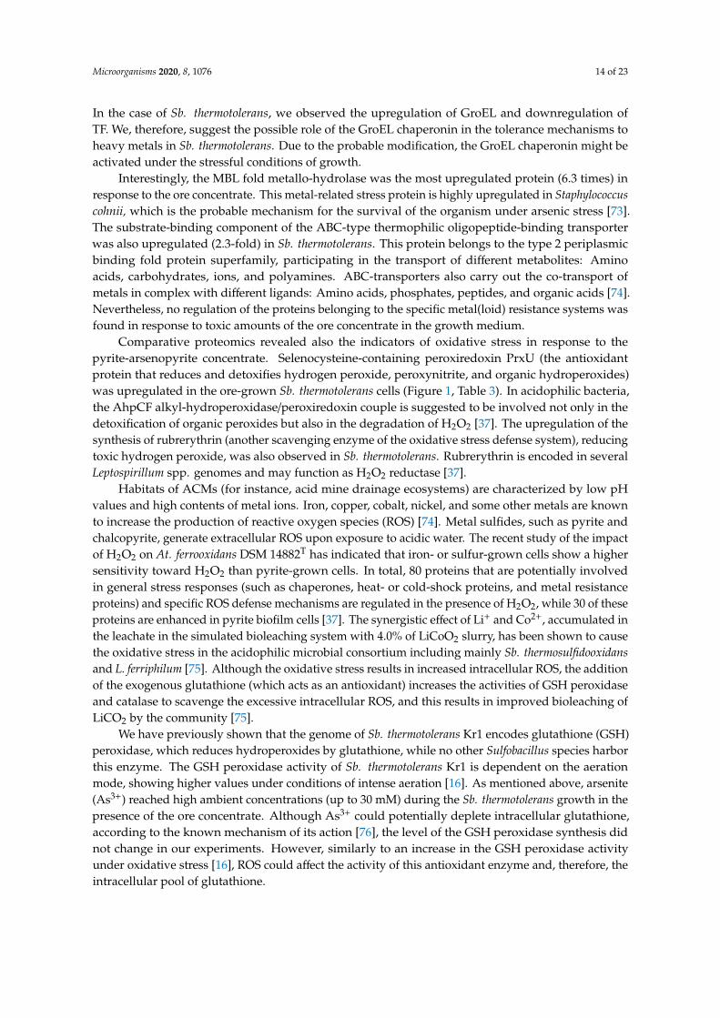
In the case of Sb. thermotolerans, we observed the upregulation of GroEL and downregulation ofTF. We, therefore, suggest the possible role of the GroEL chaperonin in the tolerance mechanisms toheavy metals in Sb. thermotolerans. Due to the probable modification, the GroEL chaperonin might beactivated under the stressful conditions of growth.
Interestingly, the MBL fold metallo-hydrolase was the most upregulated protein (6.3 times) inresponse to the ore concentrate. This metal-related stress protein is highly upregulated in Staphylococcuscohnii, which is the probable mechanism for the survival of the organism under arsenic stress [73].The substrate-binding component of the ABC-type thermophilic oligopeptide-binding transporterwas also upregulated (2.3-fold) in Sb. thermotolerans. This protein belongs to the type 2 periplasmicbinding fold protein superfamily, participating in the transport of different metabolites: Aminoacids, carbohydrates, ions, and polyamines. ABC-transporters also carry out the co-transport ofmetals in complex with different ligands: Amino acids, phosphates, peptides, and organic acids [74].Nevertheless, no regulation of the proteins belonging to the specific metal(loid) resistance systems wasfound in response to toxic amounts of the ore concentrate in the growth medium.
Comparative proteomics revealed also the indicators of oxidative stress in response to thepyrite-arsenopyrite concentrate. Selenocysteine-containing peroxiredoxin PrxU (the antioxidantprotein that reduces and detoxifies hydrogen peroxide, peroxynitrite, and organic hydroperoxides)was upregulated in the ore-grown Sb. thermotolerans cells (Figure 1, Table 3). In acidophilic bacteria,the AhpCF alkyl-hydroperoxidase/peroxiredoxin couple is suggested to be involved not only in thedetoxification of organic peroxides but also in the degradation of H2O2 [37]. The upregulation of thesynthesis of rubrerythrin (another scavenging enzyme of the oxidative stress defense system), reducingtoxic hydrogen peroxide, was also observed in Sb. thermotolerans. Rubrerythrin is encoded in severalLeptospirillum spp. genomes and may function as H2O2 reductase [37].
Habitats of ACMs (for instance, acid mine drainage ecosystems) are characterized by low pHvalues and high contents of metal ions. Iron, copper, cobalt, nickel, and some other metals are knownto increase the production of reactive oxygen species (ROS) [74]. Metal sulfides, such as pyrite andchalcopyrite, generate extracellular ROS upon exposure to acidic water. The recent study of the impactof H2O2 on At. ferrooxidans DSM 14882T has indicated that iron- or sulfur-grown cells show a highersensitivity toward H2O2 than pyrite-grown cells. In total, 80 proteins that are potentially involvedin general stress responses (such as chaperones, heat- or cold-shock proteins, and metal resistanceproteins) and specific ROS defense mechanisms are regulated in the presence of H2O2, while 30 of theseproteins are enhanced in pyrite biofilm cells [37]. The synergistic effect of Li+ and Co2+, accumulated inthe leachate in the simulated bioleaching system with 4.0% of LiCoO2 slurry, has been shown to causethe oxidative stress in the acidophilic microbial consortium including mainly Sb. thermosulfidooxidansand L. ferriphilum [75]. Although the oxidative stress results in increased intracellular ROS, the additionof the exogenous glutathione (which acts as an antioxidant) increases the activities of GSH peroxidaseand catalase to scavenge the excessive intracellular ROS, and this results in improved bioleaching ofLiCO2 by the community [75].
We have previously shown that the genome of Sb. thermotolerans Kr1 encodes glutathione (GSH)peroxidase, which reduces hydroperoxides by glutathione, while no other Sulfobacillus species harborthis enzyme. The GSH peroxidase activity of Sb. thermotolerans Kr1 is dependent on the aerationmode, showing higher values under conditions of intense aeration [16]. As mentioned above, arsenite(As3+) reached high ambient concentrations (up to 30 mM) during the Sb. thermotolerans growth in thepresence of the ore concentrate. Although As3+ could potentially deplete intracellular glutathione,according to the known mechanism of its action [76], the level of the GSH peroxidase synthesis didnot change in our experiments. However, similarly to an increase in the GSH peroxidase activityunder oxidative stress [16], ROS could affect the activity of this antioxidant enzyme and, therefore, theintracellular pool of glutathione.
Microorganisms 2020, 8, 1076 15 of 23
3.3.2. Metabolic Reorganization
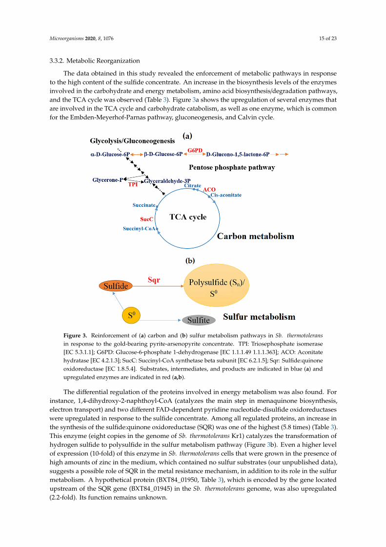
The data obtained in this study revealed the enforcement of metabolic pathways in responseto the high content of the sulfide concentrate. An increase in the biosynthesis levels of the enzymesinvolved in the carbohydrate and energy metabolism, amino acid biosynthesis/degradation pathways,and the TCA cycle was observed (Table 3). Figure 3a shows the upregulation of several enzymes thatare involved in the TCA cycle and carbohydrate catabolism, as well as one enzyme, which is commonfor the Embden-Meyerhof-Parnas pathway, gluconeogenesis, and Calvin cycle.
Microorganisms 2020, 8, x FOR PEER REVIEW 14 of 22
involved in the carbohydrate and energy metabolism, amino acid biosynthesis/degradation pathways, and the TCA cycle was observed (Table 3). Figure 3a shows the upregulation of several enzymes that are involved in the TCA cycle and carbohydrate catabolism, as well as one enzyme, which is common for the Embden-Meyerhof-Parnas pathway, gluconeogenesis, and Calvin cycle.
The differential regulation of the proteins involved in energy metabolism was also found. For instance, 1,4-dihydroxy-2-naphthoyl-CoA (catalyzes the main step in menaquinone biosynthesis, electron transport) and two different FAD-dependent pyridine nucleotide-disulfide oxidoreductases were upregulated in response to the sulfide concentrate. Among all regulated proteins, an increase in the synthesis of the sulfide:quinone oxidoreductase (SQR) was one of the highest (5.8 times) (Table 3). This enzyme (eight copies in the genome of Sb. thermotolerans Kr1) catalyzes the transformation of hydrogen sulfide to polysulfide in the sulfur metabolism pathway (Figure 3b). Even a higher level of expression (10-fold) of this enzyme in Sb. thermotolerans cells that were grown in the presence of high amounts of zinc in the medium, which contained no sulfur substrates (our unpublished data), suggests a possible role of SQR in the metal resistance mechanism, in addition to its role in the sulfur metabolism. A hypothetical protein (BXT84_01950, Table 3), which is encoded by the gene located upstream of the SQR gene (BXT84_01945) in the Sb. thermotolerans genome, was also upregulated (2.2-fold). Its function remains unknown.
Figure 3. Reinforcement of (a) carbon and (b) sulfur metabolism pathways in Sb. thermotolerans in response to the gold-bearing pyrite-arsenopyrite concentrate. TPI: Triosephosphate isomerase [EC 5.3.1.1]; G6PD: Glucose-6-phosphate 1-dehydrogenase [EC 1.1.1.49 1.1.1.363]; ACO: Aconitate hydratase [EC 4.2.1.3]; SucC: Succinyl-CoA synthetase beta subunit [EC 6.2.1.5]; Sqr: Sulfide:quinone oxidoreductase [EC 1.8.5.4]. Substrates, intermediates, and products are indicated in blue (a) and upregulated enzymes are indicated in red (a,b).
The S-adenosylmethionine synthetase was also shown to be regulated in Sb. thermotolerans. This enzyme was represented by two protein spots both of which were upregulated (2.4- and 5.0-fold). Overexpression of the S-adenosylmethionine synthetase that plays a pivotal role in the central metabolism of all organisms is expected to produce increased levels of S-adenosylmethionine. The
Figure 3. Reinforcement of (a) carbon and (b) sulfur metabolism pathways in Sb. thermotoleransin response to the gold-bearing pyrite-arsenopyrite concentrate. TPI: Triosephosphate isomerase[EC 5.3.1.1]; G6PD: Glucose-6-phosphate 1-dehydrogenase [EC 1.1.1.49 1.1.1.363]; ACO: Aconitatehydratase [EC 4.2.1.3]; SucC: Succinyl-CoA synthetase beta subunit [EC 6.2.1.5]; Sqr: Sulfide:quinoneoxidoreductase [EC 1.8.5.4]. Substrates, intermediates, and products are indicated in blue (a) andupregulated enzymes are indicated in red (a,b).
The differential regulation of the proteins involved in energy metabolism was also found. Forinstance, 1,4-dihydroxy-2-naphthoyl-CoA (catalyzes the main step in menaquinone biosynthesis,electron transport) and two different FAD-dependent pyridine nucleotide-disulfide oxidoreductaseswere upregulated in response to the sulfide concentrate. Among all regulated proteins, an increase inthe synthesis of the sulfide:quinone oxidoreductase (SQR) was one of the highest (5.8 times) (Table 3).This enzyme (eight copies in the genome of Sb. thermotolerans Kr1) catalyzes the transformation ofhydrogen sulfide to polysulfide in the sulfur metabolism pathway (Figure 3b). Even a higher levelof expression (10-fold) of this enzyme in Sb. thermotolerans cells that were grown in the presence ofhigh amounts of zinc in the medium, which contained no sulfur substrates (our unpublished data),suggests a possible role of SQR in the metal resistance mechanism, in addition to its role in the sulfurmetabolism. A hypothetical protein (BXT84_01950, Table 3), which is encoded by the gene locatedupstream of the SQR gene (BXT84_01945) in the Sb. thermotolerans genome, was also upregulated(2.2-fold). Its function remains unknown.

Microorganisms 2020, 8, 1076 16 of 23
The S-adenosylmethionine synthetase was also shown to be regulated in Sb. thermotolerans. Thisenzyme was represented by two protein spots both of which were upregulated (2.4- and 5.0-fold).Overexpression of the S-adenosylmethionine synthetase that plays a pivotal role in the centralmetabolism of all organisms is expected to produce increased levels of S-adenosylmethionine. Thelatter is a precursor for the synthesis of polyamines, including spermidine, which is involved in thiolmetabolism, and a metabolite in the trans-sulfuration pathway to cysteine. S-adenosylmethionine is theprimary methyl group donor in methylation reactions; it regulates cell signaling, gene expression, andmetabolic pathways [77]. The upregulation of this enzyme (3.7-fold) has been found in the cells of At.caldus (another member of communities of ACMs) grown in the presence of 200 mM ZnSO4 [28]. In thecells of Sb3+-resistant clinical isolates of Leishmania panamensis, the S-adenosylmethionine synthetase isupregulated exclusively, playing a central role in the upstream synthesis of precursors of trypanothione(a key molecule involved in the Sb-resistance in Leishmania parasites), indicating the importance ofthiol metabolism in resistance to the antimony in Leishmania [78]. In plants, the S-adenosylmethioninesynthetase plays an essential role in response to stress, including metal stress [79,80].
Several enzymes participating in amino acid degradation in Sb. thermotolerans were alsoupregulated (Figure 4, Table 3). They included two subunits of the 2-oxoisovalerate dehydrogenaseenzyme, which is involved in valine, leucine, and isoleucine degradation, as well as propionatemetabolism. The upregulation of this protein has been found in Sphingomonas sp. cells in responseto Cd2+ [70]. Other enzymes of amino acid metabolism were also upregulated, including cysteinedesulfurase (5.4-fold; it catalyzes the removal of elemental sulfur and selenium atoms from L-cysteine,L-cystine, L-selenocysteine, and L-selenocystine to produce L-alanine) and D-3-phosphoglyceratedehydrogenase (2-fold). Amino acids are known to be potential ligands for heavy metals and contributeto tolerance and detoxification [66].
Microorganisms 2020, 8, x FOR PEER REVIEW 15 of 22
latter is a precursor for the synthesis of polyamines, including spermidine, which is involved in thiol metabolism, and a metabolite in the trans-sulfuration pathway to cysteine. S-adenosylmethionine is the primary methyl group donor in methylation reactions; it regulates cell signaling, gene expression, and metabolic pathways [77]. The upregulation of this enzyme (3.7-fold) has been found in the cells of At. caldus (another member of communities of ACMs) grown in the presence of 200 mM ZnSO4 [28]. In the cells of Sb3+-resistant clinical isolates of Leishmania panamensis, the S-adenosylmethionine synthetase is upregulated exclusively, playing a central role in the upstream synthesis of precursors of trypanothione (a key molecule involved in the Sb-resistance in Leishmania parasites), indicating the importance of thiol metabolism in resistance to the antimony in Leishmania [78]. In plants, the S-adenosylmethionine synthetase plays an essential role in response to stress, including metal stress [79,80].
Several enzymes participating in amino acid degradation in Sb. thermotolerans were also upregulated (Figure 4, Table 3). They included two subunits of the 2-oxoisovalerate dehydrogenase enzyme, which is involved in valine, leucine, and isoleucine degradation, as well as propionate metabolism. The upregulation of this protein has been found in Sphingomonas sp. cells in response to Cd2+ [70]. Other enzymes of amino acid metabolism were also upregulated, including cysteine desulfurase (5.4-fold; it catalyzes the removal of elemental sulfur and selenium atoms from L-cysteine, L-cystine, L-selenocysteine, and L-selenocystine to produce L-alanine) and D-3-phosphoglycerate dehydrogenase (2-fold). Amino acids are known to be potential ligands for heavy metals and contribute to tolerance and detoxification [66].
Figure 4. Upregulation of the components of the amino acid metabolism in Sb. thermotolerans in response to the gold-bearing pyrite-arsenopyrite concentrate. PaaF: Enoyl-CoA hydratase [EC 4.2.1.17]; DBT: 2-Oxoisovalerate dehydrogenase E2 component (dihydrolipoyl transacylase) [EC 2.3.1.168]; BCKDHB: 2-Oxoisovalerate dehydrogenase E1 component beta subunit [EC 1.2.4.4]; BCKDHA: 2-Oxoisovalerate dehydrogenase E1 component alpha subunit [EC 1.2.4.4]; MetK: S-adenosylmethionine synthetase [EC 2.5.1.6]; SerA: D-3-phosphoglycerate dehydrogenase/2-oxoglutarate reductase [EC 1.1.1.95 1.1.1.399]. Substrates, intermediates, and products are indicated in blue and upregulated enzymes are indicated in red.
The general response of Sb. thermotolerans Kr1 cells involved also changes in the transcription, translation, cell biogenesis, coenzyme transport and metabolism, as well as the metabolism of fatty acids and secondary metabolites (Figure 2, Table 3). For instance, the YebC/PmpR family DNA-binding transcriptional regulator was found to be upregulated (2-fold) in response to the ore concentrate. The YebC family regulator proteins are widespread and conserved in many bacteria.
Figure 4. Upregulation of the components of the amino acid metabolism in Sb. thermotolerans in responseto the gold-bearing pyrite-arsenopyrite concentrate. PaaF: Enoyl-CoA hydratase [EC 4.2.1.17]; DBT:2-Oxoisovalerate dehydrogenase E2 component (dihydrolipoyl transacylase) [EC 2.3.1.168]; BCKDHB:2-Oxoisovalerate dehydrogenase E1 component beta subunit [EC 1.2.4.4]; BCKDHA: 2-Oxoisovaleratedehydrogenase E1 component alpha subunit [EC 1.2.4.4]; MetK: S-adenosylmethionine synthetase [EC2.5.1.6]; SerA: D-3-phosphoglycerate dehydrogenase/2-oxoglutarate reductase [EC 1.1.1.95 1.1.1.399].Substrates, intermediates, and products are indicated in blue and upregulated enzymes are indicatedin red.
The general response of Sb. thermotolerans Kr1 cells involved also changes in the transcription,translation, cell biogenesis, coenzyme transport and metabolism, as well as the metabolism of fatty

Microorganisms 2020, 8, 1076 17 of 23
acids and secondary metabolites (Figure 2, Table 3). For instance, the YebC/PmpR family DNA-bindingtranscriptional regulator was found to be upregulated (2-fold) in response to the ore concentrate. TheYebC family regulator proteins are widespread and conserved in many bacteria. YebC may serve as amulti-functional transcription regulator; it is predicted to regulate the resolvase complex RuvABC, mostlikely the RuvC subunit, and may participate in other biological processes [81]. The YebC protein isresponsible for the quorum-sensing (QS) and virulence in Edwardsiella piscicida, the negative regulationof the QS response regulator in Pseudomonas aeruginosa, and, probably, proteolysis in Lactobacillus [82].In the metal-tolerant bacterium Alishewanella sp., the YebC family protein RuvR is involved in Cr6+,As3+, Sb3+, and Cd2+ resistance [83]. The NusA protein that participates in transcriptional elongation,termination, anti-termination, as well as cold shock and stress-induced mutagenesis [84], was alsoupregulated (2.2-fold) in response to the arsenic-rich sulfide concentrate. NusA is suggested to serveas a molecular chaperone in addition to its functions as a transcription factor [84]. In the case of Sb.thermotolerans, the upregulation of this protein could also be associated with the stress effect of toxiccompounds in the medium of growth.
Thus, on the basis of differential proteomic analysis, a total of 33 proteins were regulated inSb. thermotolerans in response to the high-arsenic sulfide concentrate. Although higher numbersof regulated proteins may be expected under metal(loid) stress [23], the results of our study are inagreement with the previously reported proteomic data for other acidophiles. For instance, in At.ferrooxidans, At. caldus, Am. ferrooxidans, and F. acidarmanus, 7–21 proteins were upregulated and 1–22proteins were downregulated in response to zinc, arsenic, copper, and uranium stress [24,26,28,85].
It should be mentioned that mainly planktonic late-exponential vegetative cells (already detachedor not yet attached to the mineral particles) and single forespores (~1–1.5%) from the liquid phase,which contained iron, arsenic, and other dissolved elements, were analyzed in this study. According toother proteomic reports, differences in the proteomic responses of the planktonic and sessile (biofilm)cell subpopulations of At. ferrooxidans, oxidizing pyrite, have been revealed [37,41]. Therefore, theaccumulation of elements by sessile cells of Sb. thermotolerans and their proteomic response could varyfrom those of the planktonic cell population.
Overall, the results of our research imply that changes in the protein expression in Sb. thermotoleranscells were closely related to the accumulation of the toxic arsenic. Several proteins that seem toconfer resistance to arsenic in other microorganisms [66,73,76,83,84], but do not belong to any specificarsenic-resistance systems, were shown to be upregulated in this acidophilic bacterium. In other studiedacidophiles, responses to arsenic are associated with specific arsenic resistance proteins [31,33,34]. Thelatter, as well as the proteins involved in phosphate metabolism, protection from reactive oxygenspecies, GSH metabolism, DNA synthesis and repair, protein synthesis, folding and refolding, areregulated in L. ferriphilum in response to the arsenic stress [33]. In acidophilic archaeon F. acidarmanus,the proteomic analysis has not detected increased expression of the ArsB pump; the proteomic responseto arsenic involves a number of protein repair and modification enzymes, as well as metabolic andelectron transport proteins [85]. However, few studies on the proteomic responses of acidophilesto arsenic have been reported in the literature [33,85], and future proteomic research would be ofinterest to provide a better understanding of the molecular mechanisms for arsenic resistance in thismicrobial group.
It is noteworthy that according to our proteomic data on the metabolic reorganization, no pathwayswere inhibited in Sb. thermotolerans in response to the high-arsenic sulfide concentrate. On the contrary,some pathways were reinforced, while iron oxidation rates remained rather high. These results suggestthat the components of carbon, energy, and amino acid metabolism, as well as those of stress responsesystems, may be essential in the resistance mechanisms of Sb. thermotolerans to the toxic effects of thegold-containing pyrite-arsenopyrite concentrates. The efficient iron oxidation and reinforcement ofcatabolic pathways indicate the advantages of the mixotrophic lifestyle of Sb. thermotolerans duringadaptation to stress factors. This trophic strategy makes it possible to meet the increased metabolicdemands of the bacterium under adverse conditions and enhance its growth. Due to the utilization
Microorganisms 2020, 8, 1076 18 of 23
of organic compounds, Sb. thermotolerans may also play an essential role in the detoxification ofthe environment for chemolithoautotrophs (removal of microbial metabolites and lysis products) inacidophilic microbial communities exposed to metal(loid) stress.
4. Conclusions
The results of our research provide first insights into the mechanisms of resistance to thegold-bearing arsenopyrite concentrates in the genus Sulfobacillus. It was shown that:
• Sb. thermotolerans rapidly adapted to the toxic amount of the high-arsenic gold-bearing sulfideconcentrate and proved to be among the most As-tolerant organisms known to date;
• although the cellular adsorption and intracellular accumulation of metal(loid)s occurred to highlevels, the bacterium retained its growth and efficient substrate oxidation;
• in total, 30 upregulated proteins were involved in the response to the toxic content of the sulfideconcentrate in the growth medium, and only three proteins, which, however, did not affect thevital activity of the preliminarily nonadapted Sb. thermotolerans cells, were downregulated;
• Sb. thermotolerans cells responded to adverse conditions by metabolic changes, includingreinforcement of pathways of constructive and energy metabolism, and by activation of defensesystems against unfavorable factors. At the same time, no specific metal-resistance componentswere regulated in response to metal(loid)s accumulated in the culture medium and by the cells;
• proteins of the stress response, such as the metal-related stress protein MBL fold metallo-hydrolaseand GroEL chaperonin, probably, played crucial roles in the tolerance to the high-arsenic sulfideore concentrate and arsenic, in particular;
• the markedly upregulated sulfide:quinone oxidoreductase, cysteine desulfurase, andS-adenosylmethionine synthetase were other main contributors to the bacterial response. Apartfrom the enzymatic function in sulfur metabolism, sulfide:quinone oxidoreductase potentiallyfulfilled the second function of a resistance-conferring protein in Sb. thermotolerans.
This study and previous works indicate a high resistance potential of Sb. thermotolerans, whichis of interest to both the fundamental science and industrial applications. Our results may open upnew perspectives on the investigation of the roles of certain proteins in the tolerance mechanisms todifferent metal(loid)s in Sulfobacillus bacteria and other biomining acidophiles.
Supplementary Materials: The following are available online at http://www.mdpi.com/2076-2607/8/7/1076/s1.Figure S1: Growth of Sb. thermotolerans Kr1 and iron oxidation under different cultivation conditions: (a) In themedium containing ferrous iron (control); in the presence of 10 (b), 20 (c), and 30 g/L (d) of the gold-containingpyrite-arsenopyrite concentrate. Arrows (b,d) indicate parameters of the samples used in the experiments.
Author Contributions: Conceptualization, A.P.; methodology, A.P., D.M., and O.P.; validation, A.P., D.M., andO.P.; formal analysis, investigation, and resources, A.P., D.M., and O.P.; data curation, A.P., D.M., and O.P.;writing—original draft preparation, A.P.; writing—review and editing, A.P., D.M., and O.P.; visualization, A.P.,D.M., and O.P.; supervision, A.P.; project administration, A.P.; funding acquisition, A.P. All authors have read andagreed to the published version of the manuscript.
Funding: This research was funded by the Ministry of Science and Higher Education of the Russian Federation.
Acknowledgments: We thank the Center for Precision Genome Editing and Genetic Technologies for Biomedicine,the Federal Research and Clinical Center of Physical-Chemical Medicine of Federal Medical Biological Agency forthe equipment for mass spectrometric analysis.
Conflicts of Interest: The authors declare no conflict of interest.
References
1. Kaksonen, A.H.; Boxall, N.J.; Gumulya, Y.; Khaleque, H.N.; Morris, C.; Bohu, T.; Cheng, K.Y.; Usher, K.M.;Lakaniemi, A.-M. Recent progress in biohydrometallurgy and microbial characterisation. Hydrometallurgy2018, 180, 7–25. [CrossRef]
2. Rawlings, D.E.; Johnson, D.B. The microbiology of biomining: Development and optimization ofmineral-oxidizing microbial consortia. Microbiology 2007, 153, 315–324. [CrossRef]

Microorganisms 2020, 8, 1076 19 of 23
3. Bond, P.L.; Druschel, G.K.; Banfield, J.F. Comparison of acid mine drainage microbial communities inphysically and geochemically distinct ecosystems. Appl. Environ. Microbiol. 2000, 66, 4962–4971. [CrossRef]
4. d’Hugues, P.; Joulian, C.; Spolaore, P.; Michel, C.; Garrido, F.; Morin, D. Continuous bioleaching of a pyriteconcentrate in stirred reactors: Population dynamics and exopolysaccharide production vs. bioleachingperformance. Hydrometallurgy 2008, 94, 34–41. [CrossRef]
5. Dopson, M.; Lindström, E.B. Analysis of community composition during moderately thermophilic bioleachingof pyrite, arsenical pyrite, and chalcopyrite. Microb. Ecol. 2004, 48, 19–28. [CrossRef] [PubMed]
6. Bogdanova, T.I.; Tsaplina, I.A.; Kondrat’eva, T.F.; Duda, V.I.; Suzina, N.E.; Melamud, V.S.; Tourova, T.P.;Karavaiko, G.I. Sulfobacillus thermotolerans sp. nov., a thermotolerant, chemolithotrophic bacterium. Int. J.Syst. Evol. Microbiol. 2006, 56, 1039–1042. [CrossRef] [PubMed]
7. Tan, G.L.; Shu, W.S.; Hallberg, K.B.; Li, F.; Lan, C.Y.; Zhou, W.H.; Huang, L.N. Culturable and molecularphylogenetic diversity of microorganisms in an open-dumped, extremely acidic Pb/Zn mine tailings.Extremophiles 2008, 12, 657–664. [CrossRef]
8. Tsaplina, I.A.; Krasil’nikova, E.N.; Zhuravleva, A.E.; Egorova, M.A.; Zakharchuk, L.M.; Suzina, N.E.;Bogdanova, T.I.; Kondrat’eva, T.F. Phenotypic properties of Sulfobacillus thermotolerans: Comparative aspects.Microbiology 2008, 77, 654–664. [CrossRef]
9. Fomchenko, N.V.; Muravyov, M.I. Two-step biohydrometallurgical technology of copper-zinc concentrateprocessing as an opportunity to reduce negative impacts on the environment. J. Environ. Manag. 2018, 226,270–277. [CrossRef]
10. Muravyov, M.; Panyushkina, A. Distinct roles of acidophiles in complete oxidation of high-sulfur ferric leachproduct of zinc sulfide concentrate. Microorganisms 2020, 8, 386. [CrossRef]
11. Hao, C.; Wang, L.; Dong, H.; Zhang, H. Succession of acidophilic bacterial community during bio-oxidationof refractory gold-containing sulfides. Geomicrobiol. J. 2010, 27, 683–691. [CrossRef]
12. Tsaplina, I.A.; Panyushkina, A.E.; Melamud, V.S.; Grigor’eva, N.V.; Kondrat’eva, T.F. Leaching ofpyrite–arsenopyrite concentrate in bioreactors during continuous cultivation of a thermoacidophilic microbialcommunity. Microbiology 2014, 83, 568–576. [CrossRef]
13. Bulaev, A.; Belyi, A.; Panyushkina, A.; Solopova, N.; Pivovarova, T. Microbial population of industrialbiooxidation reactors. Solid State Phenom. 2017, 262, 48–52. [CrossRef]
14. Tsaplina, I.A.; Zhuravleva, A.E.; Egorova, M.A.; Bogdanova, T.I.; Krasil’nikova, E.N.; Zakharchuk, L.M.;Kondrat’eva, T.F. Response to oxygen limitation in bacteria of the genus Sulfobacillus. Microbiology 2010, 79,13–22. [CrossRef]
15. Panyushkina, A.E. Metabolic potential of Sulfobacillus thermotolerans: Pathways for assimilation of nitrogencompounds and the possibility of lithotrophic growth in the presence of molecular hydrogen. Microbiology2019, 13, 759–763. [CrossRef]
16. Panyushkina, A.E.; Babenko, V.V.; Nikitina, A.S.; Selezneva, O.V.; Tsaplina, I.A.; Letarova, M.A.;Kostryukova, E.S.; Letarov, A.V. Sulfobacillus thermotolerans: New insights into resistance and metaboliccapacities of acidophilic chemolithotrophs. Sci. Rep. 2019, 9, 15069. [CrossRef]
17. Tuffin, I.M.; Hector, S.B.; Deane, S.M.; Rawlings, D.E. Resistance determinants of a highly arsenic-resistantstrain of Leptospirillum ferriphilum isolated from a commercial biooxidation tank. Appl. Environ. Microbiol.2006, 72, 2247–2253. [CrossRef]
18. Navarro, C.A.; von Bernath, D.; Jerez, C.A. Heavy metal resistance strategies of acidophilic bacteria andtheir acquisition: Importance for biomining and bioremediation. Biol. Res. 2013, 46, 363–371. [CrossRef][PubMed]
19. Orell, A.; Navarro, C.A.; Arancibia, R.; Mobarec, J.C.; Jerez, C.A. Life in blue: Copper resistance mechanismsof bacteria and archaea used in industrial biomining of minerals. Biotechnol. Adv. 2010, 28, 839–848.[CrossRef]
20. Orellana, L.H.; Jerez, C.A. A genomic island provides Acidithiobacillus ferrooxidans ATCC 53993 additionalcopper resistance: A possible competitive advantage. Appl. Microbiol. Biotechnol. 2011, 92, 761–767.[CrossRef]
21. Salazar, C.; Acosta, M.; Galleguillos, P.; Shmaryahu, A.; Quatrini, R.; Holmes, D.S.; Demergasso, C. Analysisof gene expression in response to copper stress in Acidithiobacillus ferrooxidans strain D2, isolated from acopper bioleaching operation. Adv. Mat. Res. 2013, 825, 157–161. [CrossRef]

Microorganisms 2020, 8, 1076 20 of 23
22. Wu, X.; Zhang, Z.; Liu, L.; Deng, F.; Liu, X.; Qiu, G. Metal resistance-related genes are differently expressed inresponse to copper and zinc ion in six Acidithiobacillus ferrooxidans strains. Curr. Microbiol. 2014, 69, 775–784.[CrossRef] [PubMed]
23. Almárcegui, R.J.; Navarro, C.A.; Paradela, A.; Albar, J.P.; von Bernath, D.; Jerez, C.A. New copper resistancedeterminants in the extremophile Acidithiobacillus ferrooxidans: A quantitative proteomic analysis. J. ProteomeRes. 2014, 13, 946–960. [CrossRef] [PubMed]
24. Dekker, L.; Arsène-Ploetze, F.; Santini, J.M. Comparative proteomics of Acidithiobacillus ferrooxidans grown inthe presence and absence of uranium. Res. Microbiol. 2016, 167, 234–239. [CrossRef] [PubMed]
25. Fatemi, F.; Miri, S.; Jahani, S. Effect of metal sulfide pulp density on gene expression of electron transportersin Acidithiobacillus sp. FJ2. Arch. Microbiol. 2017, 199, 521–530. [CrossRef]
26. Baker-Austin, C.; Dopson, M.; Wexler, M.; Sawers, R.G.; Bond, P.L. Molecular insight into extreme copperresistance in the extremophilic archaeon ‘Ferroplasma acidarmanus’ Fer1. Microbiology 2005, 151, 2637–2646.[CrossRef] [PubMed]
27. Orell, A.; Remonsellez, F.; Arancibia, R.; Jerez, C.A. Molecular characterization of copper and cadmiumresistance determinants in the biomining thermoacidophilic archaeon Sulfolobus metallicus. Archaea 2013,2013, 289236. [CrossRef]
28. Mangold, S.; Potrykus, J.; Björn, E.; Lövgren, L.; Dopson, M. Extreme zinc tolerance in acidophilicmicroorganisms from the bacterial and archaeal domains. Extremophiles 2013, 17, 75–85. [CrossRef]
29. Martínez-Bussenius, C.; Navarro, C.A.; Jerez, C.A. Microbial copper resistance: Importance inbiohydrometallurgy. Microb. Biotechnol. 2017, 10, 279–295. [CrossRef]
30. Soto, D.F.; Recalde, A.; Orell, A.; Albers, S.-V.; Paradela, A.; Navarro, C.A.; Jerez, C.A. Global effect of thelack of inorganic polyphosphate in the extremophilic archaeon Sulfolobus solfataricus: A proteomic approach.J. Proteomics. 2019, 191, 143–152. [CrossRef]
31. Dopson, M.; Holmes, D. Metal resistance in acidophilic microorganisms and its significance forbiotechnologies. Appl. Microbiol. Biotechnol. 2014, 98, 8133–8144. [CrossRef]
32. Dopson, M.; Ossandon, F.J.; Lövgren, L.; Holmes, D.S. Metal resistance or tolerance? Acidophiles confronthigh metal loads via both abiotic and biotic mechanisms. Front. Microbiol. 2014, 5, 157. [CrossRef] [PubMed]
33. Li, B.; Lin, J.; Mi, S.; Lin, J. Arsenic resistance operon structure in Leptospirillum ferriphilum and proteomicresponse to arsenic stress. Bioresour. Technol. 2010, 101, 9811–9814. [CrossRef]
34. Jiang, H.; Liang, Y.; Yin, H.; Xiao, Y.; Guo, X.; Xu, Y.; Hu, Q.; Liu, H.; Liu, X. Effects of arsenite resistance onthe growth and functional gene expression of Leptospirillum ferriphilum and Acidithiobacillus thiooxidans inpure culture and coculture. BioMed Res. Int. 2015, 2015, 203197. [CrossRef] [PubMed]
35. Ramírez, P.; Guiliani, N.; Valenzuela, L.; Beard, S.; Jerez, C.A. Differential protein expression during growthof Acidithiobacillus ferrooxidans on ferrous iron, sulfur compounds, or metal sulfides. Appl. Environ. Microbiol.2004, 70, 4491–4498. [CrossRef] [PubMed]
36. Felício, A.P.; Oliveira, E.; Odena, M.A.; Garcia, O., Jr.; Bertolini, M.C.; Ferraz, L.F.C.; Ottoboni, L.M.M.;Novo, M.T.M. Differential proteomic analysis of Acidithiobacillus ferrooxidans cells maintained in contactwith bornite or chalcopyrite: Proteins involved with the early bacterial response. Process Biochem. 2011, 46,770–776. [CrossRef]
37. Bellenberg, S.; Huynh, D.; Poetsch, A.; Sand, W.; Vera, M. Proteomics reveal enhanced oxidative stressresponses and metabolic adaptation in Acidithiobacillus ferrooxidans biofilm cells on pyrite. Front. Microbiol.2019, 10, 592. [CrossRef]
38. Carlos, C.; Reis, F.C.; Vicentini, R.; Madureira, D.J.; Ottoboni, L.M.M. The rus operon genes are differentiallyregulated when Acidithiobacillus ferrooxidans LR is kept in contact with metal sulfides. Curr. Microbiol. 2008,57, 375–380. [CrossRef]
39. Christel, S.; Herold, M.; Bellenberg, S.; Hajjami, M.E.; Buetti-Dinh, A.; Pivkin, I.V.; Sand, W.; Wilmes, P.;Poetsch, A.; Dopson, M. Multi-omics reveals the lifestyle of the acidophilic, mineral-oxidizing model speciesLeptospirillum ferriphilumT. Appl. Environ. Microbiol. 2018, 84, e02091-17. [CrossRef]
40. Sand, W.; Gehrke, T. Extracellular polymeric substances mediate bioleaching/biocorrosion via interfacialprocesses involving iron(III) ions and acidophilic bacteria. Res. Microbiol. 2006, 157, 49–56. [CrossRef]
41. Vera, M.; Krok, B.; Bellenberg, S.; Sand, W.; Poetsch, A. Shotgun proteomics study of early biofilm formationprocess of Acidithiobacillus ferrooxidans ATCC 23270 on pyrite. Proteomics 2013, 13, 1133–1144. [CrossRef][PubMed]

Microorganisms 2020, 8, 1076 21 of 23
42. Belnap, C.P.; Pan, C.; VerBerkmoes, N.C.; Power, M.E.; Samatova, N.F.; Carver, R.L.; Hettich, R.L.; Banfield, J.F.Cultivation and quantitative proteomic analyses of acidophilic microbial communities. ISME J. 2010, 4,520–530. [CrossRef] [PubMed]
43. Wilmes, P.; Bowen, B.P.; Thomas, B.C.; Mueller, R.S.; Denef, V.J.; VerBerkmoes, N.C.; Hettich, R.L.;Northen, T.R.; Banfield, J.F. Metabolome-proteome differentiation coupled to microbial divergence. MBio2010, 1, e00246-10. [CrossRef] [PubMed]
44. Mosier, A.C.; Li, Z.; Thomas, B.C.; Hettich, R.L.; Pan, C.; Banfield, J.F. Elevated temperature alters proteomicresponses of individual organisms within a biofilm community. ISME J. 2015, 9, 180–194. [CrossRef]
45. Shevchenko, A.; Wilm, M.; Vorm, O.; Mann, M. Mass spectrometric sequencing of proteins from silver-stainedpolyacrylamide gels. Anal. Chem. 1996, 68, 850–858. [CrossRef]
46. Jensen, O.N.; Wilm, M.; Shevchenko, A.; Mann, M. Peptide sequencing of 2-DE gel-isolated proteins bynanoelectrospray tandem mass spectrometry. Methods Mol. Biol. 1999, 112, 571–588. [CrossRef] [PubMed]
47. Kanehisa, M.; Sato, Y.; Kawashima, M.; Furumichi, M.; Tanabe, M. KEGG as a reference resource for geneand protein annotation. Nucleic Acids Res. 2016, 44, D457–462. [CrossRef] [PubMed]
48. Melamud, V.S.; Pivovarova, T.A.; Kondrat’eva, T.F.; Karavaiko, G.I. Study of oxidation by bacteria of adifficult-to-dress gold-containing pyrite-arsenopyrite concentrate under moderately thermophilic conditions.Appl. Biochem. Microbiol. 1999, 35, 161–167.
49. Goyal, N.; Jain, S.C.; Banerjee, U.C. Comparative studies on the microbial adsorption of heavy metals. Adv.Environ. Res. 2003, 7, 311–319. [CrossRef]
50. Pagani, M.A.; Casamayor, A.; Serrano, R.; Atrian, S.; Ariño, J. Disruption of iron homeostasis in Saccharomycescerevisiae by high zinc levels: A genome-wide study. Mol. Microbiol. 2007, 65, 521–537. [CrossRef]
51. Watling, H.R.; Perrot, F.A.; Shiers, D.W. Comparison of selected characteristics of Sulfobacillus speciesand review of their occurrence in acidic and bioleaching environments. Hydrometallurgy 2008, 93, 57–65.[CrossRef]
52. Dave, S.R.; Gupta, K.H.; Tipre, D.R. Characterization of arsenic resistant and arsenopyrite oxidizingAcidithiobacillus ferrooxidans from Hutti gold leachate and effluents. Bioresour. Technol. 2008, 99, 7514–7520.[CrossRef]
53. Casas-Flores, S.; Gómez-Rodríguez, E.Y.; García-Meza, J.V. Community of thermoacidophilic and arsenicresistant microorganisms isolated from a deep profile of mine heaps. AMB Express 2015, 5, 54. [CrossRef][PubMed]
54. Kotze, A.A.; Tuffin, I.M.; Deane, S.M.; Rawlings, D.E. Cloning and characterization of the chromosomalarsenic resistance genes from Acidithiobacillus caldus and enhanced arsenic resistance on conjugal transfer ofars genes located on transposon TnAtcArs. Microbiology 2006, 152, 3551–3560. [CrossRef] [PubMed]
55. Dopson, M.; Baker-Austin, C.; Koppineedi, P.R.; Bond, P.L. Growth in sulfidic mineral environments: Metalresistance mechanisms in acidophilic micro-organisms. Microbiology 2003, 149, 1959–1970. [CrossRef]
56. Banerjee, S.; Datta, S.; Chattyopadhyay, D.; Sarkar, P. Arsenic accumulating and transforming bacteriaisolated from contaminated soil for potential use in bioremediation. J. Environ. Sci. Health A. Tox. Hazard.Subst. Environ. Eng. 2011, 46, 1736–1747. [CrossRef]
57. Yin, X.X.; Wang, L.H.; Bai, R.; Huang, H.; Sun, G.X. Accumulation and transformation of arsenic in theblue-green alga Synechocystis sp. PCC6803. Water Air Soil Pollut. 2012, 223, 1183–1190. [CrossRef]
58. Mayzel, B.; Aizenberg, J.; Ilan, M. The elemental composition of Demospongiae from the Red Sea, Gulf ofAqaba. PLoS ONE 2014, 9, e95775. [CrossRef]
59. Keren, R.; Mayzel, B.; Lavy, A.; Polishchuk, I.; Levy, D.; Fakra, S.C.; Pokroy, B.; Ilan, M. Sponge-associatedbacteria mineralize arsenic and barium on intracellular vesicles. Nat. Commun. 2017, 8, 14393. [CrossRef]
60. Dorian, A.B.H.; Landy, I.R.B.; Enrique, D.-P.; Luis, F.-L. Zinc and lead biosorption by Delftia tsuruhatensis:A bacterial strain resistant to metals isolated from mine tailings. J. Water Resour. Prot. 2012, 4, 207–216.[CrossRef]
61. Celaya, R.J.; Noriega, J.A.; Yeomans, J.H.; Ortega, L.J.; Ruiz-Manríquez, A. Biosorption of Zn(II) by Thiobacillusferrooxidans. Bioprocess Eng. 2000, 22, 539–542. [CrossRef]
62. Liu, H.-L.; Chen, B.-Y.; Lan, Y.-W.; Cheng, Y.-C. Biosorption of Zn(II) and Cu(II) by the indigenous Thiobacillusthiooxidans. Chem. Eng. J. 2004, 97, 195–201. [CrossRef]

Microorganisms 2020, 8, 1076 22 of 23
63. Aston, J.E.; Apel, W.A.; Lee, B.D.; Peyton, B.M. Effects of cell condition, pH, and temperature on lead, zinc,and copper sorption to Acidithiobacillus caldus strain BC13. J. Hazard. Mater. 2010, 184, 34–41. [CrossRef][PubMed]
64. Frees, D.; Chastanet, A.; Qazi, S.; Soerensen, K.; Hill, P.; Msadek, T.; Ingmer, H. Clp ATPases are required forstress tolerance, intracellular replication and biofilm formation in Staphylococcus aureus. Mol. Microbiol. 2004,54, 1445–1462. [CrossRef] [PubMed]
65. Sitthisak, S.; Kitti, T.; Boonyonying, K.; Wozniak, D.; Devreese, B.; Mongkolsuk, S.; Jayaswal, R.K. McsA andthe roles of metal binding motif in Staphylococcus aureus. FEMS Microbiol. Lett. 2012, 327, 126–133. [CrossRef][PubMed]
66. Morgante, V.; Mirete, S.; de Figueras, C.G.; Cacho, M.P.; González-Pastor, J.E. Exploring the diversity ofarsenic resistance genes from acid mine drainage microorganisms. Environ. Microbiol. 2015, 17, 1910–1925.[CrossRef]
67. Easton, J.A.; Thompson, P.; Crowder, M.W. Time-dependent translational response of E. coli to excess Zn(II).J. Biomol. Tech. 2006, 17, 303–307.
68. Ferianc, P.; Farewell, A.; Nystrom, T. The cadmium-stress stimulon of Escherichia coli K-12. Microbiology 1998,144, 1045–1050. [CrossRef]
69. Babu, M.M.G.; Sridhar, J.; Gunasekaran, P. Global transcriptome analysis of Bacillus cereus ATCC 14579 inresponse to silver nitrate stress. J. Nanobiotechnol. 2011, 9, 49. [CrossRef]
70. Khan, A.L.; Ullah, I.; Hussain, J.; Kang, S.M.; Al-Harrasi, A.; Al-Rawahi, A.; Lee, I.J. Regulations of essentialamino acids and proteomics of bacterial endophytes Sphingomonas sp. Lk11 during cadmium uptake. Environ.Toxicol. 2016, 31, 887–896. [CrossRef]
71. Siripornadulsil, S.; Thanwisai, L.; Siripornadulsil, W. Changes in the proteome of the cadmium-tolerantbacteria Cupriavidus taiwanensis KKU2500-3 in response to cadmium toxicity. Can. J. Microbiol. 2014, 60,121–131. [CrossRef] [PubMed]
72. Hoffmann, A.; Bukau, B.; Kramer, G. Structure and function of the molecular chaperone Trigger Factor.Biochim. Biophys. Acta 2010, 1803, 650–661. [CrossRef] [PubMed]
73. Shah, S.; Damare, S. Differential protein expression in a marine-derived Staphylococcus sp. NIOSBK35 inresponse to arsenic(III). 3Biotech 2018, 8, 287. [CrossRef] [PubMed]
74. Lemire, J.A.; Harrison, J.J.; Turner, R.J. Antimicrobial activity of metals: Mechanisms, molecular targets andapplications. Nat. Rev. Microbiol. 2013, 11, 371–384. [CrossRef]
75. Liu, X.; Liu, H.; Wu, W.; Zhang, X.; Gu, T.; Zhu, M.; Tan, W. Oxidative stress induced by metal ions inbioleaching of LiCoO2 by an acidophilic microbial consortium. Front. Microbiol. 2020, 10, 3058. [CrossRef]
76. Ehrlich, H.L. Geomicrobiology, 3rd ed.; Marcel Dekker: New York, NY, USA, 1996.77. Loenen, W.A. S-adenosylmethionine: Jack of all trades and master of everything? Biochem. Soc. Trans. 2006,
34, 330–333. [CrossRef]78. Walker, J.; Gongora, R.; Vasquez, J.-J.; Drummelsmith, J.; Burchmore, R.; Roy, G.; Ouellette, M.; Gomez, M.A.;
Saravia, N.G. Discovery of factors linked to antimony resistance in Leishmania panamensis through differentialproteome analysis. Mol. Biochem. Parasitol. 2012, 183, 166–176. [CrossRef]
79. Dai, H.; Cao, F.; Chen, X.; Zhang, M.; Ahmed, I.M.; Chen, Z.-H.; Li, C.; Zhang, G.; Wu, F. Comparativeproteomic analysis of aluminum tolerance in Tibetan wild and cultivated barleys. PLoS ONE 2013, 8, e63428.[CrossRef]
80. Hego, E.; Bes, C.M.; Bedon, F.; Palagi, P.M.; Chaumeil, P.; Barré, A.; Claverol, S.; Dupuy, J.-W.; Bonneu, M.;Lalanne, C.; et al. Differential accumulation of soluble proteins in roots of metallicolous and nonmetallicolouspopulations of Agrostis capillaris L. exposed to Cu. Proteomics 2014, 14, 1746–1758. [CrossRef]
81. Zhang, Y.; Lin, J.; Gao, Y. In silico identification of a multi-functional regulatory protein involved in Hollidayjunction resolution in bacteria. BMC Syst. Biol. 2012, 6, S20. [CrossRef]
82. Wei, L.; Wu, Y.; Qiao, H.; Xu, W.; Zhang, Y.; Liu, X.; Wang, Q. YebC controls virulence by activating T3SSgene expression in the pathogen Edwardsiella piscicida. FEMS Microbiol. Lett. 2018, 365, fny137. [CrossRef][PubMed]
83. Wu, S.; Xia, X.; Wang, D.; Zhou, Z.; Wang, G. Gene function and expression regulation of RuvRCAB inbacterial Cr(VI), As(III), Sb(III), and Cd(II) resistance. Appl. Microbiol. Biotechnol. 2019, 103, 2701–2713.[CrossRef] [PubMed]

Microorganisms 2020, 8, 1076 23 of 23
84. Li, K.; Jiang, T.; Yu, B.; Wang, L.; Gao, C.; Ma, C.; Xu, P.; Ma., Y. Escherichia coli transcription termination factorNusA: Heat-induced oligomerization and chaperone activity. Sci. Rep. 2013, 3, 2347. [CrossRef] [PubMed]
85. Baker-Austin, C.; Dopson, M.; Wexler, M.; Sawers, R.G.; Stemmler, A.; Rosen, B.P.; Bond, P.L. Extremearsenic resistance by the acidophilic archaeon ‘Ferroplasma acidarmanus’ Fer1. Extremophiles 2007, 11, 425–434.[CrossRef]
© 2020 by the authors. Licensee MDPI, Basel, Switzerland. This article is an open accessarticle distributed under the terms and conditions of the Creative Commons Attribution(CC BY) license (http://creativecommons.org/licenses/by/4.0/).