Embed Size (px)
Citation preview

symposium: cardiovascular medicine
Understanding and treatment of heart failureJennifer Barker
charles Williams
robert Tulloh
Abstractdespite ever-improving management options, paediatric heart failure
continues to be a significant global health problem. The young age of
presentation increases the number of causes of heart failure, which in-
clude congenital defects, primary or secondary heart muscle disease and
high output systemic failure. Therapy is aimed towards correcting the
underlying cause, or at least halting symptoms and progression. recent
drug trials have highlighted the importance of paediatric randomised
control trials to determine the effectiveness of established adult heart
failure medications in children, in whom the underlying pathology is dif-
ferent. This review aims to describe the main causes and treatments of
heart failure in children.
Keywords cardiomyopathy; heart muscle; inotrope medication; left
ventricle dysfunction
DefinitionHeart failure is defined as inability of the heart to supply suf-ficient oxygenated blood to meet the metabolic demands of the body. It is a clinical syndrome characterised by an alteration in cardiac output and an increase in venous pressure, which may be associated with progressive chronic deterioration in structure and function of the heart muscle.1
Aetiology
Heart failure can be broadly divided into systemic diseases and diseases that arise from within the heart itself. Systemic causes include states of high output failure such as thiamine deficiency2 or anaemia, where the cardiac output is maintained but cannot compensate for an increased circulatory demand.3 A reduction or an increase in the circulatory volume can also provoke cardiac failure.
Problems arising from within the heart include those that cause reduced filling, and those that cause reduced ejection (although
Jennifer Barker BA (Hons) is a medical student at the John Radcliffe
Hospital, Oxford, UK.
Charles Williams BA (Hons) is a medical student at the John Radcliffe
Hospital, Oxford, UK.
Robert Tulloh MA DM FRCP FRCPH is a Consultant Paediatric Cardiologist at
the Bristol Royal Hospital for Children, Bristol, UK.
paediaTrics and cHild HealTH 19:1 1
the two are frequently linked). Decreased filling is due to reduced capacity, caused by hypertrophic cardiomyopathy (where relax-ation is impaired during diastole4) or structural problems such as pericardial tamponade or constrictive pericarditis. A decrease in ejection fraction is due to obstruction (for example due to coarctation of the aorta on the left, or pulmonary hypertension on the right side) or problems with myocyte contraction (due to ischaemic damage or to intrinsic heart muscle disease5).
Physiological response
Neurohormonal responses to reduced cardiac output result in systemic vasoconstriction and fluid retention. Peripheral vasoconstriction reduces cardiac output and can result in a compensatory ventricular hypertrophy. Stroke volume initially improves with fluid retention, but falls as the heart passes a certain point on the Starling curve and becomes overloaded.
Inside the heart, beta-adrenergic receptors (β1) are stimulated acutely and cause increased inotropy and chronotropy, which may compensate acutely, but with time this process becomes maladaptive as the receptors become downregulated and desensitised.
Myocytes are terminally differentiated cells, electrically linked to contract simultaneously in response to pacemaker generated currents. Cell depolarisation opens membrane calcium channels and the resulting calcium influx activates ryanodine receptors in the membrane of the sarcoplasmic reticulum (an intracellu-lar calcium store). This causes calcium-induced calcium release into the cell cytoplasm, which acts on the sarcomeres to generate the force needed for cellular contraction. In order for relaxation to occur, calcium must be ejected from the cell membrane in exchange for sodium or re-sequestered into the sarcoplasmic reticulum via an adenosine triphosphate (ATP)-driven pump.
In heart failure there is an increase in cardiac workload. This results in myocyte hypertrophy and remodelling. Structural changes can also arise due to a direct cause for the failure such as a cardiomyopathy, or in response to valvular regurgitation or a restricted outflow tract. The changes are very diverse depending on the underlying pathology and include dilatation and an altera-tion in the thickness of the chamber walls. There is also fibrosis of the heart muscle where cell death occurs. These structural changes predispose the individual to develop arrhythmias.1
The available oxygen supply becomes limited, leading to a change from fatty acid oxidation to glycolysis.6 This change coupled to the increased workload of each cell results in and increase in the rate of necrosis and apoptosis of the myocytes.7 Since they cannot be regenerated, a vicious circle of cell death and increased workload on the remaining cells forms.
The acidosis that occurs as a result of glycolysis interferes with excitation-contraction coupling and with cellular relaxation. There are also complex changes within the cell proteins, includ-ing enzymes and membrane proteins that alter intracellular cal-cium handling (see Figure 1).8 It has recently been postulated that there are changes in the cell contractile machinery, where rat models show a reversion of myosin back to a foetal pheno-type.9 The microbiological changes in heart failure appear to be extremely complex and varied depending on the aetiology of the failure. In addition, most studies are limited to autopsies or to animal models for obvious reasons.
published by elsevier ltd.

symposium: cardiovascular medicine
Chronic activation of beta 1 receptors can lead to receptor
down regulation, desensitisation and uncoupling
Adrenoreceptors
Increased activity
Disturbances of
calcium handling
Increased apoptosis and cell death
Mitochondrium
Sarcolemma
Contractile unit
Changes in contractile protein expression
Reduced contractile forceRyanodine receptor
Sarcoplasmic reticulum
Ca2+ ATPase
Ca2+
Na+
Na+
H+
Transverse tubule
L type Ca2+ channel
Decreased function leads
to decreased [Ca 2+]
Reduced energy substrates and
increased demand.
May cause switch from fatty
acid oxidation to glycolysis
leading to build up of toxins
Figure 1 effects of heart failure on myocardial cells.
Antenatal heart failure
Heart failure occurring in utero will result in ascites and restricted growth or in intrauterine death (see Table 1 for likely causes of heart failure for the different age groups). The most likely causes for antenatal heart failure are: • anaemia – caused by rhesus sensitisation or foetal–maternal
transfusion • arrhythmias – usually supraventricular tachycardias that are
an important reversible cause of heart failure, which can be managed medically
• intrinsic heart muscle disease – such as myocarditis or car-diomyopathies
• structural problems – in particular valvular regurgitation.
Age of presentation and likely causes of heart failure
Age of presentation Differential diagnoses for cause of
heart failure
antenatal arrhythmias and anaemia
<1 week duct-dependent structural defects
� months to 1 year left to right shunts
0 years to 18 years cardiomyopathies
Table 1
paediaTrics and cHild HealTH 19:1
Congenital heart diseaseThe incidence of congenital heart disease, such as ventricular sep-tal defects and atrioventricular septal defects, is approximately eight per 1000 live births. Routine foetal ultrasonography is per-formed at 18–20 weeks and will show most structural problems. Screening is also carried out if there is a positive family history or a chromosomal abnormality associated with an increased incidence of congenital heart disease. Management is largely surgical after delivery, and mortality rates have improved to 4%10 in the UK. As a result, an increasing number of children with heart defects previously considered lethal are now surviving into adult life.
Simple heart defects may mask more complex structural prob-lems, and these should always be ruled out in children present-ing with heart failure where the clinical picture is more severe than can be explained by the diagnosed defect – for example, a simple atrial septal defect alone should not cause heart failure.11
Heart failure in the first week
• Usually an acute presentation of an obstructed heart associ-ated with the closure of the arterial duct
• Arrhythmias • Cardiomyopathies (consider metabolic problems)
Duct closureIn the foetus, the arterial duct channels blood between the pul-monary artery and the aorta. If there is a congenital heart defect
� published by elsevier ltd.

symposium: cardiovascular medicine
obstructing the left ventricle outflow tract, the systemic circula-tion is initially maintained by the patent duct, which typically closes 48 hours after birth. With duct closure, acute heart failure occurs and these infants need immediate emergency care and prostaglandins to re-open the duct. They will eventually require intervention with operation or cardiac catheterisation.
Structural problems resulting in duct-dependent circulation • Hypoplastic left heart syndrome • Critical stenosis of the aortic valve • Coarctation of the aorta • Interruption of the aortic arch
Heart failure in the first 3 months
• Left-to-right shunts produce a gradually worsening clinical picture
• Arrhythmias • Cardiomyopathies (consider metabolic problems)At birth, the pulmonary vascular resistance is very high, gradually falling in the first few months of life. As it falls, blood bypasses the higher resistance systemic circulation and as a result structural defects cause blood to mix between the systemic and the pulmo-nary circulation – a left to right shunt. These infants present with chronic, worsening heart failure usually characterised by non- specific features, such as faltering growth, difficulty in feeding and failure to meet expected developmental milestones. Without treat-ment, pulmonary vascular disease (Eisenmenger’s syndrome) will develop. Hence, surgical or interventional catheterisation at a few months of age will occlude the defect, reduce or abolish the left- to-right shunt and avoid the need for a heart and lung transplant.
Shunts causing heart failure • Ventricular septal defect • Atrioventricular septal defect • Persistent arterial duct • Aorto-pulmonary window • Arterio-venous malformations
Heart failure from 0–18 years
• Cardiomyopathies • Arrhythmias • OtherThe most common cause of heart failure in this population is cardiomyopathy. If presenting in infancy, faltering growth is again seen, but if presenting at a later age such signs will not be as immediately apparent, but poor exercise tolerance is the predominant symptom.
Children with congenital heart disease may also present with heart failure, either following surgery or due to struc-tural abnormalities, such as anomalous coronary artery from the pulmonary artery, which causes poor left ventricular function.10,12
CardiomyopathiesCardiomyopathies are diseases of heart muscle. The most com-mon type of cardiomyopathy is dilated cardiomyopathy, which is usually idiopathic (70% of cases), although an increasing number
paediaTrics and cHild HealTH 19:1 �
of genetic causes are being identified. In this condition there is ventricular dilatation and impaired systolic and diastolic func-tion, leading to cardiac failure, conduction defects and arrhyth-mias. It is important to remember structural causes of dilated cardiomyopathy, such as anomalous origin of the left coronary artery from the pulmonary artery. In this condition, the falling pulmonary vascular resistance gradually leads to infarction of the left ventricle. The infant will present with heart failure, poor growth and screaming episodes on feeding (due to angina).13 The importance of this and similar conditions is that it can be surgically treated once recognised. Similarly, valvular regurgita-tion may lead to heart failure and dilated cardiomyopathy, which may benefit from surgical intervention.14
The other main forms of cardiomyopathy – as classified by the World Health Organisation – are restrictive cardiomyopathy and hypertrophic cardiomyopathy. There is often considerable over-lap between the three, and the severity of the clinical presentation can be very varied.
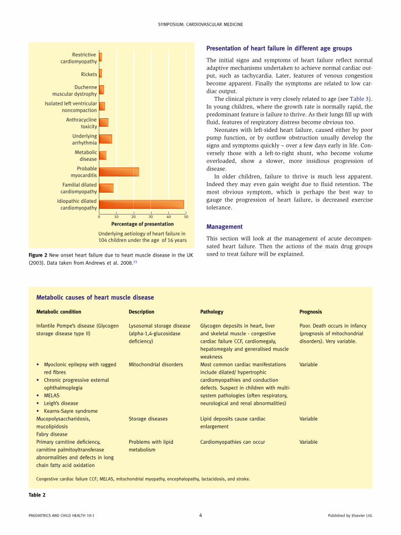
A recent prospective study of heart muscle disease in UK and Ireland has shown the incidence of new onset heart failure due to cardiomyopathy in children under 16 years to be 0.87/100 000 (see Figure 2).15
Other causes of heart muscle disease
• Myocarditis – spontaneous inflammation of the myocardium either due to an infective cause (such as Coxsackie or rubella) or because of an autoimmune or idiopathic process. The out-come is variable, and can lead to cardiac failure, which may be fatal or require heart transplantation. The most common cause of myocarditis worldwide is Chagas’ disease (caused by trypanosome cruzi), which is endemic in South America. The underlying causes of myocarditis are often not discovered, and myocardial biopsy (which is the gold standard investigation) is not routinely performed
• Infection and inflammation • Rheumatic fever • Infective endocarditis leading to acute valvular regurgita-
tion and long-term mixed valve disease • Drugs and toxins (e.g. anthracyclines) • Degenerative – primary myopathies or neurodegenerative dis-
ease, such as Friedreich’s ataxia • Metabolic (see Table 2)
Signs and symptoms
• Tachycardia • Venous congestion
• Ascites • Hepatosplenomegaly • Tachypnoea • Difficult breathing – grunting, chest wall retraction and na-
sal flare in young children • Fatigue • Failure to thrive • Cool peripheries • Exercise intolerance • Pallor • Altered consciousness
published by elsevier ltd.
symposium: cardiovascular medicine
paediaTrics and cHild HealTH 19:1 � published by elsevier ltd.
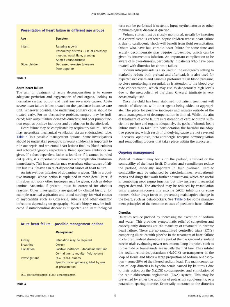
Presentation of heart failure in different age groups
The initial signs and symptoms of heart failure reflect normal adaptive mechanisms undertaken to achieve normal cardiac out-put, such as tachycardia. Later, features of venous congestion become apparent. Finally the symptoms are related to low car-diac output.
The clinical picture is very closely related to age (see Table 3). In young children, where the growth rate is normally rapid, the predominant feature is failure to thrive. As their lungs fill up with fluid, features of respiratory distress become obvious too.
Neonates with left-sided heart failure, caused either by poor pump function, or by outflow obstruction usually develop the signs and symptoms quickly – over a few days early in life. Con-versely those with a left-to-right shunt, who become volume overloaded, show a slower, more insidious progression of disease.
In older children, failure to thrive is much less apparent. Indeed they may even gain weight due to fluid retention. The most obvious symptom, which is perhaps the best way to gauge the progression of heart failure, is decreased exercise tolerance.
Management
This section will look at the management of acute decompen-sated heart failure. Then the actions of the main drug groups used to treat failure will be explained.
Metabolic causes of heart muscle disease
Metabolic condition Description Pathology Prognosis
infantile pompe’s disease (Glycogen
storage disease type ii)
lysosomal storage disease
(alpha-1,�-glucosidase
deficiency)
Glycogen deposits in heart, liver
and skeletal muscle - congestive
cardiac failure ccF, cardiomegaly,
hepatomegaly and generalised muscle
weakness
poor. death occurs in infancy
(prognosis of mitochondrial
disorders). very variable.
• myoclonic epilepsy with ragged
red fibres
• chronic progressive external
ophthalmoplegia
• melas
• leigh’s disease
• Kearns-sayre syndrome
mitochondrial disorders most common cardiac manifestations
include dilated/ hypertrophic
cardiomyopathies and conduction
defects. suspect in children with multi-
system pathologies (often respiratory,
neurological and renal abnormalities)
variable
mucopolysaccharidosis,
mucolipidosis
storage diseases lipid deposits cause cardiac
enlargement
variable
Fabry disease
primary carnitine deficiency,
carnitine palmitoyltransferase
abnormalities and defects in long
chain fatty acid oxidation
problems with lipid
metabolism
cardiomyopathies can occur variable
congestive cardiac failure ccF; melas, mitochondrial myopathy, encephalopathy, lactacidosis, and stroke.
Table 2
Underlying aetiology of heart failure in104 children under the age of 16 years
Percentage of presentation
0 10 20 30 40 50
Restrictive
cardiomyopathy
Rickets
Duchenne
muscular dystrophy
Isolated left ventricular
noncompaction
Anthracycline
toxicity
Underlying
arrhythmia
Metabolic
disease
Probable
myocarditis
Familial dilated
cardiomyopathy
Idiopathic dilated
cardiomyopathy
Figure 2 new onset heart failure due to heart muscle disease in the uK
(�00�). data taken from andrews et al. �008.15
symposium: cardiovascular medicine
Acute heart failureThe aim of treatment of acute decompensation is to ensure adequate perfusion and oxygenation of end organs, looking to normalise cardiac output and treat any reversible causes. Acute severe heart failure is best treated on the paediatric intensive care unit. Wherever possible, the underlying primary cause should be treated early. For an obstructive problem, surgery may be indi-cated; high output failure demands diuretics; and poor pump func-tion requires positive inotropes and a reduction in the afterload.
Heart failure may be complicated by respiratory failure – which may necessitate mechanical ventilation via an endotracheal tube. Table 4 lists possible management options. Some investigations should be undertaken promptly: in young children it is important to rule out sepsis and structural heart lesions first, by blood cultures and echocardiography respectively. Broad spectrum antibiotics are given. If a duct-dependent lesion is found or if it cannot be ruled out quickly, it is important to commence a prostaglandin E1infusion immediately. This intervention may exacerbate other causes of fail-ure but it is lifesaving in duct-dependent causes of heart failure.
An intravenous infusion of dopamine is given. This is a posi-tive inotrope, whose action is explained in more detail later. If this does not work other inotropes may be given, such as dobu-tamine. Anaemia, if present, must be corrected for obvious reasons. Other investigations are guided by clinical history, for example tracheal aspiration and blood serology for viral causes of myocarditis such as Coxsackie, rubella and other endemic infections depending on geography. Muscle biopsy may be indi-cated if mitochondrial disease is suspected and immunological
Acute heart failure – possible management options
Management
airway intubation may be required
Breathing oxygen
circulation positive inotropes - dopamine first line
central line to monitor fluid volume
investigations ecG, ecHo, bloods
specific investigations guided by age
at presentation
ecG, electrocardiogram; ecHo, echocardiogram.
Table 4
Presentation of heart failure in different age groups
Age Symptom
infant Faltering growth
respiratory distress - use of accessory
muscles, nasal flare, grunting
altered consciousness
older children decreased exercise tolerance
poor appetite
Table 3
paediaTrics and cHild HealTH 19:1
tests can be performed if systemic lupus erythematosus or other rheumatological disease is queried.
Volume status must be closely monitored, usually by insertion of a central venous catheter. Septic children whose heart failure is due to cardiogenic shock will benefit from fluid resuscitation. Others who have had chronic heart failure for some time and acutely decompensate may require furosemide, which can be given by intravenous infusion. An important complication to be aware of is over-diuresis, particularly in patients who have been treated with diuretics for chronic failure.
Sodium nitroprusside is also used in the emergency setting to markedly reduce both preload and afterload. It is also used for hypertensive crises and causes a profound fall in blood pressure, so close monitoring is essential, as is attention to the blood cya-nide concentration, which may rise to dangerously high levels due to the metabolism of the drug. Glyceryl trinitrate is very occasionally used.
Once the child has been stabilised, outpatient treatment will consist of diuretics, with other agents being added as appropri-ate. The place for positive inotropes and nitrates outside of the acute management of decompensation is limited. Whilst the aim of treatment of acute failure is restoration of cardiac output suffi-cient to perfuse end organs adequately, the goals of chronic heart failure must also take into consideration the harmful maladap-tive processes, which result if underlying cause are not reversed – necrosis, apoptosis, beta-adrenergic receptor downregulation and remodelling process that takes place within the myocytes.
Ongoing management
Medical treatment may focus on the preload, afterload or the contractility of the heart itself. Diuretics and venodilators reduce the preload, especially important in fluid overload. Cardiac contractility may be enhanced by catecholamines, sympathomi-metics and drugs that work further downstream, which are useful in combating poor pump function but may increase myocardial oxygen demand. The afterload may be reduced by vasodilation using angiotensin-converting enzyme (ACE) inhibitors or some nitrates. Other drugs focus on preventing maladaptive change in the heart, such as beta-blockers. See Table 5 for some manage-ment principles of the common causes of paediatric heart failure.
DiureticsDiuretics reduce preload by increasing the excretion of sodium and water. This provides symptomatic relief of congestion and consequently diuretics are the mainstay of treatment in chronic heart failure. There are no randomised controlled trials (RCTs) comparing diuretics with placebo in the treatment of heart failure in children, indeed diuretics are part of the background standard care in trials evaluating newer treatments. Loop diuretics, such as furosemide or bumetanide are usually the first line. They inhibit the sodium/chloride/potassium (Na2ClK) co-transporter in the loop of Henle and block a large proportion of sodium re-absorp-tion – some 20% of the filtered sodium load. The main complica-tion of loop diuretics is hypokalaemia caused by kaliuresis due to their action on the Na2ClK co-transporter and stimulation of the renin-aldosterone-angiotensin (RAA) system. This may be prevented by either the addition of potassium supplements, or a potassium sparing diuretic. Eventually tolerance to the diuretics
5 published by elsevier ltd.

symposium: cardiovascular medicine
may develop due to activation of the RAA system, sympathetic innervation and distal tubule hypertrophy counteracting the natriuresis. This problem is generally dealt with by the addition of another diuretic that acts elsewhere in the tubules. It is usual to add a thiazide diuretic or metolazone. They block a NaCl co-transporter in the distal convoluted tubule. Other diuretics that may be added to a loop diuretic are acetazolamide, dopamine and theophylline.
Sometimes apparent tolerance may be due to other reasons, such as inadequate perfusion of the kidneys. This may be rem-edied by increasing the cardiac output by the addition of a beta agonist. Low albumin, metabolic alkalosis and low chloride have also been noted to cause diuretic resistance.
ACE inhibitorsACE inhibitors are used to good effect in the reduction of after-load – this makes them particularly effective in the treatment of heart failure caused by left-to-right shunt, regurgitant aortic flow and left ventricular dysfunction.
In heart failure, the RAA system is activated. Renin is pro-duced in the juxtaglomerular apparatus of the kidney, in response to a fall in blood pressure or blood volume. It cleaves a plasma globulin called angiotensinogen to form angiotensin I. Angiotensin I, itself reasonably inert, is converted by angioten-sin-converting enzymes to angiotensin II, which acts primarily via the AT1 receptor to cause generalised vasoconstriction, secre-tion of aldosterone and the re-absorption of sodium in the proxi-mal convoluted tubule. ACE inhibitors prevent the conversion of angiotensin I to active angiotensin II, thus ACE inhibitors reduce cardiac afterload by vasodilatation, improve tissue perfusion and reduce congestion by natriuresis. Not only are ACE inhibitors very useful symptomatically, they also reduce cardiac myocyte remodelling by preventing fibrosis. The long-term use of ACE inhibitors may be limited by a phenomenon known as ‘aldoste-rone escape’– aldosterone secretion is stimulated independent of angiotensin II production and thus the positive effects of ACE
Management principles of the common causes of paediatric heart failure
Underlying cause Management
duct-dependent structural
defect
prostaglandin e1 to maintain patent
duct
stent or shunt insertion
left-to-right shunt if premature neonate, give i.v.
indomethacin to close pda
diuretics to prevent overload
and development of pulmonary
hypertension
surgical repair of defect
cardiomyopathy Beta-blockers
icd for arrhythmia
Heart transplant
pda, patent ductus arteriosus; icd, implantable cardioverter defibrillator.
Table 5
paediaTrics and cHild HealTH 19:1 �
inhibition are reversed. The effectiveness of ACE inhibitors has been shown by large multicentre trials in adults. One systematic review that compared mortality in populations with severe heart failure between 3 and 42 months showed a reduction in mortality of 23% in those treated with ACE inhibitors compared with pla-cebo.16 In children, studies have been more limited – one showed improved mortality17 and another showed reduced end-diastolic volume.18 A variety of ACE inhibitors are used in the treatment of heart failure in children: captopril, lisinopril and enalapril are routinely used. They are all available as oral preparations. As in adults, ACE inhibitors are contraindicated in children with bilat-eral renal artery stenosis or severe stenosis of a single function-ing kidney. Particular care must be taken when a child is also taking diuretics – the first dose of an ACE inhibitor may cause very severe hypotension. Therefore, the first dose must be very low and, where possible, the diuretic dose should be reduced, or the child must be put under close observation. In neonates, par-ticularly those who are premature, ACE inhibitors are generally avoided – the response is very unpredictable, which may be a result of a different composition of AT1 and AT2 receptors.
Angiotensin II receptor antagonistsAngiotensin receptor antagonists act in a similar way to ACE inhibitors. There are no problems of aldosterone escape with these drugs, nor do they cause the cough due to bradykinin accumulation. Some difference in their effect compared with ACE inhibitors may be postulated since the sartans are specific AT1 blockers, and young children have a relatively higher concentra-tion of AT2 receptors. These drugs are not widely used in clinical practice, and are reserved for children who cannot tolerate ACE inhibitors as there is much less evidence for them, although the limited number of adult studies suggest they are just as effec-tive.19 Losartan is the most commonly used angiotensin receptor antagonist in clinical practice.
Inotropic medicationsA variety of drugs act primarily to increase the force of cardiac contraction. They may act directly on the beta-adrenergic recep-tors, downstream in the cyclic adenosine monophosphate (AMP) cascade, on the sodium ion channels or on the sodium-calcium exchanger – all of which act to increase the amount of intracellu-lar calcium available for myocyte contraction, or to sensitise the contractile machinery to the calcium already present. • Dobutamine and adrenaline stimulate β1 receptors directly
acting in the same way as endogenous catecholamines, and have the same limitations – the eventual receptor downregu-lation, necrosis and apoptosis of the myocytes and an increase in oxygen demand.
• Phosphodiesterase inhibitors – such as amrinone, enoximone and milrinone – work downstream. They prevent the break-down of cAMP by phosphodiesterase III to reduce the after-load. Despite the initial symptomatic benefit of these drugs, a Cochrane review of phosphodiesterases in adults showed that they are associated with increased mortality due to a pro-ar-rhythmic effect.20 Nonetheless, oral enoximone is commonly used in paediatric specialist centres for its combination of ino-tropic and vasodilatory effects.
• Levosimendan is a positive inotrope that acts independently of the adenylyl cyclase pathway. Levosimendan binds troponin
published by elsevier ltd.

symposium: cardiovascular medicine
C to sensitise the cardiac myocytes to calcium. It also activates KATP channels in vascular smooth muscle – opening them to cause relaxation – and thus reducing preload and afterload. The Survival of Patients With Acute Heart Failure in Need of Intravenous Inotropic Support (SURVIVE) trial – an RCT comparing dobutamine with levosimendan in 1327 adults re-quiring inotropic support – showed levosimendan was equal-ly as effective with no significant difference in mortality at 6 months.21 Retrospective studies in small numbers of children have shown the drug to be safe and to allow a reduction in the use of conventional inotropes – which are associated with increased long-term mortality.22,23
Cardiac glycosidesIt is thought that cardiac glycosides, such as digoxin, inhibit the sodium-potassium pump – the consequent increase in intracel-lular sodium reduces the inward sodium current, which is also what drives the sodium-calcium exchanger – so there is also an increase in intracellular calcium. Digoxin thus has positive ino-tropic effects. Digoxin also causes an increase in vagal outflow, which slows the rate and allows the heart to fill up adequately; however, this increase may further drive the maladaptive sympa-thetic nervous system response. Digoxin has a narrow therapeu-tic window and its levels in the bloodstream must be monitored to avoid toxicity. It is not routinely used in children.
NitratesDrugs which are metabolised to nitric oxide – such as glyceryl trinitrate, isosorbide mononitrate and nitroprusside – cause a fall in peripheral vascular resistance, which may translate to a fall in preload or afterload or both depending on the type of nitrate used. Nitric oxide activates guanylate cyclase to increase the formation of cGMP, activating protein kinase G leading to the relaxation of vascular smooth muscle. These drugs have been limited so far to the acute setting, but an increased use for them may be found in chronic heart failure. The VHeFT trial showed that nitrates and hydralazine improved survival in adult patients with symptomatic heart failure.24 Nitrates are particularly useful in patients who cannot tolerate ACE inhibitors.
Beta-blockersIt seems somewhat counterintuitive to use negative inotropes in the treatment of heart failure. Nonetheless beta-blockers are invaluable in treating some of the counter-regulatory features of the syndrome, namely the activation of the sympathetic nervous system and RAA system. Reduction in RAA system activation reduces the preload and consequently reduces ventricular wall stress and cardiomyocyte remodelling. Initially beta-blockers may exacerbate the symptoms of heart failure, so it is impor-tant to start on a very low dose and titrate carefully. The initial relapse is less severe with drugs like carvedilol, compared with propranolol, as it causes a degree of peripheral vasodilation. It is known that beta-blockers improve symptoms and survival in adults – one systematic review showed a mortality reduction of 8% compared with placebo in heart failure of any severity25; this was increased to 25% when the heart failure was more severe.26 When different beta-blockers were compared with each other, it was found that carvedilol extends survival com-pared with metoprolol.27 This is probably due to the peripheral
paediaTrics and cHild HealTH 19:1 �
vasodilatation caused by carvedilol – the drug also reduces pre-load. A recent RCT failed to show a statistically different out-come for carvedilol compared with placebo in 161 children and adolescents with symptomatic heart failure.28 This trial illus-trates the potential danger of extrapolating results from adult RCTs to paediatrics where the pathophysiology is not equivalent, yet also highlights the challenges of developing evidence-based therapeutics in children – it is not simple to design well-powered and ethical trials.
AnticoagulationChronic heart failure puts children at an increased risk of ventric-ular mural thrombus. Aspirin prophylaxis may be indicated after cardiac surgery and in patients with diastolic heart failure, who are at particular risk of emboli. However, there have been some reports of aspirin exacerbating heart failure, by reducing the effects of ACE inhibitors and diuretics.29 Warfarin may also be used prophylactically, although this requires frequent monitoring of international normalised ratio.
SurgeryCongenital cardiac problems can generally be treated definitively with surgery or catheterisation in developed countries. Cardio-myopathies and myocarditis are treated supportively with drugs and most patients recover or are stabilised with medication. If this does not control symptoms, cardiac transplantation is the only effective treatment. Whilst waiting for a donor heart, bridg-ing devices may be used. The most common is a left ventricular assist device, a pump which replaces the left ventricle. Blood is pulled from the left ventricle into a pump that sits in the upper abdomen. From there the blood is forcibly ejected into the aorta. Occasionally, the left ventricular assist device prevents the need for future transplantation and the function of the failing heart returns.
The Fontan operation may be performed for children with only one functioning ventricle. Although this procedure is life-saving in infants, it is associated with development of dilated cardiomyopathy and subsequent heart failure later in life.
Conclusion
Heart failure, although rare, is still a cause of considerable mor-bidity and mortality in children. It may present at any stage from foetus onwards. Heart failure is a clinically defined syn-drome, just as in adults. Its causes, however, could not be more different, including: a wide variety of congenital structural defects, metabolic disease, infection, and autoimmune disease. As ever, knowledge of aetiology is imperative for successful man-agement. Medical and surgical advances have increased survival substantially, but present complications of their own, making the management of paediatric heart failure a complex but rewarding field. The challenge in the future will be to find evidence-based therapies for children. ◆
REFERENCES
1 Fenton m, Burch m. understanding chronic heart failure. Arch Dis
Child �00�; 92: 81�–81�.
published by elsevier ltd.

symposium: cardiovascular medicine
2 attas m, Hanley HG, stultz d, et al. Fulminant beriberi heart disease
with lactic acidosis: presentation of a case with evaluation of left
ventricular function and review of pathophysiologic mechanisms.
Circulation 19�8; 58(� pt 1): 5��–5��.
3 Wexler d, silverberg d, Blum m, et al. anaemia as a contributor to
morbidity and mortality in congestive heart failure. Nephrol Dial
Transplant �005; 20(suppl. �): vii11–5.
4 Haddad F, doyle r, murphy dJ, et al. right ventricular function in
cardiovascular disease, part ii: pathophysiology, clinical importance,
and management of right ventricular failure. Circulation �008; 117:
1�1�–1��1.
5 chatterjee K, rame Je. systolic heart failure: chronic and acute
syndromes. Crit Care Med �008; 36(1 suppl): s��–s51.
6 Taha m, lopaschuk Gd. alterations in energy metabolism in
cardiomyopathies. Ann Med �00�; 39: 59�–�0�.
7 Huss Jm, Kelly dp. mitochondrial energy metabolism in heart failure:
a question of balance. J Clin Invest �005; 115: 5��–555.
8 Bers dm, despa s. cardiac myocytes ca�+ and na+ regulation in
normal and failing hearts. J Pharm Sci �00�; 100: �15–���.
9 miyata s, minobe W, Bristow mr, et al. myosin heavy chain isoform
expression in the failing and nonfailing human heart. Circ Res �000;
86: �8�–�90.
10 Tulloh r. in: lissauer T, clayden G, eds. �rd edn Illustrated textbook
of paediatrics; chapter 1� cardiac disorders; vol. 1. edinburgh:
mosby, �00�.
11 andrews r, Tulloh r. atrial septal defect with failure to thrive in
infancy: hidden pulmonary vascular disease? Pediatr Cardiol �00�;
23(5): 5�8–5�0.
12 Johnson WH, moller JH. paediatric cardiology. 1st edn Core
Handbooks in Paediatrics ; vol. 1. london: lipincott Williams and
Wilkins, �001.
13 Walsh ma, duff d, oslizlok p, et al. a review of 15-year experience
with anomalous origin of the left coronary artery. Ir J Med Sci �008;
177(�): 1��–1�0.
14 cooper Ha, Gersh BJ. Treatment of chronic mitral regurgitation. Am
Heart J 1998; 135(� pt 1): 9�5–9��.
15 andrews re, Fenton mJ, ridout da, et al. new-onset heart failure
due to heart muscle disease in childhood: a prospective study in
the united Kingdom and ireland. Circulation �008; 117: �9–8�.
16 Flather m, yusuf s, Kober l, et al. for the ace-inhibitor myocardial
infarction collaborative Grouplong-term ace-inhibitor therapy
in patients with heart failure or left-ventricular dysfunction: a
systematic overview of data from individual patients. Lancet �000;
355: 15�5–1581.
17 lewis aB, chabot m. The effect of treatment with angiotensin-
converting enzyme inhibitors on survival of pediatric patients with
dilated cardiomyopathy. Pediatr Cardiol 199�; 14: 9–1�.
18 stern H, Weil J, Genz T, et al. captopril in children with dilated
cardiomyopathy: acute and long-term effects in a prospective study
of hemodynamic and hormonal effects. Pediatr Cardiol 1990; 11:
��–�8.
paediaTrics and cHild HealTH 19:1 8
19 lee vc, rhew dc, dylan m, et al. meta-analysis: angiotensin-
receptor blockers in chronic heart failure and high risk acute
myocardial infarction. Ann Intern Med �00�; 141: �9�–�0�.
20 amsallem e, Kasparian c, Haddour G, Boissel Jp, nony p.
phosphodiesterase iii inhibitors for heart failure. Cochrane Database
Syst Rev �005(1) cd00���0.
21 mebazaa a, nieminen ms, packer m, et al. for the survive
investigators. levosimendan vs dobutamine for patients with acute
decompensated heart failure. The survive randomized trial. JAMA
�00�; 297: 188�–1891.
22 namachivayam p, crossland ds, Butt WW, shekerdemian ls.
early experience with levosimendan in children with ventricular
dysfunction. Pediatr Crit Care Med �00�; 7: ��5–8.
23 egan Jr, clarke aJ, Williams s, et al. levosimendan for low cardiac
output: a pediatric experience. J Intensive Care Med �00�; 21:
18�–�.
24 cohn Jn, Johnson G, Ziesche s, et al. a comparison of enalapril
with hydralazine-isosorbide dinitrate in the treatment of chronic
congestive heart failure. N Engl J Med 1991; 325: �0�–�10.
25 Brophy Jm, Joseph l, rouleau Jl. Beta-blockers in congestive heart
failure: a Bayesian meta-analysis. Ann Intern Med �001; 134:
550–5�0.
26 Whorlow sl, Krum H. meta-analysis of effect of beta-blocker therapy
on mortality in patients with new york Heart association class iv
chronic congestive heart failure. Am J Cardiol �000; 86: 88�–889.
27 poole-Wilson pa, swedberg K, cleland JG, et al. comparison of
carvedilol and metoprolol on clinical outcomes in patients with
chronic heart failure in the carvedilol or metoprolol european Trial
(comeT): randomised controlled trial. Lancet �00�; 362: �–1�.
28 shaddy re, Boucek mm, Hsu dT, et al. pediatric carvedilol study
Group. carvedilol for children and adolescents with heart failure: a
randomized controlled trial. JAMA �00�; 298: 11�1–11�9.
29 pitt B, yusuf s. studies of left ventricular dysfunction (solvd):
subgroup results. J Am Coll Cardiol 199�; 19: �15a.
Practice points
• paediatric heart failure is a clinical diagnosis.
• age of onset helps determine aetiology.
• The main aim of acute management is to optimise cardiac
output.
• surgical management is the only definitive treatment for
structural congenital heart lesions.
• long term medical management aims to reduce maladaptive
processes, manage symptoms and where possible reverse the
underlying cause.
• existing medical management is largely based on data from
adult trials and well designed, high powered trials need to be
carried out in children.
published by elsevier ltd.