Embed Size (px)
Citation preview
Page 1/13
E�cacy of conventional �uoridated toothpastes containing arginine andhigh-�uoride toothpastes on the control of root caries in patientsundergone radiotherapy of head-and-neck: a randomized controlled trialMarilia Velo
Universidade de Sao Paulo Faculdade de Odontologia de BauruMarina Giacominni
Universidade de Sao Paulo Faculdade de Odontologia de BauruLeticia Brianezzi
Universidade de Sao Paulo Faculdade de Odontologia de BauruRafael Gonçalves
Universidade de Sao Paulo Faculdade de Odontologia de BauruGiovanna Zabeu
Universidade de Sao Paulo Faculdade de Odontologia de BauruPaulo Santos
Universidade de Sao Paulo Faculdade de Odontologia de BauruSergio Ishikiriama
Universidade de Sao Paulo Faculdade de Odontologia de BauruHeitor Honório
Universidade de Sao Paulo Faculdade de Odontologia de BauruLinda Wang ( [email protected] )
Universidade de Sao Paulo Faculdade de Odontologia de Bauru
Research Article
Keywords: Arginine, High-�uoride toothpaste, Radiotherapy of head-and-neck, Root caries.
Posted Date: May 3rd, 2021
DOI: https://doi.org/10.21203/rs.3.rs-360231/v1
License: This work is licensed under a Creative Commons Attribution 4.0 International License. Read Full License

Page 2/13
AbstractThis parallel, triple-blind RCT evaluated the restorative performance of a resin-modi�ed glass-ionomer-cement (RMGIC) in irradiated patients and theprevention of root caries lesions adjacent to restoration, comparing the effect of conventional (control) concentration, high-�uoride (F) containingfTCP and arginine-based toothpastes. A total of 63 lesions was screened and 60 were included into randomized distribution into three groups (N-participants in baseline/n- root caries lesions): G1 = 1,450 ppm F (N = 10/n = 17); G2 = 5,000 ppm F + fTCP (N = 7/n = 18) and G3 = 1,450 ppm F + arginine + CaCO3 (N = 6/n = 25). Based on eligibility criteria, all patients were mandatory enrolled after completed 3-month of radiotherapy of head-and-neck. Two calibrated operators performed the restorative procedures (RMGIC - Vitremer) and two calibrated examiners (Kappa = 0.94) evaluatedthe restorations based on modi�ed USPHS criteria at baseline, 1, 3 and 6-month follow-up. Data was collected and statistically assessed withKruskal-Wallis test (p < 0.05). There were no statistically signi�cance differences among the performance of the restoration among the three groupsregarding the criteria retention, marginal adaptation, marginal staining, post-operative sensitivity, adjacent caries, color alteration, anatomic form andsurface texture (p > 0.05). Even with oral complications caused by radiation-therapy, if the restorations are properly performed and patients are underprofessional supervision, high-F presented similar e�cacy of arginine and conventional-containing toothpastes to prevent secondary caries. Clinicalrelevance: This clinical trial brings new evidences about the regular use of high-F, arginine-based and conventional-F containing toothpastes inirradiated patients under supervision of a multidisciplinary team and the encouragement of self-cooperation.
IntroductionWhile the decline in the prevalence and incidence of coronal dental caries has been observed worldwide in children and adults, an increase in theprevalence of root caries, especially in patients such as the elderly population and patients undergone radiotherapy of head-and-neck has beennotable [1]. As life expectancy is increasing, older adults often retain a higher number of teeth in their oral cavity, which increases the risk of cariesdevelopment and progression on their exposed root surfaces in the case of the presence of etiological factors [2]. In addition, though not at allsubsites, head-and-neck cancer becomes more frequent with increasing age [2], although the incidence of oral cancer disease in people under the ageof 45 appears to be increasing [3] and, these patients are also often submitted to radiation treatment.
Despite radiotherapy plays a vital role in curative and palliative cancer therapy [4], its adverse effects in the oral cavity are of clinical concern asusually complex oral complications are inevitable, including mucositis, hyposalivation, osteoradionecrosis, dentition breakdown, and radiation-related caries [5, 6]. Radiation-related caries lesions develop mainly in the cervical area, which include root caries as a notable clinical complication inthese patients. As human dentin is highly soluble because of its less mineral and higher carbonate and magnesium content [7], dentin root cariesrapidly progresses and the risk of radiation-related caries development with its sudden onset is a lifelong threat [8]. As consequence, it can developas early as three-month post-radiotherapy resulting in a devastation of a healthy dentition within one year of treatment [9, 10].
The prevalence of dental caries has previously been estimated at 24% for cancer patients treated with radiotherapy and the etiology of this increasedrisk is multifactorial [2, 11]. Beside the factors that control the risk of dental caries development such as the presence of a cariogenic bio�lm,fermentable carbohydrate intake and low �uoride exposure [12], radiotherapy-induced salivary gland acinar degeneration and interstitial �brosis,which means that these patients present chronic hyposalivation and are at high-risk for radiation-related caries development and progression [2,].Alterations in dental tissues and modi�cation in the composition or structural pattern of root dentin may also be important contributing factors forradiation-related caries development [7, 13]. All these complications affect their feeding process, especially due to the sensitivity, di�cult tomastication and dental cleaning [6]. Therefore, efforts should be focused from prevention to manage patients with severe caries, which can beaccomplished by preventive and therapeutic strategies or reconstruction of caries lesion in these patients to reestablish their quality-of-life.
Resin-modi�ed glass ionomer cements (RMGIC) are a great choice for restoration of root caries lesions [14]. However, restorative management ofradiation-caries can be challenging, as the changes presented by radiation affect the bonding of adhesive materials [15]. Owing to the bondingcapacity to both enamel and dentin substrates and ability to release �uoride (F), RMGIC is expected to be more resistant to avoid caries lesionsadjacent to restorations. This restorative material is considered the gold standard material, but based on the current best of knowledge, theinvolvement of the patients and their self-cooperation is of great relevance to the successful approach overtime, since the effect of F-released can bereplaced by F delivered from other sources, such as toothpastes [16, 17]. The mechanism of action of F toothpastes combines a mechanicaldisruption of tooth bio�lm and F delivery, interfering with the demineralization and remineralization episodes, but this effect is in�uenced by Fconcentration [18].
Some in situ and in vivo evidences demonstrated the bene�t of high-F containing toothpastes (5,000 ppm F) to prevent initial caries developmentand to repair root caries lesions when compared with the use of conventional-F toothpastes [19–21]. When high-F concentration toothpaste isapplied, beside its mechanism of action, F also develop a calcium (Ca)-�uoride reservoir (CaF2-like particles) and provides a physical barrier on thesubstrate, inducing remineralization by maintaining high concentrations of F in the oral environment [22]. The common protocol used to reduce postradiation-related caries is the daily topical application of 1% neutral sodium F gel (5,000 ppm F) with custom-made F carriers [23]. However,compliance of patients is frequently poor because of the inconvenient method of application and many patients with head-and-neck cancer may bedebilitated to use both conventional toothpaste and topical gel application [24]. Whereas toothbrushing with �uoridated toothpastes is the most cost-

Page 3/13
effective tool to prevent dental caries [25], in patients undergoing radiotherapy of head-and-neck, the daily use of high-F toothpaste could be a greatchoice to prevent new caries development around restorations with RMGIC.
Furthermore, attempts to improve remineralization e�cacy from toothpastes include the calcium phosphate-based delivery systems such asfunctionalized tri-calcium phosphate (fTCP), calcium phosphate nanocomplexes (CPP-ACP) or insoluble Ca compound associated with arginine.Some in vitro studies demonstrated that toothpastes containing fTCP prevented Ca from prematurely interacting with ionic F, enhancing their effect[26, 27]. However, even available as a commercial product, the remineralization e�cacy regarding fTCP products are limited to in vitro researches andfocused on enamel substrate [28]. A previous in situ design showed that toothpaste containing fTCP presented similar effect to prevent early rootcaries lesions, although it could be interesting to repair active dentin caries lesions and should be evaluated in patients at high-risk of caries [19].
The toothpaste containing an insoluble Ca compound, 1.5% arginine and conventional-F concentration was also introduced to prevent cariesdevelopment at an earlier stage targeting the residual bio�lm through arginine metabolism [29]. The association with Ca ions provided by CaCO3
compound inhibits mineral loss during low-pH periods and repair it when pH returns to neutral condition. An in vivo study demonstrated that patientswho used arginine-based toothpaste had signi�cantly higher plaque pH values before and after the sucrose challenge [30]. Other investigations haveshown positive bene�ts by using arginine-based pastes to manage root caries when compared to conventional ones [31], which boost for newevidences of this technology in post-radiation caries in patients undergone radiotherapy of head-and-neck.
Thus, the purpose of this clinical trial was to test the effectiveness of high-F toothpaste (5000 ppm F) and arginine-based toothpaste on root carieslesions around RMGIC evaluating the performance of the restorations.
MethodsTrial design
This single-center, interventional, parallel, triple-blind 6-month randomized controlled clinical trial was conducted in the Clinical Research Center ofthe Bauru School of Dentistry, University of São Paulo, Brazil (44944915.4.0000.5417). The study was approved by Human Ethics in ClinicalResearch Committees of the Bauru School of Dentistry and registered at the Registro Brasileiro de Ensaios Clínicos – REBEC website under theregistration number U1111-1171-2738. This randomized controlled trial is reported in accordance with the CONSORT (Consolidated Statement OfReporting Trials) statement [32] (Fig. 1). A consent form was shown for the participants that explained the nature of the study and only after theyassigned it the study has initiated.Study population and sampling procedure
Based on eligibility criteria, all patients were enrolled after at least 3-month completed the radiotherapy. Adult patients diagnosed with untreated rootcaries were selected to be included in the trial. The sample size was determined considering the 80% of power, 25% of drop out and signi�cance leveland statistical power were adopted at 5%. The main response variables were related to the caries adjacent to the restoration and marginal leakage,considering the difference of 1 level of USPHS system recording. The randomly allocated sequence was implemented using Microsoft Excel for Mac2011, Version 14.3.5 (Microsoft, Chicago, IL, USA) based on simple randomization strategy.
To compensate for the expected number of drop-outs over 6-month the Intention-to-Treat (ITT) analysis was performed to include every patient in theprimary analysis within the respective treatment group they have been assigned to at randomization. The experimental unit was the restored tooth.
At baseline, participants were from 18 to 80 years old. The eligibility criteria for the recruitment are listed in Table 1. Before inclusion and exclusioncriteria evaluation, all patients were clinical analyzed by a multidisciplinary team, including nutritionist, speech therapist and dentist. Patientspresenting post-radiation root caries were recruited and the included subjects received instructions on oral health, particularly regarding oral hygieneand sugar consumption. Their oral cavity was evaluated and if they presented active coronal decay or other disease, they were treated accordingly.

Page 4/13
Table 1Inclusion and exclusion criteria for the recruitment of subjects
Inclusion criteria
Age group minimum of 18 years
Must have at least one root caries lesion with pulp vitality
Teeth included must be not be compromised or crowned
Patients who have their teeth treated before radiotherapy sections
Exclusion criteria
Patients undergoing radiation therapy
Patients ongoing �xed orthodontic appliance therapy, with acute periodontitis, severe bruxism and other parafunctional habits
Patients with serious systemic complications
Pregnancy, lactating or hypersensitive to the trial test products or ingredients
Undergone high-F therapy 3-month prior to this trial
Local or systemic antibiotic therapy 3-month prior to this trial or within the 6-month of the trial
Interventions
The selected patients were allocated to one of three groups using a sequence of codes randomly generated by the blind-study administrator in anExcel program. The control group participants (Group 1) were administered with conventional-F toothpaste: G1 = 1,450 ppm F (Colgate Total 12®;Colgate-Palmolive Company, São Paulo, SP, Brazil). The tested groups (Groups 2 and 3) used toothpastes containing: G2 = 5,000 ppm F + fTCP(Clinpro® 5000; 3M, Sumaré, SP, Brazil) and G3 = 1,450 ppm F + arginine + CaCO3 (Neutraçucar®, Colgate-Palmolive Company, São Paulo, SP, Brazil).
The compositions of toothpastes and all material used in this study are better described in Table 2. To blind the volunteers and examiners, thetoothpastes were packed in blank tubes and labeled with colors to identify them only for the study administrator. All patients received separatepackets containing two tubes of toothpastes and a standard soft bristled adult toothbrush (CS 5460B, Curaprox). Participants were instructed tobrush at least twice daily with their assigned toothpaste. Two tubes of toothpastes, and a new toothbrush were supplied to each patient after 45days, but additional tubes of toothpaste were available upon their request.
Table 2Composition and classi�cation of all materials used in this study.
MATERIAL COMPOSITION MANUFACTURER CLASSIFICATION
Vitremer® Primer: polyalkenoic acid, methacrylate groups, water, HEMA, camphorquinone.Powder: �uoraluminium silicate crystals, potassium persulfate, ascorbic acid,pigments. Liquid: polyalkenoic acid, methacrylate groups, water, HEMA,camphorquinone
3M ESPE, StPaul, MN, USA
Resin-modi�edglass ionomercement
Colgate Total12®
1,450 ppm F (sodium �uoride-NaF) Colgate-Palmolive®, SãoPaulo, SP, Brazil
Dentifrice 1,450ppm F(conventionalconcentration)
Clinpro5000®
1.1% sodium �uoride, water, sorbitol, hydrated
silica, glycerin, polyethylene-polypropylene glycol, �avor, polyethylene
glycol, sodium lauryl sulfate, titanium dioxide, carboxymethyl cellulose,
sodium saccharin and tri-calcium phosphate
3M ESPE, StPaul, MN, USA
High-�uoridetootpaste (5,000ppm F) plusfunctionalized tri-calciumphosphate (fTCP)
Neutraçucar® 1,450 ppm F (Sodium Mono�uorphosphate - MFP), calcium carbonate, sodium laurylsulfate, sodium saccharin, tetrasodium pyrophosphate, sodium silicate, polyethyleneglycol, sorbitol, carboxymethyl cellulose, methylparaben, propylparaben, aromaticcomposition and water
Colgate-Palmolive®, SãoPaulo, SP, Brazil
Dentifrice(conventional Fconcentration)plus 1.5% arginineplus CaCO3
Restorative procedure
One or more active root caries lesion for each subject was selected for inclusion in the study and restorations were performed by two calibrateddentist operators (MMACV and RSG).

Page 5/13
All selected patients received a prophylaxis with pumice stone and distilled water, applied with a Robinson brush prior to the intervention. Firstly,carious tissue was carefully removed using spherical bur nos. 1, 2, and 3 (KG Sorensen, Cotia, SP, Brazil) at low speed and hand excavators removingall demineralized dentin on lateral walls. Cavity preparation was limited to carious tissue removed, based on the selective carious dentin removal.Manual instruments (gingival margin cutters) were used for �nishing the cervical and extern margins. All surfaces were cleaned, and the cavitiesrestored using a RMGIC (Table 2) based on the manufacturer's instructions. Calcium hydroxide (Dycal, Dentsply Caulk, Germany) was inserted whendeep cavities were presented. The exposed dentin surface was kept visibly moist for the primer application (30s of application followed by 15s ofsoft air jet), and then it was photo-activated for 30s. (1: 1 ratio), The restorative material (1:1 ration) was positioned over lesions using an appliedsyringe (Centrix) and photo-activated by 40 s. A layer of the Finishing Gloss component was applied to the restored surface, preventing theoccurrence of synergy and imbibition. Possible excesses of restorative material were removed with a scalpel blade and after 7 days, �nishing andpolishing were performed in the restorations. Then, participants received their packets with respective toothpastes to beginning the study.Clinical examination methods
The restorations were examined at baseline, 1, 3 and 6 months by two calibrated examiners (LW and SKY, Kappa = 0.94). Mouth mirrors, woodenspatulas, and the CPI (Community Periodontal Index) probe were the dental instruments used. The clinical assessment has begun with anexamination of the soft and hard tissues of the mouth and then, the modi�ed USPHS criteria was used for restorations evaluation (Table 3). Allrestorations were scored as follows: alpha as the ideal clinical situation; Bravo was clinically acceptable (satisfactory success); Charlie representedclinically unacceptable situations where the restoration had to be replaced or lost retention (unsatisfactory fail) (Table 3).

Page 6/13
Table 3Modi�ed USPHS criteria
Criterion Code Description
Anatomicform
Alpha(A)
Restoration maintains continuity with dental surface
Bravo(B)
Presence of sub-contour without dentin exposure
Charlie(C)
Loss of material exposing dentin
Marginaladaptation
Alpha(A)
Continuity at the margin (without protrusion or gap)
Bravo(B)
Small discontinuity detectable with explorer probe, but does not require replacement
Charlie(C)
Marginal gap that requires replacement
Coloralteration
Alpha(A)
The restoration appears to match the shade and translucency of adjacent tooth structure
Bravo(B)
The restoration does not match the shade and translucency of adjacent tooth structure, but the mismatch is within thenormal range of tooth shades
Charlie(C)
The restoration does not match the shade and translucency of adjacent tooth structure, and the mismatch is outsidethe normal range of tooth shades and translucency
Marginalstaining
Alpha(A)
There was no visual evidence of marginal discoloration different from the color
of the restorative material and from the color of the adjacent tooth structure
Bravo(B)
There was visual evidence of marginal discoloration at the junction of the tooth structure and the restoration that hasnot penetrated along the restoration
Charlie(C)
There was visual evidence of marginal discoloration at the junction of the tooth structure and the restoration, and thediscoloration has penetrated along the restoration
Retention Alpha(A)
No loss of restorative material
Bravo(B)
Restorative material was partially lost
Charlie(C)
Absence of restorative material
Sensibility Alpha(A)
Not present
Charlie(C)
Present (constant sensitivity, not diminishing in intensity)
Adjacentcaries
Alpha(A)
The restoration is a continuation of existing anatomic form adjacent to the restoration and there was no cariesevidence
Charlie(C)
There is visual evidence of dark, deep discoloration adjacent to the restoration
Photographic documentation with a Digital Camera (EOS 60D Macro-Lens EF 100 mm f/2.8 USM) and Canon Flash Macro Ring Lite MR-14EX(Canon do Brasil Indústria e Comércio, São Paulo, SP, Brazil) with the aid of a photo mirror was performed before and after treatment and at allevaluation points.
Statistical methodsThe results were analyzed using SPSS software (Statistical Package for Social Sciences, IBM Inc., USA).
To analyze the distribution of the scores according to the modi�ed USPHS criteria, the Kruskal-Wallis test was used (p < 0.05). Intention to treatanalysis (ITT) was used to analyze the results.
Results

Page 7/13
A total of 68 subjects was examined and 26 was screened for the study. As root caries lesion was considered the experimental unit, a total of 63lesions was screened but only 60 were included to randomization (Fig. 1). After allocation, they were distributed into the 3 groups: G1 = 10participants in the baseline (n = 17 root caries lesions); G2 = 7 participants (n = 18 root caries lesions) and G3 = 6 participants (n = 25 root carieslesions). There were no observed or reported adverse reactions to the use of either product.
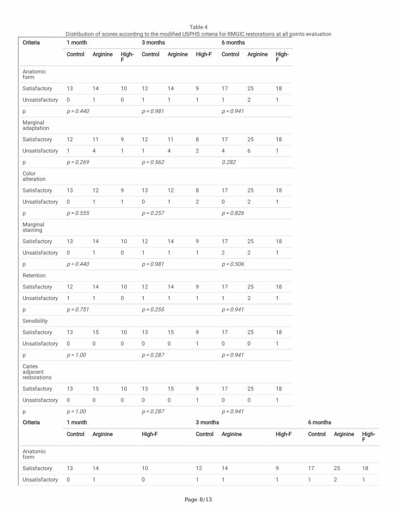
The changes in the modi�ed USPHS are tabulated in Table 4. There were no statistically signi�cance differences among the performance of therestoration among the three groups regarding the criteria retention, marginal adaptation, marginal staining, post-operative sensitivity, adjacent caries,color alteration, anatomic form and surface texture (p > 0.05).
Page 8/13
Table 4Distribution of scores according to the modi�ed USPHS criteria for RMGIC restorations at all points evaluation
Criteria 1 month 3 months 6 months
Control Arginine High-F
Control Arginine High-F Control Arginine High-F
Anatomicform
Satisfactory 13 14 10 12 14 9 17 25 18
Unsatisfactory 0 1 0 1 1 1 1 2 1
p p = 0.440 p = 0.981 p = 0.941
Marginaladaptation
Satisfactory 12 11 9 12 11 8 17 25 18
Unsatisfactory 1 4 1 1 4 2 4 6 1
p p = 0.269 p = 0.562 0.282
Coloralteration
Satisfactory 13 12 9 13 12 8 17 25 18
Unsatisfactory 0 1 1 0 1 2 0 2 1
p p = 0.555 p = 0.257 p = 0.826
Marginalstaining
Satisfactory 13 14 10 12 14 9 17 25 18
Unsatisfactory 0 1 0 1 1 1 2 2 1
p p = 0.440 p = 0.981 p = 0.506
Retention
Satisfactory 12 14 10 12 14 9 17 25 18
Unsatisfactory 1 1 0 1 1 1 1 2 1
p p = 0.751 p = 0.255 p = 0.941
Sensibility
Satisfactory 13 15 10 13 15 9 17 25 18
Unsatisfactory 0 0 0 0 0 1 0 0 1
p p = 1.00 p = 0.287 p = 0.941
Cariesadjacentrestorations
Satisfactory 13 15 10 13 15 9 17 25 18
Unsatisfactory 0 0 0 0 0 1 0 0 1
p p = 1.00 p = 0.287 p = 0.941
Criteria 1 month 3 months 6 months
Control Arginine High-F Control Arginine High-F Control Arginine High-F
Anatomicform
Satisfactory 13 14 10 12 14 9 17 25 18
Unsatisfactory 0 1 0 1 1 1 1 2 1

Page 9/13
Criteria 1 month 3 months 6 months
Control Arginine High-F
Control Arginine High-F Control Arginine High-F
p p = 0.440 p = 0.981 p = 0.941
Marginaladaptation
Satisfactory 12 11 9 12 11 8 17 25 18
Unsatisfactory 1 4 1 1 4 2 4 6 1
p p = 0.269 p = 0.562 0.282
Coloralteration
Satisfactory 13 12 9 13 12 8 17 25 18
Unsatisfactory 0 1 1 0 1 2 0 2 1
p p = 0.555 p = 0.257 p = 0.826
Marginalstaining
Satisfactory 13 14 10 12 14 9 17 25 18
Unsatisfactory 0 1 0 1 1 1 2 2 1
p p = 0.440 p = 0.981 p = 0.506
Retention
Satisfactory 12 14 10 12 14 9 17 25 18
Unsatisfactory 1 1 0 1 1 1 1 2 1
p p = 0.751 p = 0.255 p = 0.941
Sensibility
Satisfactory 13 15 10 13 15 9 17 25 18
Unsatisfactory 0 0 0 0 0 1 0 0 1
p p = 1.00 p = 0.287 p = 0.941
Cariesadjacentrestorations
Satisfactory 13 15 10 13 15 9 17 25 18
Unsatisfactory 0 0 0 0 0 1 0 0 1
p p = 1.00 p = 0.287 p = 0.941
DiscussionA daily topical application of 1% neutral sodium F gel (5,000 ppm F) with custom-made F carriers and the use of neutral F-containing mouth rinseshave demonstrated to be bene�cial in preventing caries occurrence in irradiated patients [9, 28]; however, the compliance of patients to theseprotocols is poor because of the inconvenient method of application. The preference for any effective approach usually relies on a simpler, feasiblepreventive intervention that is easily applied at-home. Moreover, many patients with head-and-neck cancer are frequently debilitated [24] to useconventional toothpastes to brush their teeth followed by topical application of �uoride. On the other hand, toothbrushing with �uoridatedtoothpastes has been considered the most cost-bene�cial tool as it combines the mechanical disruption of dental bio�lm with additional F deliverythat will act in demineralization and remineralization process [25].
Literature has shown bene�ts of using conventional �uoridated toothpastes in the prevention and treatments for dental caries in children and youngadults [18]. Despite such advantages, its effect is in�uenced by F concentration and evidences until now suggest that high-F toothpaste providesbetter control on root caries lesions than conventional F-containing toothpaste [18]. According to Duane (2012), high-F toothpaste has a greater
Page 10/13
impact on individuals at high-risk who do not use toothpaste regularly or do not brush their teeth twice a day, which highlights the importance toevaluate their effect in patients undergone radiotherapy [33]. The current clinical trial found no difference among the treatment groups (p < 0.05), i.e,our �ndings suggest that the use of 5,000 ppm F-containing toothpaste is similar to the remineralizing dentifrice (containing 1.5% arginine, Cacompound and conventional-F concentration) and conventional containing-toothpaste on preventing the development of root caries lesions aroundrestorations in patients undergone radiotherapy, but reasons could in�uence such results.
Although all careful have been done to design this clinical trial, the course of the research showed a dropout rate of 24% after 6-month.Notwithstanding, we have performed the analysis considering the surface level to increase the power of the study. All efforts were made by the teamto evaluate all participants and to �nd some of them who not returned for the evaluation points. Unfortunately, loss of follow-up was due to theworsening of the initial condition in relation to the cancer disease or death of the participants. For this reason, our follow-up period was limited to thisfeasible follow-up times. The effects of �uoride toothpaste are usually underestimated in 'short-term' clinical trials since the greater cumulative effectis conferred over time as �uoride toothpastes are used throughout life [34]. In addition, some authors believe that the use of �uoridated toothpaste inareas with community water �uoridation offers more protection than either alone [34].
To preserve a balance in prognostic factors achieved by randomization, which is important for avoiding selection bias and establishing causation, anintention-to-treat analysis (ITT) was conducted in the statistical analysis of this study. ITT keeps participants in the treatment groups to which theywere randomized regardless of whether they withdraw following randomization as a strategy to maintain the integrity of randomization andstrengthening the trial's internal validity [35].
Another point that should be addressed is that participants had all cavitated lesions restored at baseline, removing the present bio�lm accumulatedonto surfaces surrounding the cavities. Unlike some previous studies that evaluated primary root caries lesions, the objective of our clinical trial wasto test the effectiveness the toothpastes on root caries lesions around RMGIC evaluating the performance of the restorations, which di�cultcomparisons with other studies. The current evidences shown that daily use of dentifrice containing 5,000 ppm F should be recommended for elderlypatients as it presents more e�cacious in reducing active root caries lesions than dentifrice containing 1,100 to 1,450 ppm F [36].
Similarly, a systematic review has also demonstrated that daily use of dentifrices containing 1.5% arginine plus 1,450 ppm F inactivates 21% moreroot caries lesions than patients using dentifrice containing 1,100 to 1,450 ppm F, although evidence level was graded as very low [36]. The main actmechanism of arginine-based toothpastes it to prevent caries development at an earlier stage, targeting the residual bio�lm. Arginine metabolism isconverted in ammonia, carbon dioxide and acetate which, in turn, neutralize bio�lm acids after sugar challenge. Such effect in association with Caions provided by CaCO3 presented in toothpaste composition inhibits mineral loss during low pH periods and repair it when pH returns to neutralconditions [37]. As mentioned elsewhere, despite such evidences regarding arginine-based toothpastes, it is di�cult to compare the present trial withother studies, as they did not evaluate the development of root caries around restorations but focused on primary root caries lesions.
In this clinical trial, RMGIC was the restorative material of choice since it has been indicated to restore cervical and root caries lesions [14]. RMGICpresents bonding ability to the substrate, excellent coe�cient of linear thermal expansion and of modulus of elasticity which is similar to that of thetooth [38]. They are also biocompatible, bioactive and releases F, besides of the formation of a good seal around the restoration, which provide anadvantage in reducing the development of caries lesions adjacent to the restoration [38, 39]. Despite these properties of RMGIC, it is known that theeffect of F-released and cariostatic action is as a result of its sustained release of F from other sources, such as toothpastes, since F toothpastes caninterfere with caries lesion progression adjacent to dental materials [16]. In the present study, we expected that the constant use of high-F toothpastewould present better results in terms of retention, marginal staining, sensibility and caries adjacent to restorations since the ability of a restoration toact as a F reservoir is dependent on the type and permeability of �lling material, the frequency of F exposure and concentration of F product [40].
Although the addition of fTCP to F toothpaste occurred to increase F-retention in the substrate and facilitate remineralization, the present results didnot show more e�ciency of this toothpaste for prevent root caries lesions in patients at high-risk. Other in vitro and in situ studies have alsodemonstrated that the effect of topical �uoride varnishes added or not with fTCP in enamel [41] or in high-F toothpastes in dentin presented similarresults than conventional products to control dental caries [19]. This is relevant as the inclusion of biomaterials such as fTCP on toothpaste mightnot be a cost-bene�t strategy. However, it was shown that toothpastes containing fTCP could be bene�cial to repair active dentin caries lesions asthese toothpastes act mainly in the subsurface layer [42] and, in a long-term process, their continuous using can be an interesting tool to patients athigh-risk, but further clinical trials with greater follow-up than 6-month is need to evaluate it.
Finally, other factors may also have contributed to our �ndings. As patients were under treatment with a multidisciplinary team, they were constantinstructed for diet by nutritionists to reduce the frequency of sugar intake. It should be noted that only the reestablishment of the habits in patientsundergone radiotherapy of head-and-neck is not su�ciently able to control carious process as besides the indirect changes caused by radiation,radiation also leads to direct changes in biological and mechanical structures, which can imply in deleterious consequences to future toothrestorations.
Therefore, the interpretation of our results must be done with caution taking into consideration all these conditions related herein. Nevertheless, it iscertainly that even with oral complications caused by radiotherapy of head-and-neck, if the restorations are properly performed and patients are

Page 11/13
under professional control and supervised, conventional concentration toothpaste can be as effectiveness as high-F and arginine-based toothpastesto prevent secondary caries.
ConclusionHigh-�uoride toothpaste (5,000 ppm F) containing fTCP presented similar e�cacy of arginine-based and conventional toothpastes to prevent thedevelopment of secondary caries around RMGIC ionomer cements restorations in patients undergone radiotherapy of head-and-neck in a 6-monthfollow-up.
DeclarationsFunding: ‘N/A’
Con�icts of interest/Competing interests: Authors declare that there is no con�ict of interest in this study.
Availability of data and material: The authors thank the volunteers for their participation, Audria Veronez, Sueli Ribeiro, Anderson Prestes and allteam of the Clinical Research Center of the Bauru School of Dentistry for their technical support during the study. We also thank 3M-ESPE for thedonation of all soft bristled adult toothbrush (CS 5460B, Curaprox) and Clinpro 5000 toothpastes used in this study.
Code availability: 'N/A’
Ethics approval: The study was approved by Human Ethics in Clinical Research Committees of the Bauru School of Dentistry and registered at theRegistro Brasileiro de Ensaios Clínicos – REBEC website under the registration number U1111-1171-2738.
Consent to participate: all volunteers assigned for participate in this study
Consent for publication: 'N/A’
References1. Tan H, Richards L, Walsh T, Worthington HV, Clarkson JE, Wang L, Mattar de Amoedo Campos Velo M (2017) Interventions for managing root
caries. The Cochrane Database of Systematic Reviews 2017(8):CD012750. https://doi.org/10.1002/14651858.CD012750
2. Ismail AI, Pitts NB, Tellez M, Benerjee A, Deery C, Douglas G et al (2015) The International Caries Classi�cation and Management System(ICCMS™) an example of a caries management pathway. BMC Oral Health 15(Suppl 1):S9
3. Hussein AA, Helder MN, de Visscher JG et al (2017) Global incidence of oral and oropharynx cancer in patients younger than 45 years versusolder patients: A systematic review. Eur J Cancer 82:115–127. doi:10.1016/j.ejca.2017.05.026
4. Smith GL, Smith BD (2014) Radiation treatment in older patients: a framework for clinical decision making. J Clin Oncol 32:2669–2678
5. Craddock HL (2006) Treatment and maintenance of a dentate patient with ‘radiation caries’. Dent Update 33:462–468
�. Campos Velo MMA, Farha ALH, da Silva Santos PS, Shiota A, Sansavino SZ, Souza ATF, Honório HM, Wang L (2017) Gamma radiationincreases the risk of radiation-related root dental caries. Oral Oncol 71:184–185. doi:10.1016/j.oraloncology.2017.06.007
7. Velo MMAC, Farha ALH, da Silva Santos PS et al (2018) Radiotherapy alters the composition, structural and mechanical properties of root dentinin vitro. Clin Oral Investig 22:2871–2878. doi:10.1007/s00784-018-2373-6
�. Kielbassa AM, Hinkelbein W, Hellwig E, Meyer-Lückel H (2006) Radiation-related damage to dentition. Lancet Oncol 7:326–335
9. Dreizen S, Daly TE, Drane JB, Brown LR (1977) Oral complications of cancer radiotherapy. Postgrad Med 61:85–92
10. Hong CH, Napeñas JJ, Hodgson BD et al (2010) A systematic review of dental disease in patients undergoing cancer therapy. Support CareCancer 18:1007–1021
11. Kumar N (2019) The oral management of oncology patients requiring radiotherapy, chemotherapy and/or bone marrow transplantation –Clinical guidelines. R Coll Surg Engl/Br Soc Disabil Oral Heal 1–82
12. Fejerskov O (2004) Changing paradigms in concepts on dental caries: consequences for oral health care. Caries Res 38:182–191
13. Springer IN, Niehoff P, Warnke PH, Böcek G, Kovács G, Suhr M, Wiltfang J, Açil Y (2005) Radiation caries-radiogenic destruction of dentalcollagen. Oral Oncol 41:723–728
14. McComb D, Erickson RL, Maxymiw WG, Wood RE (2002) A clinical comparison of glass ionomer, resin-modi�ed glass ionomer and resincomposite restorations in the treatment of cervical caries in xerostomic head and neck radiation patients. Oper Dent 27:430–437
15. Rodrigues RB, Soares CJ, Junior PCS, Lara VC, Arana-Chavez VE, Novais VR (2018) In�uence of radiotherapy on the dentin properties and bondstrength. Clin Oral Investig 22:875–883
1�. Hara AT, Turssi CP, Ando M, Gonzalez-Cabezas C, Zero DT, Rodrigues AL Jr et al (2006) In�uence of �uoride-releasing restorative material on rootdentine secondary caries in situ. Caries Res 40:435–439

Page 12/13
17. Cury JA, de Oliveira BH, dos Santos AP, Tenuta LM (2016) Are �uoride releasing dental materials clinically effective on caries control? Dent Mater32:323–333. doi:10.1016/j.dental.2015.12.002
1�. Walsh T, Worthington HV, Glenny AM, Appelbe P, Marinho VC, Shi X (2010) Fluoride toothpastes of different concentrations for preventing dentalcaries in children and adolescents. Cochrane Database Syst Ver:CD007868. https://doi.org/10.1002/14651858. CD007868
19. de Amoêdo Campos Velo MM, Agulhari MAS, Rios D, Magalhães AC, Honório HM, Wang L (2020) Root caries lesions inhibition and repair usingcommercial high-�uoride toothpastes with or without tri-calcium phosphate and conventional toothpastes containing or not 1.5% arginineCaCO3: an in situ investigation. Clin Oral Investig 24:2295–2304. doi:10.1007/s00784-019-03084-8
20. Baysan A, Lynch E, Ellwood R, Davies R, Petersson L, Borsboom P (2001) Reversal of primary root caries using dentifrices containing 5,000 and1,100 ppm �uoride. Caries Res 35:41–46
21. Ekstrand KR, Poulsen JE, Hede B, Twetman S, Qvist V, Ellwood RP (2013) A randomized clinical trial of the anti-caries e�cacy of 5,000 comparedto 1,450 ppm �uoridated toothpaste on root caries lesions in elderly disabled nursing home residents. Caries Res 47:391–398
22. Buzalaf MA, Pessan JP, Honório HM, ten Cate JM (2011) Mechanisms of action of �uoride for caries control. Monogr Oral Sci 22:97–114
23. Jansma J, Vissink A, Gravenmade EJ, Visch LL, Fidler V, Retief DH (1989) In vivo study on the prevention of postradiation caries. Caries Res23:172–178
24. Thariat J, Ramus L, Darcourt V et al (2012) Compliance with �uoride custom trays in irradiated head and neck cancer patients. Support CancerCare 20:1811–1814
25. Rølla G, Ogaard B, Cruz RA (1991) Clinical effect and mechanism of cariostatic action of �uoride containing toothpastes: a review. Int Dent J11:442–447
2�. Karlinsey RL, MacKey AC, Walker ER, Frederick KE (2010) Surfactant-modi�ed -TCP: structure, properties, and in vitro remineralization ofsubsurface enamel lesions. J Mat Sci 21:2009–2020
27. Karlinsey RL, Pfarrer AM (2012) Fluoride plus functionalized b-TCP: a promising combination for robust remineralization. Adv Dent Res 24:48–52
2�. Philip N (2019) State of the art enamel Remineralization systems: the next frontier in caries management. Caries Res 53:284–295
29. Cummins D (2016) The superior anti-caries e�cacy of �uoride toothpaste containing 1.5% arginine. J Clin Dent 27:27–38
30. Wolff M, Corby P, Klaczany G et al (2013) In vivo effects of a new dentifrice containing 1.5% arginine and 1450 ppm �uoride on plaquemetabolism. J Clin Dent 24 Spec no A:A45-A54
31. Souza ML, Cury JA, Tenuta LM et al (2013) Comparing the e�cacy of a dentifrice containing 1.5% arginine and 1450 ppm �uoride to a dentifricecontaining 1450 ppm �uoride alone in the management of primary root caries. J Dent 41(Suppl 2):S35–S41
32. Schulz KF, Altman DG, Moher D (2010) Consort 2010 statement:updated guidelines for reporting parallel group randomized trials. Ann InternMed 152:726–732
33. Duane B (2012) 5,000 ppm F dentifrice for caries prevention in adolescents. Evid Based Dent 13:43–44
34. Walsh T, Worthington HV, Glenny AM, Marinho VC, Jeroncic A (2019) Fluoride toothpastes of different concentrations for preventing dentalcaries. Cochrane Database Syst Rev 3(3):CD007868. doi:10.1002/14651858.CD007868.pub3 Published 2019 Mar 4.
35. Polit DF, Gillespie BM (2010) Intention-to-treat in randomized controlled trials: recommendations for a total trial strategy. Res Nurs Health33:355–368
3�. Wierichs RJ, Meyer-Lueckel H (2015) Systematic review on noninvasive treatment of root caries lesions. J Dent Res 94:261–271
37. Cummins D (2013) The development and validation of a new technology, based upon 1.5% arginine, an insoluble calcium compound and�uoride, for everyday use in the prevention and treatment of dental caries. J Dent 41:S1–S11
3�. Wilson AD (1990) Resin-modi�ed glass-ionomer cements. Int J Prosthodont 3(5):425–429
39. Sidhu SK, Nicholson JW (2016) A review of glass-ionomer cements for clinical dentistry. J Funct Biomater 7:16
40. Wiegand A, Buchalla W, Attin T (2007) Review on �uoride-releasing restorative materials-�uoride release and uptake characteristics, antibacterialactivity and in�uence on caries formation. Dent Mater 23:343–362
41. Mohd Said SN, Ekambaram M, Yiu CK (2017) Effect of different �uoride varnishes on remineralization of arti�cial enamel carious lesions. Int JPaediatr Dent 27:163–173
42. Balakrishnan A, Jonathan R, Benin P, Kuumar A (2013) Evaluation to determine the caries remineralization potential of three dentifrices: an invitro study. J Conserv Dent 16:375–379. doi:10.4103/0972-0707.114347
Figures

Page 13/13
Figure 1
A �owchart of the study based on CONSORT for clinical trials. N= individuals; n= root caries. Note: Reasons for loss or follow-up: participants whochanged status or gave up participating in the survey.