Embed Size (px)
Citation preview
Ultrasound use in the management of intrathecal delivery systems
Michael Saulino, MD PhD
Physiatrist
MossRehab Elkins Park

Disclosures
• Speaker’s bureau and clinical investigator for Jazz Pharmaceuticals
• Speaker’s bureau and clinical investigator for Medtronic, Inc
• Clinical investigator for Mallinckrodt
• Consultant for SPR therapeutics
• NANS Board of Directors
Objectives
• Understand the basic underpinnings of diagnostic ultrasound
• Recognize the potential utility of using ultrasound in the management of intrathecal therapy systems
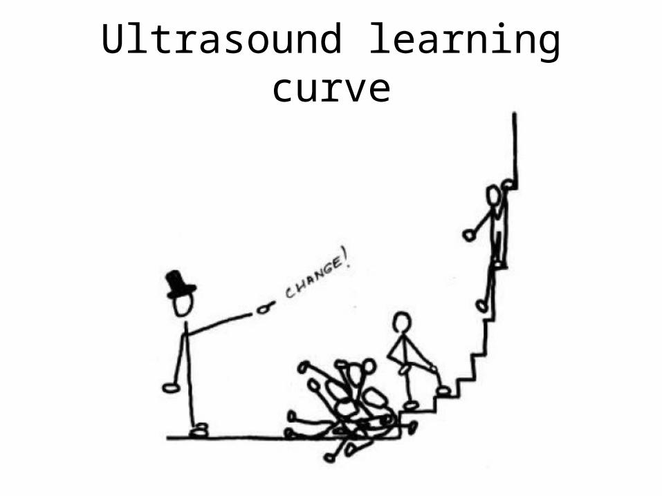
Ultrasound learning curve

Advantages of ultrasound • More accurate than palpation techniques
• More precise needle placement
• Potentially less risk for inadvertent trauma
• Can view soft tissues better than x-ray
• “Affordable”
• More portable than fluoroscopy

Advantages of ultrasound • Doppler mode visualization of flow
• No radiation exposure
• Decreased associated expense of upkeep
• Billable procedure 76942 (Caveat emptor)
• Potentially svoids the dreaded pocket fill
• Diagnoses the flipped pump

Disadvantages of ultrasound • Needle visualization has to be in-plane with
transducer
• 2d representation of 3d structure
• Poor penetration > 4-5 cm, unusable after 15cm
• Deeper views have poorer resolution

Disadvantages of ultrasound • Artifact from air and other density changes
• Anatomic variations
• Limited field of view
• Sometimes need an extra pair of hands
Practical considerations
“Thanksgiving – 8 hours of preparation for 10 minutes of eating”
Sarah Saulino – Mike’s Mom
Ultrasound refills can be the same way

Practical considerations
• Know pump anatomy
• Know the refill procedure – intrathecal delivery systems were not designed for ultrasound guidance
• Use ultrasound as an adjunct
• Consider patient positioning – can be creative here (standing, side lying, beach chair)
• Know the machine
Practical considerations
• Consider ergonomics
• Lighting can can make a world of difference
• Have all of materials within arms reach
• No such thing as too much gel
• Watch the screen, not the patient
• Use hydro dissection
• Need to retain an image for billing

Things to keep an eye on
• Echogenic needles
• Needle finding software
• Ultrasound refill kits

Ultrasound: applied reflectivity
• Transducer is both a speaker and microphone
• Piezoelectric crystal emits a sonic frequency in the 3-40 MHz range
• Human hearing stops at 20 kHz, canine hearing stops 60 kHz, dolphins use echolocation up to 150 kHz
• Microphone picks up reflected signals
• Computer decodes the reflections to create an image


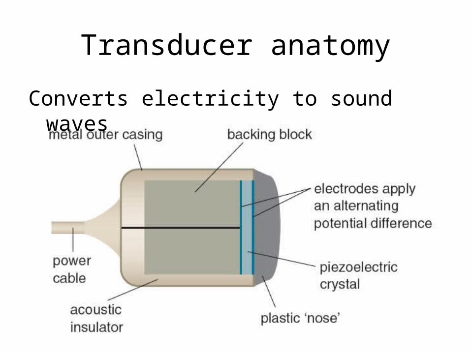
Transducer anatomy
Converts electricity to sound waves


Ultrasound is applied reflectivity
• Air reflects nothing
• Soft tissue sort of reflect
• Bone and metal reflect a lot
Ultrasound is applied reflectivity
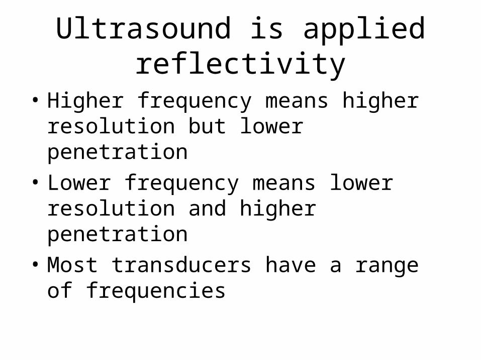
• Higher frequency means higher resolution but lower penetration
• Lower frequency means lower resolution and higher penetration
• Most transducers have a range of frequencies

Terminology
Make sure you know your right from left

Basic Knobology
• Depth
• Gain
• Doppler
• Screen capture

Depth
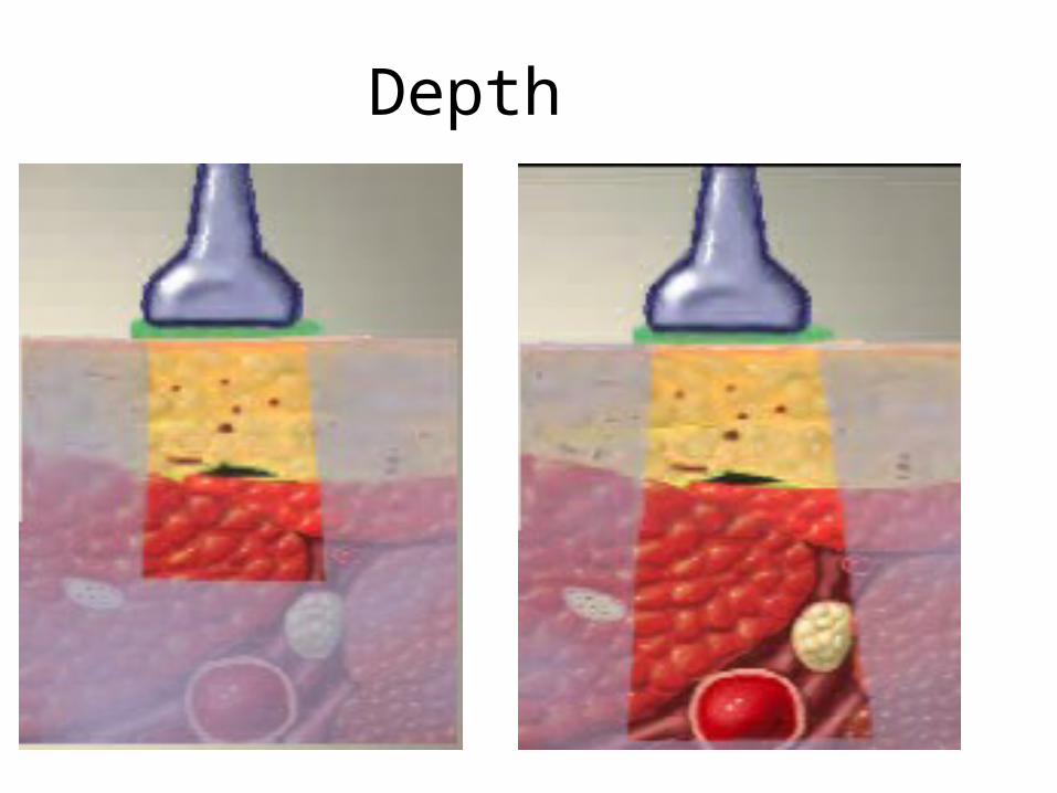
• Ultrasound can be set to penetrate a certain distance
• Set the depth a little deep to begin so you don’t waste time looking for your target
• When you have your bearings, set the depth so it is enough to cover your area of interest
• Excessive depth will degrade the picture unnecessarily
Depth

Gain
• Gain is the volume control of ultrasound
• Not enough is too dark (soft)
• Too much is too bright (loud)
• Similar to equalizer in audio systems, gain can be adjusted at different frequencies

Gain

Doppler
Ultrasound has the capacity to see fluid in motion

Visualization of current systems
• “Not-for-human-use” pumps were obtained from all three device manufacturers: SynchroMed II (Medtronic), MedStream (Codman) and Prometra (Flowonix).
• Each pump was placed in 3-inch deep baking tray and covered with an ultrasound phantom gel

Methods
• The anterior surface of each pump was then scanned with 50 mm high-frequency linear transducer HFL50 attached to the portable ultrasound machine MTurbo.
• The sonographic features of each pump was then observed and recorded.
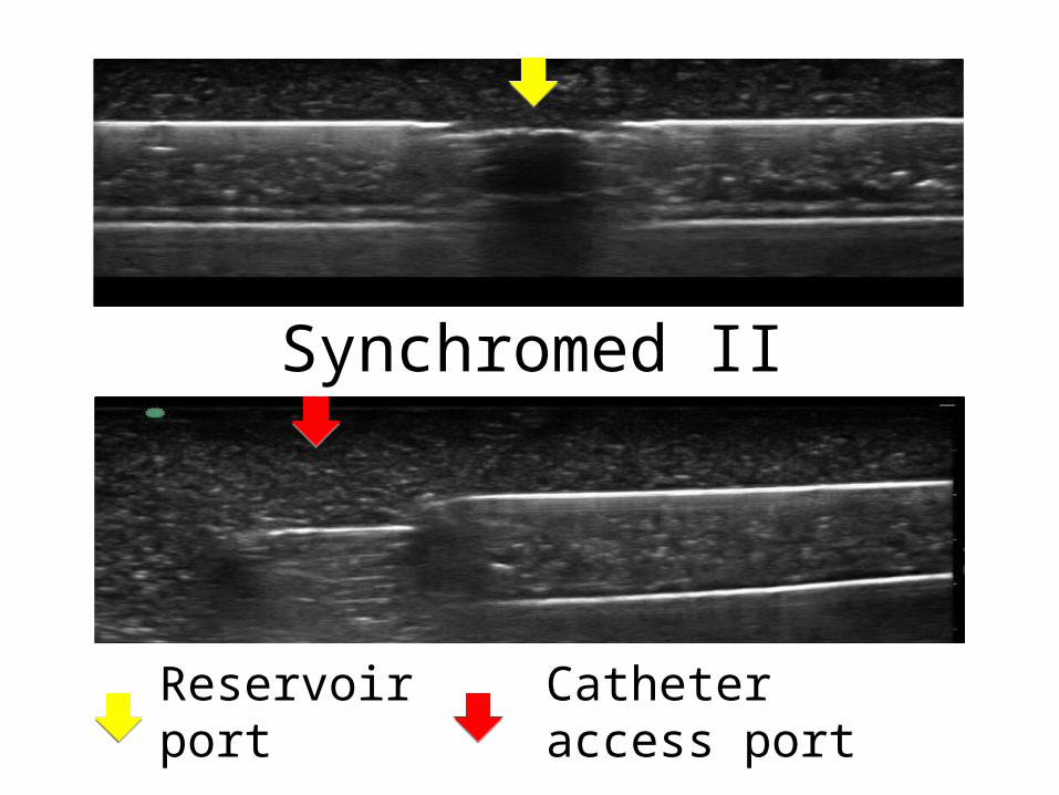
Synchromed II
Reservoir port Catheter access port

MedStream
Reservoir port Catheter access port

Prometra
Reservoir port Catheter access port

Conclusions
• All reservoir ports can be readily identified by portable sonography
• Potentially, the dome-shaped configuration of reservoir access ports for the Prometra and MedStream systems make the access easier, when compared to SynchroMed system.
• Each of the 3 systems has a unique sonographic appearance which allows for prompt identification

Flipped Synchromed II

Subcutaneous Injection

Executing the refill
• X marks the spot technique
• Real time technique

The in plane problem

The X marks the spot solution

The X marks the spot solution

Real Time refill

Doppler mode
Flow within reservoir
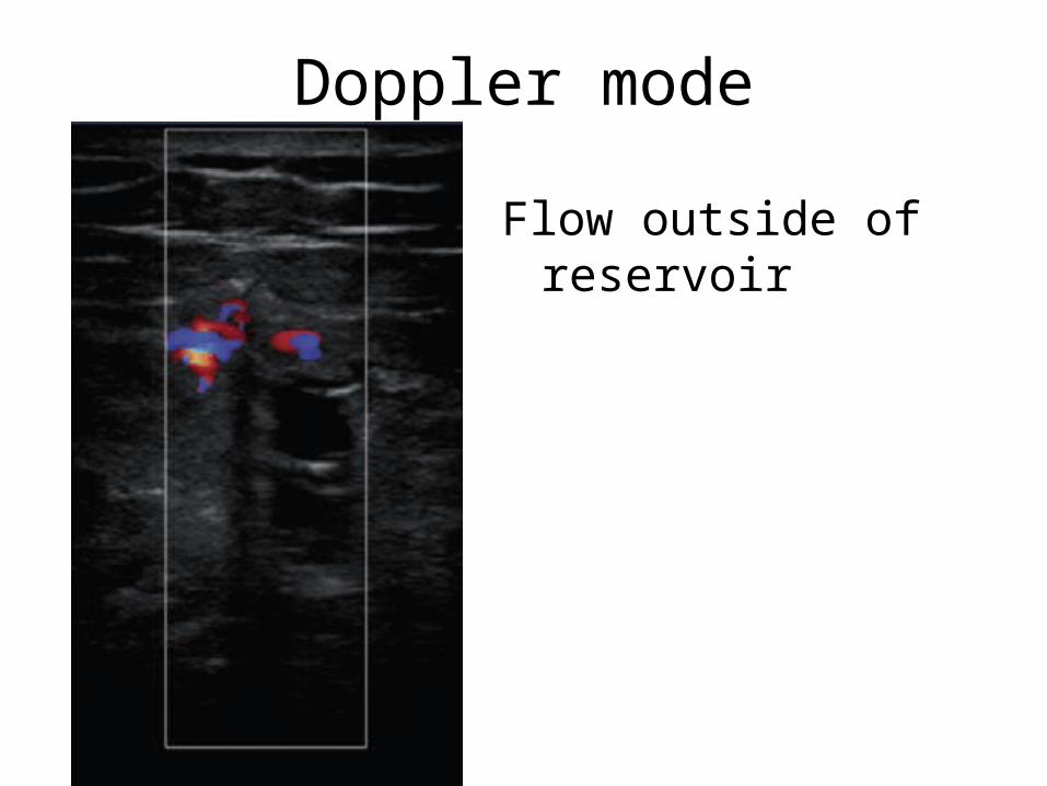
Doppler mode
Flow outside of reservoir


