Embed Size (px)
Citation preview

Lutz et al. J EXP ORTOP (2021) 8:76 https://doi.org/10.1186/s40634-021-00401-7
ORIGINAL PAPER
Ultrasound-based evaluation revealed reliable postoperative knee stability after combined acute ACL and MCL injuriesPatricia M. Lutz1, Louisa S. Höher1, Matthias J. Feucht1,3, Jan Neumann2, Daniela Junker2, Klaus Wörtler2, Andreas B. Imhoff1* and Andrea Achtnich1
Abstract
Purpose: Anterior cruciate ligament (ACL) injuries are often combined with lesions of the medial collateral ligament (MCL). The aim of this study was to evaluate treatment outcome of combined acute ACL and MCL lesions using func-tional US and clinical examination.
Methods: Patients aged > 18 years undergoing primary ACL reconstruction with concomitant operative (group 1) or non-operative treatment of the MCL (group 2) between 2014 and 2019 were included after a minimum follow-up of 12 months. Grade II MCL injuries with dislocated tibial or femoral avulsions and grade III MCL ruptures underwent ligament repair whereas grade II injuries without dislocated avulsions were treated non-operatively. Radiological outcome was assessed with functional US examinations. Medial knee joint width was determined in a supine posi-tion at 0° and 30° of knee flexion in unloaded and standardized loaded (= 15 Dekanewton) conditions using a fixation device. Clinical examination was performed and patient-reported outcomes were assessed by the use of the subjec-tive knee form (IKDC), Lysholm score, and the Tegner activity scale.
Results: A total of 40 patients (20 per group) met inclusion criteria. Mean age of group 1 was 40 ± 12 years (60% female) with a mean follow-up of 33 ± 17 months. Group 2 showed a mean age of 33 ± 8 years (20% female) with a mean follow-up of 34 ± 15 months. Side-to-side differences in US examinations were 0.4 ± 1.5 mm (mm) in 0° and 0.4 ± 1.5 mm in 30° knee flexion in group 1, and 0.9 ± 1.1 mm in 0° and 0.5 ± 1.4 mm in 30° knee flexion in group 2, with no statistically significant differences between both groups. MCL repair resulted in lower Lysholm scores (75 ± 19 versus 86 ± 15; p < 0.05). No significant differences could be found for subjective IKDC or Tegner activity scores among the two groups.
Conclusion: A differentiated treatment concept in combined ACL and MCL injuries based on injury patterns leads to reliable postoperative ligamentous knee stability in US-based and clinical examinations. However, grade II and III MCL lesions with subsequent operative MCL repair (group 1) result in slightly poorer subjective outcome scores.
Level of evidence: Retrospective cohort study; Level III
Keywords: MCL, ACL, Multiligamentous injuries, Ultrasound, Medial joint space, Valgus instability
© The Author(s) 2021. Open Access This article is licensed under a Creative Commons Attribution 4.0 International License, which permits use, sharing, adaptation, distribution and reproduction in any medium or format, as long as you give appropriate credit to the original author(s) and the source, provide a link to the Creative Commons licence, and indicate if changes were made. The images or other third party material in this article are included in the article’s Creative Commons licence, unless indicated otherwise in a credit line to the material. If material is not included in the article’s Creative Commons licence and your intended use is not permitted by statutory regulation or exceeds the permitted use, you will need to obtain permission directly from the copyright holder. To view a copy of this licence, visit http:// creat iveco mmons. org/ licen ses/ by/4. 0/.
Open Access
Journal ofExperimental Orthopaedics
*Correspondence: [email protected] Department for Orthopedic Sports Medicine, Technical University Munich, Ismaningerstrasse 22, 81675 Munich, GermanyFull list of author information is available at the end of the articleThe research was performed at the Department for Orthopedic SportsMedicine, Technical University Munich, Germany.

Page 2 of 8Lutz et al. J EXP ORTOP (2021) 8:76
IntroductionAnterior cruciate ligament (ACL) injuries are in up to 35% of cases combined with lesions to the medial side of the knee [9, 31] and available evidence of treatment con-cepts for combined lesions is limited. Whereas anatomic reconstruction of the ACL (ACL-R) using an autologous tendon graft represents the current gold standard, treat-ment strategies for MCL injuries remain inconsistent [5, 8, 13, 19, 25, 36, 37]. Time of surgery, surgical tech-niques (repair or reconstruction), and indication as well as strategy of non-operative treatment are controver-sially discussed [4, 5, 8, 10, 28, 34, 40]. From a biome-chanical point of view, there is growing evidence that MCL deficiency is a risk factor for ACL graft failure [1, 21, 23]. Therefore, postoperative MCL stability remains an important goal in the combined treatment of ACL and MCL injuries. Quantification of medial instability is commonly reported by clinical outcome, but radiologi-cal assessment is often missing. Functional ultrasound (US) examination can be used as a diagnostic tool to enhance postoperative radiological outcome measure-ment. Recently, mean values of medial joint space width in unloaded and standardized loaded conditions using a fixation device in healthy knees have been published [20]. However, results of functional US examinations for radi-ographic assessment following ACL-R and concomitant operative or non-operative treatment of the MCL are missing.
Therefore, the purpose of the present study was to eval-uate the radiological and clinical outcomes after ACL-R with concomitant MCL repair or non-operative MCL treatment. It was hypothesized that surgical repair of the MCL would contribute to higher rates of valgus instabil-ity and worse functional outcomes when compared to non-operative treatment, since the indication for surgical treatment in the authors’ department was a high-grade MCL injury.
MethodsThis retrospective cohort study was conducted to evalu-ate the clinical and radiological outcome after anatomic ACL-R with or without early MCL repair in patients with combined ACL and MCL lesions. The study was approved by the institutional review board of the Techni-cal University of Munich (235/19 S).
Patient cohortAll patients presenting with combined acute ACL and MCL injuries at our institution between February 2014 and February 2019 were included in this study. Inclusion criteria were: subjects aged > 18 years, early ACL-R with autologous hamstring tendon and MCL
repair (group 1) in the first two weeks after trauma or non-operative treatment of the MCL (group 2) for six weeks followed by staged ACL-R with autologous hamstring tendon. Diagnosis of combined ACL and MCL injuries were made using magnet resonance imaging and clinical examinations. Indication for MCL treatment was dependent on MCL grading according to Fetto and Marshall [9]. In grade II MCL injuries, an increased laxity at 30 degrees of flexion could be found, whereas in grade III MCL injuries an increased laxity at both, 0 and 30 degrees of flexion was present [9]. Grade II MCL injuries with dislocated tibial or femo-ral avulsions and grade III MCL injuries underwent ligament repair (group 1) due to the limited capacity to heal non-operatively [2]. Grade II MCL injuries with-out avulsions (partial ruptures) were treated non-oper-atively with a brace for six weeks with limited range of motion (ROM) and partial weight bearing on crutches (group 2).
Exclusion criteria for the present study were: further ligamentous or osseous injuries of the affected knee as well as previous injuries and surgical interventions on the other knee, and lack of German language skills.
Clinical notes of all patients were reviewed to collect demographic data.
Operative techniquePrior to surgery all patients had undergone a thorough clinical and radiological (X-rays and MRI) examination to ensure ligament injuries to the ACL and MCL with or without concomitant meniscus lesions.
In case of concomitant meniscus lesions, menis-cus suture systems (Arthrex, Naples, USA or Smith&Nephew, London, UK) were used or partial resection of meniscus was performed.
In both groups, an arthroscopic, anatomic single-bundle ACL technique with autologous hamstring graft was performed. The femoral tunnel was drilled via an anteromedial portal according to the diameter of the graft. A cortical suspension device (ACL tight-rope, Arthrex, Naples, USA) was used for femoral graft fixa-tion. A K-wire was then placed in the center of the tib-ial ACL footprint and was overdrilled according to the diameter of the graft, creating the tibial tunnel. A bio-absorbable interference screw (Arthrex, Naples, USA) was used for tibial fixation.
In group 1, the medial collateral ligamentous struc-tures were repaired by the use of suture anchors (Cork-screw 5,5 mm Biocomposite, Arthrex, Naples, USA) in case of femoral- or tibial-sided injuries with or without suture tape augmentation. Suture tape augmentation

Page 3 of 8Lutz et al. J EXP ORTOP (2021) 8:76
(FiberTape, Arthrex, Naples, USA) was used in grade III MCL injuries if medial collateral ligamentous struc-tures were badly damaged resulting in poor tissue qual-ity accompanied by limited success of isolated MCL repair by suture anchors. Additionally, MCL augmenta-tion was performed in case of insufficient ligamentous stability after MCL repair intra-operatively.
Postoperative rehabilitationThe postoperative protocol of group 1 consisted of 6 weeks of partial weight-bearing on crutches with limitation in ROM: in the first two weeks an active extension(ex)/flexion(flex) of 0°/20°/60°, in the next two weeks of 0°/10°/90°, and in the last two weeks of 0°/0°/90° was allowed. After six weeks, ROM was no longer lim-ited. A brace (Medi M4, Medi Bayreuth, Germany) was provided for at least 12 weeks.
In group 2, partial weight bearing on crutches with the same limitations in ROM was allowed in the six weeks of non-operative treatment in a brace. After six weeks, ACL-R was performed. Postoperatively, partial weight bearing on crutches was allowed for two weeks and ROM was only limited if meniscus suturing was performed (ex/flex 0°/0°/90°).
Return to running on the treadmill and front crawl swimming was allowed after 6 weeks, trail running after 3 months, return to sport-specific training in both groups was allowed after 6 months and full return to contact and/or pivoting sports activities after at least 9 months postoperatively.
Radiological evaluationFunctional US examinations were performed by two board-certified radiologists with at least 5 years of expe-rience in musculoskeletal imaging at follow-up. All acquired images of the knees were evaluated on picture archiving and communication system PACS workstations (Sectra Medical Systems, Sweden).
Ultrasound examinationFor evaluation of medial ligament laxity, the width of the medial joint space was assessed by ultrasound (ACUSON NX3 Ultrasound System, Siemens Erlangen, Germany) using a linear transducer (4.0–12.0 MHz, Maximum Field of View: 153 mm, Maximum Display Depth: 160 mm), placed in a longitudinal direction over the medial aspect of the knee. Subjects were positioned supine with extended leg in 0° with and without repro-ducible applied valgus stress (loaded condition) through a fixation device (TELOS, Wölfersheim-Berstadt, Ger-many) with 15 dekanewton (daN), and in a second step with a 30° bended knee with and without valgus stress
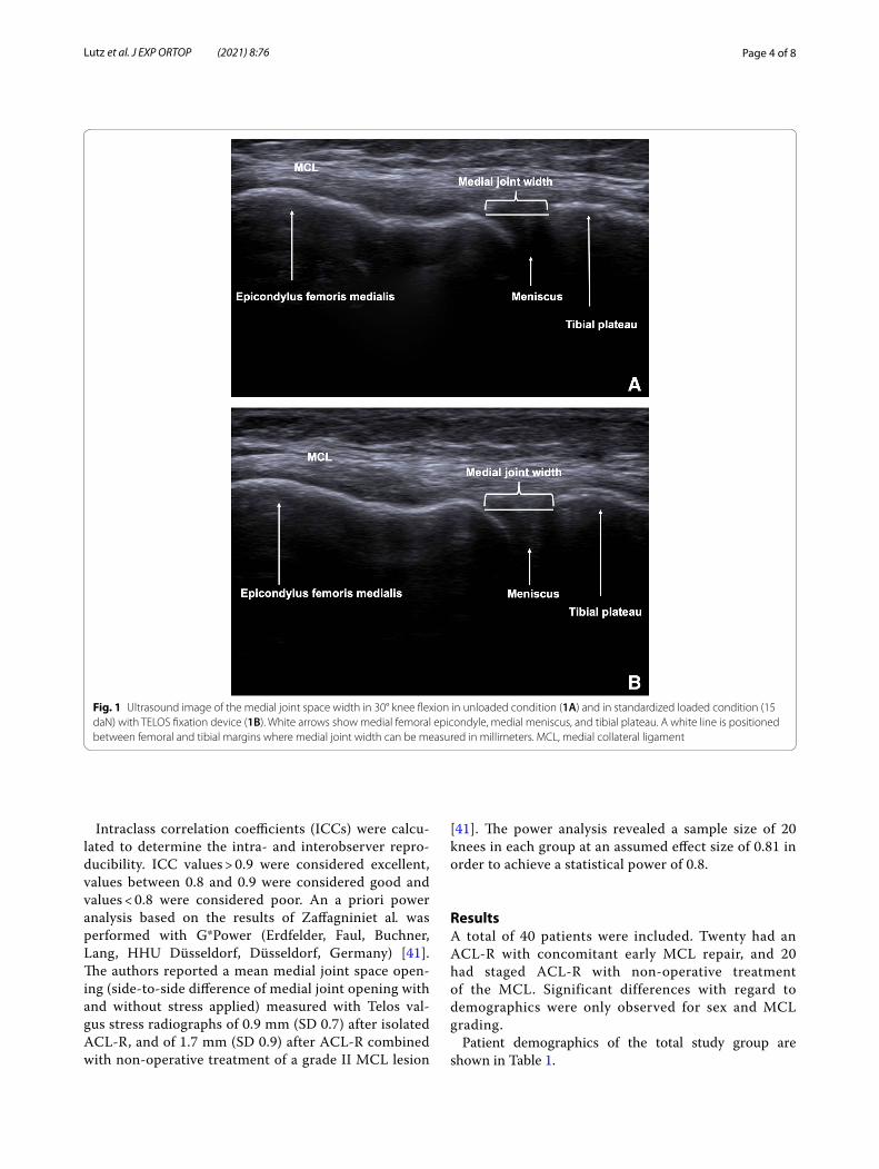
[11, 20]. For standardized measurements, the medial epicondyle was palpated and the transducer was placed in the longitudinal direction. Since the presentation of the hyperechoic bony outline of femur and tibia has been considered as an important quality assessment for standardized measurement of medial joint width [42], the medial femoral epicondyle and the proximal tibial plateau were used as bony landmarks, as described in the literature (Fig. 1A and B) [11, 20]. The distance between corresponding points on the femoral and tibial articular margins was measured in millimeters (mm). All measurements were performed by a specifically trained orthopedic sports medicine resident (rater 1). Intra- and interrater reliability was tested in 20 randomly assigned and blinded cases after an interval of six weeks by rater 1 and by one senior orthopedic surgeon (rater 2). Medial joint space width in 0° and 30° knee flexion was then compared between unloaded and standardized loaded conditions for each group. Furthermore, the mean change (delta Δ) of medial joint space width between unloaded and loaded conditions for each state of flexion and side-to-side differences of the average change (delta Δ) were compared between both groups.
Clinical examination and patient‑reported outcome measuresAll subjects underwent standardized clinical examina-tion of the knee. The International Knee Documenta-tion Committee (IKDC) valgus subscore was used for objective MCL stability assessment. Meniscus was evalu-ated by joint space tenderness and Steinmann test [35]. Patient-reported outcomes were measured with Visual Analogue Scale for pain (VAS), the subjective knee form of the IKDC, the Lysholm score, and the Tegner activity scale at follow-up [14, 38].
Statistical analysisStatistical analysis was performed by use of SPSS soft-ware (IBM, Armonk, New York, USA) and Microsoft Excel Version 2019 (Microsoft, Redmond, Washington, USA). For all statistical tests, p values less than 0.05 were considered as significant.
Descriptive statistics are presented as mean ± stand-ard deviation (SD) with a measurement accuracy of one decimal. Kolmogorov–Smirnov univariate normality test was used for continuous variables to confirm data normality. Mean change of medial joint space width between unloaded and loaded conditions and side-to-side differences were compared between the two groups using paired t-tests. Group comparison was performed with Mann–Whitney U test and unpaired t-test, as appropriate.
Page 4 of 8Lutz et al. J EXP ORTOP (2021) 8:76
Intraclass correlation coefficients (ICCs) were calcu-lated to determine the intra- and interobserver repro-ducibility. ICC values > 0.9 were considered excellent, values between 0.8 and 0.9 were considered good and values < 0.8 were considered poor. An a priori power analysis based on the results of Zaffagniniet al. was performed with G*Power (Erdfelder, Faul, Buchner, Lang, HHU Düsseldorf, Düsseldorf, Germany) [41]. The authors reported a mean medial joint space open-ing (side-to-side difference of medial joint opening with and without stress applied) measured with Telos val-gus stress radiographs of 0.9 mm (SD 0.7) after isolated ACL-R, and of 1.7 mm (SD 0.9) after ACL-R combined with non-operative treatment of a grade II MCL lesion
[41]. The power analysis revealed a sample size of 20 knees in each group at an assumed effect size of 0.81 in order to achieve a statistical power of 0.8.
ResultsA total of 40 patients were included. Twenty had an ACL-R with concomitant early MCL repair, and 20 had staged ACL-R with non-operative treatment of the MCL. Significant differences with regard to demographics were only observed for sex and MCL grading.
Patient demographics of the total study group are shown in Table 1.
Fig. 1 Ultrasound image of the medial joint space width in 30° knee flexion in unloaded condition (1A) and in standardized loaded condition (15 daN) with TELOS fixation device (1B). White arrows show medial femoral epicondyle, medial meniscus, and tibial plateau. A white line is positioned between femoral and tibial margins where medial joint width can be measured in millimeters. MCL, medial collateral ligament

Page 5 of 8Lutz et al. J EXP ORTOP (2021) 8:76
Postoperative complicationsIn total, three patients suffered from postoperative stiff-ness with limitations to ROM because of arthrofibrosis. Re-arthroscopy with the aim of arthrolysis was necessary to regain full ROM after 5 and 7 months (group 1), and after 3 months (group 2).
Clinical outcomeGroup comparisons of the patient-reported outcome scores are shown in Table 2. With regard to the inter-vention, patients with MCL repair showed signifi-cantly higher VAS values at rest and lower Lysholm score results, whereas no significant difference was observed for IKDC subjective score and Tegner activ-ity score. The mean return-to-sports time in group 1 was 22.3 ± 14.7 (range, 6–60) weeks, whereas in group 2 the mean return-to-sports time was 20.2 ± 8.8 (range, 6–36) weeks (n.s.).
Clinical examinationsConcerning the ACL, one patient of group 2 showed positive pivot-shift test. Lachman test was positive in one patient of group 1 and in three patients of group 2. Val-gus stress testing manually revealed 4 patients with IKDC grade B in 0° and in 7 patients with IKDC grade B in 30° knee flexion of group 1. All patients of group 2 showed IKDC grade A concerning valgus stability.
Ultrasound examinationsExcellent intrarater reliability was observed for all meas-urements. The ICC values were 0.96 for unloaded states
Table 1 Patient demographics of the total study group. Continuous variables are shown as mean ± standard deviation (range), categorical variables are shown as percentages
BMI, body mass index; MCL, medial collateral ligament; n.s., not significanta Statistically significant difference between both groups
Group 1 Group 2 p value
Number of patients, n 20 20
Follow‑up (months) 33.0 ± 16.7 (13–68) 34.3 ± 15.2 (12–52) n.s
Age (years) 39.8 ± 12.1 (20–55) 33.1 ± 8.1 (23–60) n.s
Sex, n (%) Male 8 (40%) 16 (80%) < 0.05 a
Female 12 (60%) 4 (20%) < 0.05 a
BMI (kg/m2) 25.0 ± 5.7 (18.3–42.4) 27.4 ± 5.6 (20.1–39.4) n.s
Laterality, n (%) Right 12 (60%) 6 (30%) n.s
Left 8 (40%) 14 (70%) n.s
MCL grading, n (%) Grade II 8 (40%) 20 (100%) < 0.001 a
Grade III 12 (60%)
MCL repair, n (%) Anchor 14 (70%)
Anchor + tape 6 (30%)
Meniscus‑Suturing, n (%) Lateral meniscus 9 (45%) 5 (25%) n.s
Medial meniscus 0 (0%) 1 (5%) n.s
Lateral and medial meniscus 4 (20%) 3 (15%) n.s
Table 2 Results of outcome scores
Continuous variables are shown as mean ± standard deviation (range)
VAS Visual Analogue Scale (pain), IKDC International Knee Documentation Committee, n.s. not significanta Values are medianb Statistically significant difference between both groups
Group 1 Group 2 p value
VASa
Rest 1 0 < 0.001b
Move 2 1 n.s
IKDC 78.0 ± 16.5(49.4 – 99.0)
85.1 ± 12.8(46.0 – 99.0)
n.s
Lysholm score 74.5 ± 18.5(29.0 – 100.0)
85.6 ± 14.8(37.0 – 100.0)
< 0.05b
Tegner activity scalea 4 5 n.s
Page 6 of 8Lutz et al. J EXP ORTOP (2021) 8:76
and 0.95 for loaded states. Interrater reliability was excel-lent with ICC values of 0.94 for unloaded and 0.95 for loaded states, respectively.
Results of US measurements are summarized in Table 3. First, the average change of medial joint space width between unloaded and standardized loaded condi-tions in 0° and 30° of knee flexion is shown. Second, side-to-side differences of the average change of medial joint space width between unloaded and standardized loaded conditions in each degree of flexion is explained. No statistically significant differences between both groups could be found.
DiscussionThe most important finding of the present study was that comparable results regarding radiological quantifi-cation of MCL laxity can be achieved after operative or non-operative treatment of grade II and grade III MCL injuries when concomitant ACL injury is treated with reconstruction. After a mean follow-up of 34 months acceptable clinical and radiological outcomes could be found for both groups. Further findings were that patients after non-operative MCL treatment reached sig-nificant higher Lysholm scores, as well as superior IKDC and Tegner activity scores, although not reaching statisti-cal significance.
Concerning the radiological method, functional US examination of medial structures of the knee has been shown to be a suitable method to describe medial joint space width [11, 17, 20, 30, 33]. In the present study, a fixation device was used to allow standardized and reproducible measurement of the medial joint space width [11, 20].
Mean side-to-side differences after combined ACL and MCL injuries were < 1 mm in both groups in the present study. In a systematic review, DeLonget al. reported an average side-to-side difference of 1.3 ± 0.9 mm after MCL repair [6]. Similar to our study, no significant side-to-side differences between groups after nonoperative grade II-III MCL treatment or MCL repair could be found by the use of valgus stress radiographs in previous research [12, 25, 41]. In our study, the average change (Δ) between unloaded and loaded conditions in 0° and 30° knee
flexion was 2.3 mm in group 1 and 2.1 mm in group 2. Previous US examinations of healthy knee joints showed similar results [11, 20, 33]. The US results of the present study revealed that valgus stability after combined ACL and MCL injuries could be completely restored in both groups.
Since 100% of both groups were identified with IKDC grade A or B when valgus stability was tested manually in clinical examination, restoration of valgus stability could be confirmed. In general, findings of improved valgus sta-bility were similar to those of previous research after dif-ferent MCL treatment approaches [6, 7, 10, 12, 15, 16, 18, 19, 24, 27, 41].
Considering that combined ACL and MCL inju-ries mostly affect the active and young to middle-aged patient population, an important purpose of different treatment approaches is not only to achieve good to excellent objective ligamentous stability, but also sub-jective outcome scores and activity levels. In our total cohort, pain did not seem to play a major role postop-eratively (VAS 0–2).
The presented findings revealed lower subjective out-come scores after MCL repair as compared to non-oper-ative MCL treatment. This is in line with recent results of Westermannet al., who reported superior patient reported outcomes after non-operative grade III MCL injuries compared to operative MCL treatment in com-bined ACL and MCL injuries [39]. With regard to the subjective IKDC score, superior outcomes (94 and 89) were reported by Canata et al. and Desai et al. after ACL-R and MCL repair [3, 7]. Multiple authors reported good to excellent Lysholm scores after ACL-R and MCL repair [3, 6, 7, 18, 27], whereas only fair results could be found in the presented study. MCL repair was therefore not only seen to result in excellent objective outcomes, but was also associated with lower patient-reported out-come scores as compared to the non-operative MCL group.
Tegner activity scale outcomes of our entire cohort showed no statistically significant differences between the two groups and therefore corresponds to the observations of several authors who reported similar results [6, 19, 29].
Table 3 Analysis of medial joint space width in Ultrasound examinations for groups (mean ± SD); average change (Δ) between unloaded and loaded conditions in 0° and 30° knee flexion, as well as side-to-side difference of the average change (Δ) in 0° and 30° knee flexion
mm millimeter, SD standard deviation, Δ average change of medial joint space width between unloaded and loaded conditions, n.s. not significant
Δ of medial joint space width (mm) in 0°
Δ of medial joint space width (mm) in 30°
Side-to-side difference (mm) in 0°
Side-to-side difference (mm) in 30°
p value
Group 1 2.3 ± 1.2 2.3 ± 1.3 0.4 ± 1.5 0.4 ± 1.5 n.s
Group 2 2.1 ± 0.7 2.1 ± 1.1 0.9 ± 1.1 0.5 ± 1.4 n.s

Page 7 of 8Lutz et al. J EXP ORTOP (2021) 8:76
Comparable to previous research [24, 29, 39], non-operative MCL treatment combined with ACL-R resulted in good Lysholm and subjective IKDC outcomes. As stated by Halinen et al. [12], a 10-point difference in the Lysholm score can be set clinically significant. This leads to the assumption that the impact of a more invasive surgical intervention (ACL-R and MCL repair) leads to clinically significant differences in subjective outcomes compared to ACL-R and non-operative MCL treatment.
Reasons for subjective lower scores after ACL-R and MCL repair are likely that higher grade MCL lesions resulted in marked extra-articular soft tissue injury. Symptoms could be associated with damaged MCL structures that are not addressed sufficiently by MCL repair. Hence, injuries to deep MCL structures may explain the lower subjective scores of the MCL repair group found in the present study.
MCL repair during ACL-R has been described as important risk factor for loss of knee function [26]. Postoperative stiffness due to arthrofibrosis in our cohort occurred in 2 patients of group 1 (10%) and in one patient of group 2 (5%) and was therefore lower as in previous research of Westermann et al. [39]. This may be attributed to ACL-R, as postoperative limited ROM is the most common complication [22, 32], which was supported by current findings. However, in 3 of 3 patients, re-arthroscopy with arthrolysis was clinically successful with normal ROM postoperatively.
Along with certain strengths, there are some limi-tations of this study. First, an experienced clinician examined all patients but was not blinded to this study. No specific measurements were taken to reduce bias. Second, the cohort included inhomogeneous injury patterns concerning the MCL. However, this fact rep-resents this patient cohort. Third, additional meniscus injuries were not excluded from this study. Fourth, the study design was retrospective. Fifth, although all func-tional US examinations were performed in a standard-ized fashion by experienced radiologists, US remains an operator-dependent imaging method. Furthermore, the average final follow-up (33 months in group 1 and 34 months in group 2) might not be sufficient to evalu-ate the long-term success rate after combined ACL and MCL injuries. Further research is therefore indicated.
ConclusionA differentiated treatment concept in combined ACL and MCL injuries based on injury patterns leads to reliable postoperative ligamentous knee stability in US-based and clinical examinations. However, grade II and III MCL lesions with subsequent operative MCL repair (group 1) result in slightly poorer subjective outcome scores.
AcknowledgementsWe would like to acknowledge our subjects for their participation.
Informed consentAll subjects gave their written informed consent to participate in this investigation.
Authors’ contributionsAA and PML designed the study. PML, LSH, JN, DF, and KW collected data. PML and AA performed the statistical analysis and wrote the manuscript. MF and KW helped to design the study, assisted with statistical analysis and data interpretation, and critically reviewed the manuscript. ABI conceived of the study, helped with data interpretation and critically reviewed the manuscript. All authors read and approved the final manuscript.
FundingOpen Access funding enabled and organized by Projekt DEAL.
Availability of data and materialsAll raw data (anonymized) is available from the corresponding author on request.
Declarations
Ethics approval and consent to participateEthical approval was obtained from the Ethics Committee of the technical University Munich. The study was approved by the institutional review board of the Technical University of Munich (235/19 S). All procedures performed were in accordance with the ethical standards of the institutional and/or national research committee and with the 1964 Declaration of Helsinki and its later amendments or comparable ethical standards.
Consent for publicationWas obtained from all subjects involved.
Competing interestsAndreas B. Imhoff is a consultant for Arthrosurface and Medi Bayreuth and receives royalties from Arthrex and Arthrosurface. All other authors declare that they have no competing interests related to this study.
Author details1 Department for Orthopedic Sports Medicine, Technical University Munich, Ismaningerstrasse 22, 81675 Munich, Germany. 2 Department of Diagnostic and Interventional Radiology, Technical University of Munich, Ismaninger-strasse 22, 81675 Munich, Germany. 3 Orthopädische Klinik Paulinenhilfe, Diakonie-Klinikum Stuttgart, Rosenbergstraße 38, 70176 Stuttgart, Germany.
Received: 6 August 2021 Accepted: 30 August 2021
References 1. Battaglia MJ, Lenhoff MW, Ehteshami JR, Lyman S, Provencher MT, Wick-
iewicz TL et al (2009) Medial collateral ligament injuries and subsequent load on the anterior cruciate ligament: a biomechanical evaluation in a cadaveric model. Am J Sports Med 37:305–311
2. Bollier M, Smith PA (2014) Anterior cruciate ligament and medial col-lateral ligament injuries. J Knee Surg 27:359–368
3. Canata GL, Chiey A, Leoni T (2012) Surgical technique: does mini-invasive medial collateral ligament and posterior oblique ligament repair restore knee stability in combined chronic medial and ACL injuries? Clin Orthop Relat Res 470:791–797
4. Cinque ME, Chahla J, Kruckeberg BM, DePhillipo NN, Moatshe G, LaPrade RF (2017) Posteromedial corner knee injuries: diagnosis, management, and outcomes: a critical analysis review. JBJS Rev 5:e4
5. Dale KM, Bailey JR, Moorman CT 3rd (2017) Surgical management and treatment of the anterior cruciate ligament/medial collateral ligament injured knee. Clin Sports Med 36:87–103

Page 8 of 8Lutz et al. J EXP ORTOP (2021) 8:76
6. DeLong JM, Waterman BR (2015) Surgical repair of medial collateral liga-ment and posteromedial corner injuries of the knee: a systematic review. Arthroscopy 31:2249-2255.e2245
7. Desai VS, Wu IT, Camp CL, Levy BA, Stuart MJ, Krych AJ (2020) Midterm outcomes following acute repair of grade III distal MCL avulsions in multiligamentous knee injuries. J Knee Surg 33:785–791
8. Elkin JL, Zamora E, Gallo RA (2019) Combined anterior cruciate ligament and medial collateral ligament knee injuries: anatomy, diagnosis, man-agement recommendations, and return to sport. Curr Rev Musculoskelet Med 12:239–244
9. Fetto JF, Marshall JL (1978) Medial collateral ligament injuries of the knee: a rationale for treatment. Clin Orthop Relat Res 1978;(132):206–18
10. Grant JA, Tannenbaum E, Miller BS, Bedi A (2012) Treatment of combined complete tears of the anterior cruciate and medial collateral ligaments. Arthroscopy 28:110–122
11. Gruber G, Martens D, Konermann W (1998) Stellenwert der sonographis-chen Untersuchung bei Läsion des medialen Knie-Seitenbandapparates. Z Orthop Ihre Grenzgeb 136:337–342
12. Halinen J, Lindahl J, Hirvensalo E, Santavirta S (2006) Operative and nonoperative treatments of medial collateral ligament rupture with early anterior cruciate ligament reconstruction: a prospective randomized study. Am J Sports Med 34:1134–1140
13. Herbort M, Michel P, Raschke MJ, Vogel N, Schulze M, Zoll A et al (2017) Should the ipsilateral hamstrings be used for anterior cruciate ligament reconstruction in the case of medial collateral ligament insufficiency? Biomechanical investigation regarding dynamic stabilization of the medial compartment by the hamstring muscles. Am J Sports Med 45:819–825
14. IKDC (2000) Formblätter International Knee Documentation Committee. https:// www. sport smed. org/ AOSSM IMIS/ membe rs/ downl oads/ resea rch/ IKDCG erman. pdf
15 Jokela MA, Mäkinen TJ, Koivikko MP, Lindahl JM, Halinen J, Lindahl J (2020) Treatment of medial-sided injuries in patients with early bicruciate liga-ment reconstruction for knee dislocation. Knee Surg Sports Traumatol Arthrosc 29:1872–1879. https:// doi. org/ 10. 1007/ s00167- 020- 06207-x
16. Kannus P (1988) Long-term results of conservatively treated medial collateral ligament injuries of the knee joint. Clin Orthop Relat Res (226):103–112.
17. Kleinbaum Y, Blankstein A (2008) Mild to moderate medial collateral liga-ment (MCL) injuries of the knee: sonographic findings and sonographic valgus stress test. J Musculoskelet Res 11:9–14
18. Koga H, Muneta T, Yagishita K, Ju YJ, Sekiya I (2012) Surgical management of grade 3 medial knee injuries combined with cruciate ligament injuries. Knee Surg Sports Traumatol Arthrosc 20:88–94
19. Lind M, Jacobsen K, Nielsen T (2020) Medial collateral ligament (MCL) reconstruction results in improved medial stability: results from the Danish knee ligament reconstruction registry (DKRR). Knee Surg Sports Traumatol Arthrosc 28:881–887
20. Lutz PM, Feucht MJ, Wechselberger J, Rasper M, Petersen W, Wörtler K et al (2020) Ultrasound-based examination of the medial ligament complex shows gender- and age-related differences in laxity. Knee Surg Sports Traumatol Arthrosc 29:1960–1967. https:// doi. org/ 10. 1007/ s00167- 020- 06293-x
21. Mancini EJ, Kohen R, Esquivel AO, Cracchiolo AM, Lemos SE (2017) Comparison of ACL strain in the MCL-deficient and MCL-reconstructed knee during simulated landing in a cadaveric model. Am J Sports Med 45:1090–1094
22. Mayr HO, Weig TG, Plitz W (2004) Arthrofibrosis following ACL reconstruc-tion—reasons and outcome. Arch Orthop Trauma Surg 124:518–522
23. Mehl J, Otto A, Kia C, Murphy M, Obopilwe E, Imhoff FB, et al (2019) Osse-ous valgus alignment and posteromedial ligament complex deficiency lead to increased ACL graft forces. Knee Surg Sports Traumatol Arthrosc 28(4):1119–29. https:// doi. org/ 10. 1007/ s00167- 019- 05770-2.
24. Millett PJ, Pennock AT, Sterett WI, Steadman JR (2004) Early ACL recon-struction in combined ACL—MCL injuries. J Knee Surg 17:94–98
25. Nakamura N, Horibe S, Toritsuka Y, Mitsuoka T, Yoshikawa H, Shino K (2003) Acute grade III medial collateral ligament injury of the knee associ-ated with anterior cruciate ligament tear: the usefulness of magnetic resonance imaging in determining a treatment regimen. Am J Sports Med 31:261–267
26. Noyes FR, Barber-Westin SD (1995) The treatment of acute combined ruptures of the anterior cruciate and medial ligaments of the knee. Am J Sports Med 23:380–391
27. Osti L, Papalia R, Del Buono A, Merlo F, Denaro V, Maffulli N (2010) Simul-taneous surgical management of chronic grade-2 valgus instability of the knee and anterior cruciate ligament deficiency in athletes. Knee Surg Sports Traumatol Arthrosc 18:312–316
28 Papalia R, Osti L, Del Buono A, Denaro V, Maffulli N (2010) Management of combined ACL–MCL tears: a systematic review. Br Med Bull 93:201–215
29. Petersen W, Laprell H (1999) Combined injuries of the medial collateral ligament and the anterior cruciate ligament. Arch Orthop Trauma Surg 119:258–262
30. Schricker T, Hien N, Wirth C (1987) Klinische Ergebnisse sonographis-cher Funktionsuntersuchungen bei Kapselbandläsionen am Knie-und Sprunggelenk. Ultraschall Med 8:27–31
31. Shirakura K, Terauchi M, Fukasawa N, Kimura M, Shimizu T (1995) Clinical and arthroscopic findings of acute anterior cruciate ligament tears of the knee. Diagn Ther Endosc 2:107–112
32. Shirakura K, Terauchi M, Katayama M, Watanabe H, Yamaji T, Takagishi K (2000) The management of medial ligament tears in patients with combined anterior cruciate and medial ligament lesions. Int Orthop 24:108–111
33. Slane LC, Slane JA, Scheys L (2017) The measurement of medial knee gap width using ultrasound. Arch Orthop Trauma Surg 137:1121–1128
34. Smyth MP, Koh JL (2015) A review of surgical and nonsurgical outcomes of medial knee injuries. Sports Med Arthrosc 23:e15–e22
35. Steinmann F (1929) Referat über Meniskusverletzungen. Schweiz Med Wochenschr 10:1355–1356
36. Svantesson E, Hamrin Senorski E, Alentorn-Geli E, Westin O, Sundemo D, Grassi A et al (2019) Increased risk of ACL revision with non-surgical treatment of a concomitant medial collateral ligament injury: a study on 19,457 patients from the Swedish National Knee Ligament Registry. Knee Surg Sports Traumatol Arthrosc 27:2450–2459
37. Svantesson E, Hamrin Senorski E, Östergaard M, Grassi A, Krupic F, Westin O et al (2020) Graft choice for anterior cruciate ligament reconstruction with a concomitant non-surgically treated medial collateral ligament injury does not influence the risk of revision. Arthroscopy 36:199–211
38. Tegner Y, Lysholm J (1985) Rating systems in the evaluation of knee liga-ment injuries. Clin Orthop Relat Res 198:43–49
39. Westermann RW, Spindler KP, Huston LJ, Wolf BR (2019) Outcomes of grade III medial collateral ligament injuries treated concurrently with anterior cruciate ligament reconstruction: a multicenter study. Arthros-copy 35:1466–1472
40. Wijdicks CA, Griffith CJ, Johansen S, Engebretsen L, LaPrade RF (2010) Injuries to the medial collateral ligament and associated medial struc-tures of the knee. J Bone Joint Surg 92:1266–1280
41. Zaffagnini S, Bonanzinga T, Marcheggiani Muccioli GM, Giordano G, Bruni D, Bignozzi S et al (2011) Does chronic medial collateral ligament laxity influence the outcome of anterior cruciate ligament reconstruction?: a prospective evaluation with a minimum three-year follow-up. J Bone Joint Surg Br 93:1060–1064
42. Zhu J, Li B, Qiu L, Liu H, Zhang M, Wang Y et al (2020) A measurement method of knee joint space width by ultrasound: a large multicenter study. Quant Imaging Med Surg 10:979–987
Publisher’s NoteSpringer Nature remains neutral with regard to jurisdictional claims in pub-lished maps and institutional affiliations.


![The analgesic efficacy of ultrasound-guided abdominis ... · TAP block.[8.9] Real-time ultrasound provides reliable imaging of three muscular layers of anterolateral abdominal wall](https://img.dokumen.tips/doc/110x75/5f27fe2048e0882a2533e16b/the-analgesic-efficacy-of-ultrasound-guided-abdominis-tap-block89-real-time.jpg)

![Methicillin-Resistant Staphylococcus aureus [Mrsa] …...physicians 12 months later with progression to CKD stage 5. An ultrasound of her kidneys at this time revealed gross right](https://img.dokumen.tips/doc/110x75/5e52ff27e5ad431e9925409f/methicillin-resistant-staphylococcus-aureus-mrsa-physicians-12-months-later.jpg)