Embed Size (px)
Citation preview

Ultrashort TE Imaging With Off-Resonance SaturationContrast (UTE-OSC)
Jiang Du,1* Atsushi M. Takahashi,2 Mark Bydder,1 Christine B. Chung,1 andGraeme M. Bydder1
Short T2 species such as the Achilles tendon and cortical bonecannot be imaged with conventional MR sequences. They havea much broader absorption lineshape than long T2 species,therefore they are more sensitive to an appropriately placedoff-resonance irradiation. In this work, a technique termed ul-trashort TE (UTE) with off-resonance saturation contrast (UTE-OSC) is proposed to image short T2 species. A high powersaturation pulse was placed �1 to �2 kHz off the water peak topreferentially saturate signals from short T2 species, leavinglong T2 water and fat signals largely unaffected. The subtractionof UTE images with and without an off-resonance saturationpulse effectively suppresses long T2 water and fat signals, cre-ating high contrast for short T2 species. The UTE-OSC tech-nique was validated on a phantom, and applied to bone samplesand healthy volunteers on a clinical 3T scanner. High-contrastimages of the Achilles tendon and cortical bone were generatedwith a high contrast-to-noise ratio (CNR) of the order of 12 to 20between short T2 and long T2 species within a total scan time of4 to 10 min. Magn Reson Med 62:527–531, 2009. © 2009 Wiley-Liss, Inc.
Key words: ultrashort TE; off-resonance saturation; projectionreconstruction; short T2 species; magnetization transfer
The human body contains a variety of short T2 species,including cortical and trabecular bone, tendons, liga-ments, menisci, short T2 components in white matter,macromolecules, and membranes in biological tissues(1,2). Magnetization from these species cannot be spatiallyencoded with conventional MR pulse sequences before thesignal has completely decayed. The short T2 species havea much broader absorption lineshape than the long T2
species, making them as much as 106 times more sensitiveto appropriately placed off-resonance irradiation (3). Thispreferential saturation of the short T2 species can be trans-ferred to the liquid spins and be detected indirectly withconventional MR sequences through the mechanism ofmagnetization transfer (MT) effect (3–5).
MT has been applied to musculoskeletal (MSK) MRI,MR angiography, and characterization of brain white mat-ter diseases such as multiple sclerosis (MS) (6–8). TypicalMT imaging includes an off-resonance pulsed radio fre-quency (RF) preirradiation, followed by conventional
spin-echo or gradient-echo imaging. An ideal saturationpulse should only saturate short T2 species, leaving long T2
species unaffected. This is achieved by choosing appropri-ate saturation pulse and frequency offset to minimize theoverlap between the saturation pulse and long T2 speciesin the spectral domain. The RF power required to effec-tively saturate the short T2 species is typically large, butalso has to fulfill the specific absorption ratio (SAR) guide-lines and to avoid tissue heating.
However, typical MT imaging can only detect the signalchange from free mobile water protons as a result of satu-ration transfer in which restricted macromolecule protonsare selectively irradiated. The short T2 species still cannotbe directly detected because of the long TE used in con-ventional pulse sequences (several milliseconds [�sec] orlonger). Through the combination of half RF pulse, vari-able-rate selective excitation (VERSE), which synchro-nizes RF excitation and gradient ramp-down, radial pro-jection, and ramp sampling, TEs �100 �s have beenachieved (9–17). Such ultrashort TE (UTE) sequences candetect signals from short T2 species.
The combination of off-resonance saturation and UTEacquisition may provide high contrast for short T2 imag-ing. In this work, we devised an approach termed UTEwith off-resonance saturation contrast (UTE-OSC), inwhich UTE images with and without an off-resonancesaturation pulse were subtracted to suppress signals fromlong T2 water and fat and to create high contrast for shortT2 species. Feasibility studies were performed on a phan-tom, bone samples, and healthy volunteers on a clinical 3Tscanner.
MATERIALS AND METHODS
Figure 1 shows the UTE-OSC sequence with a minimumTE � 8 �s achieved through the combination of half-pulseexcitation, VERSE, radial ramp sampling, and fast trans-mit/receive (T/R) switching (18). It requires two acquisi-tions (number of excitations [NEX] � 2) performed foreach radial line of k-space, by collecting data with theslice-selection gradient in one direction and adding this todata collected with the slice-selection gradient reversed. Ahard or Fermi pulse with high amplitude and long dura-tion (or large flip angle) followed by gradient dephasing(Fig. 1; dotted box) was employed for effective off-reso-nance saturation of short T2 species, with minimal satura-tion of long T2 species. In comparison, dual echo UTEimaging acquires two echoes followed by subtraction ofthe second echo (Fig. 1; dashed box) from the first one, tosuppress long T2 species and improve short T2 contrast.
Figure 2 shows a schematic diagram on how short T2
contrast is created in UTE-OSC. Long T2 species such as
1Department of Radiology, University of California, San Diego, San Diego,California, USA.2Global Applied Science Laboratory, GE Healthcare, Menlo Park, California,USA.*Correspondence to: Jiang Du, Ph.D. University of California, San Diego,Department of Radiology, 200 West Arbor Drive, San Diego, CA 92103-8756.E-mail: [email protected] 4 October 2008 ; revised 15 January 2009; accepted 9 February2009.DOI 10.1002/mrm.22007Published online 15 May 2009 in Wiley InterScience (www.interscience.wiley.com).
Magnetic Resonance in Medicine 62:527–531 (2009)
© 2009 Wiley-Liss, Inc. 527

muscle and fat have a relatively narrow spectral linewidth(typically �10 Hz) (19,20). Short T2 species such as theAchilles tendon and cortical bone have much a broaderlinewidth (hundreds to thousands of Hertz) (5,12). A sat-uration pulse placed at an appropriately placed off-reso-nance frequency may only overlap with short T2 species inthe spectral domain. High-contrast images of short T2 spe-cies were created via the following three steps. First, aconventional UTE acquisition was employed to detect sig-nal from both long and short T2 species. Second, each UTEreadout was preceded by a saturation pulse with large flipangle placed at an empirically determined frequency offsetof �1 kHz (for tendon) to �2 kHz (for bone) from the waterpeak, to selectively suppress signals from short T2 species,with long T2 water and fat signals minimally affected.Third, a difference image was formed by subtracting thesecond image from the first image to suppress signals fromlong T2 water and fat, leaving high contrast for the short T2
species.The UTE-OSC technique was first validated on a phan-
tom consisting of four tubes, including a tube of distilledwater, a tube of vegetable oil, and two tubes of distilledwater doped with two levels of MnCl2 (2.5 mM and12 mM), resulting in a short T*2 of 2 ms (simulating theAchilles tendon) and 0.4 ms (simulating cortical bone),respectively. This technique was then performed on threepig bone samples bought from a nearby slaughter houseand five healthy human volunteers. Institutional reviewboard permission was obtained for this study. All thescans were performed on a 3T Signa TwinSpeed scanner(GE Healthcare Technologies, Milwaukee, WI, USA) with amaximum gradient performance of 40 mT/m and 150 mT/m/ms. A single-channel 3-inch coil was used for signalreception. Typical imaging parameters included: field ofview [FOV] � 10 cm, slice thickness � 2 to 3 mm, read-out � 512 (260 to 284 sampling points for each half pro-jection), bandwidth (BW) � �62.5 kHz, TR � 200 to300 ms, TE � 8 �s, and 299 to 511 half projections. For
tendon imaging, a Fermi pulse with a duration of 16 mswas placed �1 kHz from the water peak. For bone imaging,a hard pulse with a duration of 10 ms was placed �2 kHzfrom the water peak. The SAR was under 1.4 W/kg. Noneof the subjects felt tissue warming due to the appliedsequences. Two acquisitions without and with off-reso-nance saturation pulse were performed for each study,resulting in a total scan time of 4 min to 10 min. Conven-tional dual-echo UTE acquisition and a clinical gradient-echo MT sequence with similar imaging parameters werealso performed for comparison. TE for the second echoUTE acquisition was kept at the minimal value for the fatand water in-phase imaging, i.e., 4.4 ms or 6.6 ms depend-ing on the FOV, acquisition matrix size, and BW. Theclinical gradient-echo MT sequence has a minimal TE of4.7 ms, with frequency offset � 1200 Hz and flip angle �670° for the Fermi MT pulse (8 ms in duration).
For quantitative assessment, direct saturation ratio(DSR) and contrast-to-noise ratio (CNR) measurementswere performed. DSR refers to the direct saturation effectdue to the off-resonance saturation pulse and was calcu-lated as the ratio of the mean signal intensity inside auser-drawn region-of-interest (ROI) with and without thesaturation pulse. CNR between the short T2 and long T2
species were calculated as their signal difference overbackground noise, which was calculated as the standarddeviation (SD) of the signal in an ROI placed in the back-ground.
RESULTS
Figure 3 shows the phantom study using conventionaldual-echo UTE as well as UTE-OSC. UTE provides highsignal for all four tubes, with ring artifact around the oiltube due to off-resonance effect. There is a significant fatsignal decay during echo spacing � 4.4 ms, resulting inreduced contrast between short T2 and fat tubes in thesubtraction image. A Fermi pulse with duration �16 ms
FIG. 2. UTE-OSC includes two acquisitions with and without anoff-resonance saturation pulse. The saturation pulse was placed �1to �2 kHz away from the on-resonance water peak to minimize itsspectral overlap with that of long T2 water and fat. Short T2 specieshave a broad spectrum and thus suppressed by the saturationpulse. Subtraction of UTE images with and without off-resonancesaturation pulse suppresses long T2 water and fat signals, leavinghigh contrast for short T2 species.
FIG. 1. UTE-OSC sequence employs UTE acquisition with a mini-mal TE � 8 �s, preceded by a hard (solid line) or Fermi (dashed line)saturation pulse centered �1 to �2 kHz from the water peak toselectively saturation short T2 species with little effect on long T2
water and fat. In comparison, dual-echo UTE imaging acquires twoechoes followed by subtraction of the second echo from the firstecho to suppress long T2 water and fat and improve short T2
contrast.
528 Du et al.
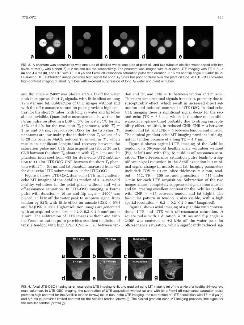
and flip angle � 2400° was placed �1.5 kHz off the waterpeak to suppress short T2 signals, with little effect on longT2 water and fat. Subtraction of UTE images without andwith the off-resonance saturation pulse provides high con-trast for the short T2 tubes, with long T2 water and fat tubesalmost invisible. Quantitative measurement shows that theFermi pulse resulted in a DSR of 3% for water, 1% for fat,37% and 8% for the two short T2 phantoms, with T*2 �2 ms and 0.4 ms, respectively. DSRs for the two short T2
phantoms are low mainly due to their short T1 values of 5to 20 ms because MnCl2 reduces T2 as well as T1, whichresults in significant longitudinal recovery between thesaturation pulse and UTE data acquisition (about 20 ms).CNR between the short T2 phantom with T*2 � 2 ms and fatphantom increased from –55 for dual-echo UTE subtrac-tion to 119 for UTE-OSC. CNR between the short T2 phan-tom with T*2 � 0.4 ms and fat phantom increased from –23for dual-echo UTE subtraction to 17 for UTE-OSC.
Figure 4 shows UTE-OSC, dual-echo UTE, and gradient-echo MT imaging of the Achilles tendon of a 54-year-oldhealthy volunteer in the axial plane without and withoff-resonance saturation. In UTE-OSC imaging, a Fermipulse with duration � 16 ms and flip angle � 2400° wasplaced �1 kHz off the water peak to suppress signal fromtendon by 82% with little effect on muscle (DSR � 5%)and fat (DSR � 3%). High-resolution images are generatedwith an acquired voxel size � 0.2 � 0.2 � 2.0 mm3 under3 min. The subtraction of UTE images without and withthe Fermi saturation pulse provides excellent depiction oftensile tendon, with high CNR: CNR � �20 between ten-
don and fat, and CNR � 18 between tendon and muscle.There are some residual signals from skin, probably due tosusceptibility effect, which result in increased direct sat-uration and reduced contrast in UTE-OSC. In dual-echoUTE imaging there is significant signal decay for the sec-ond echo (TE � 6.6 ms, which is the shortest possiblewater-fat in-phase time) probably due to strong suscepti-bility effect, resulting in reduced CNR: CNR � 3 betweentendon and fat, and CNR � 5 between tendon and muscle.The clinical gradient-echo MT imaging provides little sig-nal for tendon because of a long TE � 4.7 ms.
Figure 5 shows sagittal UTE imaging of the Achillestendon of a 30-year-old healthy male volunteer without(Fig. 5; left) and with (Fig. 5; middle) off-resonance satu-ration. The off-resonance saturation pulse leads to a sig-nificant signal reduction in the Achilles tendon but mini-mal signal change in muscle and fat. Imaging parametersincluded: FOV � 10 cm, slice thickness � 2 mm, read-out � 512, TR � 300 ms, and projections � 511 under5 min for each UTE acquisition. Subtraction of the twoimages almost completely suppressed signals from muscleand fat, creating excellent contrast for the Achilles tendonwith CNR � �15 between tendon and fat (right). Thefascicular pattern in tendon is also visible, with a highspatial resolution � 0.2 � 0.2 � 2.0 mm3 (acquired).
Figure 6 shows axial imaging of a pig tibia with conven-tional UTE and UTE with off-resonance saturation. Asquare pulse with a duration � 10 ms and flip angle �2400° was centered at �2 kHz off the water peak foroff-resonance saturation, which significantly reduced sig-
FIG. 3. A phantom was constructed with one tube of distilled water, one tube of plant oil, and two tubes of distilled water doped with twolevels of MnCl2 with a short T*2 � 2 ms and 0.4 ms, respectively. The phantom was imaged with dual-echo UTE imaging with TE � 8 �s(a) and 4.4 ms (b), and UTE with TE � 8 �s and Fermi off-resonance saturation pulse with duration � 16 ms and flip angle � 2400° (c). d:Dual-echo UTE subtraction image provides high signal for short T2 tubes but poor contrast over the plant oil tube. e: UTE-OSC provideshigh-contrast imaging of short T2 tubes with excellent suppression of long T2 water and plant oil tubes.
FIG. 4. Axial UTE-OSC imaging (a–c), dual-echo UTE imaging (d–f), and gradient echo MT imaging (g) of the ankle of a healthy 54-year-oldmale volunteer. In UTE-OSC imaging, the subtraction of UTE acquisition without (a) and with (b) a Fermi off-resonance saturation pulseprovides high contrast for the Achilles tendon (arrow) (c). In dual-echo UTE imaging, the subtraction of UTE acquisition with TE � 8 �s (d)and 6.6 ms (e) provides limited contrast for the Achilles tendon (arrow) (f). The clinical gradient-echo MT imaging provides little signal forthe Achilles tendon (arrow) (g).
UTE-OSC 529

nal from cortical bone. There is no visible change in fatsignal, suggesting that the off-resonance saturation pulsewas properly placed to reduce direct saturation of long T2
species. Subtraction of these two sets of images effectivelysuppressed signals from bone marrow fat, providing highnominal spatial resolution with FOV � 10 cm, readout �512, and slice thickness � 3 mm, and excellent contrast forcortical bone with a CNR � �12 between bone and fatwithin a total scan time of 4 min. Periosteum is partlysuppressed by the off-resonance saturation pulse due to itsrelatively short T2. The strong susceptibility effect near theair periosteum interface may also contribute to the signalreduction in UTE imaging with off-resonance saturation.As a result, periosteum appears as high signal in UTE-OSC.
DISCUSSION
UTE-OSC has been shown to be an effective technique increating high-contrast images of short T2 species, such asthe Achilles tendon and cortical bone, that have a broadspectral response and are therefore very sensitive to satu-ration pulses placed at off-resonance frequencies. The longT2 species have a narrow spectral response and are mini-mally affected by the off-resonance saturation pulse.Therefore, the subtraction of UTE images with and withoutoff-resonance saturation pulse effectively suppresses longT2 water and fat signals and selectively depicts the short T2
species.Although UTE-OSC bears some similarity to the conven-
tional MT imaging (3–8), they are fundamentally differenttechniques. MT requires at least two distinct proton pools:the free mobile protons and restricted protons. If these twopools or reservoirs are strongly coupled, relaxation of oneof the proton groups will influence the relaxation of theother. The MT pulse will reduce signals from the mobilewater protons when a saturation pulse is placed off-reso-nance to selectively irradiate the restricted water protons.No MT effect can be observed if there is no couplingbetween these two pools of protons. UTE-OSC can effec-tively and robustly detect signal changes from the re-stricted water protons, even if there is no coupling be-tween these two pools of protons. UTE-OSC can detect MTeffect, which results in increased residual long T2 signalsin the subtraction image and thus reduced short T2 con-trast.
Conventional UTE imaging typically employs a dual-echo acquisition and subtraction technique or long T2 sat-
uration techniques to suppress long T2 signals and im-prove short T2 contrast (12–14). The long T2 saturationpulses may saturate the short T2 signals directly (12). Thedual-echo approach is subject to susceptibility, off-reso-nance, and gradient distortion artifact, especially for thesecond echo with TE � 5 ms (2,12). The UTE-OSC tech-nique employs the same short TE � 8 �s for both acquisi-tions, and is therefore less sensitive to these artifacts. Thesaturation pulse was placed �1 to �2 kHz off the on-resonance frequency, minimizing the saturation effect onlong T2 species. However, the susceptibility effect near thetissue interface, such as the skin air interface in the Achil-les tendon study shown in Fig. 4 and periosteum-air inter-face in the pig tibia study shown in Fig. 6, still results inincreased residual signal in UTE-OSC. Tailored saturationpulses with more localized spectral response and opti-mized off-resonance frequencies will help reduce this sus-ceptibility effect. UTE-OSC requires prolonged total scantime over conventional UTE imaging due to the require-ment of repeated UTE acquisitions with and without off-resonance saturation pulse. Any motion occurring be-tween the two scans leads to misregistration in the sub-traction image.
Future work will focus on designing and optimizing theoff-resonance saturation pulses, including hard pulses,Fermi pulses, and Gaussian pulses. Pulse duration, flipangle, and off-resonance frequency offset will be investi-gated for maximal short T2 saturation and minimal long T2
suppression. UTE imaging with a series of off-resonancefrequency offsets may provide information on bound waterand bulk water in short T2 species such as the Achillestendon and cortical bone (21). Further work is needed toinvestigate this application.
In conclusion, UTE-OSC provides high-contrast imagesof short T2 species by subtracting UTE images with andwithout a properly placed high-power off-resonance satu-ration pulse. The UTE-OSC technique can be readily ap-plied to image the short T2 components in brain whitematter and other short T2 components or species.
REFERENCES
1. Harrison R, Bronskill MJ, Henkelman RM. Magnetization transfer andT2 relaxation components in tissue. Magn Reson Med 1995;33:490–496.
2. Gatehouse PD, Bydder GM. Magnetic resonance imaging of short T2components in tissue. Clin Radiol 2003;58:1–19.
3. Wolff SD, Balaban RS. Magnetization transfer contrast (MTC) and tissuewater proton relaxation in vivo. Magn Reson Med 1989;10:135–144.
4. Graham SJ, Henkelman RM. Pulsed magnetization transfer imaging:evaluation of technique. Radiology 199;212:903–910.
5. Henkelman RM, Stanisz GJ, Graham SJ. Magnetization transfer in MRI:a review. NMR Biomed 2001;14:57–64.
FIG. 5. Sagittal UTE imaging of the Achilles tendon of a 30-year-oldmale volunteer without (a) and with (b) a saturation pulse, and thecorresponding subtraction image (c), which shows high contrast forthe Achilles tendon (arrow).
FIG. 6. UTE imaging without (a) and with (b) a hard saturation pulseshifted �2 kHz from the water peak, and the corresponding sub-tracted image (c), which shows high contrast for cortical bone.
530 Du et al.

6. Kim DK, Ceckler TL, Hascall VC, Calabro A, Balaban RS. Analysis ofwater-macromolecule proton magnetization transfer in articular carti-lage. Magn Reson Med 1993;29:211–215.
7. Parker DL, Buswell HR, Goodrich KG, Alexander AL, Keck N, TsurudaJS. The application of magnetization transfer in MR angiography withreduced total power. Magn Reson Med 1995;34:283–286.
8. Stanisz GJ, Kecojevic A, Bronskill MJ, Henkelman RM. Characterizingwhite matter with magnetization transfer and T2. Magn Reson Med1999;42:1128–1136.
9. Bergin CJ, Pauly JM, Macovski A. Lung parenchyma: projection recon-struction MR imaging. Radiology 1991;179:777–781.
10. Conolly S, Nishimura D, Macovski A, Glover G. Variable-rate selectiveexcitation. J Magn Reson 1988;78:440–458.
11. Gold GE, Pauly JM, Macovski A, Herfkens RJ. MR spectroscopic imag-ing of collagen: tendons and knee menisci. Magn Reson Med 1995;34:647–654.
12. Robson MD, Gatehouse PD, Bydder M, Bydder GM. Magneticresonance: an introduction to ultrashort TE (UTE) imaging. J ComputAssist Tomogr 2003;27:825–846.
13. Du J, Takahashi A, Bydder M, Chung CB. Two dimensional ultrashortecho time imaging using a spiral trajectory. Magn. Reson. Imaging2008;26:304–312.
14. Larson PE, Gurney PT, Nayak K, Gold GE, Pauly JM, Nishimura DG.Designing long-T2 suppression pulses for ultrashort echo time imaging.Magn Reson Med 2006;56:94–103.
15. Ngo JT, Morris PG. NMR pulse symmetry. J Magn Reson 1987;74:122–133.
16. Magland, J, Epstein CL. Exact half pulse synthesis via the inversescattering transform. J Magn Reson 2004;171:305–313.
17. Staehle F, Nielles-Vallespin S, Bongers A, Schad LR. Slice profilemeasurements of half pulse excitation for MR-imaging with ultra-shortecho times (UTE). Z Med Phys 2006;16:200–207.
18. Du J, Takahashi A, Chung CB, Bydder GM. Ultrshort TE (UTE) Imagingwith off-resonance saturation: creating high contrast for short T2 tis-sues. In: Proceedings of the 16th Annual Meeting of ISMRM, Toronto,Ontario, Canada, 2008 (Abstract 3639).
19. Stanisz GJ, Odrobina EE, Pun J, Escaravage M, Graham SJ, Bronskill MJ,Henkelman RM. T1, T2 relaxation and magnetization transfer in tissueat 3T. Magn Reson Med 2005;54:507–512.
20. Gold GE, Han E, Stainsby J, Wright GA, Brittain J, Beaulieu C. Muscu-loskeletal MRI at 3.0T: relaxation times and image contrast. AJNR Am JNeuroradiol 2004;183:343–350.
21. Fullerton GD, Rahal A. Collagen structure: the molecular source of thetendon magic angle effect. J Magn Reson Imaging 2007;25:345–361.
UTE-OSC 531