Embed Size (px)
Citation preview

Ultrahigh-resolution imaging reveals formationof neuronal SNARE/Munc18 complexes in situAlexandros Pertsinidisa,b,1, Konark Mukherjeec,2, Manu Sharmac, Zhiping P. Pangc, Sang Ryul Parkb, Yunxiang Zhangb,Axel T. Brungerc,d,e,1, Thomas C. Südhofc,e, and Steven Chuc,f
aStructural Biology Program, Memorial Sloan–Kettering Cancer Center, New York, NY 10065; bCalifornia Institute for Quantitative Biosciences (QB3),University of California, Berkeley, CA 94720; cDepartment of Molecular and Cellular Physiology, Stanford University, Stanford, CA 94305; dDepartments ofNeurology and Neurological Science, Structural Biology, and Photon Science, Stanford University, Stanford, CA 94305; eHoward Hughes Medical Institute,Stanford University, Stanford, CA 94305; and fDepartment of Physics, Stanford University, Stanford, CA 94305
Contributed by Axel T. Brunger, June 7, 2013 (sent for review February 5, 2013)
Membrane fusion is mediated by complexes formed by SNAP-receptor (SNARE) and Secretory 1 (Sec1)/mammalian uncoordi-nated-18 (Munc18)-like (SM) proteins, but it is unclear when andhow these complexes assemble. Here we describe an improvedtwo-color fluorescence nanoscopy technique that can achieve ef-fective resolutions of up to 7.5-nm full width at half maximum(3.2-nm localization precision), limited only by stochastic photonemission from single molecules. We use this technique to dissectthe spatial relationships between the neuronal SM protein Munc18-1and SNARE proteins syntaxin-1 and SNAP-25 (25 kDa synaptosome-associated protein). Strikingly, we observed nanoscale clusters consist-ing of syntaxin-1 and SNAP-25 that contained associated Munc18-1.Rescue experiments with syntaxin-1 mutants revealed that Munc18-1recruitment to the plasma membrane depends on the Munc18-1 bind-ing to the N-terminal peptide of syntaxin-1. Our results suggestthat in a primary neuron, SNARE/SM protein complexes containingsyntaxin-1, SNAP-25, and Munc18-1 are preassembled in microdo-mains on the presynaptic plasma membrane. Our superresolutionimaging method provides a framework for investigating interac-tions between the synaptic vesicle fusion machinery and other sub-cellular systems in situ.
neurotransmission | exocytosis | single-molecule | colocalization |active stabilization
Intracellular trafficking as well as transmission of signals acrosscell membranes, such as release of neurotransmitters at neu-
ronal synapses, is mediated by fusion of vesicles with target mem-branes. The energy required for membrane juxtaposition andfusion is provided by folding of cognate vesicular- and target-membrane SNAP (soluble NSF attachment protein) receptors(SNAREs) into tight helical bundles that bring the two lipidbilayers into close apposition (1). However, in vitro and in vivoneuronal SNAREs do not efficiently overcome transition bar-riers to proceed to full fusion, requiring the action of Secretory 1(Sec1)/mammalian uncoordinated-18 (Munc18)-like (SM) pro-teins (2), as deletion of SM protein genes results in severe fusiondefects in yeast (3), flies (4), worms (5, 6), and mice (7).Moreover, at a synapse the SNARE/SM protein fusion ma-chinery is regulated by the Ca2+ sensor synaptotagmin-1 (8) incoordination with complexin (9) to efficiently trigger neuro-transmitter release (10, 11). Despite their central role, the exactmechanisms by which SM and SNARE proteins mediate fusionin vivo have been the subject of considerable debate.At least four modes of interactions between SM proteins and
SNAREs have been reported: (i) Many SM proteins bind to theircognate syntaxins via a conserved N-terminal peptide that isexposed in the SNARE complex, resulting in SM protein/SNARE complex assemblies (12–14); note that this interactionplays an important role when syntaxin is in either binary or ter-nary SNARE complexes. (ii) In addition to engaging in the firstmode, SM proteins involved in exocytosis (Munc18-1, -2, and -3)also bind to their cognate syntaxins in a closed conformation that
kinetically impedes binary or ternary SNARE complex formation(15, 16) and that is largely independent of the interaction withthe syntaxin N-peptide; note that this interaction also occurs forthe L165A, E166A LE mutant of syntaxin that is predominantlyopen in solution (15). (iii) Munc18-1 binds to the neuronalternary SNARE four-helix bundle, although the interactionwith the syntaxin-1 N-peptide is essential for tight binding (17,18), whereas Sec1p in yeast (but not the corresponding SMproteins in other organisms) also binds to assembled SNAREcomplexes, but independent of the syntaxin N terminus (19). (iv)The homotypic fusion and vacuole protein sorting (HOPS)complex containing the SM protein vacuolar protein sorting-associated protein 33 (VPS33) binds to its cognate SNAREcomplexes by an unknown mechanism that may involve inter-actions of other components of the HOPS complex with theSNARE. Of these interactions, only (i) is shared among severaldiverse fusion reactions, and all SM proteins are known to bindto assembled SNARE complexes.Although these interactions are well characterized in vitro and
are likely to occur in vivo, it is unknown especially for synapticexocytosis when during fusion Munc18-1 associates with SNAREcomplexes and when and how the initial association with theclosed syntaxin-1 conformation allows SNARE complex forma-tion. Results from coimmunoprecipitation or biochemical frac-tionation assays, often used to characterize protein–proteininteractions, can be skewed due to formation of inappropriatecomplexes after detergent solubilization, dissociation of unstable
Significance
Synaptic vesicle fusion is catalyzed by multiprotein complexesthat bring two lipid bilayers into close opposition. Several as-sembly mechanisms have been proposed for the synaptic ves-icle fusion machinery, but exactly how these proteins interactin vivo remains unclear. We developed two-color fluorescencenanoscopy to directly visualize molecular interactions in situ anddiscovered that syntaxin-1, SNAP-25, and Munc18-1 (mammalianuncoordinated-18), three essential components for neurotrans-mission, closely colocalize on the plasma membrane, sug-gesting possible pathways for SNARE-mediated membranefusion. Our superresolution method provides a framework fordelineating the molecular underpinnings of the synapticvesicle fusion machinery.
Author contributions: A.P., K.M., M.S., Z.P.P., A.T.B., T.C.S., and S.C. designed research;A.P., K.M., M.S., Z.P.P., and S.R.P. performed research; Z.P.P., S.R.P., and Y.Z. contributednew reagents/analytic tools; A.P., M.S., and Y.Z. analyzed data; and A.P., K.M., M.S.,A.T.B., T.C.S., and S.C. wrote the paper.
The authors declare no conflict of interest.1To whom correspondence may be addressed. E-mail: [email protected] or [email protected].
2Present address: Virginia Tech Carilion Research Institute, Roanoke, VA 24016.
This article contains supporting information online at www.pnas.org/lookup/suppl/doi:10.1073/pnas.1310654110/-/DCSupplemental.
E2812–E2820 | PNAS | Published online July 2, 2013 www.pnas.org/cgi/doi/10.1073/pnas.1310654110
Dow
nloa
ded
by g
uest
on
Apr
il 8,
202
0

complexes, and other artifacts, whereas electron-microscopicanalysis of immunogold particle distributions is cumbersome andnot always easy to interpret.Modern subdiffraction fluorescence imaging techniques (20)
could overcome such caveats to gain insights into the nativeorganization of SM and SNARE proteins in situ. Measurementsof relative positions of fluorescent probes used for labeling thesample (21) can provide structural information accurate downto the molecular scale, achieving subnanometer precision andaccuracy, significantly below the few-nanometer size of a proteinmolecule. Imaging extended structures by localizing multipleclosely spaced fluorophores typically can achieve a remarkable∼10-fold improvement over conventional microscopy—withan effective full-width at half-maximum (FWHM) resolution ofd ∼ 20–40 nm in the focal plane of a single lens (22–24) and d ∼10–20 nm for the combined focal plane of two opposed ob-jective lenses (25–27). Despite these notable advances, however,the ultimate potential of such techniques in deciphering mo-lecular structures and interactions inside a cell has yet to befully realized.Although single photoswitchable dyes are bright enough to be
theoretically localized to a few nanometers, systematic effectssuch as apparatus drift (22–24) often result in a significantlyworse experimentally obtainable resolution. Additionally, severalmulticolor proof-of-concept experiments have been described insystems with well-defined spatial organization (28–33), in whichinterpreting images of a priori known structures has beenstraightforward. However, due to experimental inaccuracies inregistering multicolor single-molecule localizations and the lackof an analytical framework to extract biologically meaningfulinformation from such data, colocalizing two irregularly distrib-uted protein species on length scales below ∼100 nm, down tothe length scales associated with formation of distinct molecularcomplexes, has proved technically challenging.Here we extend a previously reported (21) dual-color super-
resolution imaging approach and demonstrate two-colorsubdiffraction imaging based on photoswitchable probes withphoton-noise–limited d ∼ 7.5-nm FWHM resolution and with∼3-nm registration accuracy extended over cellular length scales(∼15 μm). With these unique capabilities we assay nanometer-scale correlations between two endogenous intracellular pro-teins. We observe that SNAP-25 and syntaxin-1 are partiallycolocalized in <100-nm clusters on the plasma membrane. Afraction of soluble Munc18-1 is also associated with such SNAP-25 and syntaxin-1 clusters. Our data provide unexpected insightsinto the spatial organization and association of Munc18-1,SNAP-25 and syntaxin-1 on the neuronal plasma membrane andsuggest that recruitment of an SM protein to an on-pathwaytripartite SM–syntaxin-1–SNAP-25 association could be a gen-eral mechanism for setting the stage for SNARE-mediatedmembrane fusion reactions.
ResultsActively Stabilized Superresolution Fluorescence Imaging at thePhoton-Noise Limit. We used active feedback systems that stabi-lize the position of the microscope stage in 3D during super-resolution data acquisition (21), thus eliminating systematiclocalization errors due to long-term drift (Fig. S1). Microspheres(0.5 μm) attached on the coverslip or approximately circularsubmicrometer features of cellular structures were used to obtainan accurate position of the sample by real-time processing theirbright-field images on a sensitive CCD camera (Fig. S1). Suchhigh-contrast fiduciary marks were tracked to a few nanometersrms at 5 Hz (Fig. S1) whereas a digital feedback loop imple-mented in the software controlled a three-axis piezoelectric stageto lock the xyz coordinates at the desired set point. Using a0.5-μm bead as reference, we demonstrated (21) subnanometerlocalization precisions for Cyanine-5 (Cy5)/Alexa 647 attached to
dsDNA and E-cadherin dimers. For cellular imaging, the long-term stability achieved, as evidenced by tracking an out-of-loopfiducial as reference, is 1.5 nm in xy and <4.0 nm in z (rms) over>1 h (Fig. S1). Notably, fluorescent spheres or gold nanoparticlesthat are imaged on the same CCD used for single-molecule de-tection can also serve as fiducials (Fig. S1), achieving similar long-term stability (1.5 nm in xy and 8 nm in z, rms) while allowingflexible feedback stabilization strategies.Two popular subdiffraction imaging benchmarks demonstrate
the improved resolution afforded by our actively stabilized sys-tem. To characterize the effective point-spread function we im-aged short (20-bp) DNA duplexes containing a Cy5 fluorescentprobe at the 3′ end of one strand, whereas the other end of theduplex was attached to a coverslip through biotin–streptavidininteractions. Under stochastic switching conditions, each Cy5created a Lorentzian distribution of xy points of FWHM 2√2σ0 ∼2.828σ0 = 9 nm (Fig. 1A), as expected from the total signal andbackground counts (SI Methods and Fig. S2), demonstrating thatour active stabilization approach successfully eliminates system-atic errors due to drift. For standard photoactivated localizationmicroscopy (PALM)/stochastic optical reconstruction micros-copy experiments without feedback stabilization, the achieved
Fig. 1. Superresolution imaging of point-like (20 bp dsDNA) and one-dimensional (actin filaments) objects. (A) xy clusters of localization points forindividual Cy5 molecules attached to the surface-tethered DNA. The clustersfor each dye molecule were aligned by their respective center of mass andsuperimposed. Lorentzian fits through the distributions show 2√2σ0 = 8.8nm and 9.3 nm FWHM resolution in x and y, respectively. (B) Refinement ofthe xy distribution by selecting progressively better-localized moleculesincreases the effective resolution down to ≤d = 2.35s ∼ 7.5-nm FWHM (s ∼3.2 nm). Selecting random subsets of the localization points does not resultin increased resolution (gray curve). (C) (Top) Diffraction-limited image of anAlexa 647-phalloidin decorated filament. (Scale bar: 1 μm.) (Middle) Locali-zation points (centers of observed spots) for the filament in Top (note dif-ferent scales in x and y). (Bottom) Close-up of red rectangle in Top. The redline is a fifth-order polynomial fit. (D) Refinement of xy distributions bykeeping progressively better-localized dye molecules decreases the mea-sured filament width down to d = 2.35s ∼ 11-nm FWHM (s ∼ 4.5 nm).
Pertsinidis et al. PNAS | Published online July 2, 2013 | E2813
CELL
BIOLO
GY
APP
LIED
PHYS
ICAL
SCIENCE
SPN
ASPL
US
Dow
nloa
ded
by g
uest
on
Apr
il 8,
202
0
resolution is significantly worse (23, 29, 34) than with our sta-bilization system (see also Fig. S1, showing how Cy5-DNApositions cannot be corrected as well if the microscope is allowedto drift).We note that because each Cy5 molecule undergoes multiple
switching cycles, further improvement in the resolution is pos-sible by taking into account only those cycles during which a dyeemitted enough photons to be localized better than a certainerror cutoff (SI Methods), improving the precision to s = 3.2 nm[corresponding to d = 2 √(2ln 2) s = 7.5-nm FWHM resolutionfor a Gaussian peak, Fig. 1B]. Although decreasing the precisioncutoff yields more accurate localization of the dye molecules,there is a trade-off because some dye molecules and hence somefeatures of the reconstructed image may be missed with a toostringent precision cutoff (Fig. S3).As a further test, we imaged 8-nm-thick actin filaments that
were decorated by Alexa 647-labeled phalloidin. At length scalesof 50–500 nm, each actin filament appeared slightly curved (Fig.1C), consistent with an expected persistence length of ∼10–20 μm(35). We fitted the localization points for contiguous 1- to 2-μm-long segments to a low-degree polynomial. The apparent rmswidth of each such segment (deviation of points from the fittedcurve) was σ0 ∼ 6 nm, due to the smaller photon count in each oncycle for Alexa 647-phalloidin vs. DNA-attached Cy5. However,the elimination of systematic errors in our apparatus allows us toobtain a measurement of the filament width down to s = 4.6-nm
rms (d ∼ 11-nm FWHM, Gaussian peak) by refining the dis-tributions on the basis of 20% of the points that were localizedbetter than 3 nm (Fig. 1D).
SNAP-25 and Syntaxin-1 Form Clusters of <100 nm on the PlasmaMembrane. The ∼10-nm FWHM focal-plane optical resolutionafforded by our technique can be extended to imaging of cellularultrastructure in situ. We imaged the distribution of the endog-enous SNARE proteins SNAP-25 and syntaxin-1 in culturedneurons by direct immunofluorescence (Figs. 2 and 3). In ourmeasurements, individual primary antibodies labeled with Alexa647 appeared as distinct clusters of localization points with rmswidths of σ0 = 4.5 nm (13-nm FWHM).Strikingly, the distribution of antibodies along axons was not
uniform; often we observed clusters of antibodies in closeproximity (Fig. 2C and Fig. S4), suggestive of organization ofSNAREs in discrete ≤100-nm–sized domains. We verified thatthe clustered pattern of localization points was not due to in-complete data acquisition by repeatedly imaging the same regionof interest and comparing successive datasets (Fig. S4). Also,saturation of available epitopes rules out incomplete staining(Fig. S4). Finally, in our experiments syntaxin-1 and SNAP-25appeared clustered irrespective of the exact fixation protocolsused (formaldehyde vs. −20 °C methanol).Previous subdiffraction optical studies in neurons (31, 36) and
rat adrenal gland phaeochromocytoma (PC12) cells (37, 38),using stimulated emission depletion (STED) imaging in bothfixed and live specimens, have also indicated the presence ofnanometer-sized clusters for a variety of membrane or mem-brane-associated proteins, including syntaxin-1 and SNAP-25.Notably, although sequestration of such proteins seems to bea general paradigm, the resolution (30- to 80-nm FWHM) ofthose experiments was inadequate to directly resolve multiplecopies of a protein in each nanodomain or to directly revealdetails of the architecture of such domains. Rather, clusteringwas exclusively inferred by the apparent increase (less thantwofold vs. isolated antibodies) in the size (37) and/or intensity(36) of resolution-limited spots in the STED images. In contrast,our observations at fivefold higher spatial resolution, akin toelectron microscopy of direct or silver-enhanced immunogoldstaining (39), resolve individual antigens in close proximity. Arecent localization-based imaging approach (40) also providedevidence for syntaxin-1 and SNAP-25 clustering in PC12 cells;however, the effects of multiple fluorophore localizations, theuse of secondary vs. primary antibodies, and the actual experi-mentally obtainable resolution were not quantitatively charac-terized. Thus, the positions of individual molecules could not beunambiguously resolved.We determined the SNAP-25 and syntaxin-1 distributions in
cultured neurons, using our superresolution approach. The pair-distribution function gðxÞ ¼ hρðXÞρðX − xÞi=ρ2, where ρðxÞ ¼∑kδðx− xkÞ and ρ ¼ hρðxÞi, reports the density profile of thelocalization points centered on each point x = (x, y). For a ran-dom distribution of points, g(x) = 1, whereas g(x) > 1 if clus-tering occurs (41, 42). For both SNAP-25 and syntaxin-1, theradial profile g(r), based of the estimated (x, y) coordinates ofeach single antibody, decays over a short distance from the ori-gin, indicating a characteristic cluster size of radius <100 nm(Fig. S5). We note that the ability to localize individual mole-cules within clusters could enable a more thorough analysis ofthe physicochemical forces that drive membrane protein in-homogeneities. Here we focus on the development of two-colorimaging capabilities and the characterization of the associationsof distinct molecular species.
A Fraction of Munc18-1 Is Associated with SNAP-25/Syntaxin-1 Mem-brane Clusters. We probed interactions between two intracellu-lar proteins at nanometer scales, using simultaneous two-color
Fig. 2. Superresolution imaging of SNAP-25 organization along neuronalaxons. (A) Localization points of Alexa 647-labeled SNAP-25 antibodies. Weimaged a thin region of the culture, slightly above the coverslip, to minimizespurious signal from antibodies nonspecifically bound to the glass as well asout-of-focus fluorescence background. The diffraction-limited summed-TIRreconstruction (Inset) discerns individual ∼300-nm-diameter axons, but failsto reveal the high-resolution information present in the xy localization data.(Scale-bar: 3 μm.) (B) Effective superresolved Point Spread Function (PSF),determined by aligning the localization clusters from n = 439 individual(well-resolved, in dilute staining conditions) SNAP-25 antibodies, showing2√2σ0 =13-nm FWHM resolution. (C) Molecular probability density surfaceρ(x, y) determined from the localization data in the region enclosed by thered square in A. Distinct clusters of localization points (black dots) corre-sponding to individual SNAP-25 antibodies can be resolved to d ∼ 13-nmFWHM resolution (Fig. S4). The red circles indicate the estimated locationsof individual SNAP-25 antibodies (Methods), which are not distributed uni-formly along the axons. Instead, individual antibodies are spaced in closeproximity, consistent with the organization of multiple SNAP-25 moleculesin 50- to 100-nm nanodomains.
E2814 | www.pnas.org/cgi/doi/10.1073/pnas.1310654110 Pertsinidis et al.
Dow
nloa
ded
by g
uest
on
Apr
il 8,
202
0
superresolution imaging of Alexa 647 and Atto 532. Using twospectrally distinct fluorescent probes ensures negligible crosstalkbetween detection channels, enabling unambiguous identification ofeach detected molecule. We extended our previous two-colorregistration method (21) to calibrate the whole field of view ofthe CCD with an accuracy of ∼3 nm (Fig. S6). Achieving ac-curate registration between the two detection channels was es-sential to measure the relative distribution of Munc18-1 andt-SNAREs at nanometer scales.In conventional diffraction-limited confocal images, both
SNAP-25 and syntaxin-1 as well as Munc18-1 appeared along thelength of axons, following almost identical distributions (Fig. 3 Aand E). In two-color superresolution images, the two proteinsform small clusters, with a fraction of the clusters containing lo-calization points from both species (Fig. 3 B and F). We performeda statistical analysis to measure the degree of overlap betweenSNAP-25 and syntaxin-1. Similarly to g(x) for a single species, thecross-species pair-distribution function gijðxÞ ¼ hρiðXÞρjðX − xÞi=ðρiρjÞ reports the density profile of species i centered on moleculesof species j. For two completely randomly intermixed species weexpect gij(x) = 1, whereas if the two species associate at shortscales, gij(x) > 1 toward the origin.The cross-correlation function for pairwise combinations of
Munc18-1, SNAP-25, and syntaxin-1 showed a peak at the origin,indicating that these three proteins are associated at lengthscales <100 nm (Fig. 3 C and G). The radial profile gij(r) hasa roughly exponential decay gij(r) ∼ exp(−r/ξ), with ξ ∼ 80–100nm for the various combinations (Fig. 3 D and H).The pair-distribution analysis provides only a statistical mea-
sure of correlations averaged over the whole dataset. Inspectionof the two-color superresolution images suggests that the syn-taxin-1, Munc18-1, and SNAP-25 clusters overlap. We per-formed a local-density–based analysis (43, 44), calculating foreach SNAP-25 localization point i the radii (core distances)cdist iGG and cdist iGR of neighborhoods centered on i that con-tain SNAP-25 or syntaxin-1 clusters of size Minpts (minimumnumber of points required forming a cluster). An ordered plot of
cdist iGG and cdist iGR reveals very similar density-based underlyingclustering structures. Also, cdist iGG and cdist iGR are significantlycorrelated for a range of cluster sizes Minpts ∈ [2, 19] (Pearson’sr ∼ 0.3–0.5, P value negligible) (Fig. S5).The density-based analysis allows further quantification of the
degree of clustering and colocalization between syntaxin-1,SNAP-25, and Munc18-1. The majority (67–75%) of syntaxin-1,SNAP-25, or Munc18-1 is assigned to clusters that containon average N ∼10 detected molecules within an area of less than100 nm, whereas 49% of syntaxin-1 and 37% of Munc18-1 mole-cules overlap with the nearest SNAP-25 cluster, and 62% and 34%of SNAP-25 overlap with the nearest syntaxin-1 and Munc18-1cluster, respectively (Fig. S7). The observed cross-correlationoriginates from spatial overlap of clusters that contain multiplecopies of syntaxin-1, Munc18-1, and SNAP-25. Munc18-1 is thuslikely recruited to plasma membrane SNAP-25 clusters throughinteractions with syntaxin-1.The ability to measure spatial correlations between two pro-
teins at nanometer scales allows further analysis of the Munc18-1–syntaxin-1–SNAP-25 interactions, using mutagenesis. Syntaxin-1,thought to be the main binding partner of Munc18-1, exists intwo isoforms, syntaxin-1A and -1B. In neurons from syntaxin-1Aknockout mice (45), the level of Munc18-1 immunostaining wasslightly reduced (approximately twofold), but the overall distri-bution and the relationship to SNAP-25 remained similar to theWT case (Fig. 4A). Observed with two-color superresolution im-aging, the cross-correlation between Munc18-1 and SNAP-25persisted (Fig. 4 D and E), indicating a redundant role for the twosyntaxin-1 isoforms.Because direct interactions between Munc18-1 and SNAP-25
do not occur in vitro (46), we used the observed nanometercorrelation between Munc18-1 and SNAP-25 as a reference fordissecting the finer details of the tripartite Munc18-1, syntaxin-1,and SNAP-25 association. Knockdown of syntaxin-1B on thesyntaxin-1A knockout background (47) resulted in a cross-correlation function for Munc18-1–SNAP-25 that showed nopeak at the origin, indicating a complete loss of association (Fig. 4
Fig. 3. Colocalization of SNAP-25, syntaxin-1, and Munc18-1 in clusters. (A) Overlaid confocal images of syntaxin-1 (red) and SNAP-25 (green) show almostcomplete overlap. (B) xy-scatter plots of the syntaxin-1 (red dots) and SNAP-25 (green dots) superresolution localizations. (C and D) The localizations ofsyntaxin-1 and SNAP-25 are correlated at nanometer scales, shown by the cross-correlation map g(x, y) (C) and the radial pair distribution function g(r) (D).The red line is an exponential fit with ξ = 98 nm. (E) Overlaid confocal images of Munc18-1 (red) and SNAP-25 (green) show almost complete overlap. (F) xy-scatter plots of the Munc18-1 (red dots) and SNAP-25 (green dots) superresolution localizations. (G and H) The localizations of Munc18-1 and SNAP-25 arecorrelated at nanometer scales, shown by the cross-correlation map g(x, y) (G) and the radial pair distribution function g(r) (H). The red line is an exponentialfit with ξ = 87 nm. [Scale bars: 32 μm (A and E), 1 μm (B and F), and 100 nm (C and G and Insets in B and F).]
Pertsinidis et al. PNAS | Published online July 2, 2013 | E2815
CELL
BIOLO
GY
APP
LIED
PHYS
ICAL
SCIENCE
SPN
ASPL
US
Dow
nloa
ded
by g
uest
on
Apr
il 8,
202
0
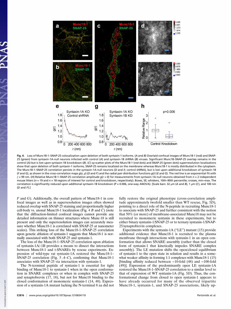
F and G). Additionally, the overall pattern of Munc18-1 in con-focal images as well as in superresolution images often showedreduced overlap with SNAP-25 staining and proportionally highercell-body vs. axonal Munc18-1 localization (Fig. 4 B and C) (notethat the diffraction-limited confocal images cannot provide anydetailed information on thinner structures where Munc-18 is stillpresent and only the superresolution images can accurately mea-sure whether Munc18-1 is correlated with SNAP-25 at nanometerscales). This striking loss of the Munc18-1–SNAP-25 correlationupon genetic ablation of syntaxin-1 suggests that Munc18-1 is nor-mally associated with both SNAP-25 and syntaxin-1.The loss of the Munc18-1–SNAP-25 correlation upon ablation
of syntaxin-1A/-1B provides a means to dissect the interactionsbetween Munc18-1 and t-SNAREs by rescue experiments. Ex-pression of wild-type rat syntaxin-1A restored the Munc18-1–SNAP-25 correlation (Fig. 5 A–C), confirming that Munc18-1associates with SNAP-25 via interaction with syntaxin-1.The N-terminal peptide of syntaxin-1 is essential for tight
binding of Munc18-1 to syntaxin-1 when in the open conforma-tion in SNARE complexes or when in complex with SNAP-25and synaptobrevin (17, 18), but not for Munc18 binding to theclosed conformation of monomeric syntaxin-1 (14, 48). Expres-sion of a syntaxin-1A mutant lacking the N-terminal 8 aa did not
fully restore the original phenotype (cross-correlation ampli-tude approximately twofold smaller than WT rescue, Fig. 5D),pointing to a direct role of the N-peptide in recruiting Munc18-1to associate with SNAP-25 and further consistent with the notionthat 50% (or more) of membrane-associated Munc18 may not berecruited to monomeric syntaxin in these experiments, but toeither binary syntaxin-1/SNAP-25 or to ternary syntaxin-1/SNAP-25/synaptobrevin complexes.Experiments with the syntaxin-1A (“LE”) mutant (15) provide
additional evidence that Munc18-1 is recruited to the plasmamembrane through interactions with syntaxin-1 in an open con-formation that allows SNARE assembly (rather than the closedform of syntaxin-1 that kinetically impedes SNARE complexassembly). The LE mutation shifts the open/closed equilibriumof syntaxin-1 to the open state in solution and results in a some-what weaker affinity in forming 1:1 complexes with Munc18-1 (15)[binding affinity reduced between ∼10-fold (48) and ∼100-fold(49)]. Expression of the predominantly open LE syntaxin-1Arestored the Munc18-1–SNAP-25 correlation to a similar level tothat of expression of WT syntaxin-1A (Fig. 5D). Thus, the con-formational change from closed to open syntaxin-1 appears tohave already occurred for many of the observed tripartiteMunc18-1, syntaxin-1, and SNAP-25 associations, likely up-
Fig. 4. Loss of Munc18-1–SNAP-25 colocalization upon deletion of both syntaxin-1 isoforms. (A and B) Overlaid confocal images of Munc18-1 (red) and SNAP-25 (green) from syntaxin-1A-null neurons infected with control (A) and syntaxin-1B shRNA (B) viruses. Significant Munc18–SNAP-25 overlap remains in thecontrol (A) but is lost upon syntaxin-1B knockdown (B). (C) xy-scatter plots of the Munc18-1 (red dots) and SNAP-25 (green dots) superresolution localizationsshow that upon deletion of both syntaxin-1 isoforms, SNAP-25 remains localized on the membrane whereas Munc18-1 is mostly distributed in the cytoplasm.The Munc18-1–SNAP-25 correlation persists in the syntaxin-1A-null neurons (D and E; control shRNA), but is lost upon additional knockdown of syntaxin-1B(F and G), as shown in the cross-correlation maps g(x, y) (D and F) and the radial pair distribution functions g(r) (E and G). The red line is an exponential fit withξ = 99 nm. (H) Relative Munc18-1–SNAP-25 correlation amplitude g(r = 0) for measurements from syntaxin-1A-null neurons obtained from n = 2 independentmouse litters (n = 19 and n = 18 regions of interest for control and knockdown, respectively). Boxes, SE; whiskers, 10th–90th percentile; crosses, min–max. Thecorrelation is significantly reduced upon additional syntaxin-1B knockdown (P = 0.006, one-way ANOVA). [Scale bars: 32 μm (A and B), 1 μm (C), and 100 nm(D and F).]
E2816 | www.pnas.org/cgi/doi/10.1073/pnas.1310654110 Pertsinidis et al.
Dow
nloa
ded
by g
uest
on
Apr
il 8,
202
0

stream of the full SNARE bundle formation in the neuronalmembrane fusion reaction.Biochemical assays further corroborate our nanometer-scale
imaging results on the associations of Munc18-1, syntaxin-1, andSNAP-25 (Fig. S8). Munc18-1 can be pulled down from WTmouse brain lysate together with the SNARE protein SNAP-25but not with monomeric SNAP-25. Also, in cortical neuronscultured from syntaxin-1A KO mice, the syntaxin-1B knock-downinhibits coimmunoprecipitation of Munc18-1 and SNAP-25. Thisloss of coimmunoprecipitation is rescued by reexpression ofWT rat syntaxin-1A but not of mutant syntaxin-1A lacking theN-terminal peptide. Consistent with the notion that the observedtripartite associations involve mostly the open syntaxin confor-mation, expression of LE syntaxin-1A results in enhanced pull-down of both Munc-18 and SNAP-25.
DiscussionFluorescence Nanoscopy with Photon-Limited Spatial Resolution.Dissecting the complex molecular assemblies and interactionsthat regulate neurotransmitter release has been hindered by theunavailability of high-resolution/high-sensitivity in situ imagingtools. Here we demonstrate fluorescence nanoscopy of biologicalspecimens at ambient conditions down to the photon-noise limit
of d ∼ 7.5-nm FWHM resolution (3-nm localization precision).The performance achieved is an approximately two- to threefoldimprovement compared with conventional single-marker switchingapproaches (22) that demonstrated ∼20-nm FWHM resolutionbenchmarks using similar fluorophores, photoswitching schemes,and photon efficiencies to those used here (23, 29) and thatrepresent a widely adopted opto-mechanical platform forsuperresolution microscopy implementations. Our results are aunique example of using superresolution cross-correlation anal-ysis and mutagenesis to characterize details of protein–proteininteractions in vivo.Higher resolution requires minimizing systematic effects while
maximizing the photon budget. Three recent implementationsthat collect twice as many photons by imaging the sample usingtwo opposed objective lenses have shown focal plane FWHMresolutions ∼10 nm (27) and ∼20 nm (25, 26), respectively.Faster acquisition reduces the effect of long-term instabilitiesand ref. 26 reports an ∼2-nm short-term stability; however, thefaster on–off dye cycling might have limited the photon budgetand thus the obtainable resolution. Brighter fluorophores enablehigher resolution but require correspondingly longer acquisitiontimes, thus resulting in correspondingly higher susceptibility toinstrument drift, and although the extra improvement in ref. 27 is
Fig. 5. Rescue of Munc18-1–SNAP-25 cross-correlation by syntaxin-1A overexpression. (A) xy-scatter plots of superresolution localization of Munc18-1 (red)and SNAP-25 (green) in syntaxin-1A-null neurons that were infected with a virus encoding shRNA for mouse syntaxin-1 and overexpressing a rat syntaxin-1A.Overexpression of rat syntaxin-1A rescues the Munc18-1–SNAP-25 cross-correlation at nanometer scales, as shown in the cross-correlation map g(x, y) (B) andpair-distribution function g(r) (C). The red line is an exponential fit with ξ = 56 nm. (D) The syntaxin-1 N-peptide is important for recovering Munc18-1–SNAP-25 cross-correlation. Shown is the relative Munc18-1–SNAP-25 correlation amplitude g(r = 0) for measurements from syntaxin-1A-null neurons obtained fromn = 2 independent mouse litters (n = 28, 14, 18, and 21 regions of interest for N-terminal deletion rescue, wild-type rescue, LE rescue, and knockdown re-spectively). Boxes, SE; whiskers, 10th–90th percentile; crosses, min–max. The correlation is significantly higher for WT rescue compared with the knockdownand the NTD rescue and for LE rescue compared with the knockdown (P = 0.018, P = 0.044, and P = 0.022, respectively, one-way ANOVA). [Scale bars: 1 μm (A)and 100 nm (B).] (E) A dynamic equilibrium model for Munc18-1, SNAP-25, and syntaxin-1 associations on the plasma membrane. Wild-type syntaxin-1 (yellow)can interconvert between (Top Left) an open conformation that can associate with SNAP-25 (green) and can bind Munc18-1 (gray) in an N-peptide-dependentinteraction and (Top Right) a closed conformation that displaces SNAP-25 and binds tightly to Munc18-1. Our results indicate that the closed-syntaxin-1/Munc18-1 complex is not the only major configuration outside the sites of fusion but rather that the N-peptide-dependent tripartite open-syntaxin-1/SNAP-25/Munc18-1 state is significantly populated. After Munc18-1 recruitment, a fusion-competent complex can be formed with the addition of synaptobrevin-2 (red).Munc18-1 can participate in fusion pore opening by interacting with the ternary SNARE complex.
Pertsinidis et al. PNAS | Published online July 2, 2013 | E2817
CELL
BIOLO
GY
APP
LIED
PHYS
ICAL
SCIENCE
SPN
ASPL
US
Dow
nloa
ded
by g
uest
on
Apr
il 8,
202
0

noteworthy, this performance is significantly worse than thetheoretical limit. Our active stabilization scheme is simpler andmore efficient than these alternative approaches, while, moreimportantly, minimizing systematic errors due to long-term driftand achieving a currently unique to our knowledge performancevery close to the theoretic resolution limit (Table S1).In addition to our active feedback system, further increase in the
raw resolution is possible. We note that with additional optimiza-tion of fluorescent probes (50) and/or switching kinetics (51), for∼105 collected photons in each on cycle, localization accuracydown to s ∼ 0.5 nm (d ∼ 1-nm FWHM resolution) is possible in ourexperimental apparatus (21), although statistical errors of ∼0.2 nmin the interprobe distances could be achieved by repetitive meas-urements over multiple (e.g., ∼10) successive on cycles. Furtherrefinement of the sample preparation procedures to achieve denselabeling, possibly via genetically encoded (50), enzymatically in-corporated (52), or chemically attached (53) fluorescent tags,would further use the potential of our approach to reveal the exactmolecular-scale architecture of subcellular structures.We also developed optimized calibration standards and algo-
rithms for two-color subdiffraction imaging, using spectrallyseparable fluorescent probes. Featuring negligible crosstalk and∼3-nm spatial registration accuracy over the size of a cell (∼15μm), our approach enables measuring the relative intracellulardistributions of two distinct protein species. This result estab-lishes a methodological framework to analyze, in situ, inter-actions between protein components that form macromolecularassemblies. Our work significantly extends the scope of the earlyproof-of-principle demonstrations of well-characterized and spa-tially defined structures, like the cytoskeleton or clathrin cages, anddiscerns the unknown organization of irregularly distributedintracellular molecules.Due to the demonstrated enhanced imaging performance, we
foresee that refined approaches to carefully eliminate extraneouserrors, such as the active feedback-stabilization and the two-color mapping calibration procedures we report here, will beused to improve the resolution of fluorescence “nanoscopes”.Importantly, deciphering molecular interactions on the basis ofdiffraction-limited imaging is often not possible as proteins thatappear colocalized with a conventional microscope can be wellseparated on the nanoscale. The improved spatial resolution andmulticolor registration accuracy afforded by our approach opensup the possibility for further applications to characterize in-tracellular protein–protein interactions and formation of definedmolecular complexes in situ.
Association of Syntaxin-1, SNAP-25, and Munc18 at the PlasmaMembrane. Here we show at 13-nm FWHM resolution that syn-taxin-1, SNAP-25, and Munc18-1 are present in clusters on theplasma membrane, and we demonstrate in pairwise double-labeling experiments that these three proteins colocalize. Thisis to the best of our knowledge a unique two-color experimentdemonstrating that syntaxin-1, SNAP-25, and SM proteins can befound in the same nanometer-sized clusters. The clusters havea size of 50–100 nm and contain up to ∼10 detected copies ofeach molecule (note that the actual number of molecules presentmay be higher, because steric hindrance can preclude efficientbinding of multiple antibodies on closely packed complexes). Weobserved a striking colocalization of syntaxin-1, SNAP-25,and Munc18-1.Assembly of the neurotransmitter release machinery during
membrane fusion reactions is thought to proceed in severalsteps. Often, subsets of SNARE proteins can bind to each otherin kinetically trapped, “dead-end” configurations, vivid examplesbeing the 2:1 syntaxin-1–SNAP-25 (54, 55) complexes. Munc18-1binds to closed syntaxin-1 in a binary complex that is thought tocontrol the beginning of SNARE-complex assembly (45). Re-constitution experiments with Munc18-1, neuronal SNAREs,
NSF, SNAP, and Munc13 suggest that the binary syntaxin-1/SNAP-25 complex is readily dissociated by NSF and SNAP,allowing Munc18-1 to capture the closed state of syntaxin, ki-netically blocking assembly of binary or ternary SNARE complex(56). Upon action of Munc13 (57), presumably in conjunctionwith an approaching synaptic vesicle, binary or ternary SNAREcomplex formation is enabled, setting the stage for Ca2+-trig-gered fusion. Specifically, when syntaxin opens up, the modeof Munc18 binding to syntaxin is dependent on the syntaxin-1N terminus for tight binding but does not require the autonomouslyfolded three-helix “abc” bundle (Habc) domain anymore (14).Of these two binding modes, the first is observed only for SMproteins in exocytosis, whereas the second is generally present inmany SM-protein/SNARE interactions. The second bindingmode also involves a direct interaction of Munc18 with the four-helix bundle of the SNARE complex (17, 18). Munc18’s mostimportant role, however, is probably in fusion directly by an asyet unknown mechanism because Munc18 and other SM proteinsare generally essential for the fusion reactions in which theyparticipate, more so often than synaptobrevin and SNAP-25 (7,58), suggesting that their function is not that of a chaperone orSNARE booster, but truly intrinsic to the fusion process.Because we observe a tripartite association of syntaxin-1,
SNAP-25, and Munc18-1 in neurons, in principle, this suggeststhree possibilities: a close association of the closed syntaxin–Munc18-1 complex with SNAP-25, a complex involving the openconformation of syntaxin with both Munc18-1 and SNAP-25, ora dynamic equilibrium between these states. In either case, theMunc18-1 interaction with the N terminus of syntaxin would playa role: This interaction is essential for tight binding betweenMunc18 and the ternary SNARE complex (17, 48), whereas itstrengthens the interaction between the closed conformation ofMunc18 and syntaxin (48). This conclusion is further supportedby our finding that deletion of the N terminus of syntaxin-1largely abrogates the colocalization of Munc18-1 with SNAP-25(Fig. 5D).The observation that the Habc domain of syntaxin is not es-
sential for exocytosis but that the N terminus is essential for bothspontaneous and evoked release (47) does not directly rule outeither state (Munc18–closed syntaxin-1 complex with SNAP-25nearby or Munc18-1/syntaxin/SNAP-25 complex). However, ourfinding that rescue with the N-terminal peptide deletion (NTD)mutant of syntaxin, which prevents tight binding of Munc18-1 tosyntaxin-1 in a binary or ternary SNARE complex but not toclosed monomeric syntaxin-1 (14), does not fully recover theMunc18/SNAP-25 cross-correlation (Fig. 5D), suggests that theobserved tripartite Munc18, syntaxin-1, SNAP-25 associationsinvolve binary or ternary SNARE complexes or a dynamic equi-librium involving these states.Our results support the notion that in neurons, SNARE pro-
teins are normally arranged in membrane patches, consistent withthe overall subcellular organization of the neuronal plasmamembrane, where channels and receptors are arranged into par-ticular subdomains. This notion is surprising given that SNAREseffectively primarily function in presynaptic active zones, whichcoincide only with a tiny percentage if any of the patches con-taining SNAREs. Even though a single SNARE complex is suffi-cient to dock liposomes to membranes and promote spontaneouslipid and content mixing in vitro (59), conferring fast exocytosis incombination with the Ca2+ sensor synaptotagmin requires morethan one synaptobrevin molecule (60) and more than threeSNAP-25 molecules (61). The organization of SNAREs in ≤100-nmmembrane domains, similar to the size corresponding to individualpresynaptic active zones, may enhance the efficiency and speed ofsynaptic vesicle fusion reaction.A certain fraction of the clusters of Munc18-1, syntaxin-1, and
SNAP-25 observed here likely contain binary (syntaxin-1/SNAP-25)or ternary (syntaxin-1/SNAP-25/synaptobrevin) SNARE complexes
E2818 | www.pnas.org/cgi/doi/10.1073/pnas.1310654110 Pertsinidis et al.
Dow
nloa
ded
by g
uest
on
Apr
il 8,
202
0
or they represent averages over dynamic states involving thesecomplexes. It has been known for some time that SNARE andSM proteins involved in synaptic vesicle exocytosis are not ac-tually enriched at sites of exocytosis (62), but the existence ofstructured clusters of SM/SNARE protein complexes throughoutneurons is nevertheless surprising. It suggests that these complexesmay operate in other fusion reactions, but more importantlyindicates that the specificity and regulation of synaptic vesiclefusion operate at a level different from that of SNARE and SMproteins. How these clusters are formed and why SNARE andSM protein complexes are not randomly distributed in membranesremain unknown. A plausible hypothesis is that the biophysicalproperties of the phospholipid membrane with lipid subdomainscontaining enrichment of cholesterol may contribute to the for-mation of these clusters, but it seems unlikely that these clustersare only a consequence of such physicochemical forces and morelikely that they represent the result of organizing proteins thatare generally involved in controlling membrane fusion.Given these previous observations, our finding that syntaxin-1
and SNAP-25 are abundantly present in a tripartite associationwith Munc18-1 is unexpected and raises a number of questions.It is unexpected because—as mentioned above—Munc18-1 alsobinds to syntaxin-1 in a different, independent mode, namely theclosed conformation of syntaxin-1 before it assembles into anycomplex with another SNARE protein. Thus, it is surprising thatour observations suggest that this complex might not be the onlypredominant complex outside of the sites of synaptic fusion—instead, we detected recruitment of Munc18-1 through a tri-partite association with SNAP-25 and syntaxin that is N-peptidedependent (Fig. 5E). At the same time, our results independentlyvalidate the notion that Munc18-1 binding to the N-peptide ofsyntaxin-1 plays a central role in fusion as proposed previously(14, 17, 47, 49, 63–65).
MethodsActively StabilizedMicroscope. The two-color superresolution imaging setup isshown in Fig. S1. A feedback loop that tracked the bright-field image ofa fiduciary in real time actuated a three-axis nanopositioning stage (PhysikInstrumente; 561-3DD, E-710 controller) and locked the sample at a fixed x,y, z set point during data acquisition. For cellular imaging, in cases where nofiducials of high-enough contrast for z tracking existed in the field of view,we used an alternative focus-stabilization scheme (21), based on a dedicatedstabilized near-infrared (NIR) laser beam that undergoes total internal re-flection (TIR) on the sample and is projected on a position-sensitive detec-tor (Quadrant Photo-Detector, QPD).
Stochastic Single-Molecule Switching Imaging. We used ∼5-kW/cm2 and∼20-kW/cm2 continuous-wave (CW) ∼640-nm and 532-nm laser illumination forCy5/Alexa 647 and Atto 532, respectively. We found that simultaneous il-lumination with both lasers resulted in irreversible bleaching of the Alexa647, as well as higher autofluorescence background in the red channel.Therefore, during cellular imaging, Alexa 647 was measured first for ∼30min, followed by ∼40–50 min for Atto 532. The presence of an enzymaticoxygen scavenging system and a millimolar concentration of thiol enabledphotoswitching (23, 66), each molecule undergoing a rapid transition toa dark state upon emitting a distribution of N0 mean collected photons(Fig. S2). To achieve the highest possible contrast ratio (molecules on:molecules off) we imaged Cy5/Alexa 647 without the presence of an acti-vator dye (23, 29) or a high-power shorter-wavelength laser (34), allowingslow, spontaneous recovery of each molecule from the dark state (rate∼10−2 s−1). This cycle could be repeated several times, providing a cluster oflocalization points (centers of observed spots) from each on state for eachmolecule. Aligning such localization clusters from multiple moleculescreated a Lorentzian distribution of xy points with FWHM 2√2σ0 ∼ 2.828σ0(Figs. 1 and 2), as expected from the total signal and background counts (67,68) (SI Methods), demonstrating that our active stabilization approach suc-cessfully eliminates systematic errors due to drift. (Note that σ0 is used only
to parameterize the FWHM, as the SD for a Lorentzian distributionis infinite.)
Resolution Refinement. To determine the increased resolution afforded bykeeping progressively more precisely localized molecules, only points inFig. 1 A and C were kept for which the calculated precision σ was better thana certain cutoff σcutoff. The xy distributions (Fig. 1A) or the deviations from thefitted line (Fig. 1C) for the subset of points were fitted to a Gaussian peak.The refined resolution vs. σcutoff was determined from the fitted peakwidths as d = 2.35s. We note that in previous refinement attempts (22, 69)similar fractions of the data were kept (∼1–10%); however, the accuracy forthe refined set of points was not quantitatively validated against a knownstructure. Practically, this procedure is expected to improve the resolutionby approximately two- to threefold (SI Methods and Fig. S3) whereas incontrast, selecting a random subset of the original data does not improvethe resolution.
Identification of Individual SNAP-25 Antibodies. To independently determinethe superresolution image of individual Alexa 647-labeled SNAP-25 anti-bodies, we imaged a neuronal sample at dilute staining conditions, ensuringwell-spaced antibodies. Because a single Alexa 647 dye can undergo multipleswitching cycles, each SNAP-25 antibody appears as a cluster of xy localizationpoints (approximately five points per hour of acquisition time). The FWHMof the distribution of points in each cluster is d = 13 nm, determined byaligning individual clusters by their center of mass (Fig. 2B).
Individual antibodies could also be identified as distinct clusters of Alexa647 xy localization points in densely stained specimens (Fig. 2A). Neighborantibodies could be readily resolved from the xy scatter plots, at minimumseparations of ∼30–50 nm, with <15-nm FWHM resolution (Fig. S4). To es-timate a molecular probability density function ρ(x, y) we rendered each xyAlexa 647 localization point (Fig. 2C) as a normalized 2D Gaussian (22) of rmssize σ given by Eq. S1a. Individual SNAP-25 antibodies were identified aslocal maxima of the resulting 2D ρ(x, y) surface (Fig. 2C), using a peak searchalgorithm. The xy coordinates of each SNAP-25 antibody were then de-termined from the centroid of ρ(x, y) around each peak.
Two-Color Registration over Extended Field of View. Two types of referenceobjects were used to register the coordinates between the two CCD channels:(i) 20-bp Cy3-Cy5 DNA duplexes, randomly distributed in the field of view,and (ii) a regular nanofabricated pattern of subwavelength holes on an Al-coated quartz wafer (SI Methods and Fig. S6). A set of reference coordinatesobtained from multiple such objects and from sampling the whole field ofview was used to obtain a mapping transformation (21), using a low-orderpolynomial or spline interpolation (SI Methods and Fig. S6).
Sample Preparation and Imaging Conditions. The preparations of DNA con-structs, F-actin, dye-labeled antibodies, and neuronal cultures, as well asprocedures and conditions for confocal immunofluorescence imaging, aredetailed in SI Methods.
Quantitative Colocalization and Clustering Structure Analysis. We used theOPTICS algorithm (44) to perform a hierarchical ordering of the antibodylocalization points. For each point i we calculated core-distances (43) cdist iGGand cdist iGR with respect to points of the same and opposite colors, re-spectively (Fig. S5). Clusters were identified using the DBSCAN algorithm(43),with Minpts = 3 and « ∼ 30–40 nm. The selected values for « correspond toroughly the average core distances for the particular dataset; approximatelytwofold smaller « values failed to identify all but the densest clusterswhereas approximately twofold larger « values resulted in merging all of thepoints into just a few large ones.
ACKNOWLEDGMENTS. A.P. wishes to thank James W. Conway for sharingexpertise in Electron Beam Lithography. Work was performed in part at theStanford Nanofabrication Facility which is supported by National ScienceFoundation through the National Nanotechnology Infrastructure Networkunder Grant ECS- 9731293. This work was supported by the NationalInstitutes of Health (NIH), the National Science Foundation, the NationalAeronautics and Space Administration, and the Defense Advanced ResearchProjects Agency through awards to S.C. and by NIH Grant R37-MH63105(to A.T.B.).
1. Gao Y, et al. (2012) Single reconstituted neuronal SNARE complexes zipper in three
distinct stages. Science 337(6100):1340–1343.
2. Hata Y, Slaughter CA, Südhof TC (1993) Synaptic vesicle fusion complex contains unc-
18 homologue bound to syntaxin. Nature 366(6453):347–351.
Pertsinidis et al. PNAS | Published online July 2, 2013 | E2819
CELL
BIOLO
GY
APP
LIED
PHYS
ICAL
SCIENCE
SPN
ASPL
US
Dow
nloa
ded
by g
uest
on
Apr
il 8,
202
0
3. Novick P, Schekman R (1979) Secretion and cell-surface growth are blocked in a temperature-sensitive mutant of Saccharomyces cerevisiae. Proc Natl Acad Sci USA 76(4):1858–1862.
4. Harrison SD, Broadie K, van de Goor J, Rubin GM (1994) Mutations in the DrosophilaRop gene suggest a function in general secretion and synaptic transmission. Neuron13(3):555–566.
5. Brenner S (1974) The genetics of Caenorhabditis elegans. Genetics 77(1):71–94.6. Gengyo-Ando K, et al. (1993) The C. elegans unc-18 gene encodes a protein expressed
in motor neurons. Neuron 11(4):703–711.7. Verhage M, et al. (2000) Synaptic assembly of the brain in the absence of
neurotransmitter secretion. Science 287(5454):864–869.8. Perin MS, Fried VA, Mignery GA, Jahn R, Südhof TC (1990) Phospholipid binding by
a synaptic vesicle protein homologous to the regulatory region of protein kinase C.Nature 345(6272):260–263.
9. McMahon HT, Missler M, Li C, Südhof TC (1995) Complexins: Cytosolic proteins thatregulate SNAP receptor function. Cell 83(1):111–119.
10. Diao J, et al. (2012) Synaptic proteins promote calcium-triggered fast transition frompoint contact to full fusion. eLife 1:e00109.
11. Kyoung M, et al. (2011) In vitro system capable of differentiating fast Ca2+-triggeredcontent mixing from lipid exchange for mechanistic studies of neurotransmitterrelease. Proc Natl Acad Sci USA 108(29):E304–E313.
12. Yamaguchi T, et al. (2002) Sly1 binds to Golgi and ER syntaxins via a conserved N-terminal peptide motif. Dev Cell 2(3):295–305.
13. Dulubova I, et al. (2002) How Tlg2p/syntaxin 16 ‘snares’ Vps45. EMBO J 21(14):3620–3631.
14. Dulubova I, et al. (2007) Munc18-1 binds directly to the neuronal SNARE complex.Proc Natl Acad Sci USA 104(8):2697–2702.
15. Dulubova I, et al. (1999) A conformational switch in syntaxin during exocytosis: Roleof munc18. EMBO J 18(16):4372–4382.
16. Misura KM, Scheller RH, Weis WI (2000) Three-dimensional structure of the neuronal-Sec1-syntaxin 1a complex. Nature 404(6776):355–362.
17. Shen J, Tareste DC, Paumet F, Rothman JE, Melia TJ (2007) Selective activation ofcognate SNAREpins by Sec1/Munc18 proteins. Cell 128(1):183–195.
18. Xu Y, Su L, Rizo J (2010) Binding of Munc18-1 to synaptobrevin and to the SNAREfour-helix bundle. Biochemistry 49(8):1568–1576.
19. Carr CM, Grote E, Munson M, Hughson FM, Novick PJ (1999) Sec1p binds to SNAREcomplexes and concentrates at sites of secretion. J Cell Biol 146(2):333–344.
20. Hell SW (2007) Far-field optical nanoscopy. Science 316(5828):1153–1158.21. Pertsinidis A, Zhang Y, Chu S (2010) Subnanometre single-molecule localization,
registration and distance measurements. Nature 466(7306):647–651.22. Betzig E, et al. (2006) Imaging intracellular fluorescent proteins at nanometer
resolution. Science 313(5793):1642–1645.23. Rust MJ, Bates M, Zhuang X (2006) Sub-diffraction-limit imaging by stochastic optical
reconstruction microscopy (STORM). Nat Methods 3(10):793–795.24. Hess ST, Girirajan TP, Mason MD (2006) Ultra-high resolution imaging by fluorescence
photoactivation localization microscopy. Biophys J 91(11):4258–4272.25. Shtengel G, et al. (2009) Interferometric fluorescent super-resolution microscopy
resolves 3D cellular ultrastructure. Proc Natl Acad Sci USA 106(9):3125–3130.26. Aquino D, et al. (2011) Two-color nanoscopy of three-dimensional volumes by 4Pi
detection of stochastically switched fluorophores. Nat Methods 8(4):353–359.27. Xu K, Babcock HP, Zhuang X (2012) Dual-objective STORM reveals three-dimensional
filament organization in the actin cytoskeleton. Nat Methods 9(2):185–188.28. Subach FV, et al. (2009) Photoactivatable mCherry for high-resolution two-color
fluorescence microscopy. Nat Methods 6(2):153–159.29. Bates M, Huang B, Dempsey GT, Zhuang X (2007) Multicolor super-resolution imaging
with photo-switchable fluorescent probes. Science 317(5845):1749–1753.30. Shroff H, et al. (2007) Dual-color superresolution imaging of genetically expressed
probes within individual adhesion complexes. Proc Natl Acad Sci USA 104(51):20308–20313.
31. Punge A, et al. (2008) 3D reconstruction of high-resolution STED microscope images.Microsc Res Tech 71(9):644–650.
32. Dani A, Huang B, Bergan J, Dulac C, Zhuang X (2010) Superresolution imaging ofchemical synapses in the brain. Neuron 68(5):843–856.
33. Denker A, Kröhnert K, Bückers J, Neher E, Rizzoli SO (2011) The reserve pool ofsynaptic vesicles acts as a buffer for proteins involved in synaptic vesicle recycling.Proc Natl Acad Sci USA 108(41):17183–17188.
34. Heilemann M, et al. (2008) Subdiffraction-resolution fluorescence imaging withconventional fluorescent probes. Angew Chem Int Ed Engl 47(33):6172–6176.
35. Gittes F, Mickey B, Nettleton J, Howard J (1993) Flexural rigidity of microtubules andactin filaments measured from thermal fluctuations in shape. J Cell Biol 120(4):923–934.
36. Willig KI, Rizzoli SO, Westphal V, Jahn R, Hell SW (2006) STED microscopy reveals thatsynaptotagmin remains clustered after synaptic vesicle exocytosis. Nature 440(7086):935–939.
37. Sieber JJ, et al. (2007) Anatomy and dynamics of a supramolecular membrane proteincluster. Science 317(5841):1072–1076.
38. van den Bogaart G, et al. (2011) Membrane protein sequestering by ionic protein-lipidinteractions. Nature 479(7374):552–555.
39. Garcia EP, McPherson PS, Chilcote TJ, Takei K, De Camilli P (1995) rbSec1A and Bcolocalize with syntaxin 1 and SNAP-25 throughout the axon, but are not in a stablecomplex with syntaxin. J Cell Biol 129(1):105–120.
40. Bar-On D, et al. (2012) Super-resolution imaging reveals the internal architecture ofnano-sized syntaxin clusters. J Biol Chem 287(32):27158–27167.
41. Hansen J-P, McDonald IR (2006) Theory of Simple Liquids, 3rd edition (Academic Press,London, UK), P 29–30.
42. Sengupta P, et al. (2011) Probing protein heterogeneity in the plasma membraneusing PALM and pair correlation analysis. Nat Methods 8(11):969–975.
43. Ester MKH-P, Sander J, Xu X (1996) A density based algorithm for discovering clustersin large spatial databases with noise. Proceedings of the Second International Con-ference on Knowledge Discovery and Data Mining, eds Simoudis E, Han J, Fayyad U(Association for the Advancement of Artificial Intelligence, Menlo Park, CA), pp 226–231.
44. Ankerst M, Breunig M, Kreigel H-P, Sander J (1999) OPTICS: Ordering points toidentify clustering structure. Proceedings of the 1999 ACM SIGMOD InternationalConference on Management of Data ACM Press New York, NY, USA Alex Delis,Christos Faloutsos, Shahram Ghandeharizadeh (Eds.) pp 49–60.
45. Gerber SH, et al. (2008) Conformational switch of syntaxin-1 controls synaptic vesiclefusion. Science 321(5895):1507–1510.
46. Hata Y, Südhof TC (1995) A novel ubiquitous form of Munc-18 interacts with multiplesyntaxins. Use of the yeast two-hybrid system to study interactions between proteinsinvolved in membrane traffic. J Biol Chem 270(22):13022–13028.
47. Zhou P, et al. (2013) Syntaxin-1 N-peptide and Habc-domain perform distinct essentialfunctions in synaptic vesicle fusion. EMBO J 32(1):159–171.
48. Burkhardt P, Hattendorf DA, Weis WI, Fasshauer D (2008) Munc18a controls SNAREassembly through its interaction with the syntaxin N-peptide. EMBO J 27(7):923–933.
49. Khvotchev M, et al. (2007) Dual modes of Munc18-1/SNARE interactions are coupled byfunctionally critical binding to syntaxin-1 N terminus. J Neurosci 27(45):12147–12155.
50. Shaner NC, et al. (2008) Improving the photostability of bright monomeric orangeand red fluorescent proteins. Nat Methods 5(6):545–551.
51. Vogelsang J, Cordes T, Forthmann C, Steinhauer C, Tinnefeld P (2009) Controlling thefluorescence of ordinary oxazine dyes for single-molecule switching and superresolutionmicroscopy. Proc Natl Acad Sci USA 106(20):8107–8112.
52. Hein B, et al. (2010) Stimulated emission depletion nanoscopy of living cells usingSNAP-tag fusion proteins. Biophys J 98(1):158–163.
53. Keppler A, Pick H, Arrivoli C, Vogel H, Johnsson K (2004) Labeling of fusion proteinswith synthetic fluorophores in live cells. Proc Natl Acad Sci USA 101(27):9955–9959.
54. Fasshauer D, Bruns D, Shen B, Jahn R, Brünger AT (1997) A structural change occursupon binding of syntaxin to SNAP-25. J Biol Chem 272(7):4582–4590.
55. Xiao W, Poirier MA, Bennett MK, Shin YK (2001) The neuronal t-SNARE complex isa parallel four-helix bundle. Nat Struct Biol 8(4):308–311.
56. Ma C, Su L, Seven AB, Xu Y, Rizo J (2013) Reconstitution of the vital functions ofMunc18 and Munc13 in neurotransmitter release. Science 339(6118):421–425.
57. Ma C, Li W, Xu Y, Rizo J (2011) Munc13 mediates the transition from the closedsyntaxin-Munc18 complex to the SNARE complex. Nat Struct Mol Biol 18(5):542–549.
58. Schoch S, et al. (2001) SNARE function analyzed in synaptobrevin/VAMP knockoutmice. Science 294(5544):1117–1122.
59. van den Bogaart G, et al. (2010) One SNARE complex is sufficient for membranefusion. Nat Struct Mol Biol 17(3):358–364.
60. Sinha R, Ahmed S, Jahn R, Klingauf J (2011) Two synaptobrevin molecules aresufficient for vesicle fusion in central nervous system synapses. Proc Natl Acad Sci USA108(34):14318–14323.
61. Mohrmann R, de Wit H, Verhage M, Neher E, Sørensen JB (2010) Fast vesicle fusion inliving cells requires at least three SNARE complexes. Science 330(6003):502–505.
62. Südhof TC (2012) Calcium control of neurotransmitter release. Cold Spring HarbPerspect Biol 4(1):a011353.
63. Deák F, et al. (2009) Munc18-1 binding to the neuronal SNARE complex controlssynaptic vesicle priming. J Cell Biol 184(5):751–764.
64. Rathore SS, et al. (2010) Syntaxin N-terminal peptide motif is an initiation factor forthe assembly of the SNARE-Sec1/Munc18 membrane fusion complex. Proc Natl AcadSci USA 107(52):22399–22406.
65. Johnson JR, et al. (2009) Binding of UNC-18 to the N-terminus of syntaxin is essentialfor neurotransmission in Caenorhabditis elegans. Biochem J 418(1):73–80.
66. Fölling J, et al. (2008) Fluorescence nanoscopy by ground-state depletion and single-molecule return. Nat Methods 5(11):943–945.
67. Thompson RE, Larson DR, Webb WW (2002) Precise nanometer localization analysisfor individual fluorescent probes. Biophys J 82(5):2775–2783.
68. Mortensen KI, Churchman LS, Spudich JA, Flyvbjerg H (2010) Optimized localizationanalysis for single-molecule tracking and super-resolution microscopy. Nat Methods7(5):377–381.
69. Cronin B, de Wet B, Wallace MI (2009) Lucky imaging: Improved localization accuracyfor single molecule imaging. Biophys J 96(7):2912–2917.
E2820 | www.pnas.org/cgi/doi/10.1073/pnas.1310654110 Pertsinidis et al.
Dow
nloa
ded
by g
uest
on
Apr
il 8,
202
0