Embed Size (px)
Citation preview

ULTRASTRUCTURE AND LSU rDNA-BASED PHYLOGENYOF ESOPTRODINIUM GEMMA(DINOPHYCEAE), WITH NOTES ON FEEDING BEHAVIOR AND THE DESCRIPTION OF
THE FLAGELLAR BASE AREA OF A PLANOZYGOTE1
Antonio J. Calado,2 Sandra C. CraveiroDepartamento de Biologia, Universidade de Aveiro, P-3810-193 Aveiro, Portugal
Niels Daugbjerg and Øjvind Moestrup
Department of Phycology, Biological Institute, University of Copenhagen, Øster Farimagsgade 2D, DK-1353 Copenhagen K,
Denmark
A small, freshwater dinoflagellate with an incom-plete cingulum, identified as Esoptrodinium gemmaJavornicky (5Bernardinium bernardinense sensu au-ctt. non sensu Chodat), was maintained in mixedculture and examined using light and serial sectionTEM. Vegetative flagellate cells, large cells with twolongitudinal flagella (planozygotes), and cysts wereexamined. The cells displayed a red eyespot nearthe base of the longitudinal flagellum, made of twoor three layers of pigment globules not bounded bya membrane. Yellow-green, band-shaped chloro-plasts, bounded by three membranes and contain-ing lamella with three thylakoids, were present inboth flagellate cells and cysts. Most cells had foodvacuoles, containing phagotrophically ingestedchlamydomonads or chlorelloid green algae; inges-tion occurred through the ventral area, involving athin pseudopod apparently driven by the peduncle.The pusule was tubular, with numerous diverticulain its distal portion, and opened into the longitudi-nal flagellar canal. Three roots were associated witheach pair of flagellar bases, both in vegetative cellsand in a planozygote. The longitudinal microtubu-lar root bifurcated around the longitudinal basalbody. The planozygote contained a single peduncleand associated structures, and a single transverseflagellar canal with the two converging transverseflagella. Using two ciliates as outgroup species,phylogenetic analyses based on maximum parsimo-ny, neighbor-joining and posterior probability (Bay-esian analysis) supported a clade comprisingEsoptrodinium, Tovellia, and Jadwigia.
Key index words: Bernardinium bernardinense;Dinophyceae; Esoptrodinium gemma; eyespot; flag-ellar apparatus; LSU rDNA; phagotrophy; phylo-geny; planozygote; ultrastructure
Abbreviations: BA, Bayesian analysis; BB, basalbody; LB, longitudinal basal body; LMR, longitu-dinal microtubular root; MCMC, Monte Carlo
Markov chains; MP, maximum parsimony; NJ,neighbor-joining; PP, posterior probabilities; PSC,peduncular striated collar; SRC, striated root con-nective (TSR to LMR); TB, transverse basal body;TMR, transverse microtubular root; TMRE, TMRextension; TSR, transverse striated root; TSRM,transverse striated root microtubule; VR, ventralridge
The organism used in this study is a naked dino-flagellate that would be readily identified as Bern-ardinium bernardinense Chodat by recent dinoflagellatefloras (Starmach 1974, Matvienko and Litvinenko1977, Popovsky and Pfiester 1990). In his original de-scription of B. bernardinense, Chodat (1924, pp. 40–41)mentioned the incomplete cingulum, which disap-peared on the dorsal side, and the absence of chloro-plasts and of a distinct sulcus. The new genusBernardinium was compared with Hemidinium F. Stein,from which it was distinguished mainly by the well-de-fined sulcus in the two Hemidinium species previouslydescribed. Although Hemidinium was originally de-scribed as unarmored (Stein 1878, pp. 91 and 97,1883, pl. 2, Figs. 23–26), the type species is currentlyregarded as a thecate form with thin plates (Popovskyand Pfiester 1990). The smaller of the two cell portionsdelimited by the incomplete cingulum was placed ontop in Chodat’s (1924) original drawings; his descrip-tion shows he regarded this as the epicone (‘‘parte di-midia superiore’’). Schiller (1935) reinterpreted theorientation of the cell and reproduced Chodat’s draw-ings with the longitudinal flagellum emerging down-ward, pointing out that, with this orientation, thecingulum is directed toward the right side of thebody. Huber-Pestalozzi (1950, p. 164) regardedthe species as imperfectly described and transferredit to Hemidinium.
The second report of an organism identified as B.bernardinense is that of Thompson (1951); the cells de-scribed resembled Chodat’s (1924) in both size andgeneral morphology, but displayed the orientation ofcingulum and transverse flagellum that is common in
1Received 31 August 2005. Accepted 22 December 2005.2Author for correspondence: e-mail [email protected].
434
J. Phycol. 42, 434–452 (2006)r 2006 Phycological Society of AmericaDOI: 10.1111/j.1529-8817.2006.00195.x

dinoflagellates, i.e. to the cell’s left. A third and moredetailed description of an organism under this namecomes from Javornicky (1962), whose specimens showthe same orientation as those of Thompson (1951).Javornicky (1962) considered the orientation shown inChodat’s (1924) drawings to be an error, and Conrad’s(1939) comment that Bernardinium is an inverse Hemi-dinium to be therefore unjustified. Wawrik (1983) re-ported the species, with the name H. bernardinense,from a pond in Lower Austria (Wawrik 1983, Fig. 1g);a single cell is depicted with the cingulum extending tothe viewer’s left, although the orientation of the cellwas not mentioned and a transverse flagellum was notseen. Popovsky (1990) reported B. bernardinense with aleft hand orientation of the cingulum from ponds inCzechoslovakia, although his drawings show cells witha different general morphology and a well-markedsulcus, making the identification somewhat doubtful.
Javornicky (1997) reported a flagellate with the gen-eral features ascribed to B. bernardinense from a littlefreshwater pond in Rugen Island, North-East Germa-ny and both the cingulum and the transverse flagellumoriented toward the cell’s right; upon comparison withChodat’s (1924) drawings, Javornicky (1997) conclud-ed that this German population represented B. bernar-dinense in the original sense of Chodat. Considering thenearly mirror-symmetrical populations studied byboth Thompson (1951) and Javornicky (1962) to rep-resent not only a different species, but a different ge-nus, Javornicky (1997) described the new genusEsoptrodinium, with the type species Esoptrodinium gem-ma based on the population described by Javornicky(1962) under the name B. bernardinense. The organismstudied herein has a left-oriented cingulum and trans-verse flagellum and is therefore identifiable as E. gem-ma Javornicky.
The organization of the flagellar base area has beenstudied in detail for over 20 species of dinoflagellates(Calado and Moestrup 2002, Hansen and Daugbjerg2004). Although swimming dinoflagellate cells result-ing from the fusion of gametes are known to have twolongitudinal flagella and the parallel arrangement ofthe two sets of basal bodies and roots has been shown(Wedemayer and Wilcox 1984), a detailed descriptionof the flagellar base area of a planozygote, such as isgiven herein for Esoptrodinium, has not previously beenpublished. Additionally, we report on the phylogeny ofEsoptrodinium based on three-cell PCR determinationof nuclear-encoded large subunit (LSU) rDNA.
MATERIALS AND METHODS
E. gemma was collected from the sediment of a sidewalkcovered by a few centimeters of flowing tap water leaking froma damaged pipe, in the campus of the University of Aveiro,Portugal. Samples were taken repeatedly between June andSeptember 2002 andmixed cultures were prepared in Chu no.10 medium (Nichols 1973) and kept at 201 C with a 12:12Light:Dark photoperiod. Some cultures were initiated fromthe sediment of samples that had dried out, by adding culturemedium and exposing it to the light.
Light microscopy. The morphology and swimming mode ofcells were recorded with a Sony Video Cassette RecorderSLV-825 (Sony Corporation, Tokyo, Japan). Micrographswere taken with a Leica DMLB light microscope (Leica Mi-crosystems, Wetzlar, Germany) with differential interferencecontrast. Observations of the feeding process were made inmicrochambers prepared by attaching a rectangular Plasti-cine support onto a 24mm ! 32mm coverslip, placing adrop of the culture in the middle of the coverslip, invertingit over a slide, and pressing along the edges.
Electron microscopy. Cultured material was fixed in a mix-ture of 1% glutaraldehyde and 0.25% osmium tetroxide (finalconcentrations) in 0.1M phosphate buffer, pH 7.2, for 15minat room temperature. After centrifugation, the pellet wasrinsed in the same buffer, embedded in 1.5% agar, and post-fixed for approximately 1 h in 1% buffered osmium tetroxideat room temperature. Rinsing in buffer and water, and de-hydration through a graded ethanol series up to 70%, wasperformed at 41 C. After the final dehydration steps inethanol and propylene oxide, at room temperature, the ma-terial was embedded in Epon. Single cells of E. gemma, whichwere greatly outnumbered by green flagellates and otherprey organisms, were singled out in the blocks with the lightmicroscope and sectioned with a diamond knife on a ReichertUltracut E (Leica Microsystems, Wetzlar, Germany). The sec-tions were picked up with slot grids, placed on Formvar film,contrasted with uranyl acetate and lead citrate, and exam-ined with a JEOL JEM 1010 electron microscope (JEOL Ltd.,Tokyo, Japan). Serial sections from two vegetative cells, aplanozygote and a cyst were examined.
PCR amplification. Using a capillary pipette three cells ofE. gemma from a culture grown on a species of Chlamydomonaswere transferred through three rinses in double-distilled wa-ter and placed in the same 0.5mL PCR tube containing 8 mLof double-distilled water. Amplification of partial LSU rDNAusing the forward primer ‘‘D1F’’ (Scholin et al. 1994) and thereverse dinoflagellate specific primer ‘‘Dino-ND’’ (Hansenand Daugbjerg 2004) resulted in a PCR product of approx-imately 1800 bp in length. The temperature profile used forPCR amplification and subsequent determination of the nu-clear-encoded LSU rDNA sequence (GenBank DQ289020)was established as described previously (Hansen et al. 2003,Hansen and Daugbjerg 2004).
Sequence alignment. The LSU rDNA sequence of Eso-ptrodinium was determined in both directions and added tothe same data matrix recently compiled by Moestrup et al.(2006) to elucidate the phylogeny of woloszynskioid dino-flagellates. The data matrix analyzed here comprised a totalof 40 dinoflagellates and sequences were aligned by incor-porating information from secondary structure of LSUrDNA as suggested by de Rijk et al. (2000). The alignmentcomprised 1477 bp, including introduced gaps and covered36 bp upstream domain D1–20 bp downstream D6 (see thesecondary model for the LSU rDNA for Prorocentrum micansproposed by Lenaers et al. (1989)). Because of ambiguousalignment of domain D2, a fragment starting 8 bp down-stream D2 to the end of this domain was omitted beforephylogenetic analysis, hence leaving a fragment of 1124 bpfor the analyses. The data matrix was manually edited usingMacClade ver. 4.07 (Maddison and Maddison 2003).
Phylogenetic analyses. We used PAUP* ver. 4b10 (Swofford2003) for maximum parsimony (MP) and neighbor-joining(NJ) analyses and MrBayes ver. 3.1 (Ronquist and Huelsen-beck 2003) for Bayesian analysis (BA). MP analyses wereconducted with 1000 random additions in heuristic searchesand a branch-swapping algorithm (tree-bisection-reconnec-tion). All characters were equally weighted and unordered.Gap positions were treated as missing data. For MP bootstrapanalyses we used 1000 replications. Modeltest ver. 3.6
ESOPTRODINIUM ULTRASTRUCTURE AND PHYLOGENY 435

(Posada and Crandall 1998) was used to search for the bestmodel for the LSU rDNA sequences using hierarchical like-lihood ration tests and the best-fit model found wasTrN" I"G (Tamura and Nei 1993). Among sites rate het-erogeneity was a5 0.6328, an estimated proportion of invar-iable sites was I5 0.2569 and two substitution rate categorieswere A–G5 2.7815 and C–T5 6.6913. Base frequencieswere set as follows A5 0.288, C5 0.1698, G5 0.2713 andT5 0.2709. The Tamura and Nei model was used to com-pute dissimilarity values and the resulting distance matrixwas applied to build a tree with the NJ method. For NJ boot-strap we used 1000 replications. For the BA, a GTR substi-tution model was used with base frequencies and substitutionrate matrix estimated from the data matrix. Four simultane-ous Monte Carlo Markov chains (MCMC; Yang and Rannala1997) were run from random trees for a total of 1 ! 106
generations (Metropolis-coupled MCMC). A tree was sam-pled every 50 generations and the ‘‘burn-in’’ was evaluatedfor stationarity by examination of the plateau in log-likeli-hood over generations using Tracer ver. 1.2.1 by A. Rambaut& A. Drummond and an Excel spreadsheet. ‘‘Burn-in’’ of thechain occurred in fewer than 27,000 generations so the first540 trees were discarded, leaving 19,461 trees for estimatingposterior probabilities (PP). Thus, PP values were obtainedfrom the 50% majority rule consensus of the kept trees. Toconfirm that the analysis was not trapped at a local optimumBA was performed three times (Leache and Reeder 2002).
Outgroup species. Previous molecular studies addressingthe phylogeny of major groups of eucaryotes have shownthat ciliates form a sister group to dinoflagellates (van de Peeret al. 1996). Together with the Apicomplexa they constitutethe alveolates. The morphological synapomorphic characteris the cortical alveolae under the plasma membrane (Patter-son 1999). Hence, we used two ciliates (Tetrahymena pyriformisand Tetrahymena thermophila) to root the ingroup of dinoflag-ellates in all phylogenetic analyses.
RESULTS
Morphology. The roughly obovoid cells were most-ly 10–16 mm long and 7–11 mm wide (average of 70cells were 11.9 and 9.2 mm, respectively), with occa-sional large cells about 18 mm long and 12 mm wide,and in some batches numerous tiny cells down to8 mm long and 5 mm wide. Large cells were often seento carry two longitudinal flagella. Most cells were
slightly flattened dorsoventrally, but some were near-ly circular in apical view. The cingulum was a deepand slightly descending groove that extended fromthe ventral face to about the mid-dorsal side, sharplydelimiting on the left side of the cell the longer ep-icone from the shorter and narrower hypocone. Theright side of the cell was almost straight or smoothlycurved although some cells showed a slight depres-sion marking the lower-right edge of the epicone(Fig. 1a). The apex was broadly rounded and thesides of the epicone were nearly straight in the longercells, or slightly curved to the shape of a helmet inshorter ones (Fig. 1a); an inflexion near the lower-leftedge of the epicone produced a beak-like projectionin a few cells (not shown). The antapex was usuallyround, although the shape of the hypocone was morevariable, especially in the smaller cells (Fig. 1, a-c).Near the proximal end of the cingulum the upperedge was somewhat raised, invading the epicone (Fig.1b, short arrow). The transverse flagellum undulatedin a relatively loose helix and did not extend signif-icantly beyond the end of the cingulum. The sulcuswas surprisingly inconspicuous given its appearancein the TEM, and it was only clearly visible in slow-moving cells in favorable positions in the light mi-croscope. The longitudinal flagellum was usuallyslightly longer than the cell.
The cells swam rapidly, with velocities reaching over200mm/s as estimated from video recordings, and fre-quently changed direction. They also showed a ten-dency to swim in tight circles for minutes on end,especially in microscope preparations, often turningaround in the same spot with their ventral or dorsalside upwards, invariably with the apex moving towardthe cell’s right and the longitudinal flagellum trailingaround the right edge of the cell.
The cell contents were often obscured by food vac-uoles, containing prey cells ranging in size from ap-proximately 10mm long Chlamydomonas cells (Fig. 1d)to chlorelloid cells some 3–6mm in diameter; large Eso-ptrodinium cells were seen containing up to 10 food
FIG. 1. Morphology of Esoptrodinium gemma swarmers and cyst. Differential interference light microscopy. FV, food vacuole; N, nu-cleus. (a, b) Ventral view of a relatively large cell in optical section (a) and in surface focus (b), showing the different-sized parts delimitedby the incomplete cingulum, and the main cell structures. The short arrow in (b) indicates the proximal part of the cingulum invadingthe epicone. Scale bar as in (c). (c) Left-ventral view of a small cell. Note the band-shaped chloroplast (Ch) and the eyespot (E). (d) A cystand Chlamydomonas cells (short arrows) from a mixed culture.
ANTONIO J. CALADO ET AL.436

vacuoles. A red eyespot, shaped as a markedly curvedplate with an outward facing concavity, was visible nearthe base of the longitudinal flagellum, right below theproximal end of the cingulum (Fig. 1, b and c). Thenucleus was located in the upper part of the hypocone,on the left side of the cell (Fig. 1a). Although difficult tosee at low magnification, closer examination always re-vealed the presence of one or two band-shaped, yel-low-green chloroplasts (Fig. 1c), which appeared moreor less broken up in cells containing food vacuoles(Fig. 1, a and b).
Round, smooth-walled cysts were found in thesediment of field-collected material and on the bottomof the culture wells, where their number increased asthe cultures grew old (Fig. 1d); their cytoplasm resem-bled that of flagellate cells and often contained foodvacuoles, but an eyespot was not seen. Cyst diameterranged from 12 to 16mm (n515).
Feeding. In rapidly growing cultures with plenty offood organisms most cells of Esoptrodinium exhibited arapid, jerky and somewhat helical motion. Invertedmicroscope observations of these batches in wells ofculture plates revealed the presence of concentrationareas where groups of dinoflagellate cells swamaround some prey cells, some distance away fromother similar groups. The attracting power of someprey cells was used to increase the chances of observ-ing feeding events at higher magnification in micro-chambers. The selection of such cells was dictated bythe repeated approach by Esoptrodinium cells that re-mained for periods of seconds to minutes rotatingwith the ventral side toward the prey while movingaround it, whereas nearby prey cells were not ap-proached.
Although on most occasions the dinoflagellate cellswould leave without feeding, prey ingestion was ob-served a few times. The ingestion process lasted in theorder of 5 s and, although the cells moved more slowlywhile handling the prey, rotation out of the plane offocus often concealed all details. The prey cells wereingested through the ventral side of the hypocone. Awide and thin pseudopod embraced one side of theprey and drove it to the concave ventral face ofthe somewhat deformed dinoflagellate cell, whichsquirmed back to its previous shape after ingestion.The edge of the thin pseudopod was a distinctly thick-er cylindrical structure 1–2 mm wide that was interpret-ed as a peduncle based on its appearance and positionin the cell (Fig. 2).
General ultrastructure. The general fine-structuralfeatures of the cells are shown in Figures 3 and 4.The nucleus was a typical dinokaryon, extendingfrom the flagellar base area to the left, near the ven-tral surface, approximately at cingulum level(Fig. 3a). Mitochondrial profiles of the usual dino-flagellate type were present in all sections, in centralas well as peripheral cytoplasm (Figs. 3, a and b, and4a). Dictyosomes were located in the central part ofthe cell, especially near the flagellar base area(Fig. 3a). Trichocysts were common along the
surface (Figs. 3a and 4a). Starch grains and oil drop-lets occupied a large portion of the cytoplasm (Figs. 3,a and b, and 4a). The cells contained large food vac-uoles, each apparently with a single prey cell in var-ious degrees of degradation (Fig. 3, a and b). Theplasmalemma and the underlying amphiesmal vesi-cles, although disrupted in some places, were intactover most of the cell surface; the vesicles tightly abut-ted one another and did not contain any plate-likematerial (Fig. 3, d and e). A thin layer of electron-opaque material (about the width of a unit mem-brane) covered the cytoplasmic surface of the innermembranes of the amphiesmal vesicles (Fig. 3, d ande, arrowheads). Transversely elongated vesicles un-derlay the peripheral microtubules along most of thesurface (Figs. 3, a–d and 4a). Collared pits, about150 nm deep and 50 nm wide in their constrictedpart, were very sparsely distributed on the surface(Fig. 3e).
Chloroplast. Chloroplast profiles were present inall cells examined, either scattered throughout thecytoplasm (Fig. 3b) or restricted to a part of the cell(see Figs. 3a, no chloroplasts visible, and 4a, two sec-tions of the same cell). The chloroplasts were bound-ed by three evenly spaced membranes and thestroma was crossed by bands of three thylakoids(Fig. 4b). Thylakoid-free areas were visible in somechloroplast lobes (Fig. 4a).
Ultrastructure of the cyst. The contents of thecyst were essentially similar to the cytoplasm of flag-ellate cells, with numerous starch grains and oil drop-lets, and some apparently old food vacuoles (Fig. 5a).Chloroplast profiles had the same appearance in thecyst as in flagellate cells (Fig. 5, a and b). A continuouswall surrounded the cyst (Fig. 5a); it was about120 nm thick and had a homogeneous appearanceexcept for an outermost thin layer that was moreelectron-opaque (Fig. 5b). Two unit membranes,parallel to each other, were found in the space
FIG. 2. Esoptrodinium gemma engaged in feeding (approxi-mate dorsal view). The prey cells are chlorelloid green algae.Image taken from video recording. Note the extended peduncle(arrowhead). The double arrow indicates the position of a cyto-plasmic sheet extending from the peduncle to the cell body,visible in the footage but imperceptible in the still image.
ESOPTRODINIUM ULTRASTRUCTURE AND PHYLOGENY 437

between the wall and the plasmalemma, which wasunderlain by an electron-opaque layer of about thesame thickness as the membrane (Fig. 5b).
Pusule. The pusular system consisted of a long,extensively coiled tube wrapped in a vesicle; al-though the tube was non-ramified, in the sense that
FIG. 3. Esoptrodinium gemma. General ultrastructure and details of the amphiesma. Ch, chloroplast; FV, food vacuole; N, nucleus; nu,nucleolus; P, pusule; T, trichocyst; TF, traverse flagellum. (a) Transverse section of a cell with a single set of basal bodies, viewed from theantapex near the exit point of the transverse flagellum. (b) Oblique-longitudinal section of a cell with a double set of basal bodies (notvisible in this section), viewed from the right near the proximal end of the cingulum. (c) Ventral area of the same cell as in (a), showing thelocation of the feeding and flagellar apparatuses. Note the ventral ridge (VR). (d, e) Cell surface. The arrowheads indicate a layer ofelectron-opaque material at the cytoplasmic surface of the inner amphiesmal membrane. (e) Collared pit at the cell surface.
ANTONIO J. CALADO ET AL.438

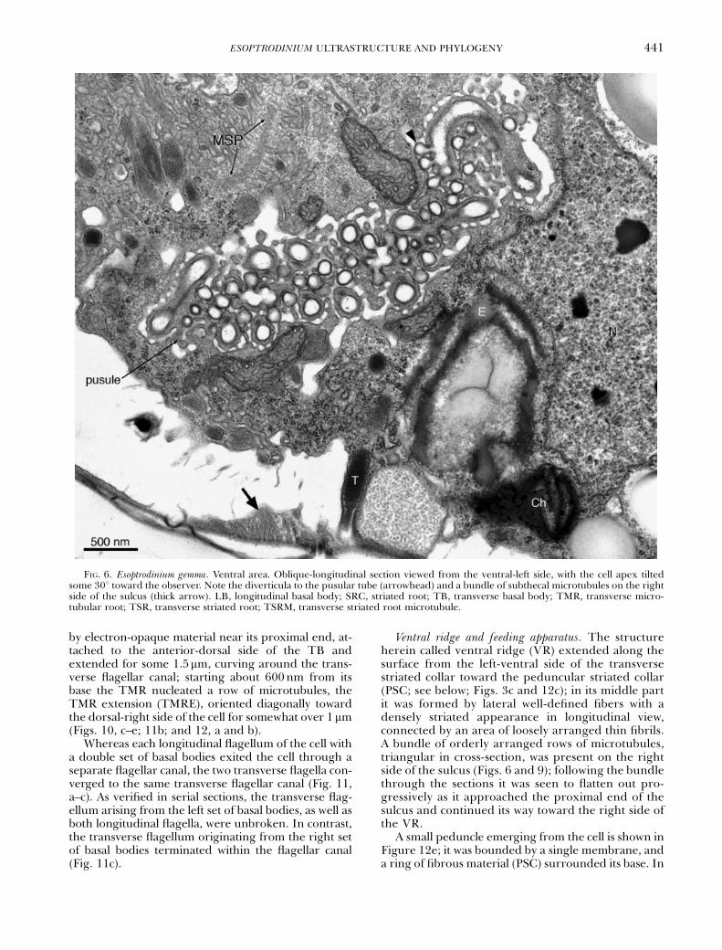
it did not divert into long branches, much of its distalpart was surrounded by numerous diverticula, eachabout 150 nm long, with a round distal end and aconstricted connection to the main tube (Fig. 6). Thewidth of the tube was 100–130 nm throughout itslength. The inner portion of the tube contained someperipheral electron-opaque material with a granularappearance (Fig. 6). Only one pusule was found inthe cells that had a single set of flagella, whereas inthe cell that had a double set of basal bodies two pus-ular tubes were present, each tube attached to one ofthe two longitudinal flagellar canals (Figs. 8b and 9).No pusule was seen attached to the transverse flag-ellar canal.
Eyespot. The eyespot was shaped as a curved plateunderlying the full width of the proximal part of thesulcus for over 2 mm (Figs. 4a, 6, and 9). It consistedof one to three layers of globules with sizes varyingbetween approximately 100 and 380 nm; in someplaces the globules appeared fused together, form-ing a continuous layer about 100 nm thick (Figs. 6and 10d). When two or three layers were presenttheir middle lines were placed approximately 150 nmapart, as measured in cross-sections of the structure.Although chloroplast lobes were present near thesulcus, they were not connected to the eyespot
(Figs. 4a and 6). In the cell with two longitudinalflagella two sulcal depressions were present; theshape of the underlying eyespot accompanied theshape of the depressions and some of the layers ofglobules were discontinuous (Fig. 9).
Flagellar apparatus. A diagrammatic view of theflagellar apparatus of a cell with two longitudinalflagella (a planozygote) is given in Figure 7. Struc-tures associated with the longitudinal basal body (LB)of a cell with a single longitudinal flagellum areshown in Figure 8. Flagella and basal body (BB)-as-sociated structures in the cell with a double set of ba-sal bodies are shown in Figures 9–12, a–c. The areawhere the peduncle exits the cell is shown in Figure12d and e, taken from the same series of sections asFigure 8.
The deeply marked sulcus closed into a tubularchamber near its proximal end, sheathing the longitu-dinal flagellum for approximately 700nm (Fig. 8a andb); a striated collar limited the innermost part of thischamber, the longitudinal flagellar canal, which wasbounded by a single membrane (Fig. 8c). In the freepart of the longitudinal flagellum a fibrous rod ranalongside the axoneme. Starting at 300–400nm fromthe base of the flagellum, an additional layered struc-ture, about 300nm wide and 20nm thick, was placed
FIG. 4. Esoptrodinium gemma. General ultrastructure and detail of the chloroplast. LF, longitudinal flagellum; T, trichocyst. (a) Trans-verse section through the middle of the sulcus (antapical view, same cell as in Fig. 3a). Note the eyespot (E) underneath the sulcus, andthe chloroplast lobes (Ch). (b) Chloroplast, showing three evenly-spaced bounding membranes (triple arrows).
ESOPTRODINIUM ULTRASTRUCTURE AND PHYLOGENY 439

beneath the flagellar membrane on the left side of theparaxonemal rod (Figs. 8a, arrowhead; 9 and 10d).The LB was associated with a single microtubularstrand, the longitudinal microtubular root (LMR; desi-gnated r1 by Moestrup 2000). As seen in transversesections, the distal part of the LMR consisted of ap-proximately 25 microtubules placed along the left-dor-sal side of the sulcus (Fig. 8, a and b). Near the LB themicrotubules of the LMR bent in different directionswhereby the structure bifurcated, with a group of six toseven microtubules arching past the dorsal side of theLB and associating with its right side (Figs. 8, e–h and10, a–c); a striated fiber (the striated root connective,SRC) attached to the dorsal side of this right branch ofthe LMR, spanned the distance to the transverse basalbody (TB) and attached to the proximal end of thetransverse striated root (TSR) (Figs. 8, h; 10, a–c, and11a and b). The microtubules associated with the right-dorsal side of the LB were nearly perpendicular to theBB triplets and at least one (perhaps two or three)continued on an almost reverse course toward the lon-gitudinal flagellar canal (Fig. 8, c–h). The remaining
LMR microtubules associated with the left side of theLB and continued past its proximal end on a nearlystraight line for some 300nm, their dorsal side linedwith electron-opaque material (Figs. 10, b–d and 11,a–c). Several thin fibers attached to three triplets on theleft-dorsal side of the TB and widened toward theventral surface of the LMR, connecting to groups ofthree to five microtubules (Figs. 8, e–h and 10, a–c).
The basal bodies were inserted at an angle of ap-proximately 1351, with the proximal end of the TBextending slightly to the right of the LB. The proximalends of the basal bodies were approximately 250nmapart and were not directly connected to each other(Figs. 10b and 11a). Two microtubule-containing rootswere associated with the TB; the TSR, with its associ-ated microtubule (TSRM; r4, sensu Moestrup 2000),was attached to the dorsal-posterior face of the TB andextended for 1.2–1.5mm along a nearly straight pathtoward the striated collar that delimited the transverseflagellar canal (Figs. 10, c–e; 11b and c; and 12, a–c).The transverse microtubular root (TMR; r3, Moestrup2000), consisting of a single microtubule surrounded
FIG. 5. Esoptrodinium gemma cyst. (a) General view. Note the chloroplast lobes (Ch) and the food vacuole (FV) with disorganizedcontents. (b) Cyst surface. Two parallel membranes (arrows) are visible between the cyst wall and the cytoplasmic membrane. Thearrowhead indicates the electron-opaque layer underlying the plasmalemma. Small slanted numbers refer to the section number.
ANTONIO J. CALADO ET AL.440
by electron-opaque material near its proximal end, at-tached to the anterior-dorsal side of the TB andextended for some 1.5mm, curving around the trans-verse flagellar canal; starting about 600nm from itsbase the TMR nucleated a row of microtubules, theTMR extension (TMRE), oriented diagonally towardthe dorsal-right side of the cell for somewhat over 1mm(Figs. 10, c–e; 11b; and 12, a and b).
Whereas each longitudinal flagellum of the cell witha double set of basal bodies exited the cell through aseparate flagellar canal, the two transverse flagella con-verged to the same transverse flagellar canal (Fig. 11,a–c). As verified in serial sections, the transverse flag-ellum arising from the left set of basal bodies, as well asboth longitudinal flagella, were unbroken. In contrast,the transverse flagellum originating from the right setof basal bodies terminated within the flagellar canal(Fig. 11c).
Ventral ridge and feeding apparatus. The structureherein called ventral ridge (VR) extended along thesurface from the left-ventral side of the transversestriated collar toward the peduncular striated collar(PSC; see below; Figs. 3c and 12c); in its middle partit was formed by lateral well-defined fibers with adensely striated appearance in longitudinal view,connected by an area of loosely arranged thin fibrils.A bundle of orderly arranged rows of microtubules,triangular in cross-section, was present on the rightside of the sulcus (Figs. 6 and 9); following the bundlethrough the sections it was seen to flatten out pro-gressively as it approached the proximal end of thesulcus and continued its way toward the right side ofthe VR.
A small peduncle emerging from the cell is shown inFigure 12e; it was bounded by a single membrane, anda ring of fibrous material (PSC) surrounded its base. In
FIG. 6. Esoptrodinium gemma. Ventral area. Oblique-longitudinal section viewed from the ventral-left side, with the cell apex tiltedsome 301 toward the observer. Note the diverticula to the pusular tube (arrowhead) and a bundle of subthecal microtubules on the rightside of the sulcus (thick arrow). LB, longitudinal basal body; SRC, striated root; TB, transverse basal body; TMR, transverse micro-tubular root; TSR, transverse striated root; TSRM, transverse striated root microtubule.
ESOPTRODINIUM ULTRASTRUCTURE AND PHYLOGENY 441
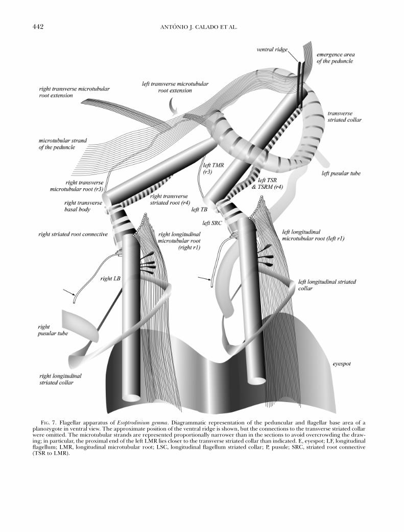
FIG. 7. Flagellar apparatus of Esoptrodinium gemma. Diagrammatic representation of the peduncular and flagellar base area of aplanozygote in ventral view. The approximate position of the ventral ridge is shown, but the connections to the transverse striated collarwere omitted. The microtubular strands are represented proportionally narrower than in the sections to avoid overcrowding the draw-ing; in particular, the proximal end of the left LMR lies closer to the transverse striated collar than indicated. E, eyespot; LF, longitudinalflagellum; LMR, longitudinal microtubular root; LSC, longitudinal flagellum striated collar; P, pusule; SRC, striated root connective(TSR to LMR).
ANTONIO J. CALADO ET AL.442

FIG. 8. Esoptrodinium gemma. Flagellar apparatus of a cell with a single set of basal bodies in antapical view. Non-adjacent serial sectionsproceeding toward the apex, showing the longitudinal flagellum and basal body (BB), and associated structures (same cell as in Figs. 3aand 4a). The sections shown in (f–h) were tilted 161 to give a vertical view of the BB. Small slanted numbers refer to the section number.The scale bar applies to all figures. E, eyespot; LF, longitudinal flagellum, LSC, longitudinal flagellum striated collar; SRC, striated rootconnective (TSR to LMR). (a, b) Longitudinal flagellum enclosed in a tubular chamber at the proximal end of the sulcus. Note thelayered structure on the left side of the paraxonemal rod (arrowhead) and the connection of the pusular tube (P) to the longitudinalflagellar canal. (c–h) Longitudinal basal body (LB) and its association with the proximal part of the longitudinal microtudinal root (LMR).One microtubule (arrowheads) is visible diverging from the branch of the LMR that contacts the right side of the LB and continuingtoward the flagellar canal. Electron-opaque material with the aspect of a hollow fiber (short arrows) approaches the right end of the LMRfrom the ventral side of the LB. Fibers extend from triplets of the LB to the ventral side of the LMR (arrows).
ESOPTRODINIUM ULTRASTRUCTURE AND PHYLOGENY 443

serial sections, a connection could be followed from thetransverse striated collar, through the VR, to the PSC,although only a very thin layer of electron-opaque ma-terial was visible in some sections. A single strand ofabout 30 microtubules, which was over 5mm long inthe large cell shown in Figure 3b, extended into thepeduncle; this microtubular strand of the peduncle(MSP) ran nearly parallel to part of the TMRE and thetwo strands of microtubules were in some places lessthan 200nm apart (Fig. 11b). Numerous elongate ves-icles, many of which with electron-opaque contents,were present in the ventral area near the MSP.
Nucleotide divergence and phylogeny of Esoptro-dinium. The phylogenetic inferences based on MP,NJ, and BA (Fig. 13) all suggest that E. gemma forms asister taxon to the two species of the woloszynskioidgenus Tovellia (viz. Tovellia coronata [Wo"oszynska]Moestrup, K. Lindberg et Daugbjerg and T. sanguin-ea sp. ined.). Thus, Esoptrodinium is recognized asthe third genus in the recently proposed family
Tovelliaceae (Lindberg et al. 2005). It should be not-ed that this family is not well supported in terms ofbootstrap values in MP and NJ analyses (51% and57%, respectively) whereas it is highly supported interms of PP (100) from Bayesian analyses. In generalthe tree topology of the deep branches is not wellsupported in terms of bootstrap values and PP andtherefore the sister group relationships of major lin-eages of dinoflagellates could not be establishedbased on the nuclear encoded LSU rDNA sequences.
Estimates of the nucleotide divergence of LSUrDNA based on 1422bp for species within the To-velliaceae are shown in Table 1. The pairwise compar-isons at the genus level are all above 20%, thusrevealing a similar substitution rate within this familyof dinoflagellates. However, see Lindberg et al. (2005)for a discussion on the two strains of Jadwigia applanataMoestrup, K. Lindberg et Daugbjerg (CCAC 0021 andFW 145).
DISCUSSION
Identity of the organism. The morphological rangereported herein fits the description of the populationfrom which the type figure of E. gemma was selected(Javornicky 1962, 1997). Javornicky (1962) foundmostly colorless cells, and originally described thepopulation as lacking chloroplasts, although the dif-fuse and very pale yellow–green matter found insome specimens was reinterpreted by Javornicky(1997) as either chloroplasts or the remnants of in-gested algae; the band-shaped structure labeled‘‘ch?’’ in his Figure 1a–c (Javornicky 1997, pl. 2) sug-gests the type of chloroplast we observed. In contrast,Thompson (1951) found ‘‘many small, diffuse, pari-etal, very pale yellow–green chromatophores’’ in allspecimens but one, and this contained three largeyellow to brown masses in the epicone. The observa-tion in our material of clearly band-shaped chloro-plasts in cells with no food vacuoles (see Fig. 1c) pro-vided a basis for comparison without which the iden-tity of the fragmented chloroplasts mixed with cellinclusions might be doubted (Fig. 1, a and b). Theelectron microscopical observations confirmed thenarrowness of some chloroplast bands. It is interest-ing to note that all cells of the mirror-symmetricalB. bernardinense from Rugen Island had greenish–yellow chloroplasts (Javornicky 1997).
Whereas Javornicky (1962) described an eyespotsimilar to what we found, Thompson (1951) reportedinstead a dark spot produced by the sharp margins ofthe deep longitudinal flagellar pore. Interestingly, inthe original description of B. bernardinense, Chodat(1924) mentions hematochrome granules, which heinterpreted as foreign bodies and not a true eyespot,and his drawings show a dark spot in an analogousposition to the eyespot of Esoptrodinium.
The similarities between B. bernardinense and E. gem-ma extend to the swimming mode observed in thepopulation we studied, in particular the lengthy rota-
FIG. 9. Esoptrodinium gemma. Oblique-longitudinal view of thesulcal area of the cell with a double set of basal bodies. The twolongitudinal flagella (LF) and one of the pusular tubes (P) arevisible. The eyespot (E) follows the wavy shape of the two sulcaldepressions. A bundle of subthecal microtubules on the rightside of the sulcus is marked by a thick arrow.
ANTONIO J. CALADO ET AL.444

tion on one spot, which would be adequately describedas ‘‘tournant sans cesse dans unmeme sens, c’est-a-direselon une ligne qui va du sommet de la moitie pluspetite vers le sillon’’ (Chodat 1924, p. 41); four ofChodat’s (1924) drawings show cells with the longitu-dinal flagellum around the non-indented lateral mar-
gin, the same general appearance we found in rotatingspecimens.
The nearly perfect symmetry between Bernardiniumand Esoptrodinium, and the uniqueness of a right-ori-ented transverse flagellum in Bernardinium, are indeedsurprising and the possibility of a mistake must be ad-
FIG. 10. Esoptrodinium gemma. Flagellar apparatus of the cell with a double set of basal bodies. Non-adjacent serial sections progressingfrom right to left. The cell is viewed from the right, with the apex tilted about 401 away from the observer. The series is presented withthe sulcus oriented to the top of the images to facilitate comparison with Fig. 8. Small slanted numbers refer to the section number. Thescale bar applies to all figures. See text for description.
ESOPTRODINIUM ULTRASTRUCTURE AND PHYLOGENY 445

dressed. The image formed by a traditional light mi-croscope, with the ocular directly aligned with the ob-jective, is rotated 1801, but it is not flipped; this meansthat looking through the ocular the image is seen withthe same chiral orientation as the object. In contrast,inverted microscopes introduce a number of reflec-tions and changes in viewpoint, each of which produc-es a mirror-symmetrical version of the previous image,the end result depending on the number of such in-versions. Some years ago, while attempting to docu-ment feeding with a Leitz Labovert FS invertedmicroscope, we recorded on video some would-be mir-ror-symmetrical cells of Prosoaulax lacustris (F. Stein)Calado et Moestrup. As not all inverted microscopesproduce mirror images (e.g. the older Wild M40 doesnot) a microscopist may be caught off guard againstsuch a possibility. Javornicky (1997) studied live mate-rial collected in northeast Germany and one may spec-ulate that he observed the organism with somemicroscope unfamiliar to him, obtained in a nearbylaboratory. We could not confirm this possibility, how-ever, and therefore have no ground to assume a mis-taken observation.
Whereas the eventual rediscovery of a Bernardinium-like organism with a right-oriented transverse flag-ellum would prove its existence, the possibility of it
being the product of erroneous observations may bedifficult, if not impossible, to demonstrate. As the char-acteristics associated with the name E. gemma are clear-ly defined, as opposed to the ambiguity inherent to thedifferent uses of the name B. bernardinense, the use ofthe former name for the organisms we report on seemsthe best option.
Feeding. The clusters of rapidly swimmingEsoptrodinium cells observed in dense mixed cultures,and the attractive power of some prey cells, betray achemosensory system for detection of food items,although it was not clear from the observationswhat features characterized attractive cells. In so-called histophagous species such as Gymnodiniumfungiforme Anisimova (5Katodinium fungiforme[Anisimova] Fott ex A.R. Loeblich) and Peridiniopsisberolinensis (Lemmermann) Bourrelly, chemosensiti-vity is used to find injured or dying prey; it is accu-rate to the point of directing the predators to theexact areas on the surface of prey where ingestiblecontents may flow out. These species take up food bysuction through a feeding tube and, except forsmall items that may be taken up whole, ingest onlypart of their prey (Spero 1982, 1985, Calado andMoestrup 1997, see Hansen and Calado 1999, for areview). Detection of prey cells in poor physiological
FIG. 11. Flagellar apparatus of Esoptrodinium gemma. Continuation of the series shown in Fig. 10. Small slanted numbers refer to thesection number. The scale bar applies to all figures. See text for description.
ANTONIO J. CALADO ET AL.446

condition, perhaps easier to capture or more digest-ible, may be speculated as the function of chemosen-sitivity in Esoptrodinium.
Although the rotating motion of Esoptrodinium cellsnear potential prey closely resembled the spinning dis-played by P. berolinensis before attachment to food par-ticles (Calado and Moestrup 1997), the emission of anattachment filament was never observed. The contin-uous motion of the cells throughout the engulfmentprocess constantly shifted the observation point ofview, giving rise to different appearances often diffi-
cult to interpret. Dangeard’s (1892) account of fooduptake in Katodinium vorticella (F. Stein) A.R. Loeblich(as Gymnodinium vorticella F. Stein) aptly describes whatwas visible during some observations on feeding Eso-ptrodinium cells. The few feeding events recorded onvideo that show the cell in focus for most of the pro-cess, combined with the ultrastructural information,suggest the involvement of the peduncular microtu-bules in driving forward the wide, thin pseudopod.The presence of a single membrane, not underlain byamphiesmal vesicles, extending down from the pedun-
FIG. 12. Esoptrodinium gemma. Transverse flagellum, ventral ridge (VR) and microtubular strand of the peduncle (MSP). (a–c)Continuation of the series shown in Figures 10 and 11. Scale bar as in (d). (d, e) Same series as Fig. 8. Small slanted numbers referto the section number. Note the thin layer of electron-opaque material (arrowheads) that connects the peduncular striated collar (PSC)to the VR.
ESOPTRODINIUM ULTRASTRUCTURE AND PHYLOGENY 447

cle along the axis of the VR, highlights this area as thepotential place of ingestion, thereby suggesting a func-tion for this enigmatic structure.
As seen with the EM, the peduncle and associatedstructures of E. gemma are essentially similar to whatwas found in Prosoaulax lacustris (as Amphidinium lacustreF. Stein non auctt.; Calado et al. 1998, Calado and
Moestrup 2005). However, in P. lacustris food is ingest-ed through a narrower area, which was interpreted asthe place of emergence of the peduncle based on itslocation in the cell. The apparent peduncle-assistedpseudopod formation and direct engulfment in Eso-ptrodinium illustrate a different use for the same cellstructures.
FIG. 13. Phylogeny of Esoptrodinium gemma inferred from unweighted parsimony analysis based on 1124bp of nuclear-encoded LSUrDNA. Of the characters included 540 were parsimony informative and the parsimony analyses produced three equally parsimonioustrees each 2632 steps (CI50.436, RI50.543). The tree topology illustrated is a strict consensus of the three parsimonious trees. Boot-strap values or support from posterior probabilities (PP) of 50% or above are written to the left of internal nodes. The first numbers arefrom parsimony analyses (1000 replications, characters unweighted), the second numbers are from neighbor-joining analyses based onthe maximum likelihood settings obtained using Modeltest (TrN" I"G model and with 1000 replications). The third numbers are PPfrom Bayesian analyses and based on 19,461 trees. The two ciliates assigned to Tetrahymena comprised the outgroup. The familyTovelliaceae (woloszynskioid group I sensu Lindberg et al. 2005), including Esoptrodinium, is marked.
ANTONIO J. CALADO ET AL.448

Morphology and ultrastructure. The cells of E. gemmadisplayed the general ultrastructural features typicalof dinoflagellates. Chloroplast organization was of theusual peridinin-containing type, although the colorperceived with the light microscope hints at a differ-ent pigment composition. However, the high propor-tion of prey cells in the mixed cultures and the smallvolume of the batches precluded pigment analysis ofthe dinoflagellate. Judging from their ultrastructuralintegrity, the chloroplasts seemed functional, al-though their relatively small size and the presenceof food vacuoles in nearly all cells suggest that phago-trophy is the essential mode of nutrition. All attemptsto grow Esoptrodinium without food organisms wereunsuccessful.
The eyespot of Esoptrodinium belongs to Dodge’s(1984) type A and to the ‘‘Woloszynskia coronata type’’ ofKawai and Kreimer (2000), and is characteristic of thefamily Tovelliaceae (Lindberg et al. 2005). The pres-ence of a similar type of eyespot in Katodinium campylops(T.M. Harris) A.R. Loeblich (Wilcox 1989) indicates aclose affinity with members of the Tovelliaceae. As seenwith the light microscope, the closely related K. vorti-cella is strongly reminiscent of Esoptrodinium, both forthe appearance of the eyespot and for the generalstructure of the cell; in addition, it produces a restingcyst with paracingulum and two axial horns (unpub-lished observations), the same type of cyst produced byT. coronata (Wo"oszynska 1917, as Gymnodinium corona-tum Wo"oszynska; Lindberg et al. 2005), suggestingthat the group of K. vorticella-related species are mem-bers of the Tovelliaceae. However, the type species ofKatodinium, K. nieuportense (W. Conrad) Fott, is a poorlyknown marine species with a rather different appear-ance (Conrad 1926), possibly unrelated to most speciescurrently assigned to Katodinium.
With its numerous diverticula, the pusular tube ofE. gemma most closely resembles that of T. coronata, al-though the distal part of the tube appears somewhatmore irregular in the latter species (Crawford andDodge 1971, Figs. 13, 14; Lindberg et al. 2005, Figs.28, 30). As in vegetative cells of E. gemma, a single pus-ule was reported in T. coronata, attached to the longi-tudinal flagellar canal (Lindberg et al. 2005). A singletubular pusule per cell was also found in Prosoaulaxlacustris, either attached to the longitudinal or to the
transverse flagellar canal (Calado et al. 1998). As thenumber of cells of E. gemma examined was small, thepossibility that the pusular tube may in some cells as-sociate with the transverse flagellar canal cannot beexcluded.
Electron-opaque material with a layered appear-ance lining the dorsal surface of the proximal stretchof LMR has been reported from K. campylops and Wo-loszynskia pascheri (Suchlandt) Stosch (Wilcox 1989),two species closely related to the wolozynskioids. Therelatively wide strand of nearly straight microtubulesextending past the LB in K. campylops is similar to whatwe found in Esoptrodinium, and can also be seen in thepublished micrographs of Tovellia sp. (as Woloszynskiasp.; Roberts and Timpano 1989, Lindberg et al. 2005)and J. applanata (as Woloszynskia limnetica Bursa; Robe-rts et al. 1995). A similar arrangement is suggested inFigure 34 of Lindberg et al. (2005) for T. coronata. In allthese species the SRC is relatively wide and attaches tothe LMR near the area where the microtubules bendtoward the antapex.
A bifurcating LMR, such as the one found in Eso-ptrodinium, has not been previously reported. The mi-crotubule that extends beyond the right side of the LBis potentially confusing because this is the positionwhere the so-called single-stranded microtubular root(SMR, usually interpreted as r2; Moestrup 2000) isfound in gonyaulacoids and peridinioids; however, theSMR has its proximal end on the right ventral side ofthe LB and extends in an arc in a dorso-antapical di-rection (see Hansen andMoestrup 1998a, Calado et al.1999, Calado and Moestrup 2002) whereas in Eso-ptrodinium the right hand side microtubule extendsfrom the right-dorsal side of the LB toward the ven-tral-right. The continuity of the projectingmicrotubulewith the LMR was best observed in sections perpen-dicular to the LB (Fig. 8h) and may be difficult to per-ceive in other angles. Although the viewpoints of theseries of sections shown in Roberts et al. (1995) are notappropriate to reveal such an arrangement, it shouldbe noted that the conformation of the LMR schemat-ically shown in Figure 7 fits what is visible inJ. applanata in Roberts et al. (1995, Figs. 35–38), as-suming that the structure marked by an arrowhead (inFig. 38) is a microtubule. More detailed work on theflagellar apparatus of members of the Tovelliaceae is
TABLE 1. Sequence divergence in percentage of three genera within the Tovelliaceae (Esoptrodinium, Tovellia, and Jadwigia)based on 1422 unambiguously aligned LSU rDNA nucleotides
Esoptrodinium gemma Tovellia coronata Tovellia sanguinea Jadwigia applanata (CCAC0021) Jadwigia applanata (FW145)
E. gemma – 25.79 27.87 23.67 22.23T. coronata 32.03 – 11.85 22.5 22.35T. sanguinea 35.33 12.98 – 23.87 23.84J. applanata (CCAC0021) 28.84 27.05 29.02 – 10.45J. applanata (FW145) 26.66 26.92 28.53 11.37 –
Uncorrected distances (‘‘p’’ values in PAUP*) are given above the diagonal and distance values calculated using Kimura2-parameter model are given below the diagonal.LSU, large subunit.
ESOPTRODINIUM ULTRASTRUCTURE AND PHYLOGENY 449

needed to verify if a bifurcating LMR is a shared fea-ture in this group. It is not clear if the SMR and theprojecting microtubule of Esoptrodinium are homolo-gous structures.
Organization of the planozygote. The unequivocal es-tablishment of sexual reproduction in dinoflagellatesis usually credited to Stosch (1964, 1965), who laterestablished the identity of cells with paired longitu-dinal flagella as planozygotes (Stosch 1973). This hassince been confirmed in numerous life cycle studies(Pfiester 1984). The generally large-sized cells foundwith paired longitudinal flagella in our batches ofEsoptrodinium and the thick-walled resting cysts thatabounded in older batches are recognizable, respec-tively as the planozygote and the hypnozygote stagein the life cycle of this species. The first publishedillustrations of the parallel arrangement of basal bod-ies and roots in a dinoflagellate are in Leadbeater andDodge (1967a, Fig. 3) and Leadbeater and Dodge(1967b, Figs. 13, 18), showing what was interpretedat the time as recently divided pairs of flagellar basesin Karlodinium micrum (B. Leadbeater et J. D. Dodge)J. Larsen (as Woloszynskia micra B. Leadbeater et J.D.Dodge); it is now clear that the cells illustrated wereplanozygotes (Bergholtz et al. 2006).
In a study of flagellar transformation in Prorocent-rum micans Ehrenberg it has been shown that the newlyformed basal bodies are not parallel to the parentalpair and that the parental TB develops into a LB dur-ing cell division (Heimann et al. 1995). A large anglewas demonstrated in Peridinium cinctum (O.F. Muller)Ehrenberg between the parental and the newly formedbasal bodies, which were both oriented toward theapex of the cell (Calado et al. 1999). Similarly locatedaccessory basal bodies were shown in Peridiniella cat-enata (Levander) Balech (Hansen and Moestrup1998b, Figs. 14–16). Parallel arrangements of basalbodies and roots have been shown in planozygotes ofPeridiniopsis berolinensis (Wedemayer and Wilcox 1984,Figs. 19 and 20), Scrippsiella minima X. Gao et J. D.Dodge (Gao et al. 1989, Figs. 7 and 8; as Scrippsiella sp.,see Gao and Dodge 1991) and T. coronata (Lindberget al. 2005, Figs. 33 and 34).
We found a similar arrangement of flagellar basesand associated structures in both left and right pairs ofbasal bodies in the planozygote, and it was essentiallythe same as seen in vegetative cells; namely the rootsand connectives associated with each pair of basal bod-ies were the same. However, it is interesting to notethat not all structures in the ventral area were doubled.Whereas each longitudinal flagellar canal was connect-ed to an independent pusular system, the eyespotswere partially fused, although two longitudinalgrooves were maintained. A single peduncle andMSP were present in the planozygote, associated witha single VR, in a similar position relative to the left pairof BBs as the one found in vegetative cells. Unexpect-edly, we found that both transverse flagella convergedto the same flagellar canal, in contrast with the two in-dependent longitudinal flagellar canals. As seen in
Figure 7, the structures that are not doubled in theplanozygote (i.e. transverse flagellar canal, VR, emer-gence point of the peduncle; also the triangular arrayof microtubules on the right side of the sulcus notshown in Fig. 7) are nearly aligned longitudinally andit may be speculated that this axial area is pivotal in therearrangements taking place during gamete fusion,because the two gametal flagellar systems seem to con-verge in this zone. However, the mechanisms drivingthe migration of some structures and the loss of othersduring the final stages of gamete fusion are unknown.
Although occasionally reported with two transverseflagella (Coats et al. 1984), planozygotes are more of-ten seen with a single flagellum in the cingulum(Stosch 1973, Gao et al. 1989). Convergence to a sin-gle flagellar pore may perhaps render more difficultthe retention of both transverse flagella in Eso-ptrodinium. Examination of planozygotes of other spe-cies is necessary to determine whether the organizationdescribed herein is shared by other dinoflagellates.
Phylogenetic affinities. The close affinity between E.gemma and the genera Tovellia and Jadwigia is stronglyindicated by both the molecular data used and thegeneral similarities in eyespot, flagellar apparatusand pusule construction, and Esoptrodinium is there-fore assigned to the family Tovelliaceae. In contrast,the recently investigated Woloszynskia halophila (Bie-cheler) Elbrachter et Kremp contains a brick-likeeyespot and has closer affinities with a group ofspecies which includes symbiotic forms and the ma-rine polar genus Polarella Montresor, Procacciniet Stoecker (Kremp et al. 2005). Interestingly, noplate material was found in E. gemma, a feature thatwould dictate its assignment to the Gymnodiniaceaein traditional taxonomical schemes. The value ofamphiesmal vesicle contents as a phylogenetic mark-er is, however, rather uncertain as some gym-nodinioids (including the type species ofGymnodinium, G. fuscum [Ehrenberg] F. Stein; Dodgeand Crawford 1969, Hansen et al. 2000) have thinplates. One feature found in Tovellia and in Jadwigiathat was not seen in E. gemma is the apical line ofnarrow plates; fixations for SEM were tried, but theirquality was not good enough to ascertain whetherthis character is present or missing in Esoptrodinium.
Although in the phylogenetic tree shown in Figure13 E. gemma is grouped with Tovellia, we consider thatthe relationship between the genera included in theTovelliaceae is not well supported in terms of bootstrapvalues and posterior probability, and the available mor-phological characters suggest different affinities.Whereas general morphology seems closer betweenTovellia and Jadwigia, the pusular tube of J. applanata,not shown to possess diverticula, would argue for acloser relationship between Tovellia and Esoptrodinium.On the other hand, the round cyst of E. gemma is moresimilar to the cyst of J. applanata than to the hornedtype cyst of T. coronata. A detailed analysis of the flag-ellar base area of T. coronata is needed to provide othercharacters for comparison between the genera.
ANTONIO J. CALADO ET AL.450

A. J. C. was supported by the European Community’s pro-gramme ‘‘Improving the Human Research Potential and theSocio-Economic Knowledge Base’’ (through Copenhagen Bio-systematics Centre [COBICE]). Financial support was alsoprovided by the Danish Science Research Council (Grant no.21-02-0539) to Ø. M. and N. D. Prof. Jorge Rino first broughtour attention to the organism on the sidewalk and kindlyprovided the light micrographs for Figure 1, a–c.
Bergholtz, T., Daugbjerg, N., Moestrup, Ø. & Fernandez-Tejedor,M. (in press). On the identity of Karlodinium veneficum and de-scription of Karlodinium armiger sp. nov., based on light andelectron microscopy, nuclear-encoded LSU rDNA and pig-ment composition. J. Phycol. 42:170–93.
Calado, A. J., Craveiro, S. C. & Moestrup, Ø. 1998. Taxonomy andultrastructure of a freshwater, heterotrophic Amphidinium(Dinophyceae) that feeds on unicellular protists. J. Phycol.34:536–54.
Calado, A. J., Hansen, G. & Moestrup, Ø. 1999. Architecture of theflagellar apparatus and related structures in the type species ofPeridinium, P. cinctum (Dinophyceae). Eur. J. Phycol. 34:179–91.
Calado, A. J. & Moestrup, Ø. 1997. Feeding in Peridiniopsis be-rolinensis (Dinophyceae): new observations on tube feeding byan omnivorous, heterotrophic dinoflagellate. Phycologia 36:47–59.
Calado, A. J. & Moestrup, Ø. 2002. Ultrastructural study of thetype species of Peridiniopsis, Peridiniopsis borgei (Dinophyceae),with special reference to the peduncle and flagellar apparatus.Phycologia 41:567–84.
Calado, A. J. & Moestrup, Ø. 2005. On the freshwater dinoflagel-lates presently included in the genus Amphidinium, with a de-scription of Prosoaulax gen. nov. Phycologia 44:112–9.
Chodat, R. (‘‘avec la collaboration de M. J. Zender’’) 1924. Alguesde la region du Grand Saint-Bernard. III. Bull. Soc. Bot. GeneveSer. 2 15:33–48.
Coats, D. W., Tyler, M. A. & Anderson, D. M. 1984. Sexual proc-esses in the life cycle of Gyrodinium uncatenum (Dinophyceae): amorphogenetic overview. J. Phycol. 20:351–61.
Conrad, W. 1926. Recherches sur les flagellates de nos eauxsaumatres. 1e partie: dinoflagellates. Arch. Protistenk. 55:63–100, pls 1, 2.
Conrad, W. 1939. Notes protistologiques. IX. Sur trois Dinoflagel-lates de l’eau saumatre. Bull. Mus. R. Hist. Nat. Belg. 15:1–10.
Crawford, R. M. & Dodge, J. D. 1971. The dinoflagellate genusWoloszynskia. II. The fine structure of W. coronata. Nova He-dwigia 22:699–719.
Dangeard, P.-A. 1892. La nutrition animale des Peridiniens. Bota-niste 3:7–27, pls 1, 2..
de Rijk, P., Wuyts, J., van der Peer, Y., Winkelmans, T. & de Wach-ter, R. 2000. The European large subunit ribosomal RNAdatabase. Nucleic Acids Res. 28:117–8.
Dodge, J. D. 1984. The functional and phylogenetic significance ofdinoflagellate eyespots. BioSystems 16:259–67.
Dodge, J. D. & Crawford, R. M. 1969. The fine structure of Gym-nodinium fuscum. New Phytol. 68:613–8.
Gao, X. & Dodge, J. D. 1991. The taxonomy and ultrastructure of amarine dinoflagellate, Scrippsiella minima sp. Nov.. Br. Phycol. J.26:21–31.
Gao, X., Dodge, J. D. & Lewis, J. 1989. An ultrastructural study ofplanozygotes and encystment of a marine dinoflagellate, Scrip-psiella sp.. Br. Phycol. J. 24:153–65.
Hansen, P. J. & Calado, A. J. 1999. Phagotrophic mechanisms andprey selection in free-living dinoflagellates. J. Eukaryot. Micro-biol. 46:382–9.
Hansen, G. & Daugbjerg, N. 2004. Ultrastructure of Gyrodiniumspirale, the type species of Gyrodinium (Dinophyceae), includinga phylogeny of G. dominans, G. rubrum and G. spirale deducedfrom partial LSU rDNA sequences. Protist 155:271–94.
Hansen, G., Daugbjerg, N. & Franco, J. M. 2003. Morphology,toxin composition and LSU rDNA phylogeny of Alexandriumminutum (Dinophyceae) from Denmark, with some morpho-
logical observations on other European strains. Harmful Algae2:317–35.
Hansen, G. & Moestrup, Ø. 1998a. Fine-structural characterizationof Alexandrium catenella (Dinophyceae) with special emphasison the flagellar apparatus. Eur. J. Phycol. 33:281–91.
Hansen, G. & Moestrup, Ø. 1998b. Light and electron microscop-ical observations on Peridiniella catenata (Dinophyceae). Eur. J.Phycol. 33:293–305.
Hansen, G., Moestrup, Ø. & Roberts, K. M. 2000. Light and elec-tron microscopical observations on the type species of Gym-nodinium, G. fuscum (Dinophyceae). Phycologia 39:365–76.
Heimann, K., Roberts, K. R. & Wetherbee, R. 1995. Flagellar ap-paratus transformation and development in Prorocentrum mi-cans and P. minimum (Dinophyceae). Phycologia 34:323–35.
Huber-Pestalozzi, G. 1950. Cryptophyceen, Chloromonadinen,Peridineen. In Thienemann, A. [Ed.] Die Binnengewasser. Vol.16. E. Schweizerbart’sche Verlagsbuchhandlung, Stuttgart,310 pp.
Javornicky, P. 1962. Two scarcely known genera of the class Din-ophyceae: Bernardinium Chodat and Crypthecodinium Biecheler.Preslia 34:98–113.
Javornicky, P. 1997. Bernardinium Chodat (Dinophyceae), an athec-ate dinoflagellate with reverse, right-handed course of thecingulum and transverse flagellum, and Esoptrodinium genusnovum, its mirror-symmetrical pendant. Arch. Hydrobiol. Suppl.122(Algol. Stud. 87):29–42.
Kawai, H. & Kreimer, G. 2000. Sensory mechanisms. Phototaxesand light perception in algae. In Leadbeater, B. S. C. & Green,J. C. [Eds.] The Flagellates. Unity, Diversity and Evolution. Taylor& Francis, New York, pp. 124–46 (Systematics AssociationSpecial Volume No. 59.) pp. 124–146.
Kremp, A., Elbrachter, M., Schweikert, M., Wolny, J. L. & Gottsch-ling, M. 2005. Woloszynskia halophila (Biecheler) comb. nov.: abloom-forming cold-water dinoflagellate co-occurring withScrippsiella hangoei (Dinophyceae) in the Baltic Sea. J. Phycol.41:629–42.
Leache, A. D. & Reeder, T. W. 2002. Molecular systematics of theeastern fence lizard (Sceloporus undulatus): a comparison ofparsimony, likelihood, and Bayesian approaches. Syst. Biol.51:44–68.
Leadbeater, B. S. C. & Dodge, J. D. 1967a. An electron microscopestudy of nuclear and cell division in a dinoflagellate. Arch.Mikrobiol. 57:239–54.
Leadbeater, B. S. C. & Dodge, J. D. 1967b. An electron microscopestudy of dinoflagellate flagella. J. Gen. Microbiol. 46:305–14,pls. 1–4.
Lenaers, G., Maroteaux, L., Michot, B. & Herzog, M. 1989. Dino-flagellates in evolution. A molecular phylogenetic analysis oflarge subunit ribosomal RNA. J. Mol. Evol. 29:40–51.
Lindberg, K., Moestrup, Ø. & Daugbjerg, N. 2005. Studies on wo-loszynskioid dinoflagellates I: Woloszynskia coronata re-exam-ined using light and electron microscopy and partial LSUrDNA sequences, with description of Tovellia gen. nov. andJadwigia gen. nov. (Tovelliaceae fam. nov.). Phycologia 44:416–40.
Maddison, D. R. & Maddison, W. P. 2003. MacClade 4. SinauerAssociates Inc., Sunderland, MAs, USA.
Matvienko, O. M. & Litvinenko, R. M. 1977. Pirofitovi vodorosti. InHollerbach, M. M. & Kondrat’eva, N. V. [Eds.] ViznachnykPrisnovodnykh Vodorostej Ukrains’koj RSR. Vol. 3(2). NaukovaDumka, Kiev, 386 pp.
Moestrup, Ø. 2000. The flagellate cytoskeleton. Introduction of ageneral terminology for microtubular flagellar roots in pro-tists. In Leadbeater, B. S. C. & Green, J. C. [Eds.] The Flagel-lates. Unity, Diversity and Evolution (Systematics Association SpecialVolume No. 59). Taylor & Francis, New York, pp. 69–94.
Moestrup, Ø., Hansen, G., Daugbjerg, N., Flaim, G. & D’Andrea,M. Studies on woloszynskioid dinoflagellates II: on Tovelliasanguinea sp. nov., the dinoflagellate species responsiblefor the reddening of Lake Tovel, N. Italy. Eur. J. Phycol. (sub-mitted).
Nichols, H. W. 1973. Growth media—freshwater. In Stein, J. R.[Ed.] Handbook of Phycological Methods. Culture Methods and
ESOPTRODINIUM ULTRASTRUCTURE AND PHYLOGENY 451

Growth Measurements. Cambridge University Press, Cambridge,pp. 7–24.
Patterson, D. J. 1999. Diversity of eukaryotes. Am. Nat. 154:S96–S124.
Pfiester, L. A. 1984. Sexual reproduction. In Spector, D. L. [Ed.]Dinoflagellates. Academic Press, Orlando, pp. 181–99.
Popovsky, J. 1990. New knowledge on freshwater dinoflagellates ofCentral Europe; with description of a new species. Arch. Hy-drobiol. Suppl. 87(Algol. Stud. 60):1–18.
Popovsky, J. & Pfiester, L. A. 1990. Dinophyceae (Dinoflagellida).In Ettl, H., Gerloff, J., Heynig, H. & Mollenhauer, D. [Eds.]Su!wasserflora von Mitteleuropa. Vol. 6. Gustav Fischer, Jena,272 pp.
Posada, D. & Crandall, K. A. 1998. MODELTEST: testing themodel of DNA substitution. Bioinformatics 14:817–8.
Roberts, K. R., Hansen, G. & Taylor, F. J. R. 1995. General ultra-structure and flagellar apparatus architecture of Woloszynskialimnetica (Dinophyceae). J. Phycol. 31:948–57.
Roberts, K. R. & Timpano, P. 1989. Comparative analyses of thedinoflagellate flagellar apparatus. I. Woloszynskia sp. J. Phycol.25:26–36.
Ronquist, F. & Huelsenbeck, J. P. 2003. Mr. Bayes 3: Bayesianphylogenetic inference under mixed models. Bioinformatics19:1572–4.
Schiller, J. 1935. Dinoflagellatae (Peridineae) in monographischerBehandlung. In Kolkwitz, R. [Ed.] Rabenhorst’s Kryptogamen-flora von Deutschland, Osterreich und der Schweiz. 2nd ed. Vol. 10,3 part 2. Akademische Verlagsgesellschaft, Leipzig, pp. 589[The volume was finished in 1937 but the relevant pages werepublished in March 1935].
Scholin, C. A., Herzog, M., Sogin, M. & Anderson, D. M. 1994.Identification of group-and strain-specific genetic markers forglobally distributed Alexandrium (Dinophyceae). II. Sequenceanalysis of a fragment of the LSU rRNA gene. J. Phycol.30:999–1011.
Spero, H. J. 1982. Phagotrophy in Gymnodinium fungiforme (Pyr-rhophyta): the peduncle as an organelle of ingestion. J. Phycol.18:356–60.
Spero, H. J. 1985. Chemosensory capabilities in the phagotrophicdinoflagellate Gymnodinium fungiforme. J. Phycol. 21:181–4.
Starmach, K. 1974. Cryptophyceae, Dinophyceae, Rap-hidophyceae. In Starmach, K. & Sieminska, J. [Eds.] FloraS"odkowodna Polski. Vol. 4. Panstwowe Wydawnictwo Naukowe,Warszawa, 520 pp.
Stein, F. 1878. Der Organismus der Infusionsthiere nach eigenen Forsc-hungen in systematischer Reihenfolge bearbeitet. III. Abtheilung. DieNaturgeschichte der Flagellaten oder Geisselinfusorien. I. Halfte. Den
noch nicht Abgeschlossenen allgemeinen Theil nebst Erklarung dersammtlichen Abbildungen enthaltend. Wilhelm Engelmann, Leip-zig, 154 pp., 24 pls.
Stein, F. 1883. Der Organismus der Infusionsthiere . . . III. Abtheilung.II. Halfte. Die Naturgeschichte der arthrodelen Flagellaten. Einlei-tung und Erklarung der Abbildungen. Wilhelm Engelmann, Leip-zig, 30 pp., 25 pls.
von Stosch, H. A. 1964. Zum Problem der sexuellen Fortpflanzungin der Peridineengattung Ceratium. Helgol. Wiss. Meeresunters.10:140–52.
von Stosch, H. A. 1965. Sexualitat bei Ceratium cornutum(Dinophyta). Naturwissenschaften 52:112–3.
von Stosch, H. A. 1973. Observations on vegetative reproductionand sexual life cycles of two freshwater dinoflagellates, Gym-nodinium pseudopalustre Schiller and Woloszynskia apiculata sp.nov. Br. Phycol. J. 8:105–34.
Swofford, D. L. 2003. PAUP* Phylogenetic Analysis Using Parsimony(*and Other Methods). Version 4. Sinauer Associates, Sunderland,MA.
Tamura, K. & Nei, N. 1993. Estimation of the number of nucleo-tide substitutions in the control region of mitochondrial DNAin humans and chimpanzees. Mol. Biol. Evol. 10:512–26.
Thompson, R. H. 1951. A new genus and new records of fresh-water Pyrrophyta in the Desmokontae and Dinophyceae.Lloydia 13:277–99.
Van de Peer, Y., Van der Auwera, G. & de Wachter, R. 1996. Theevolution of stramenopiles and alveolates as derived by ‘‘sub-stitution rate calibration’’ of small ribosomal RNA. J. Mol. Evol.42:201–10.
Wawrik, F. 1983. Sommerliche Planktonaspekte 1981: seltene undneue Algen aus Teichen des Waldviertels in Niederosterreich.Nova Hedwigia 36:775–94.
Wedemayer, G. J. & Wilcox, L. W. 1984. The ultrastructure of thefreshwater colorless dinoflagellate Peridiniopsis berolinense(Lemm.) Bourrelly. J. Protozool. 31:444–53.
Wilcox, L. W. 1989. Multilayered structures (MLSs) in two dino-flagellates, Katodinium campylops and Woloszynskia pascheri. J.Phycol. 25:785–9.
Wo"oszynska, J. 1917. Nowe gatunki Peridineow, tudziez spostrze-zenia nad budowa okrywy u Gymnodiniow i Glenodiniow.—Neue Peridineen-Arten, nebst Bemerkungen uber denBau der Hulle bei Gymno- und Glenodinium. Bull. Int. Acad.Sci. Cracovie, Cl. Sci. Math. Nat., Ser. B 1917:114–22, pls.11–13.
Yang, Z. & Rannala, B. 1997. Bayesian phylogenetic inference us-ing DNA sequences: a Markov chain Monte Carlo method.Mol. Biol. Evol. 14:717–24.
ANTONIO J. CALADO ET AL.452


![Saizen [somatropin (rDNA origin) for injection] … · Saizen® [somatropin (rDNA origin) for injection] cool.click](https://img.dokumen.tips/doc/110x75/5b8977fc7f8b9abe1e8db089/saizen-somatropin-rdna-origin-for-injection-saizen-somatropin-rdna-origin.jpg)