Embed Size (px)
Citation preview

Issued by the Standards Unit, Microbiology Services, PHE Bacteriology | B 17 | Issue no: 6 | Issue date: 08.04.16 | Page: 1 of 24
© Crown copyright 2016
UK Standards for Microbiology Investigations
Investigation of tissues and biopsies from deep-seated sites and organs
Investigation of tissues and biopsies from deep-seated sites and organs
Bacteriology | B 17 | Issue no: 6 | Issue date: 08.04.16 | Page: 2 of 24 UK Standards for Microbiology Investigations | Issued by the Standards Unit, Public Health England
Acknowledgments UK Standards for Microbiology Investigations (SMIs) are developed under the auspices of Public Health England (PHE) working in partnership with the National Health Service (NHS), Public Health Wales and with the professional organisations whose logos are displayed below and listed on the website https://www.gov.uk/uk-standards-for-microbiology-investigations-smi-quality-and-consistency-in-clinical-laboratories. SMIs are developed, reviewed and revised by various working groups which are overseen by a steering committee (see https://www.gov.uk/government/groups/standards-for-microbiology-investigations-steering-committee). The contributions of many individuals in clinical, specialist and reference laboratories who have provided information and comments during the development of this document are acknowledged. We are grateful to the medical editors for editing the medical content. For further information please contact us at: Standards Unit Microbiology Services Public Health England 61 Colindale Avenue London NW9 5EQ E-mail: [email protected] Website: https://www.gov.uk/uk-standards-for-microbiology-investigations-smi-quality-and-consistency-in-clinical-laboratories PHE publications gateway number: 2016019 UK Standards for Microbiology Investigations are produced in association with:
Logos correct at time of publishing.
Investigation of tissues and biopsies from deep-seated sites and organs
Bacteriology | B 17 | Issue no: 6 | Issue date: 08.04.16 | Page: 3 of 24 UK Standards for Microbiology Investigations | Issued by the Standards Unit, Public Health England
Contents AMENDMENT TABLE ............................................................................................................. 4
UK SMI: SCOPE AND PURPOSE ........................................................................................... 6
SCOPE OF DOCUMENT ......................................................................................................... 9
INTRODUCTION ..................................................................................................................... 9
TECHNICAL INFORMATION/LIMITATIONS ......................................................................... 12
1 SAFETY CONSIDERATIONS .................................................................................... 13
2 SPECIMEN COLLECTION ......................................................................................... 13
3 SPECIMEN TRANSPORT AND STORAGE ............................................................... 14
4 SPECIMEN PROCESSING/PROCEDURE ................................................................. 14
5 REPORTING PROCEDURE ....................................................................................... 19
6 NOTIFICATION TO PHE, OR EQUIVALENT IN THE DEVOLVED ADMINISTRATIONS .................................................................................................. 20
APPENDIX: INVESTIGATION OF TISSUES AND BIOPSIES FROM DEEP-SEATED SITES AND ORGANS ...................................................................................................................... 21
REFERENCES ...................................................................................................................... 22
Investigation of tissues and biopsies from deep-seated sites and organs
Bacteriology | B 17 | Issue no: 6 | Issue date: 08.04.16 | Page: 4 of 24 UK Standards for Microbiology Investigations | Issued by the Standards Unit, Public Health England
Amendment table Each SMI method has an individual record of amendments. The current amendments are listed on this page. The amendment history is available from [email protected]. New or revised documents should be controlled within the laboratory in accordance with the local quality management system.
Amendment no/date. 10/08.04.16
Issue no. discarded. 5.3
Insert issue no. 6
Section(s) involved Amendment
Whole document.
Hyperlinks updated to gov.uk Title updated to include ‘from deep-seated sites and organs’. References reviewed throughout. Addition of lung tissue and biopsy for suspected infection with Legionella species.
Page 2. Updated logos added.
Scope. Scope updated to include rapid methods and links to relevant SMIs.
Introduction.
Reorganised for clarity. Specific tissue types placed into alphabetical order. Information regarding skin infection streamlined and information include in B11 – Investigation of swabs from skin and soft tissue infections.
Technical information/limitations. Section on rapid methods included.
Safety considerations.
Safety considerations regarding Hazard Group 3 organisms amended. It is recommended that all Gram-negative coccobacilli from sterile sites should be processed in a Class I or Class II microbiological safety cabinet until Hazard Group 3 pathogens (ie Brucella) have been definitively excluded.
Specimen processing. Samples for mycological examination must not be homogenised/ground.
Specimen processing/procedure.
Ideally, all grinding or homogenisation should be performed in a Class II exhaust protective cabinet.

Investigation of tissues and biopsies from deep-seated sites and organs
Bacteriology | B 17 | Issue no: 6 | Issue date: 08.04.16 | Page: 5 of 24 UK Standards for Microbiology Investigations | Issued by the Standards Unit, Public Health England
Surgically obtained specimens for fungal culture should be cut (finely sliced) rather than homogenised. Addition of fluorescent staining technique. Section 4.5.1 (culture media, conditions and organisms) media and incubation updated.
• Immunocompromised/suspected fungal infection changed to Sabouraud agar slope + chloramphenicol (35-37ºC 14d incubation, 28-30ºC 28d incubation).
• Mycetoma addition of Sabouraud agar slope + chloramphenicol.
• Nocardiosis blood agar 35-37ºC up to 7d. • Addition of Legionella species BMPA or
alternative 35-37ºC up to 10d. • Mixed infection/local policy, addition of
Mannitol Salt Agar. Section 4.6.1 (minimum level of identification) level of identification updated for β haemolytic streptococci, coagulase negative streptococci, enterobacteriaceae and pseudomonas. Consider sending staphylococci isolates from post mortem samples for toxin testing.
Reporting procedure. Culture reporting statement updated.
Appendix. Updated to reflect section 4.5.1.

Investigation of tissues and biopsies from deep-seated sites and organs
Bacteriology | B 17 | Issue no: 6 | Issue date: 08.04.16 | Page: 6 of 24 UK Standards for Microbiology Investigations | Issued by the Standards Unit, Public Health England
UK SMI#: scope and purpose Users of SMIs Primarily, SMIs are intended as a general resource for practising professionals operating in the field of laboratory medicine and infection specialties in the UK. SMIs also provide clinicians with information about the available test repertoire and the standard of laboratory services they should expect for the investigation of infection in their patients, as well as providing information that aids the electronic ordering of appropriate tests. The documents also provide commissioners of healthcare services with the appropriateness and standard of microbiology investigations they should be seeking as part of the clinical and public health care package for their population.
Background to SMIs SMIs comprise a collection of recommended algorithms and procedures covering all stages of the investigative process in microbiology from the pre-analytical (clinical syndrome) stage to the analytical (laboratory testing) and post analytical (result interpretation and reporting) stages. Syndromic algorithms are supported by more detailed documents containing advice on the investigation of specific diseases and infections. Guidance notes cover the clinical background, differential diagnosis, and appropriate investigation of particular clinical conditions. Quality guidance notes describe laboratory processes which underpin quality, for example assay validation. Standardisation of the diagnostic process through the application of SMIs helps to assure the equivalence of investigation strategies in different laboratories across the UK and is essential for public health surveillance, research and development activities.
Equal partnership working SMIs are developed in equal partnership with PHE, NHS, Royal College of Pathologists and professional societies. The list of participating societies may be found at https://www.gov.uk/uk-standards-for-microbiology-investigations-smi-quality-and-consistency-in-clinical-laboratories. Inclusion of a logo in an SMI indicates participation of the society in equal partnership and support for the objectives and process of preparing SMIs. Nominees of professional societies are members of the Steering Committee and working groups which develop SMIs. The views of nominees cannot be rigorously representative of the members of their nominating organisations nor the corporate views of their organisations. Nominees act as a conduit for two way reporting and dialogue. Representative views are sought through the consultation process. SMIs are developed, reviewed and updated through a wide consultation process.
Quality assurance NICE has accredited the process used by the SMI working groups to produce SMIs. The accreditation is applicable to all guidance produced since October 2009. The process for the development of SMIs is certified to ISO 9001:2008. SMIs represent a good standard of practice to which all clinical and public health microbiology
# Microbiology is used as a generic term to include the two GMC-recognised specialties of Medical Microbiology (which includes Bacteriology, Mycology and Parasitology) and Medical Virology.
Investigation of tissues and biopsies from deep-seated sites and organs
Bacteriology | B 17 | Issue no: 6 | Issue date: 08.04.16 | Page: 7 of 24 UK Standards for Microbiology Investigations | Issued by the Standards Unit, Public Health England
laboratories in the UK are expected to work. SMIs are NICE accredited and represent neither minimum standards of practice nor the highest level of complex laboratory investigation possible. In using SMIs, laboratories should take account of local requirements and undertake additional investigations where appropriate. SMIs help laboratories to meet accreditation requirements by promoting high quality practices which are auditable. SMIs also provide a reference point for method development. The performance of SMIs depends on competent staff and appropriate quality reagents and equipment. Laboratories should ensure that all commercial and in-house tests have been validated and shown to be fit for purpose. Laboratories should participate in external quality assessment schemes and undertake relevant internal quality control procedures.
Patient and public involvement The SMI working groups are committed to patient and public involvement in the development of SMIs. By involving the public, health professionals, scientists and voluntary organisations the resulting SMI will be robust and meet the needs of the user. An opportunity is given to members of the public to contribute to consultations through our open access website.
Information governance and equality PHE is a Caldicott compliant organisation. It seeks to take every possible precaution to prevent unauthorised disclosure of patient details and to ensure that patient-related records are kept under secure conditions. The development of SMIs is subject to PHE Equality objectives https://www.gov.uk/government/organisations/public-health-england/about/equality-and-diversity. The SMI working groups are committed to achieving the equality objectives by effective consultation with members of the public, partners, stakeholders and specialist interest groups.
Legal statement While every care has been taken in the preparation of SMIs, PHE and the partner organisations, shall, to the greatest extent possible under any applicable law, exclude liability for all losses, costs, claims, damages or expenses arising out of or connected with the use of an SMI or any information contained therein. If alterations are made by an end user to an SMI for local use, it must be made clear where in the document the alterations have been made and by whom such alterations have been made and also acknowledged that PHE and the partner organisations shall bear no liability for such alterations. For the further avoidance of doubt, as SMIs have been developed for application within the UK, any application outside the UK shall be at the user’s risk. The evidence base and microbial taxonomy for the SMI is as complete as possible at the date of issue. Any omissions and new material will be considered at the next review. These standards can only be superseded by revisions of the standard, legislative action, or by NICE accredited guidance. SMIs are Crown copyright which should be acknowledged where appropriate.
Suggested citation for this document Public Health England. (2016). Investigation of tissues and biopsies from deep-seated sites and organs. UK Standards for Microbiology Investigations. B 17 Issue 6.
Investigation of tissues and biopsies from deep-seated sites and organs
Bacteriology | B 17 | Issue no: 6 | Issue date: 08.04.16 | Page: 8 of 24 UK Standards for Microbiology Investigations | Issued by the Standards Unit, Public Health England
https://www.gov.uk/uk-standards-for-microbiology-investigations-smi-quality-and-consistency-in-clinical-laboratories
Investigation of tissues and biopsies from deep-seated sites and organs
Bacteriology | B 17 | Issue no: 6 | Issue date: 08.04.16 | Page: 9 of 24 UK Standards for Microbiology Investigations | Issued by the Standards Unit, Public Health England
Scope of document Type of specimen Tissue, biopsy This SMI describes the processing and investigation of tissues and biopsies from deep-seated sites and organs for bacteria and fungi. In addition to culture methods, rapid methods including NAAT may be used. For further information regarding investigation of infections caused by fungi, Mycobacterium species and parasites refer to: B 39 - Investigation of dermatological specimens for superficial mycoses B 40 - Investigation of specimens for Mycobacterium species B 31 - Investigation of specimens other than blood for parasites The following samples are not included in this document: Tissue associated with orthopaedic implant infection (B 44 - Investigation of prosthetic joint infection samples). Bone and soft tissue associated with osteomyelitis (B 42 - Investigation of bone and soft tissue associated with osteomyelitis). Gastric biopsies (for the presence of Helicobacter pylori) (B 55 - Investigation of gastric biopsies for Helicobacter pylori). This SMI should be used in conjunction with other SMIs.
Introduction A biopsy may be defined as a portion of tissue removed from the body for further examination. With the increasing sophistication of clinical imaging and sampling devices there are few organs in the human body that cannot be biopsied. Tissue obtained at operation is particularly precious as the sampling procedure may not be repeatable. Ideally these specimens should be discussed with the laboratory prior to sampling to ensure that transport and processing are timely and appropriate tests are performed. Biopsies and other tissue samples are obtained in 3 main ways:
• as a closed procedure usually through the skin (eg needle biopsy). Percutaneous biopsy samples are associated with particular problems; they are often very small, may miss the infected lesion and may be contaminated with skin flora
• as an open procedure at operation (eg during debridement of devitalised or infected tissue). Tissue obtained at operation is generally more rewarding to deal with, particularly when the purpose of surgery is to remove infected tissue
• at post mortem (eg tissue from the lungs of a patient with pneumonia). In many cases the primary purpose of sampling is to obtain tissue for histological examination. The microbiological yield from such samples is often low and they are commonly contaminated with enteric flora. Careful clinical interpretation of such isolates is required because they are often not significant
Investigation of tissues and biopsies from deep-seated sites and organs
Bacteriology | B 17 | Issue no: 6 | Issue date: 08.04.16 | Page: 10 of 24 UK Standards for Microbiology Investigations | Issued by the Standards Unit, Public Health England
Biopsies may be taken from chronically infected tissues and so, in addition to investigation for bacterial infection, they may also require investigation for fungi, Mycobacterium species and parasites. Histological investigation will often inform the decision to investigate for particular classes of infection. For instance, the presence of caseating granulomata should raise the suspicion of tuberculous infection; similar appearances may be caused by deep fungal infection on occasion. Tissues and biopsies are not easily repeatable specimens thus prolonged storage (1 month) of residual specimens may be critical in enabling the arrangement of any further appropriate investigations such as mycobacterial cultures or referral for 16S rDNA PCR.
Specific tissues1
Aortic aneurysm contents Aortic aneurysm contents may be sent for the exclusion of an infective cause2.
Artificial materials Artificial materials may also be sent to the laboratory for investigation. Such materials include prosthetic cardiac valves, pacemakers, grafts, artificial joints and tissue implants.
Brain biopsies Brain biopsies may sometimes be taken to differentiate non-infectious conditions from infection.
Corneas Corneas should be examined in cases where deep seated eye infection is suspected. Refer to: SMI B 2 - Investigation of bacterial eye infections.
Donor heart valves or cornea rims Donor heart valves or cornea rims need to be screened for bacterial infection prior to implantation.
Heart valves Heart valves are submitted from patients with infective endocarditis undergoing valve replacement or at post mortem. Infected prosthetic valves may also be sent for culture. Where possible the results of these cultures should be correlated with blood cultures or serology. In recent years PCR has been found useful in the diagnosis of infective endocarditis, detecting Coxiella burnetii in heart valve samples3,4. Duplex PCR has been successfully used to differentiate between Coxiella burnetii and other causes of infective endocarditis5.
Lung biopsies (percutaneous, bronchoscopic, surgical or post mortem)6 Lung biopsies are classified by the method of entry or the reason for biopsy. They may be useful for infections caused by bacteria including Actinomyces species, Nocardia
Investigation of tissues and biopsies from deep-seated sites and organs
Bacteriology | B 17 | Issue no: 6 | Issue date: 08.04.16 | Page: 11 of 24 UK Standards for Microbiology Investigations | Issued by the Standards Unit, Public Health England
species, Legionella species and Mycobacterium species and fungi, especially Aspergillus species, and Pneumocystis jirovecii. Pneumocystis pneumonia (PCP) occurs almost exclusively in patients who are immunocompromised. PCP may be diagnosed less invasively (usually with reduced sensitivity) by processing induced sputum or brochoalveolar lavage specimens. Refer to B 57 - Investigation of brochoalveolar lavage, sputum and associated specimens.
Lymph nodes Excised lymph nodes are submitted for investigation of lymphadenitis, particularly suspected mycobacterial lymphadenitis. The most common cause in children under 15 years old is mycobacteria other than Mycobacterium tuberculosis (non-tuberculous Mycobacterium (NTM)) notably Mycobacterium avium-intracellulare. However, Mycobacterium tuberculosis may also be isolated from these and older patients7. Other important causes of lymphadenitis are toxoplasmosis; cat scratch disease which is caused by Bartonella henselae, a Gram negative organism endemic among domestic cats; and lymphogranuloma venereum - a sexually transmitted chlamydial infection8. All of these conditions are perhaps best diagnosed by a combination of histological and serological investigations, coupled with molecular diagnostic testing where available (eg NAAT for Toxoplasma genome, offered by the Toxoplasma Reference Laboratory https://www.gov.uk/government/collections/toxoplasma-reference-laboratory-trl).
Placental specimens and products of conception Products of conception and placental specimens are submitted for the investigation of septic abortion and listeriosis. Listeria monocytogenes may cause serious infection in pregnant women, neonatal infants and patients who are immunocompromised9,10. In pregnant women septicaemia caused by L. monocytogenes presents as an acute febrile illness that may affect the fetus10. This may lead to systemic infection (granulomatosis infantisepticum), stillbirth and neonatal meningitis. Products of conception, placenta and neonatal screening swabs should be examined for this organism. Routine culture of vaginal swabs for L. monocytogenes is not usually performed although it may be useful in suspected cases. Blood cultures are indicated. Serological investigations have no place in the diagnosis of listeriosis (see B 28 - Investigation of genital tract and associated specimens)9. Septic abortion may result in serious maternal morbidity and may be fatal10. Uterine perforation, presence of necrotic debris, and retained placental products can lead to infection. Most infections are polymicrobial and involve anaerobes. Clostridial sepsis complicating abortion is potentially lethal. Clostridium species are part of the normal vaginal flora in some women.
Skin biopsies Skin biopsies may be submitted for the investigation of bacterial and fungal skin and soft tissue infection, and tissue parasites such as Onchocerca volvulus, Mansonella streptocerca and Leishmania species (B 31 - Investigation of specimens other than blood for parasites). They are also used to confirm cases of swimming pool or fish tank granuloma, a chronic skin infection which results from infection with Mycobacterium marinum, and is associated with injury and contact with water for swimmers and keepers of tropical fish11 (B 40 - Investigation of specimens for Mycobacterium species).

Investigation of tissues and biopsies from deep-seated sites and organs
Bacteriology | B 17 | Issue no: 6 | Issue date: 08.04.16 | Page: 12 of 24 UK Standards for Microbiology Investigations | Issued by the Standards Unit, Public Health England
Necrotising fasciitis is limited by the deep fascia. The infection spreads widely and rapidly due to the absence of internal barriers in the fascia. The infection can be fatal in a very short time. Some cases occur post-operatively or in patients with underlying clinical conditions such as malignancy. Some authorities consider that it exists as two types. Type I is due to infection by a polymicrobial mixture with aerobic and anaerobic organisms (group A streptococci, anaerobes, S. aureus and members of the Enterobacteriaceae). Type II (haemolytic streptococcal gangrene) is due to infection with group A streptococci12.
Technical information/limitations Limitations of UK SMIs The recommendations made in UK SMIs are based on evidence (eg sensitivity and specificity) where available, expert opinion and pragmatism, with consideration also being given to available resources. Laboratories should take account of local requirements and undertake additional investigations where appropriate. Prior to use, laboratories should ensure that all commercial and in-house tests have been validated and are fit for purpose.
Selective media in screening procedures Selective media which does not support the growth of all circulating strains of organisms may be recommended based on the evidence available. A balance therefore must be sought between available evidence, and available resources required if more than one media plate is used.
Specimen containers13,14 SMIs use the term “CE marked leak proof container” to describe containers bearing the CE marking used for the collection and transport of clinical specimens. The requirements for specimen containers are given in the EU in vitro Diagnostic Medical Devices Directive (98/79/EC Annex 1 B 2.1) which states: “The design must allow easy handling and, where necessary, reduce as far as possible contamination of and leakage from, the device during use and, in the case of specimen receptacles, the risk of contamination of the specimen. The manufacturing processes must be appropriate for these purposes”.
Rapid methods To improve sensitivity and reduce turnaround times, rapid identification and sensitivity tests may be performed in conjunction with routine methods where appropriate. A variety of rapid identification and sensitivity methods have been evaluated; these include molecular techniques and matrix assisted laser desorption ionisation time-of-flight (MALDI-TOF)15-17. It is important to ensure that fresh cultures of pure single isolates are tested to avoid reporting misleading results. Laboratories should follow manufacturers’ instructions and all rapid tests must be validated and be shown to be fit for purpose prior to use.

Investigation of tissues and biopsies from deep-seated sites and organs
Bacteriology | B 17 | Issue no: 6 | Issue date: 08.04.16 | Page: 13 of 24 UK Standards for Microbiology Investigations | Issued by the Standards Unit, Public Health England
1 Safety considerations13,14,18-32 1.1 Specimen collection, transport and storage13,14,18-21 Use aseptic technique. Collect specimens in appropriate CE marked leak proof containers and transport in sealed plastic bags. Compliance with postal, transport and storage regulations is essential.
1.2 Specimen processing13,14,18-32 Containment Level 2. Where infection with a Hazard Group 3 organism eg Mycobacterium tuberculosis, Brucella abortus, Histoplasma capsulatum, Coccidioides species, Blastomyces dermatitidis, Paracoccidioides brasiliensis, Talaromyces (previously Penicillium) marneffei, Cladophialophora species, Fonsecea species and Rhinocladiella mackenziei is suspected, all specimens must be processed in a microbiological safety cabinet under full Containment Level 3 conditions. It is recommended that all Gram-negative coccobacilli from sterile sites should be processed in a Class I or Class II microbiological safety cabinet until Hazard Group 3 pathogens (ie Brucella) have been definitively excluded33. Laboratory procedures that give rise to infectious aerosols must be conducted in a microbiological safety cabinet24. Grinding and homogenisation of all specimens must be undertaken in a microbiological safety cabinet. Wherever possible, the use of sterile scissors is recommended in preference to a scalpel blade. Note: Samples for mycological examination must not be homogenised/ground. Specimen containers must also be placed in a suitable holder. Refer to current guidance on the safe handling of all organisms documented in this SMI. The above guidance should be supplemented with local COSHH and risk assessments.
2 Specimen collection 2.1 Type of specimens Tissue, biopsy
2.2 Optimal time and method of collection1 For safety considerations refer to Section 1.1. Collect specimens before antimicrobial therapy where possible1. A medical practitioner will normally collect the specimen. Collect specimens into appropriate CE marked leak proof containers and place in sealed plastic bags.
Investigation of tissues and biopsies from deep-seated sites and organs
Bacteriology | B 17 | Issue no: 6 | Issue date: 08.04.16 | Page: 14 of 24 UK Standards for Microbiology Investigations | Issued by the Standards Unit, Public Health England
General If specimen is small, place it in sterile water to prevent desiccation. Note: Specimens received in formol-saline are not suitable for culture. Note: Ensure that the retention and disposal of tissues complies with the Human Tissue Act 2004.
Suspected Legionella species (lung tissue and biopsy) If specimen is small place it in sterile water to prevent desiccation. Note: This would not be appropriate for specimens undergoing processing for diagnosis by molecular methods. Note: Avoid the use of saline, as it is known to be inhibitory to Legionella species.
2.3 Adequate quantity and appropriate number of specimens1 The specimen should, ideally, be large enough to carry out all microscopy preparations and cultures. Minimum specimen size will depend on the number of investigations requested. Numbers and frequency of specimen collection are dependent on clinical condition of patient.
3 Specimen transport and storage13,14 3.1 Optimal transport and storage conditions For safety considerations refer to Section 1.1. Specimens should be transported and processed as soon as possible1. If processing is delayed, refrigeration is preferable to storage at ambient temperature1. The volume of the specimen influences the transport time that is acceptable. Larger pieces of tissue maintain the viability of anaerobes for longer34. Tissues and biopsies are not easily repeatable specimens thus prolonged storage (1 month) of residual specimens may be critical in enabling the arrangement of any further appropriate investigations such as mycobacterial cultures or referral for 16S rDNA PCR.
4 Specimen processing/procedure13,14 4.1 Test selection Select a representative portion of specimen for appropriate procedures such as culture for fungi (B 39 - Investigation of dermatological specimens for superficial mycoses) and Mycobacterium species (B 40 - Investigation of specimens for Mycobacterium species), and examination for parasites (B 31 - Investigation of specimens other than blood for parasites) depending on clinical details. If there is insufficient specimen for all investigations, they should be prioritised according to clinical indications after consultation with a medical microbiologist.
Investigation of tissues and biopsies from deep-seated sites and organs
Bacteriology | B 17 | Issue no: 6 | Issue date: 08.04.16 | Page: 15 of 24 UK Standards for Microbiology Investigations | Issued by the Standards Unit, Public Health England
4.2 Appearance N/A
4.3 Sample preparation For safety considerations refer to Section 1.2.
4.3.1 Pre-treatment
Standard Grind or homogenise specimen with, as appropriate, using a sterile tissue grinder (Ballotini beads), a sterile scalpel or (preferably) sterile scissors and petri dish. The addition of a small volume (approximately 0.5mL) of sterile, filtered water, saline, peptone or broth will aid the homogenisation process. Ideally, all grinding or homogenisation should be performed in a Class II exhaust protective cabinet. Note: Surgically obtained specimens for fungal culture should be cut (finely sliced) rather than homogenised35.
4.3.2 Supplementary N/A
4.4 Microscopy
4.4.1 Standard N/A
4.4.2 Supplementary
Gram stain Homogenised specimens (See section 4.3.1 for method of homogenisation). Place one drop of specimen on to a clean microscope slide with a sterile pipette. Spread this with a sterile loop to make a thin smear for Gram staining. Non-homogenised specimens Prepare a touch preparation - use sterile forceps to grasp pieces of specimen, touch the sides of one or more pieces of the specimen to a clean microscope slide for Gram staining. Group the touch preparations together for easier examination. This sample should not be used for culture. See TP 39 - Staining procedures.
Fluorescent staining technique Follow kit manufacturers’ instructions. Legionella For suspected Legionella species (lung tissue and biopsies) homogenise specimens as in section 4.3.1.
Investigation of tissues and biopsies from deep-seated sites and organs
Bacteriology | B 17 | Issue no: 6 | Issue date: 08.04.16 | Page: 16 of 24 UK Standards for Microbiology Investigations | Issued by the Standards Unit, Public Health England
Using a sterile pipette place one drop of homogenised specimen onto a clean PTFE microscope slide. Spread the drop with a sterile loop to make a thin smear for fluorescent staining. Suspected fungal infections For suspected fungal infections finely cut specimens as in section 4.3.1. Place a small portion of tissue in a sterile Eppendorf tube with a few drops of 20% KOH, place in a heat block for 20 min to soften. Using a sterile pipette place the softened tissue on a glass slide, add a drop of calcoflour/blankophor and view under a fluorescent microscope. Note the type of structures seen to correlate with subsequent culture results ie pseudohyphae, true hyphae, yeast forms, other fungal elements. See TP 39 - Staining procedures.
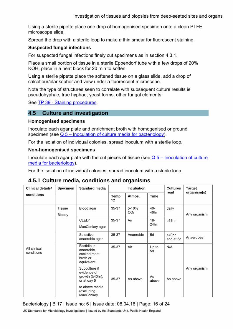
4.5 Culture and investigation Homogenised specimens Inoculate each agar plate and enrichment broth with homogenised or ground specimen (see Q 5 – Inoculation of culture media for bacteriology). For the isolation of individual colonies, spread inoculum with a sterile loop. Non-homogenised specimens Inoculate each agar plate with the cut pieces of tissue (see Q 5 – Inoculation of culture media for bacteriology). For the isolation of individual colonies, spread inoculum with a sterile loop.
4.5.1 Culture media, conditions and organisms Clinical details/
conditions
Specimen Standard media Incubation Cultures read
Target organism(s)
Temp. °C
Atmos. Time
All clinical conditions
Tissue
Biopsy
Blood agar 35-37 5-10% CO2
40-40hr
daily
Any organism
CLED/
MacConkey agar
35-37 Air 18-24hr
≥18hr
Selective anaerobic agar
35-37 Anaerobic 5d ≥40hr and at 5d Anaerobes
Fastidious anaerobic, cooked meat broth or equivalent.
Subculture if evidence of growth (≥40hr), or at day 5
to above media (excluding MacConkey
35-37
35-37
Air
As above
Up to 5d
As above
N/A
As above
Any organism
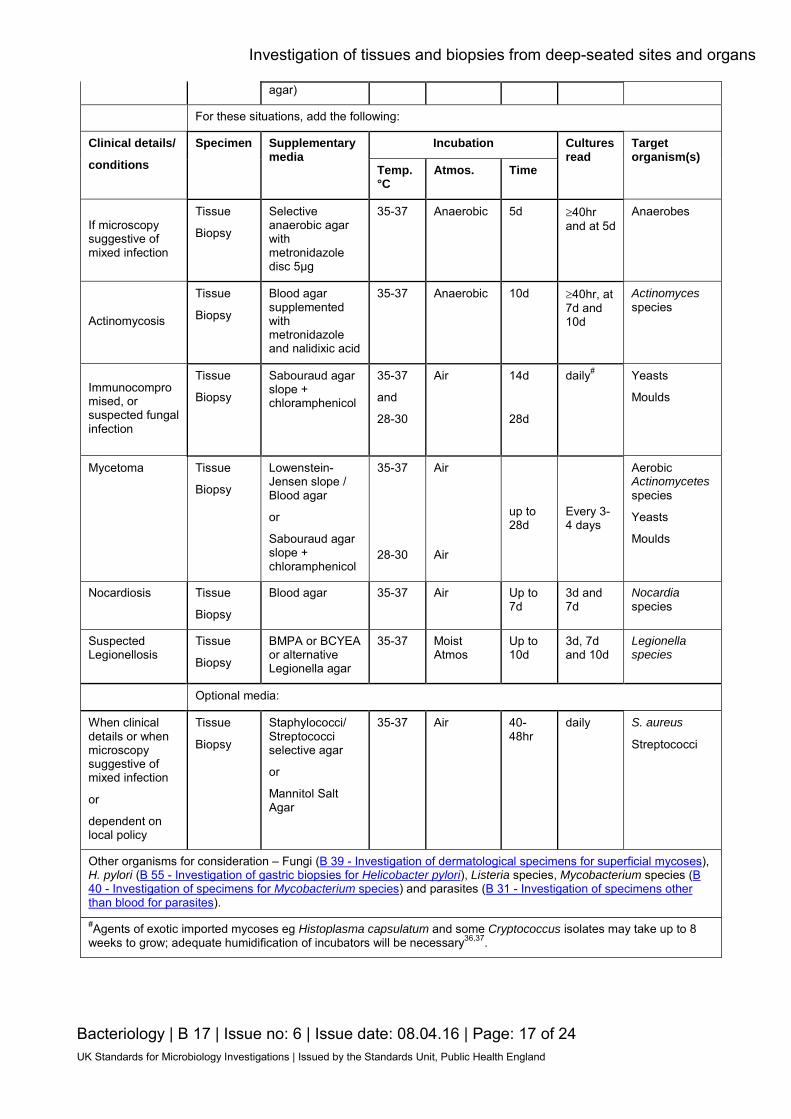
Investigation of tissues and biopsies from deep-seated sites and organs
Bacteriology | B 17 | Issue no: 6 | Issue date: 08.04.16 | Page: 17 of 24 UK Standards for Microbiology Investigations | Issued by the Standards Unit, Public Health England
agar)
For these situations, add the following:
Clinical details/
conditions
Specimen Supplementary media
Incubation Cultures read
Target organism(s)
Temp. °C
Atmos. Time
If microscopy suggestive of mixed infection
Tissue
Biopsy
Selective anaerobic agar with metronidazole disc 5μg
35-37 Anaerobic 5d ≥40hr and at 5d
Anaerobes
Actinomycosis
Tissue
Biopsy
Blood agar supplemented with metronidazole and nalidixic acid
35-37 Anaerobic 10d ≥40hr, at 7d and 10d
Actinomyces species
Immunocompromised, or suspected fungal infection
Tissue
Biopsy
Sabouraud agar slope + chloramphenicol
35-37
and
28-30
Air 14d
28d
daily# Yeasts
Moulds
Mycetoma Tissue
Biopsy
Lowenstein-Jensen slope / Blood agar
or
Sabouraud agar slope + chloramphenicol
35-37
28-30
Air
Air
up to 28d
Every 3-4 days
Aerobic Actinomycetes species
Yeasts
Moulds
Nocardiosis Tissue
Biopsy
Blood agar 35-37 Air Up to 7d
3d and 7d
Nocardia species
Suspected Legionellosis
Tissue
Biopsy
BMPA or BCYEA or alternative Legionella agar
35-37 Moist Atmos
Up to 10d
3d, 7d and 10d
Legionella species
Optional media:
When clinical details or when microscopy suggestive of mixed infection
or
dependent on local policy
Tissue
Biopsy
Staphylococci/ Streptococci selective agar
or
Mannitol Salt Agar
35-37 Air 40-48hr
daily S. aureus
Streptococci
Other organisms for consideration – Fungi (B 39 - Investigation of dermatological specimens for superficial mycoses), H. pylori (B 55 - Investigation of gastric biopsies for Helicobacter pylori), Listeria species, Mycobacterium species (B 40 - Investigation of specimens for Mycobacterium species) and parasites (B 31 - Investigation of specimens other than blood for parasites). #Agents of exotic imported mycoses eg Histoplasma capsulatum and some Cryptococcus isolates may take up to 8 weeks to grow; adequate humidification of incubators will be necessary36,37.

Investigation of tissues and biopsies from deep-seated sites and organs
Bacteriology | B 17 | Issue no: 6 | Issue date: 08.04.16 | Page: 18 of 24 UK Standards for Microbiology Investigations | Issued by the Standards Unit, Public Health England
4.6 Identification Refer to individual SMIs for organism identification.
4.6.1 Minimum level of identification in the laboratory Actinomycetes genus level
ID 10 – Identification of aerobic Actinomycetes species
ID 15 – Identification of anaerobic Actinomycetes species
Anaerobes "anaerobes" level
ID 8 - Identification of Clostridium species
β-haemolytic streptococci species level
Coagulase negative staphylococci “coagulase negative" level
Enterobacteriaceae species level
Pseudomonads species level
S. aureus species level
(consider Panton-Valentine leukocidin (PVL) and toxin testing if appropriate clinical details)
(consider toxin testing on samples from post mortems)
S. anginosus group S. anginosus group level
Yeast species level
Mould species level
Legionella species species level
Mycobacterium species species level
B 40 - Investigation of specimens for Mycobacterium species
Parasites species level
B 31 - Investigation of specimens other than blood for parasites
Organisms may be further identified if this is clinically or epidemiologically indicated.
4.7 Antimicrobial susceptibility testing Refer to British Society for Antimicrobial Chemotherapy (BSAC) and/or EUCAST guidelines. CLSI breakpoints are available for Candida species and moulds.
4.8 Referral for outbreak investigations Refer to British Society for Antimicrobial Chemotherapy (BSAC) guidelines.
4.9 Referral to reference laboratories For information on the tests offered, turnaround times, transport procedure and the other requirements of the reference laboratory click here for user manuals and request forms.
Investigation of tissues and biopsies from deep-seated sites and organs
Bacteriology | B 17 | Issue no: 6 | Issue date: 08.04.16 | Page: 19 of 24 UK Standards for Microbiology Investigations | Issued by the Standards Unit, Public Health England
Organisms with unusual or unexpected resistance, and whenever there is a laboratory or clinical problem, or anomaly that requires elucidation should be sent to the appropriate reference laboratory. Consider sending S. aureus isolates for toxin testing where appropriate clinical details are provided. For example, isolates from post mortems where the specimen is suspected to be the cause of death should be sent for toxin testing. Contact appropriate devolved national reference laboratory for information on the tests available, turnaround times, transport procedure and any other requirements for sample submission: England and Wales https://www.gov.uk/specialist-and-reference-microbiology-laboratory-tests-and-services Scotland http://www.hps.scot.nhs.uk/reflab/index.aspx Northern Ireland http://www.publichealth.hscni.net/directorate-public-health/health-protection
5 Reporting procedure 5.1 Microscopy
Gram stain Report on WBCs and organisms detected.
Legionella immunofluorescence Legionella pneumophila detected by immunofluorescence or Legionella pneumophila not detected by immunofluorescence Fungal fluorescent stain Report on type of fungal element seen.
5.1.1 Microscopy reporting time All results should be issued to the requesting clinician as soon as they become available, unless specific alternative arrangements have been made with the requestors. Urgent results should be telephoned or transmitted electronically in accordance with local policies.
5.2 Culture The following results should be reported:
• clinically significant organisms isolated • other growth with appropriate comment, eg No significant growth • absence of growth
Also, report results of supplementary investigations.
Investigation of tissues and biopsies from deep-seated sites and organs
Bacteriology | B 17 | Issue no: 6 | Issue date: 08.04.16 | Page: 20 of 24 UK Standards for Microbiology Investigations | Issued by the Standards Unit, Public Health England
5.2.1 Culture reporting time Interim or preliminary results should be issued on detection of potentially clinically significant isolates as soon as growth is detected, unless specific alternative arrangements have been made with the requestors. Urgent results should be telephoned or transmitted electronically in accordance with local policies. Final written or computer generated reports should follow preliminary and verbal reports as soon as possible. Legionella Final written or computer generated reports should follow preliminary/verbal reports within 3 - 10 days stating, if appropriate, that a further report will be issued.
5.3 Antimicrobial susceptibility testing Report susceptibilities as clinically indicated. Prudent use of antimicrobials according to local and national protocols is recommended.
6 Notification to PHE38,39, or equivalent in the devolved administrations40-43 The Health Protection (Notification) regulations 2010 require diagnostic laboratories to notify Public Health England (PHE) when they identify the causative agents that are listed in Schedule 2 of the Regulations. Notifications must be provided in writing, on paper or electronically, within seven days. Urgent cases should be notified orally and as soon as possible, recommended within 24 hours. These should be followed up by written notification within seven days. For the purposes of the Notification Regulations, the recipient of laboratory notifications is the local PHE Health Protection Team. If a case has already been notified by a registered medical practitioner, the diagnostic laboratory is still required to notify the case if they identify any evidence of an infection caused by a notifiable causative agent. Notification under the Health Protection (Notification) Regulations 2010 does not replace voluntary reporting to PHE. The vast majority of NHS laboratories voluntarily report a wide range of laboratory diagnoses of causative agents to PHE and many PHE Health protection Teams have agreements with local laboratories for urgent reporting of some infections. This should continue. Note: The Health Protection Legislation Guidance (2010) includes reporting of Human Immunodeficiency Virus (HIV) & Sexually Transmitted Infections (STIs), Healthcare Associated Infections (HCAIs) and Creutzfeldt–Jakob disease (CJD) under ‘Notification Duties of Registered Medical Practitioners’: it is not noted under ‘Notification Duties of Diagnostic Laboratories’. https://www.gov.uk/government/organisations/public-health-england/about/our-governance#health-protection-regulations-2010 Other arrangements exist in Scotland40,41, Wales42 and Northern Ireland43.

Investigation of tissues and biopsies from deep-seated sites and organs
Bacteriology | B 17 | Issue no: 6 | Issue date: 08.04.16 | Page: 21 of 24 UK Standards for Microbiology Investigations | Issued by the Standards Unit, Public Health England
Appendix: Investigation of tissues and biopsies from deep-seated sites and organs Grind or homogenise specimen
(unless fungal infection suspected)
Standard MediaSupplementary Media
Optional Media
Dirty sites or when microscopy suggestive of mixed infection, or dependent on local
policy
Staph/Strep selective agarOr
MSA
Incubate at 35-37°C Air
40-48hr Read daily
If microscopy suggestive of mixed
infectionActinomycosis
Immunocompromised or suspected fungal
infectionMycetoma
Selective anaerobe agar with
metronidazole disc 5μg
Incubate at 35-37°C Anaerobic
5 d Read at ≥ 40 hr and
at 5 d
Blood agar supplemented with metronizadole and
nalidixic acid
Incubate at 35-37°C Anaerobic
10 d Read at ≥ 40hr and
at 7 d and 10 d
Sabouraud agar slope + chloramphenicol
Incubate at 35-37°C Air
14 dRead dailyor 28-30°CUp to 28 d
LJ slope and/or Blood agar or
Sabouraud agar slope+
chloramphenicol
Incubate at 28-30°C Air
Up to 28 d Read every 3-4 d
Incubate at 35-37°C
Air5 d
Subculture toBlood agarSelective
anaerobic agarCLED
If evidence of growth (≥40hr) or
5 d
Any organismRefer to IDs
AnaerobesID 8, 14, 25
Actinomyces spRefer to ID 15
Anaerobic Actinomycetes
S. aureusID 7
StreptococciID 4
Blood agar Selective anaerobe agar
CLED agar/MacConkey
Fastidious anaerobic, cooked
meat broth or equivalent
Incubate at 35-37°C
5-10% CO2 40-48hr
Read daily
Incubate at 35-37°C
Anaerobic 5 d
Read at ≥ 40hr and at 5 d
Incubate at 35-37°C
Air 18-24hr
Read at ≥ 18hr
YeastMould
Suspected Legionellosis
BPMA/CYE/BCYEA/Legionella selective
agar
Incubate at 35-37°C Moist Atmos
10 d Read at 3 d, 7 d and
10 d
Legionella spID 18
Nocardiosis
Blood agar
Incubate at 35-37°C Air
Up to 7 d Read 3 d and 7d
Nocardia spRefer to ID 10
All Specimens
Any organismRefer to IDs
Incubate as ‘All specimens’
direct plates
AnaerobesID 8, 14, 25
Ricerca su tessuti e biopsie da sedi profonde organi
Batterologia | B 17 | Emissione no: 6 | Data emissione: 08.04.16 | Pagina: 22 di 24 UK Standards for Microbiology Investigations | Emesso da Standards Unit, Public Health England
Bibliografia
1. Baron EJ, Miller JM, Weinstein MP, Richter SS, Gilligan PH, Thomson RB, Jr. et al. A Guide to Utilization of the Microbiology Laboratory for Diagnosis of Infectious Diseases: 2013 Recommendations by the Infectious Diseases Society of America (IDSA) and the American Society for Microbiology (ASM). ClinInfectDis 2013;57:e22-e121.
2. Buckels JA, Fielding JW, Black J, Ashton F, Slaney G. Significance of positive bacterial
cultures from aortic aneurysm contents. BrJSurg 1985;72:440-2. 3. Jaton K, Peter O, Raoult D, Tissot JD, Greub G. Development of a high throughput PCR to
detect Coxiella burnetii and its application in a diagnostic laboratory over a 7-year period. New MicrobesNew Infect 2013;1:6-12.
4. Lepidi H, Houpikian P, Liang Z, Raoult D. Cardiac valves in patients with Q fever endocarditis:
microbiological, molecular, and histologic studies. JInfectDis 2003;187:1097-106. 5. Tang YW. Duplex PCR assay simultaneously detecting and differentiating Bartonella quintana,
B. henselae, and Coxiella burnetii in surgical heart valve specimens. JClinMicrobiol 2009;47:2647-50.
6. Manhire A, Charig M, Clelland C, Gleeson F, Miller R, Moss H et al. Guidelines for
radiologically guided lung biopsy. Thorax 2003;58:920-36. 7. Dandapat MC, Mishra BM, Dash SP, Kar PK. Peripheral lymph node tuberculosis: a review of
80 cases. BrJSurg 1990;77:911-2. 8. White J, O'Farrell N, Daniels D. 2013 UK National Guideline for the management of
lymphogranuloma venereum: Clinical Effectiveness Group of the British Association for Sexual Health and HIV (CEG/BASHH) Guideline development group. International journal of STD & AIDS 2013;24:593-601.
9. Schuchat A, Swaminathan B, Broome CV. Epidemiology of human listeriosis. ClinMicrobiolRev
1991;4:169-83. 10. Mylonakis E, Paliou M, Hohmann EL, Calderwood SB, Wing EJ. Listeriosis during pregnancy: a
case series and review of 222 cases. Medicine (Baltimore) 2002;81:260-9. 11. Jernigan JA, Farr BM. Incubation period and sources of exposure for cutaneous
Mycobacterium marinum infection: case report and review of the literature. ClinInfectDis 2000;31:439-43.
12. Gabillot-Carre M, Roujeau JC. Acute bacterial skin infections and cellulitis. CurrOpinInfectDis
2007;20:118-23. 13. European Parliament. UK Standards for Microbiology Investigations (SMIs) use the term "CE
marked leak proof container" to describe containers bearing the CE marking used for the collection and transport of clinical specimens. The requirements for specimen containers are given in the EU in vitro Diagnostic Medical Devices Directive (98/79/EC Annex 1 B 2.1) which states: "The design must allow easy handling and, where necessary, reduce as far as possible contamination of, and leakage from, the device during use and, in the case of specimen receptacles, the risk of contamination of the specimen. The manufacturing processes must be appropriate for these purposes". 1998.
14. Official Journal of the European Communities. Directive 98/79/EC of the European Parliament
and of the Council of 27 October 1998 on in vitro diagnostic medical devices 1998. 1-37.

Ricerca su tessuti e biopsie da sedi profonde e organi
Bateriologia | B 17 | Emissione no: 6 | Data emissione: 08.04.16 | Pagina: 23 di 24 UK Standards for Microbiology Investigations | Emesso da Standards Unit, Public Health England
15. Clark AE, Kaleta EJ, Arora A, Wolk DM. Matrix-assisted laser desorption ionization-time of flight mass spectrometry: a fundamental shift in the routine practice of clinical microbiology. ClinMicrobiolRev 2013;26:547-603.
16. Espy MJ, Uhl JR, Sloan LM, Buckwalter SP, Jones MF, Vetter EA et al. Real-time PCR in
clinical microbiology: applications for routine laboratory testing. ClinMicrobiolRev 2006;19:165-256.
17. Moore JL, Caprioli RM, Skaar EP. Advanced mass spectrometry technologies for the study of
microbial pathogenesis. Current opinion in microbiology 2014;19:45-51. 18. Health and Safety Executive. Safe use of pneumatic air tube transport systems for pathology
specimens. 2009. 19. Department for transport. Transport of Infectious Substances, 2011 Revision 5. 2011. 20. World Health Organization. Guidance on regulations for the Transport of Infectious Substances
2013-2014. 2012. 21. Home Office. Anti-terrorism, Crime and Security Act. 2001. 22. Advisory Committee on Dangerous Pathogens. The Approved List of Biological Agents. Health
and Safety Executive 2013. 1-32. 23. Advisory Committee on Dangerous Pathogens. Infections at work: Controlling the risks. Her
Majesty's Stationery Office 2003. 24. Advisory Committee on Dangerous Pathogens. Biological agents: Managing the risks in
laboratories and healthcare premises. Health and Safety Executive 2005. 25. Advisory Committee on Dangerous Pathogens. Biological Agents: Managing the Risks in
Laboratories and Healthcare Premises. Appendix 1.2 Transport of Infectious Substances - Revision. Health and Safety Executive 2008.
26. Centers for Disease Control and Prevention. Guidelines for Safe Work Practices in Human and
Animal Medical Diagnostic Laboratories. MMWR Surveill Summ 2012;61:1-102. 27. Health and Safety Executive. Control of Substances Hazardous to Health Regulations. The
Control of Substances Hazardous to Health Regulations 2002. 5th ed.: HSE Books; 2002. 28. Health and Safety Executive. Five Steps to Risk Assessment: A Step by Step Guide to a Safer
and Healthier Workplace. HSE Books. 2002. 29. Health and Safety Executive. A Guide to Risk Assessment Requirements: Common Provisions
in Health and Safety Law. HSE Books. 2002. 30. Health Services Advisory Committee. Safe Working and the Prevention of Infection in Clinical
Laboratories and Similar Facilities. HSE Books 2003. 31. British Standards Institution (BSI). BS EN12469 - Biotechnology - performance criteria for
microbiological safety cabinets 2000. 32. British Standards Institution (BSI). BS 5726:2005 - Microbiological safety cabinets. Information
to be supplied by the purchaser and to the vendor and to the installer, and siting and use of cabinets. Recommendations and guidance. 2005. 1-14.
33. Reddy S, Manuel R, Sheridan E, Sadler G, Patel S, Riley P. Brucellosis in the UK: a risk to
laboratory workers? Recommendations for prevention and management of laboratory exposure. JClinPathol 2010;63:90-2.
Ricerca su tessuti e biopsie da sedi profonde e organi
Bateriologia| B 17 | Emissione no: 6 | Data emissione: 08.04.16 | Pagina: 24 di 24 UK Standards for Microbiology Investigations | IEmesso da Standards Unit, Public Health England
34. Holden J. Collection and transport of clinical specimens for anaerobic culture. In: Isenberg HD, editor. Clinical Microbiology Procedures HandbookVol 1. Washington D.C.: American Society for Microbiology; 1992. p. 2..1-7.
35. Revankar SG, Sutton DA. Melanized fungi in human disease. ClinMicrobiolRev 2010;23:884-
928. 36. Bosshard PP. Incubation of fungal cultures: how long is long enough? Mycoses 2011;54:e539-
e45. 37. Morris AJ, Byrne TC, Madden JF, Reller LB. Duration of incubation of fungal cultures.
JClinMicrobiol 1996;34:1583-5. 38. Public Health England. Laboratory Reporting to Public Health England: A Guide for Diagnostic
Laboratories 2013. 1-37. 39. Department of Health. Health Protection Legislation (England) Guidance. 1-112. 2010. 40. Scottish Government. Public Health (Scotland) Act. 2008. 41. Scottish Government. Public Health etc. (Scotland) Act 2008. Implementation of Part 2:
Notifiable Diseases, Organisms and Health Risk States. 2009. 42. The Welsh Assembly Government. Health Protection Legislation (Wales) Guidance. 2010. 43. Home Office. Public Health Act (Northern Ireland) 1967 Chapter 36. 1967.