Embed Size (px)
Citation preview

UDP-Glucuronosyltransferases (UGTs): Overview
Anna Radominska-Pandya
Department of Biochemistry and Molecular Biology
University of Arkansas for Medical Sciences
Little Rock, Arkansas, US
October 2010; Gdansk University of Technology

General Concept of Detoxification
Endobiotics
XenobioticsMetabolism
Inactive Excretion
Products
Urine Bile
Lipid Soluble Water Soluble
Phase I
Oxidation
(P450s)
OH
Phase II
Conjugation
(UGTs)
H
O
HO
O OHH
COOH
O
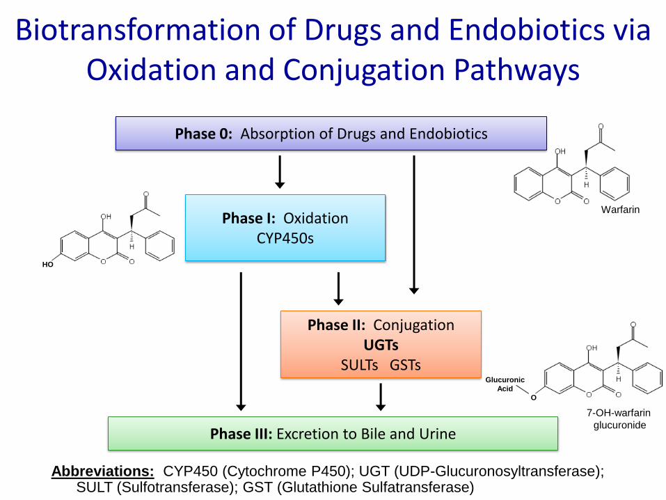
Phase 0: Absorption of Drugs and Endobiotics
Biotransformation of Drugs and Endobiotics via Oxidation and Conjugation Pathways
Phase I: OxidationCYP450s
Phase III: Excretion to Bile and Urine
Phase II: ConjugationUGTs
SULTs GSTs
Abbreviations: CYP450 (Cytochrome P450); UGT (UDP-Glucuronosyltransferase); SULT (Sulfotransferase); GST (Glutathione Sulfatransferase)
HO
7-OH-warfarin
glucuronide
Warfarin
Glucuronic
Acid
O

Biotransformation via Glucuronidation
UDP-GlcUA
Co-SubstrateUGT
Cytoplasm
Lumen
ER
_COO
-
+H3N_
Various Glucuronides
Bioactivated Glucuronides
Retinoic Acid-gluc
6-O-Morphine-gluc
3-O-Lithocholic Acid-gluc
D-ring glucuronides of estradiol,
testosterone, DHEA
Electophilic Glucuronides
Acyl glucuronides (NSAIDs)
N-O-glucuronides (Hydroxamic Acids)
Hydrophilic β-D-glucuronides
Therapeutic Drugs
Carcinogens
Environmental Toxins
Dietary Constituents
Lipophilic Substrates
Bilirubin Steroids
Bile Acids
Retinoic Acids
Fatty Acids
Prostaglandins

Substrate Specificity of UGTs
• Substrate specificity is broad and overlapping (promiscuous)
• Wide range of substrates:– Endogenous substrates
– Xenobiotic substrates• Drugs
• Dietary plant constituents
• Carcinogens
• Types of glucuronides:– O-glucuronides
• Including acyl glucuronides
– N-glucuronides
– S-glucuronides
– C-glucuronides
Tukey R.H. 2000.
Phenols
Aliphatic alcohols
Anthraquinones/flavones
Carboxylic acids
Billirubin
Bile acids
Amines
Coumarins
Opioids
Steroids
Sapogenins
Estrogens
Androgens
Progestin
Bile acids
Primary
Secondary
Tertiary
Heterocyclic
Simple
Complex

Glucuronidation Reactions Catalyzed by UGTs
O-Glucuronidation
S-Glucuronidation
N-Glucuronidation
C-Glucuronidation

UDP-Glucuronosyltransferases (UGTs)
• Large family of membrane bound glycosylatedproteins located in the ER as well as inner and outer nuclear membranes
• Conjugate a wide range of endobiotics and xenobiotics with glucuronic acid
– Glucuronic acid moiety can attach via a hydroxyl, carboxyl, amino, thiol or carbonyl group on the substrates
– Generate more polar, water soluble metabolites which can be excreted in urine and bile

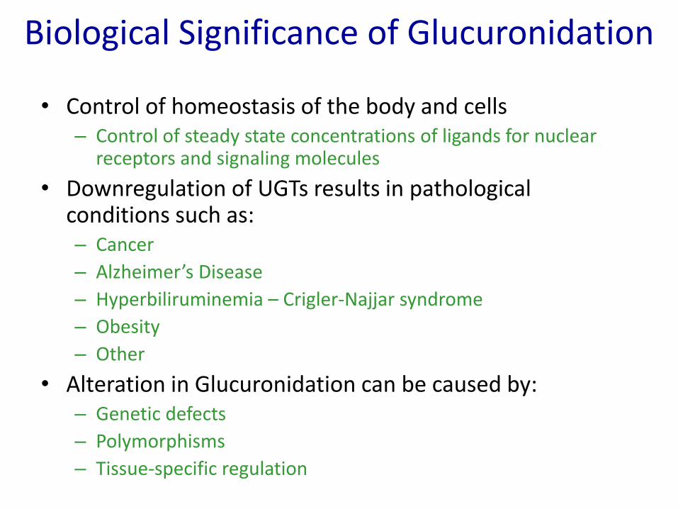
Biological Significance of Glucuronidation
• Detoxification
– increases hydrophobic properties• soluble in blood/urine
– structure different from parent compound• no favorable interaction with pharmacological target
• Detoxification leads to:
– Excretion of catabolic products
– Elimination of nucleophilic metabolites of carcinogens
– Inactivation of biologically active components• AZT

Biological Significance of Glucuronidation
• Bioactivation of the parent compound– Increases toxicity
– Increases pharmacological properties
• Metabolic activation leads to:– Cholestatic glucuronides
• Lithocholic acid glucuronide
• Estradiol glucuronide
– Chemically reactive glucuronides:• Acyl-glucuronides
• NSAID glucuronides– Ketoprofen
– Zomepirax
– Metabolically-active glucuronides• Morphine-6-O-glucuronide
• Irinotecan glucuronide
• Retinoid glucuronides
Biological Significance of Glucuronidation
• Control of homeostasis of the body and cells– Control of steady state concentrations of ligands for nuclear
receptors and signaling molecules
• Downregulation of UGTs results in pathological conditions such as:– Cancer
– Alzheimer’s Disease
– Hyperbiliruminemia – Crigler-Najjar syndrome
– Obesity
– Other
• Alteration in Glucuronidation can be caused by:– Genetic defects
– Polymorphisms
– Tissue-specific regulation
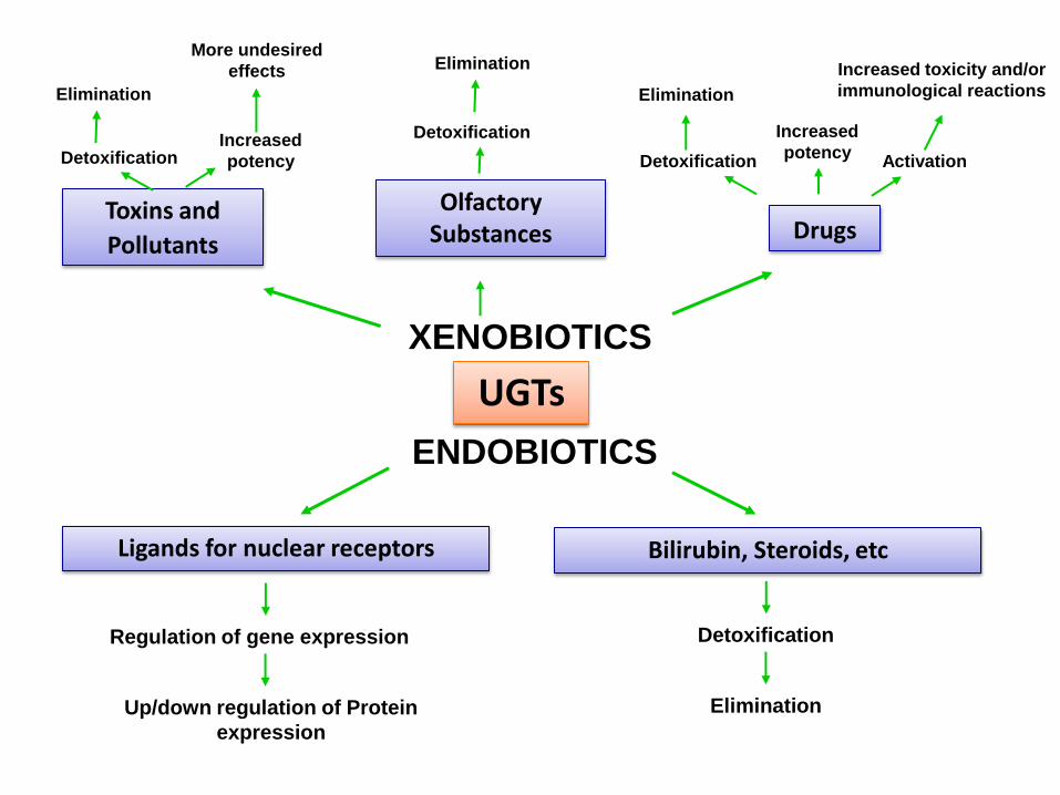
Ligands for nuclear receptors
Regulation of gene expression
Up/down regulation of Protein
expression
UGTs
ENDOBIOTICS
XENOBIOTICS
Toxins and
Pollutants
DetoxificationIncreased
potency
Elimination
More undesired
effects
Olfactory Substances
Detoxification
Drugs
Detoxification
Elimination
Increased
potencyActivation
Increased toxicity and/or
immunological reactions
Elimination
Bilirubin, Steroids, etc
Detoxification
Elimination

Phylogenetic Tree of Mammalian UGTs
• Divergence of 49 mammalian UGT proteins – Known mammalian UGTs have been separated into two families,
UGT1 and UGT2.• UGT1 family is localized on chromosome 2q37 and is divided into 2
subfamilies, UGT1A and UGT1B. • UGT2 is localized on chromosome 4q13 and is divided into 3
subfamilies, UGT2A, UGT2B, and UGT2C (not shown).
• 20 human UGTs have been identified
Guillemette C, DMR 2009

Guillemette C, DMR 2009

Ritter JK, 1992
UGT1A Gene Cluster and Putative Protein Structure in Humans
Radominska-Pandya A, 1999
Transmembrane
fragment
Substrate binding domain UDP-GlcUA binding domain
Variable N-terminal domain
amino acids 25 - 286
NH2 COO-
Retention
signal
Signal
peptideConserved C-terminal domain
amino acids 287 - 530
UDP-GlcUASubstratesUDP-
GlcUA
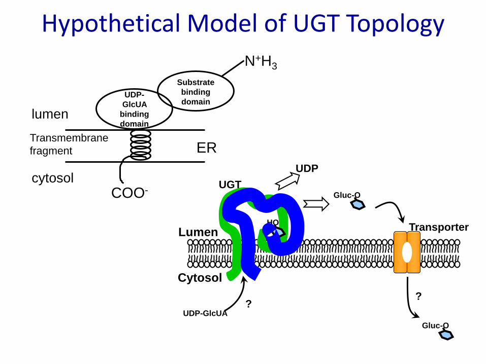
UDP-GT
UDP-
GlcUA
binding
domain
Substrate
binding
domain
ER
COO-
N+H3
cytosol
lumen
HO
Cytosol
Lumen
Gluc-O
Gluc-O
UDP-GlcUA
UGT
Transporter
??
UDP
Hypothetical Model of UGT Topology
Transmembrane
fragment

Tissue-Specific Expression of Human UGTsIsoform
Tissue
Protein Expression mRNA expression
UGT1A1 Biliary tissue, colon, intestine, liver, stomach,Kidney, trachea, adrenal gland, lung, prostate, testis, thymus,
thyroid
UGT1A3 Biliary tissue, colon, liver, stomach, brain
UGT1A4 Biliary tissue, colon, liver, intestine, lung,
UGT1A6 Biliary tissue, colon, liver, stomach, brain, kidney, larynx, lungAdrenal gland, placenta, prostate, salivary gland, small intestine,
testis, thymus, thyroid gland, trachea, uterus.
UGT1A7 Orolaryngeal tissue, esophagus, stomach
UGT1A8 Colon, esophagus, intestine, kidney, larynx
UGT1A9 Breast, colon, esophagus, liver, kidney, ovary, prostate, skin, testis
UGT1A10Orolaryngeal tissue, colon, biliary tissue, esophagus, intestine,
stomachBreast
UGT2B4Adipose tissue, adrenals, breast, ovary, liver, lung, placenta,
prostate, skin, testis, kidney
UGT2B7Breast, brain, colon, esophagus, intestine, kidney, liver, lung, and
pancreasTestis, uterus
UGT2B10 Liver Adrenal gland, colon, heart, skeletal muscle, testis, uterus
UGT2B11 Lung
UGT2B15Breast, testis, uterus, prostate, lung, ovary, esophagus, kidney, liver,
skin Colon, pancreas, small intestine, stomach, testis, trachea
UGT2B17 Liver, prostateAdrenal gland, bone marrow, brain, colon, lung, pancreas, peripheral
leukocytes, salivary gland, small intestine, spinal cord, spleen, stomach, testis, thymus, trachea
UGT2A1 Lung Trachea

UGT Polymorphisms and Genetic Deficiencies

Examples of UGT Polymorphisms
• UGT1A1– UGT1A1*28
• A common variant [A(TA)7TAA] in the TATA-box region of the UGT1A1 promoter
– UGT1A1*1 • Results in:
– Decrease level of UGT1A1 gene expression
– Increased breast cancer risk (due to estrogen metabolism), specifically in African American women
• UGT1A6– Metabolizes aspirin and other NSAIDs
– Two missense mutations leading to T181A and R184S amino acid substitution• UGT1A6*2
– Has a frequency of 30% in Caucasian pop.
– Positively modified protective effect of aspirin (decreased glucuronidation leads to higher levels of aspirin)

Examples of UGT Polymorphisms
• UGT1A7– Glucuronidates polycyclic aromatic hydrocarbons and dietary
heterocyclic aromatic amines– Three missense mutations in exon 1 result in four alleles:
• UGT1A7*1 (N129, R131, W208)• UGT1A7*2 (K129, K131, W208)• UGT1A7*3 (K129, K131, R208)
– Increased risk of orolaryngeal, liver, and colon cancer
• UGT1A7*4 (N129, R131, R208)– Increased risk of orolaryngeal, liver, and colon cancer
• UGT2B7 – Single nucleotide polymorphisms in coding and regulatory
region of UGT2B7 gene are thought to play a role in morphine glucuronidation• Cytosine to thymine polymorphism at 802 bp• UGT2B7*1 (Y268)
– 3 times more likely Asian pop than 2B7*2.
• UGT2B7*2 (H268)
Genetic Deficiencies of UGT1A1
• Crigler-Najjar disease:– Severe, chronic, non-hemolytic, unconjugated
hyperbilirubinemia– Defect in the gene encoding bilirubin UGT1A1– Caused by mutations to common exons 2-5 or by a mutation to
exon 1– Type I
• Complete loss of bilirubin-conjugating activity
– Type II• Partial loss of bilirubin-conjugating activity (typically <10% of normal) • Responds to phenobarbital treatment
• Gilbert’s syndrome:– Mild, unconjugated hyperbilirubinemia– Defect in the gene encoding bilirubin UGT1A1– Missense mutation in the coding region– Homozygous insertion into promoter
Conclusions

Practice Questions