Embed Size (px)
Citation preview

UDC: 61 ISSN 1409-9837
ЗАММ
МАММ
ACTA MORPHOLOGICA
INTERNATIONAL JOURNAL OF THE MACEDONIAN ASSOCIATION
OF ANATOMISTS AND MORPHOLOGISTS
Vol. 16 (1) 2019

ACTA MORPHOLOGICA
International Journal of the Macedonian Association of Anatomists and Morphologists (MAAM)
Member of the European Federation of Experimental Morphology (EFEM) Member of the International Symposium of Morphological Sciences (ISMS)
Published
Twice a year
EDITORIAL BOARD
Editor in Chief
Dobrila Tosovska-Lazarova
Skopje, Republic of North Macedonia
Editors
Gordana Teofilovski-Parapid
Belgrade, Serbia
Andreas H. Weiglein
Graz, Austria
Guido Macchiarelli
L’Aquila, Italy
Petru Matusz
Timisoara, Romania
Erdogan Sendemir
Bursa, Turkey
Alessandro Riva
Cagliary, Italy
Sadeta Sekic
Sarajevo, B&H
Diogo Pais
Lisboa, Portugal
Marija Vavlukis
Skopje, Republic of North Macedonia
Venko Filipce
Skopje, Republic of North Macedonia
Yavor Enchev
Varna, Bulgaria
Susana N. Biasutto
Cordoba, Argentina
Marko Kostovski
Skopje, Republic of North Macedonia
Macedonian Scientific Committee
Natasha Janevska-Nakeva
Angja Strateska-Zafiroska
Vesna Janevska
Elena Trajkovska-Dokikj
Nevena Kostovska
Gjorgji Jota
Dragica Jurkovic
Pre-Press
Marko Kostovski
CONTENT
ORIGINAL ARTICLES
5. Ozone treatment of stage II/ III of bisphosphonate-related osteonecrosis of the jaws
(BRONJ). Markovska Arsovska Mirjana, Popovic Monevska D, Popovska K,
Simjanovska Lj, Isijanovski I, Gerasimova S.
15. Lower serum potassium level is associated with mortality as confounding effect of
malnutrition in dialysis patients. Trajceska Lada, Selim Gj, Pavleska Kuzmanovska S,
Pusevski V, Sikole A.
20. Implementation and validation of the new AO classification of thoracolumbar
fractures. Ljupco Nikolov.
29. Vertical irregularities influence over the size, form and shape of the symphysis.
Bogdanovska Biljana, Pop Stefanova-Trposka M, Gavrilovic I, Curcieva-Cuckova G,
Bogdanovski S.
CASE REPORTS
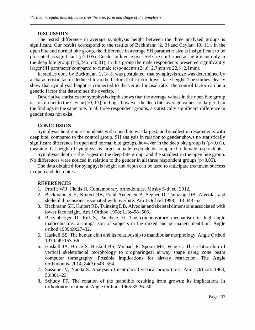
35. Perineo-vaginal reconstruction with IPAP propeller flap – A case report. Igor Peev,
Zhogovska-Mirchevska E, Alulovski I, Krstevska-Dukova I.
REVIEW ARTICLES
42. Acute chorioamnionitis, histopathologic changes of the placenta and prevalence of
microbial invasion in patients with preterm delivery – Review of literature. Kochoski
Goran, Spasova R, Kjaev I, Karadzova D, Stojoska Lazarova A, Janevska V, Spasevska
L.
53. Ethical considerations towards individual results delivery and the consequences of
incidental findings in the whole genome and exome sequencing. Kostovski Marko,
Simeonovski V.
63. Utility of exhaled nitric oxide in pediatric practice – Review of literature. Arnaudova-
Danevska Ivana, Balkanov T, Boshkovska K, Jakjovska T, Popova G, Cakalarovska I.
INSTRUCTIONS FOR AUTHORS
77. Instructions for authors
AN EXCLUSIVE STATEMENT
81. An exclusive statement


Acta morphol.2019;Vol.16(1):5–14
UDC: 615.31.03:546.214]:616.714.4-002.4-02:615.272
Page | 5
ORIGINAL ARTICLE
OZONE TREATMENT OF STAGE II/ III OF BISPHOSPHONATE-RELATED
OSTEONECROSIS OF THE JAWS (BRONJ)
Markovska Arsovska Mirjana1, Popovic Monevska D2, Popovska K3, Simjanovska Lj1,
Isijanovski I4, Gerasimova S1
1University Dental Clinical Centre”St. Panteleimon“, Department of Oral Surgery,
Skopje, Republic of North Macedonia 2University Clinic for Maxillofacial Surgery, Faculty of Dental Medicine,
Skopje, Republic of North Macedonia 3Institute of Microbiology and Parasitology, Faculty of Medicine,
Skopje, Republic of North Macedonia 4Faculty of Medicine – Skopje, Republic of North Macedonia
ABSTRACT
Bisphosphonate-related osteonecrosis of the jaws (BRONJ) is defined as exposed jawbone
(part of the jawbone) in the oral cavity that persists for more than eight weeks despite the treatment,
and patients haven’t received radiotherapy and there is no history of bone metastases. This is a
serious complication in patients receiving bisphosphonate therapy, especially in those who have
received it intravenously. Bisphosphonates are used as potent inhibitors of bone resorption in
various diseases and conditions of the bones, such as malignancies, osteoporosis, and bone
metastases. There are many studies related to the impact of medical ozone on wound healing in
bisphosphonate osteonecrosis. Several authors show the effect of ozone therapy on the wound
healing in the osteonecrosis area in patients receiving bisphosphonate therapy. The aim of this
study was to show the influence of ozone therapy on wound healing in stage II/III of
bisphosphonate-related osteonecrosis of the jaws. This study included 25 male and female patients
who received bisphosphonate therapy, and in whom II or III clinical stage of bisphosphonate-
related osteonecrosis of the jaws was diagnosed. After receiving standard antibiotic therapy
determined with an antibiogram, patients received medical ozone therapy for dental use in the form
of a gas (ozone therapy device, Ozone DTA-Dental ozone generator of Apoza Enterprise). Patients
were treated with conservative treatment or surgical treatment (sequestrectomy). In patients with
clinical stage II / III, the positive effect of this therapy was observed after the application of ozone,
as well as after treatment with an antibiotic agent, with a greater tendency of decline in patients
with ozone therapy. Reduction of pain as well as reduction of bleeding and secretion (reduction of
fetor - bad smell in oral cavity) are the benefits of the treatment with local application of ozone
gas in patients with bisphosphonate-related osteonecrosis of the jaws. Ozone therapy destroys
bacteria, or reduces their effect on wound healing. It also affects the improvement or maintenance
of the clinical stage of osteonecrosis of the jaws.
Keywords: ozone therapy, bisphosphonates, pain, osteonecrosis, wound healing.

Markovska Arsovska et аl.
Page | 6
INTRODUCTION
Bisphosphonate-related osteonecrosis of the jaws (BRONJ) was described for the first time by
Marx (2003) [1] (as exposed jawbone, part of the jawbone in the oral cavity that persists for more
than eight weeks despite the treatment, and patients haven’t received radiotherapy and there is no
history of bone metastases), which appears after dental procedures in patients that have received
or still have been receiving bisphosphonates for inhibition of the bone resorption. This is a serious
complication in patients receiving bisphosphonate therapy, especially in those who have received
it intravenously [2]. Bisphosphonates are used as potent inhibitors of bone resorption in various
diseases and conditions of the bones, such as malignancies, osteoporosis, and bone metastases [3].
There are many hypotheses about the cause of BRONJ occurrence, but very often the trigger is
tooth extraction. The hypotheses of osteonecrosis are based on the inhibitory effect of
bisphosphonates on the osteoclastic activity of bone cells, as well as their toxic effects on soft
tissue and their anti-angiogenic effect. The influence of bisphosphonates on the oral microflora, as
well as the creation of biofilm (microbiota) at the site of osteonecrosis, is one of the possible
reasons for its occurrence. Biofilm is actually a set of bacterial colonies that are interconnected
with fibronectin. They cover the necrotic bone and cause frequent and recurrent infections in these
patients [4].
According to the American Association of Oral and Maxillofacial Surgeons (AAOMS, 2009)
[5] there are several clinical stages of BRONJ.
- Patients at risk – patients have been receiving or had received bisphosphonate therapy, where
tooth extraction or other oral surgery should be performed.
- Clinical stage 0 - unspecified clinical findings and symptoms (unpleasant continuous pain with
low intensity, slow wound healing in the region were tooth was pulled out, patients complain of
bad smell in the mouth), but there is not an exposed or necrotic bone in the oral cavity.
- Clinical stage I – clinical findings are: exposed or necrotic jawbone without signs of infection
(erythema), and patients complain of persistent, not very severe pain and unpleasant smell from
the oral cavity as a result of local accumulation of deposits at the site of the exposed bone
- Clinical stage II – clinical findings are: an exposed bone, but also signs of infection, pain and
erythema, as well as an unpleasant smell from the oral cavity (fetor).
- Clinical stage III - in addition to the exposed jawbone, there are signs of infection, bad smell
(fetor) and pain, as well as the possibility of pathological fracture, extraoral or intraoral fistula,
and the possibility of creating oroanthral communication or osteolysis of the jaw bones.
There are many studies related to the impact of medical ozone on wound healing in bisphosphonate
osteonecrosis. Several authors show the effect of ozone therapy on the wound healing in the
osteonecrosis area in patients receiving bisphosphonate therapy. The influence of ozone is due to
its antibacterial [6, 7], antiviral [8, 9] and antifungal effect [10], improving tissue oxygenation, as
well as its impact on epithelialization of the wound [11], and stimulation of local immunity. Basic
forms of application of ozone in the oral cavity are: ozone gas, ozonated oil and ozonated water
[12].

Ozone treatment of stage II/III of bisphosphonate-related osteonecrosis of the jaws
Page | 7
Ripamonti et al. [13] in their study presented treatment of osteonecrosis of jaws with lesions greater
than > 2.5 cm, by using ozone (O3) as a gas insufflation in the area of the lesion.
They explained the effect of ozone gas with obvious results in wound healing, such as
demarcation and sequestration of osteonecrotic bone from healthy bone and subsequent healing of
the wound.
Agrillo et al.[14], in a five-year-research used ozone gas insufflations as a conservative treatment
or as supplemental therapy in minimal sequestrectomy in patients with bisphosphonate-related
osteonecrosis of the jaws. They also described reduction of pain and reduction of osteonecrotic
lesion, as well as secretion in osteonecrotic area.
Also, medical ozone has impact on the stimulation or suppression of the immune system and
oxidative influence when it is used in small concentrations [15].
In everyday dental practice use of antibiotics for treatment of BRONJ can be followed by
additional ozone therapy [16], which should be applied during 15 days, then followed by two ozone
insufflations during surgical treatment (sequestrectomy).Their research explains the impact of
ozone in reducing pain, as well as reduction of the bad smell (fetor) in the mouth after treatment.
The topical use of ozone on infected wound in oral cavity gives an obvious result in patients who
have received a high dose of radiotherapy (in the treatment of primary disease) [17].
The positive effects of ozone and its application as an ozonized oil can be used for different
treatments in the oral cavity, which was done by Nogales [18] in the treatment of alveolitis sicca.
He compared the effectiveness between antibiotic therapy and ozone therapy. Patients were more
satisfied with ozone therapy, and they felt no pain and other additional unpleasant symptoms.
In in vitro evaluation of wound healing in rats Borges et al. [19] showed the antimicrobial potential
of ozone therapy, as well as its antifungal action.
In the literature, there are data for application of ozone therapy for preventive purposes as
protection against postoperative infection with the application of ozone before, during and after
extraction of the teeth. Filipovic et al.[20] used ozone for preventive purposes during extraction
of the impacted third molars. They also examined postoperative pain and bad smell (fetor) in the
oral cavity in these patients.
For that purpose Calvo et al. [21] applied ozone therapy in certain concentrations for wound
healing and indicated its analgesic effect.
Besides antibiotic and ozone therapy, Passaretti et al. [22] also used low-frequency laser therapy
in the treatment of BRONJ. They applied ozone gas three times a week (eight treatments). The
concomitant use of ozone therapy and low-frequency laser were also used by Moraes et al. [23]
The future will show which therapeutic method is more effective, but in any case they both show
obvious results.
The aim of this study was to show the influence of ozone therapy on wound healing in stage
II/III of bisphosphonate-related osteonecrosis of the jaws.
Markovska Arsovska et аl.
Page | 8
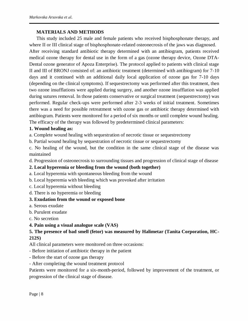
MATERIALS AND METHODS
This study included 25 male and female patients who received bisphosphonate therapy, and
where II or III clinical stage of bisphosphonate-related osteonecrosis of the jaws was diagnosed.
After receiving standard antibiotic therapy determined with an antibiogram, patients received
medical ozone therapy for dental use in the form of a gas (ozone therapy device, Ozone DTA-
Dental ozone generator of Apoza Enterprise). The protocol applied to patients with clinical stage
II and III of BRONJ consisted of: an antibiotic treatment (determined with antibiogram) for 7-10
days and it continued with an additional daily local application of ozone gas for 7-10 days
(depending on the clinical symptoms). If sequestrectomy was performed after this treatment, then
two ozone insufflations were applied during surgery, and another ozone insufflation was applied
during sutures removal. In those patients conservative or surgical treatment (sequestrectomy) was
performed. Regular check-ups were performed after 2-3 weeks of initial treatment. Sometimes
there was a need for possible retreatment with ozone gas or antibiotic therapy determined with
antibiogram. Patients were monitored for a period of six months or until complete wound healing.
The efficacy of the therapy was followed by predetermined clinical parameters:
1. Wound healing as:
a. Complete wound healing with sequestration of necrotic tissue or sequestrectomy
b. Partial wound healing by sequestration of necrotic tissue or sequestrectomy
c. No healing of the wound, but the condition in the same clinical stage of the disease was
maintained
d. Progression of osteonecrosis to surrounding tissues and progression of clinical stage of disease
2. Local hyperemia or bleeding from the wound (both together)
a. Local hyperemia with spontaneous bleeding from the wound
b. Local hyperemia with bleeding which was provoked after irritation
c. Local hyperemia without bleeding
d. There is no hyperemia or bleeding
3. Exudation from the wound or exposed bone
a. Serous exudate
b. Purulent exudate
c. No secretion
4. Pain using a visual analogue scale (VAS)
5. The presence of bad smell (fetor) was measured by Halimetar (Tanita Corporation, HC-
212S)
All clinical parameters were monitored on three occasions:
- Before initiation of antibiotic therapy in the patient
- Before the start of ozone gas therapy
- After completing the wound treatment protocol
Patients were monitored for a six-month-period, followed by improvement of the treatment, or
progression of the clinical stage of disease.
Ozone treatment of stage II/III of bisphosphonate-related osteonecrosis of the jaws
Page | 9
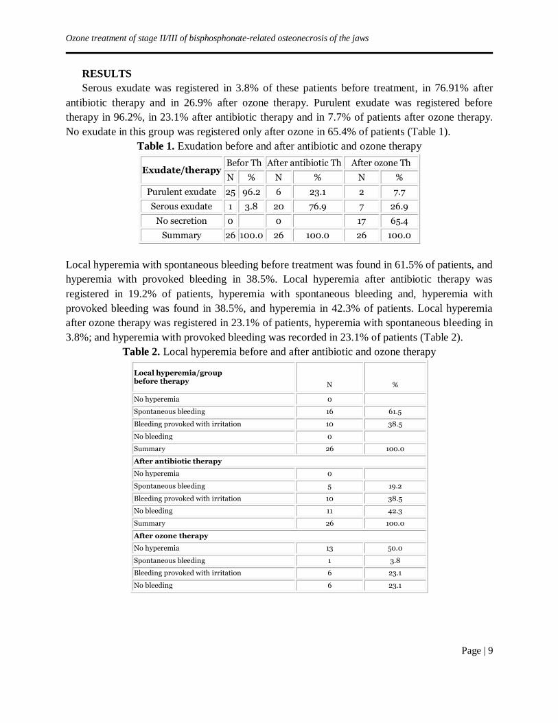
RESULTS
Serous exudate was registered in 3.8% of these patients before treatment, in 76.91% after
antibiotic therapy and in 26.9% after ozone therapy. Purulent exudate was registered before
therapy in 96.2%, in 23.1% after antibiotic therapy and in 7.7% of patients after ozone therapy.
No exudate in this group was registered only after ozone in 65.4% of patients (Table 1).
Table 1. Exudation before and after antibiotic and ozone therapy
Exudate/therapy Befor Th After antibiotic Th After ozone Th
N % N % N %
Purulent exudate 25 96.2 6 23.1 2 7.7
Serous exudate 1 3.8 20 76.9 7 26.9
No secretion 0 0 17 65.4
Summary 26 100.0 26 100.0 26 100.0
Local hyperemia with spontaneous bleeding before treatment was found in 61.5% of patients, and
hyperemia with provoked bleeding in 38.5%. Local hyperemia after antibiotic therapy was
registered in 19.2% of patients, hyperemia with spontaneous bleeding and, hyperemia with
provoked bleeding was found in 38.5%, and hyperemia in 42.3% of patients. Local hyperemia
after ozone therapy was registered in 23.1% of patients, hyperemia with spontaneous bleeding in
3.8%; and hyperemia with provoked bleeding was recorded in 23.1% of patients (Table 2).
Table 2. Local hyperemia before and after antibiotic and ozone therapy
Local hyperemia/group before therapy
N %
No hyperemia 0
Spontaneous bleeding 16 61.5
Bleeding provoked with irritation 10 38.5
No bleeding 0
Summary 26 100.0
After antibiotic therapy
No hyperemia 0
Spontaneous bleeding 5 19.2
Bleeding provoked with irritation 10 38.5
No bleeding 11 42.3
Summary 26 100.0
After ozone therapy
No hyperemia 13 50.0
Spontaneous bleeding 1 3.8
Bleeding provoked with irritation 6 23.1
No bleeding 6 23.1
Markovska Arsovska et аl.
Page | 10
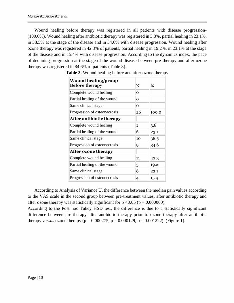
Wound healing before therapy was registered in all patients with disease progression-
(100.0%). Wound healing after antibiotic therapy was registered in 3.8%, partial healing in 23.1%,
in 38.5% at the stage of the disease and in 34.6% with disease progression. Wound healing after
ozone therapy was registered in 42.3% of patients, partial healing in 19.2%, in 23.1% at the stage
of the disease and in 15.4% with disease progression. According to the dynamics index, the pace
of declining progression at the stage of the wound disease between pre-therapy and after ozone
therapy was registered in 84.6% of patients (Table 3).
Table 3. Wound healing before and after ozone therapy
Wound healing/group Before therapy
N %
Complete wound healing 0
Partial healing of the wound 0
Same clinical stage 0
Progression of osteonecrosis 26 100.0
After antibiotic therapy
Complete wound healing 1 3.8
Partial healing of the wound 6 23.1
Same clinical stage 10 38.5
Progression of osteonecrosis 9 34.6
After ozone therapy
Complete wound healing 11 42.3
Partial healing of the wound 5 19.2
Same clinical stage 6 23.1
Progression of osteonecrosis 4 15.4
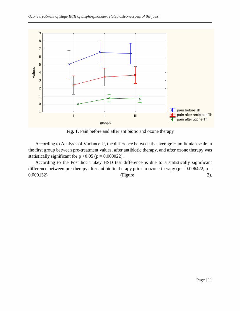
According to Analysis of Variance U, the difference between the median pain values according
to the VAS scale in the second group between pre-treatment values, after antibiotic therapy and
after ozone therapy was statistically significant for p <0.05 (p = 0.000000).
According to the Post hoc Tukey HSD test, the difference is due to a statistically significant
difference between pre-therapy after antibiotic therapy prior to ozone therapy after antibiotic
therapy versus ozone therapy (p = 0.000275, p = 0.000129, p = 0.001222) (Figure 1).
Ozone treatment of stage II/III of bisphosphonate-related osteonecrosis of the jaws
Page | 11
pain before Th
pain after antibiotic Th
pain after ozone ThI II III
groupe
-1
0
1
2
3
4
5
6
7
8
9V
alu
es
Fig. 1. Pain before and after antibiotic and ozone therapy
According to Analysis of Variance U, the difference between the average Hamiltonian scale in
the first group between pre-treatment values, after antibiotic therapy, and after ozone therapy was
statistically significant for p <0.05 (p = 0.000022).
According to the Post hoc Tukey HSD test difference is due to a statistically significant
difference between pre-therapy after antibiotic therapy prior to ozone therapy (p = 0.006422, p =
0.000132) (Figure 2).

Markovska Arsovska et аl.
Page | 12
fetor before Thfetor after antibiotic Th.fetor after ozone Th
I II III
group
-1
0
1
2
3
4
5V
alu
es
Fig. 2. Fetor before and after antibiotic and ozone therapy
DISCUSSION
Bisphosphonates are the most commonly used drugs in the treatment of bone metastases, as
well as hypercalcemia in cancer-related conditions. They perform suppression of osteoclastic
differentiation and activity of cells in bones, leading to their apoptosis (programmed cell death).
The dental interventions, such as tooth extraction, in these patients using bisphosphonates may
increase the risk of BRONJ [23].
This study was intended to illustrate the clinical effects of ozone therapy in patients with
bisphosphonate osteonecrosis.
In patients with clinical stage II / III, the positive effect of this therapy was observed after the
application of ozone, as well as after treatment with an antibiotic agent, with a greater tendency of
decline in patients with ozone therapy.
Agrillo et al. [15], in a clinical study performed on 131 patients, described the reduction of pain,
as well as the reduction of the lesion in the area of osteonecrosis. Our results have shown the same
effect of ozone therapy as in their study, with a statistical significance in pain reduction.
Ozone therapy can also be used as ozonated water or ozone oil in osteonecrotic lesions [12].
The positive effects of ozone therapy have also been explained in the research of Ripamonti et al.
[13], who used ozone gas insufflations in the treatment of bisphosphonate osteonecrosis, and in
some cases the necrotic part was sequestered and rejected or it was removed by surgery.
Reduction of pain as well as reduction of bleeding and secretion (reduction of fetor - bad smell in
oral cavity) are the benefits of treatment with local application of ozone gas in patients with
bisphosphonate-related osteonecrosis of the jaws. In our study, there was a significant decrease in
the secretion, as well as bleeding from the wound in the osteonecrotic area. Ozone therapy for
preventive purposes against postoperative infection was used by Filipovic et al. [21].
Ozone treatment of stage II/III of bisphosphonate-related osteonecrosis of the jaws
Page | 13
They did the application of ozone before, during and after the extraction of the impacted third
molars. They also examined postoperative pain and bad smell (fetor) in the oral cavity in these
patients, and they showed the positive effect of the ozone therapy.
In the treatment of BRONJ, besides antibiotic therapy, some authors like Moraes et al. [23], used
ozone oil as well as laser therapy in its management that gave positive effects.
Ozone therapy, with its antibacterial action, destroys bacteria or reduces its effect, thus improving
the wound healing in the osteonecrosis region, as has been shown with the results obtained in this
study.
CONCLUSION
Ozone therapy has a positive impact on the reduction of pain and bad smell (fetor) in the oral
cavity, destroys bacteria, or reduces their effect on wound healing. It also affects the improvement
or maintenance of the clinical stage of osteonecrosis of the jaws.
REFERENCES
1. Marx RE. Pamidronate aredia and zolendronate zometa induced avascular necrosis of the
jaws: a growing epidemic. J Oral Maxillofacial Surg. 2003;61:1115–1117.
2. Ruggiero SL, Fantasia J, Carlson E. Bisphosphonate-related osteonecrosis of the jaws:
background and guidelines for diagnosis, staging and management. Oral Surg Oral Med
Oral Path Oral Rad Endodontol. 2006;102:433–441.
3. Francesko B. Bisphosphonate-related osteonecrosis of the jaws, Med Oral Patol Oral Cir
Bucal. 2013;18(5):752–758.
4. Real CV. Role of microbiota and inflammation in osteonecrosis of the jaw: Int J Clin Exp
Pathol. 2016;9(4);418–4196.
5. American Association of oral and Maxillofacial surgeons, position paper on
bisphosphonate- related osteonecrosis of the jaws. Approved by the Board of Trustees,
Septembre 25. J Oral Maxillofac Surg. 2007; 65:369.
6. Lohr A, Gratzek J: Bactericidal and paraciticidal effects of an activated air oxidant in a
closed aquatic system. J Aquaric Aquat Sci. 1984;4(41/2):1–8.
7. Johansson E, Claesson R, Van Dijken JW. Antibacterial effect of ozone on cariogenic
bacterial species. J Dent. 2009;37:449–53.
8. Roy D, Engelbrecht RS, Chian ES: Comparative inactivation of six enteroviruses by ozone.
Am Water Works Assoc J. 1982;74(12): 660–664.
9. Riesser V, Perrich J, Silver B, Mc Cammon J: Possible mechanism of poliovirus
inactivation by ozone, in Forum on Ozone Disinfection. Proceedings of the International
Ozone Institute. Syracuse, NY, 1977; pp 186–192.
10. Matus V, Lyskova T, Konev V. Fungi growth and sporulation after a single treatment of
spores with ozone. Mokol Fitopatol. 1982;16(5):420–423.
11. Fillipi A.The influence of ozonised water on the epithelial wound healing process in the
oral cavity. Clinic of oral surgery, radiology and oral medicine. University of Basel,
Switzerland, 2011; Available at: <www. Oxyplus. Net>
12. Rajiv S. Ozone therapy in dentistry: A strategic review; J Nat Sci Biol Med. 2011;2(2):
151–153.

Markovska Arsovska et аl.
Page | 14
13. Ripamonti CL, Cislaghi E, Mariani L, Maniezzo M. Efficacy and safety of medical ozone
(O3) delivered in oil suspension applications for the treatment of osteonecrosis of the jaw
in patients with bone metastases treated with bisphosphonates: Preliminary results of a
phase I-II study, Oral Oncol. 2001;47(3):185–90.
14. Agrillo A, Filiaci V, Ramieri E, et al. Bisphosphonate- related osteonecrosis of the jaw
(BRONJ): 5 year experience in the treatment of 131 cases with ozone therapy, Eur Rev
Med Pharmacol Sci. 2012;16:1741–1748.
15. Bocci V, Larini A, Bianchi L. Oxygen-ozone therapy. A critical Evaluation, Kluwer
Academic Publishers, Dordrecht, The Netherlands, 2002; 1–440.
16. Petrucci MT, Gallucci C, Agrillo A, Mustazza MC, Foa R. Role of ozone therapy in the
treatment of osteonecrosis of the jaws in multiple myeloma patients. Haematologica J.
2007;92(09):1289–1290.
17. Stȕbinger S, Sader R, Fellipi A. The use of ozone in dentistry and maxillofacial surgery. A
review. Quintessence International. 2006;37(5):353–356.
18. Nogales CG, Ferrari PH, Kantorovich EO, Lage- Marques IL. Ozone therapy in Medicine
and Dentistry. J Cont Dent Pract. 2008;9(4):75–84.
19. Borges GA, Elias ST, et al. In vitro evaluation of wound healing and antimicrobial potential
of ozone therapy. J Cranio Max Fac Surg. 2017;45:364–370.
20. Filipovic I, Divic Z, Duski R, et al. Impact of ozone on healing after alveolotomy of
impacted lower third molars. Saudi Med J. 2011;32(6):642–4.
21. Calvo B, Catala L, Perez JL et al. Ozone therapy on cerebral blood flow and a preliminary
report. Evidence- based complementary and Alternative Medicine. 2004;1:315–319.
22. Passaretti A, Zuccarini F, Tordiglione P, Araimo Morselli FSM, Imperiale C, et al. Oxigen-
Ozone treatment in bisphosphonate related osteonecrosis of the jaw: A case report. Glob J
Anesthesiol. 2016;3(1):01–017.
23. Moraes MB, Lopes GS, Nascimento RD, Goncalves FCP, Santos LM, Raldi FV. Use of
ozone therapy together to low power laser in osteonecrosis induced bisphosphonates-
Clinical case. Braz Dent Sci. 2016;19(1):129–134.
Acta morphol.2019;Vol.16(1):15–19
UDC: 616.152.32:616.61-008.64-78-052
Page | 15
ORIGINAL ARTICLE
LOWER SERUM POTASSIUM LEVEL IS ASSOCIATED WITH MORTALITY AS
CONFOUNDING EFFECT OF MALNUTRITION IN DIALYSIS PATIENTS
Trajceska Lada, Selim Gj, Pavleska Kuzmanovska S, Pusevski V, Sikole A University Clinic of Nephrology Skopje, Republic of North Macedonia
ABSTRACT
Introduction: Obtaining normal serum potassium level is an important goal in maintenance
hemodialysis patients. Hyperkalemia is known to be associated with mortality. In this study we
aimed to assess the relationship between pre-dialysis potassium level, nutritional status and
survival in dialysis patients.
Materials and methods: This study used annual cohorts of hemodialysis patients with 36
months of follow-up. To determine the impact of potassium level on mortality, patients were
followed from the first potassium measurement until death or a censoring event; hypokalemia was
defined by potassium levels below median level - 5.5 mmol/l and albumin level below 35g/l was
considered as an index for undernourished. Time-dependent Cox proportional hazards modeling
was used to estimate the association between potassium level and mortality.
Results: A total of 199 patients were included in the study. Mean age was approximately 56
years, about 59% were men and 23% had end-stage renal disease caused by diabetes. Albumin
below 35 g/l was observed in 26 (13%) patients. In the follow-up period 53 (26%) patients died,
consisting of 31 (31%) of the 101 hypokalemic and 22 (22%) of 98 hyperkalemic patients. The
Kaplan-Meier survival rate was significantly better in the hyperkalemic population (34.300.71
vs. 31.061.16, p=0.051). Hypokalemia, when defined as serum potassium 5.5 mmol/l, was
associated with all-cause mortality (hazards ratio (HR) 1.857, 95% CI 0.986-3.496, p = 0.051).
The significance was lost in the model after adjustment for albumin level. Only albumin level
determined mortality (p=0.03).
Conclusion: Lower potassium level was associated with all-cause mortality, but only as a
confounding effect of malnutrition in dialysis patients.
INTRODUCTION
Dyskalemia is a frequent electrolyte imbalance observed among maintenance hemodialysis
patients. Hyperkalemia is recognized as a silent and a potential life threatening factor. In contrast,
much less attention has been paid to hypokalemia [1]. Sudden cardiac death and arrhythmias are
contributed by rapid electrolyte shifts during dialysis, especially in patients with pre-dialysis high
potassium level [2-4]. Serum albumin level is a predictor of nutritional status in dialysis patients
[5]. Association between serum albumin and mortality in dialysis patients is partly explained by
inflammation and nutrition [6]. Severe hypokalemia
in hemodialysis patients usually is a result from low potassium intake (malnutrition), chronic
diarrhea, etc. [1]. In this study we sought to access the relationship between pre-dialysis potassium
level, nutritional status and survival in dialysis patients.

Trajceska et al.
Page | 16
MATERIALS AND METHODS
The cohort of 199 dialysis patients from one dialysis centre was divided into two groups
according to the presence of low (below median) or high (above median) potassium serum level
before dialysis. Patients with dialysis vintage more than three months and age above 18 years were
included in the study. Those with active malignancy were excluded. Blood samples were drawn
during routine dialysis sessions. Measurements were obtained within 24 hours in a central
laboratory. The dialysis schedule was defined as Monday-Wednesday-Friday and Tuesday-
Thursday-Saturday. Blood was drawn on Mondays and Tuesdays after the long interdialytic
interval. Dialysis prescription consisted of minimum three times weekly, four to five hours
hemodialysis sessions with low flux synthetic membranes. The dialysis concentrate consisted of
2.0 mmol/L potassium. Demographic and clinical variables were obtained from medical histories.
Variables included sex, age, body mass index (BMI), dialysis vintage, and presence of diabetes.
Also, serum sodium, calcium, hemoglobin, uric acid, blood urea nitrogen (BUN), creatinine,
cholesterol, phosphorous, CRP, and albumin were monitored. Dialysis adequacy was calculated
by kt/V formula. Patients were followed for 36 months until death, or other censoring
event.Statistical analysis was performed with SPSS 16.0 for Windows: Continuous variables are
shown as mean values and categorical as percentages. Kaplan-Meier survival curves were applied
to estimate difference in survival in patients with low (below median), or high (above median)
potassium level. The median albumin level was obtained for survival correction by nutritional
status.
RESULTS
The mean age of all patients was approximately 56 years, about 59% were men and 23% had
end-stage renal disease caused by diabetes. Albumin below 35 g/l was observed in 26 (13%)
patients. On average, patients were dialyzed for more than 8 years and the vast majority achieved
good dialysis adequacy (mean Kt/V 1.570.29). Renal anemia was treated with erythropoietin and
the mean hemoglobin level of the population was well – 116 g/L. The nutritional indices as
albumin and BMI were in target ranges (38.914.8, 23.634.61, respectively). The values of C-
reactive protein (CRP) as a marker of inflammation showed distinct variations between subjects:
9.822.89, range 0.1-200 mg/l. The distribution of potassium level was bell-shaped (Figure 1).
The mean value was 5.4 ± 0.94 and median level 5.5 mmol/L with range 1.9-8.3mmol/L. Only
2.6% of patients had potassium levels below 3.6 mmol/L and 3.5% above 7.0 mmol/L.
Fig. 1. Bell-shaped curve of potassium levels distribution
Serum potassium distribution
No
of
pat
ien
ts

Lower serum potassium level is associated with mortality as confounding effect of malnutrition in dialysis patients
Page | 17
The comparative analysis of the two study groups on demographic, clinical and dialysis indices
is shown in Table 1. The only significant difference between the two groups was in albumin and
phosphorous levels 37.73±4.34 vs. 39.98±5.04, p=0.03; 1.29±0.47 vs. 1.49±0.53, p= 0.024,
respectively. BUN showed a borderline significant difference and higher values in hyperkalemic
patients (24.70±6.24 vs. 26.61±6.89, p=0.066).
Table 1. Comparative analysis of the two study groups on demographic, clinical and dialysis
indices. Potassium 5.4mmol/L
N=101
Potassium 5.5mmol/L
N=98
p-value
Men (%) 63(63%) 66 (67%) 0.473
Age (years) 57.14±13.78 55.16±12.44 0.334
Time of HD session (hours) 4.11±0.31 4.10±0.34 0.898
Kt/V 1.56±0.34 1.57±0.26 0.902
Hemoglobin (g/L) 115.81±15.13 116.61±16.29 0.751
BUN (mmol/L) 24.70±6.24 26.61±6.89 0.066
Creatinine (mol/L) 826.39±263.18 849.27±290.38 0.606
Sodium (mmol/L) 136.50±3.06 136.12±3.26 0.457
Calcium (mmol/L) 2.12±0.36 2.14±0.23 0.695
Phosphorous (mmol/L) 1.29±0.47 1.49±0.53 0.024
Albumin (g/L) 37.73±4.34 39.98±5.04 0.003
CRP (mg/L) 9.54±18.19 10.39±27.02 0.821
Uric acid (mmol/L) 411.22±116.17 400.81±79.58 0.528
BMI (Kg/m2) 23.69±4.41 23.65±4.86 0.961
CRP- C - Reactive Protein, BUN – Blood Urea Nitrogen, BMI – Body Mass Index
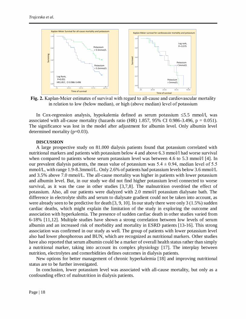
In the follow-up period 53 (27%) patients died, of those 38 (72%) were cardio-vascular deaths
(11 cerebrovascular deaths, 6 acute myocardial infarctions, 3 patients with sudden cardiac death).
The same number of patients had sepsis or cirrhosis as a cause of death (3), one died of malignancy
and five were diagnosed with malnutrition. In the high potassium level group, 22 (22.4%) patients
died and in the low potassium group 31 (30.7%). When patient’s all-cause mortality and survival
were analyzed in relation to high or low potassium level, the difference was borderline significant
(34.30 0.71 months vs. 33.7 1.16 months, p= 0.051). The number of cardiovascular deaths was
similar in both potassium groups [19 (18.8%) vs. 19 (19.3%), p>0.05, respectively], and there was
no significant difference in survival (p=0.575), as shown in Figure 2.
Trajceska et al.
Page | 18
Fig. 2. Kaplan-Meier estimates of survival with regard to all-cause and cardiovascular mortality
in relation to low (below median), or high (above median) level of potassium
In Cox-regression analysis, hypokalemia defined as serum potassium 5.5 mmol/l, was
associated with all-cause mortality (hazards ratio (HR) 1.857, 95% CI 0.986-3.496, p = 0.051).
The significance was lost in the model after adjustment for albumin level. Only albumin level
determined mortality (p=0.03).
DISCUSSION
A large prospective study on 81.000 dialysis patients found that potassium correlated with
nutritional markers and patients with potassium below 4 and above 6.3 mmol/l had worse survival
when compared to patients whose serum potassium level was between 4.6 to 5.3 mmol/l [4]. In
our prevalent dialysis patients, the mean value of potassium was 5.4 ± 0.94, median level of 5.5
mmol/L, with range 1.9-8.3mmol/L. Only 2.6% of patients had potassium levels below 3.6 mmol/L
and 3.5% above 7.0 mmol/L. The all-cause mortality was higher in patients with lower potassium
and albumin level. But, in our study we did not find higher potassium level connected to worse
survival, as it was the case in other studies [3,7,8]. The malnutrition overdried the effect of
potassium. Also, all our patients were dialyzed with 2.0 mmol/l potassium dialysate bath. The
difference in electrolyte shifts and serum to dialysate gradient could not be taken into account, as
were already seen to be predictive for death [3, 9, 10]. In our study there were only 3 (1.5%) sudden
cardiac deaths, which might explain the limitation of the study in exploring the outcome and
association with hyperkalemia. The presence of sudden cardiac death in other studies varied from
6-18% [11,12]. Multiple studies have shown a strong correlation between low levels of serum
albumin and an increased risk of morbidity and mortality in ESRD patients [13-16]. This strong
association was confirmed in our study as well. The group of patients with lower potassium level
also had lower phosphorous and BUN, which are recognized as nutritional markers. Other studies
have also reported that serum albumin could be a marker of overall health status rather than simply
a nutritional marker, taking into account its complex physiology [17]. The interplay between
nutrition, electrolytes and comorbidities defines outcomes in dialysis patients.
New options for better management of chronic hyperkalemia [18] and improving nutritional
status are to be further investigated.
In conclusion, lower potassium level was associated with all-cause mortality, but only as a
confounding effect of malnutrition in dialysis patients.
Time of survival
Surv
ivin
g
Kaplan-Meier Survival for all-cause mortality and potassium
Potassium> 5.5mmol/L
Potassium < 5.5mmol/L
Log Rank,P=0.051
HR1.857, CI 0.986-3.496
Potassium< 5mmol/L
Potassium> 5mmol/L
Log rank,P= 0.575
Kaplan-Meier survival for cardiovascular mortality and potassium
Time of survival
Surv
ive
d
Lower serum potassium level is associated with mortality as confounding effect of malnutrition in dialysis patients
Page | 19
REFERENCES
1. Choi Y. Hoon and Ha K. Sung. Potassium balances in maintenance hemodialysis.
Electrolyte Blood Press. 2013;11:9–16
2. Karaboyas A, Zee J, Brunelli S, Usvyat L, Weiner D, Maddux F, et al. Dialysate Potassium,
Serum potassium, mortality and arrhythmia events in hemodialysis: results from the
dialysis outcomes and practice patterns study. Am J Kidney Dis. 2017;69:266–77.
3. Brunelli MS, Spiegel MD, Mond DC, Oestereicher N, Winkelmayer W and Kovesdy C.
Serum to dialysate potassium gradient and its associations with short term outcomes in
hemodialysis patients. Nephrol Dial Transplant. 2018;33:1207–14.
4. Kovesdy PC, Regidor LD, Mehrotra R, Jing J, McAllister JC, Greenlad S, et al. Serum and
Dialysate Potassium Concentrations and survival in hemodialysis patients. Clin J am Soc
Nephrol. 2007;2:999–1007.
5. Axelsson TG, Heimburger O, Stenvinkel P, et al. Serum albumin level as predictor of
nutritional status in patients with ESRD. Clin J Am Soc Nephrol. 2012;7:1446–53.
6. de Musterd R, Grootendorst DC, Indemans F, et al. Association between serum albumin
and mortality in dialysis patients is partly explained by inflammation and not by
malnutrition. J Ren Nutr. 2009;19:127–35.
7. Yusuf A, Hu Y, Singh B, Menoyo H, Wetmore J. Serum potassium levels and mortality in
hemodialysis patients: a retrospective cohort study. Am j Nephrol. 2016;44:179–86.
8. Pun PP and Middleton PJ. Dialysate potassium, dialysate magnesium and hemodialysis
risk. J Am Soc Nephrol. 2017; 28:3441–51.
9. Silva B, Freitas BB G, Silva V, Abensur H, Luders C, Pereira JB et al. Hemodynamic
behavior during hemodialysis: Effects of dialysate concentrations of bicarbonate and
potassium. Kidney Blood Press Res. 2014;39:490–96.
10. Agar UB, Culleton FB, Fluck R, Leypoldt KJ. Potassium kinetics during hemodialysis.
Hemodial Int. 2014;19(1):23–32.
11. Vazqusez E, Sanchez PC, Garcia GG, et al. Sudden death in Incident Dialysis patients. Am
J Nephrol. 2014;39:331–36.
12. Herzog C. Can we prevent sudden cardiac death in dialysis patients? Clin j Am Soc
Nephrol. 2007;2:410–12.
13. Anderson CF, Wochos DN. The utility of serum albumin values in the nutritional
assessment of hospitalized patients. Mayo Clin Proc. 1982;57:181–84.
14. Reinhardt GF, Myscofski JW, Wilkens DB, Dobrin PB, Mangan JE, Jr, Stannard RT.
Incidence and mortality of hypoalbuminemic patients in hospitalized veterans. JPEN J
Parenter Enteral Nutr. 1980;4:357–59.
15. Apelgren KN, Rombeau JL, Twomey PL, Miller RA. Comparison of nutritional indices
and outcome in critically ill patients. Crit Care Med. 1982;10:305–7.
16. Kaw M, Sekas G. Long-term follow-up of consequences of percutaneous endoscopic
gastrostomy (PEG) tubes in nursing home patients. Dig Dis Sci. 1994;39:738–43
17. Ikizler TA. The Use and Misuse of Serum Albumin as a Nutritional Marker in Kidney
Disease. Clin J Am Soc Nephrol. 2012;7(9):1375–7.
18. Fried L, Kovesdy C and Palmer B. New options for the management of chronic
hyperkalemia. Kidney Int Suppl. 2017;7:164–70.

Acta morphol.2019;Vol.16(1):20–28
UDC: 616.711.5/.6-001.5
Page | 20
ORIGINAL ARTICLE
IMPLEMENTATION AND VALIDATION OF THE NEW AO CLASSIFICATION OF
THORACOLUMBAR FRACTURES
Ljupco Nikolov Zan Mitrev Clinic, Skopje, Republic of North Macedonia
ABSTRACT
Introduction: Development of a widely accepted, comprehensive and yet simple TL
classification system with clinically acceptable intra- and interobserver reliability for use in
clinical practice and researches, is necessity. AO Spine- thoracolumbar spine injury classification
system was developed using international consensus process, making it a simple and reproducible
system which consists of a morphologic classification of the fracture, a grading system for the
neurological status, and description of relevant patient-specific modifiers.
Objective: The aims of this study were to demonstrate the AO Spine thoracolumbar spine
injury classification system can be reliably applied by a group of surgeons and to detect those
injury types which are difficult for spine surgeons to classify reliably.
Materials and methods: AO Spine Thoracolumbar Spine Injury Classification system
consists of a morphologic classification of the fracture, a grading system for the neurologic status
and relevant patient-specific modifiers was applied to 40 cases by 6 spinal surgeons from
Traumatology department, twice independently, in grading sessions 1 month apart. The results
were analyzed for classification reliability using the Kappa coefficient (κ).
Results: Kappa coefficient for all 40 cases was 0.59, which represents moderate reliability.
For type A injuries, Kappa values describing interobserver agreement were 0.71, for type B injuries
0.50 and 0.61 for type C injuries. All representing substantial reliability. Fracture subtype A4 was
with the lowest level of agreement (Kappa = 0.19). Interobserver analysis demonstrated overall
average Kappa statistic for subtype grading of 0.55 also representing moderate reproducibility.
Conclusion: Study demonstrated moderate interobserver and substantial interobserver
reliability. These results suggest that most spine surgeons can reliably apply this system (AOSpine
Thoracolumbar Spine Injury Classification System) to spine trauma patients as more reliably than
previously described systems
Keywords: Implementation, Validation, AO Spine TLSIS.
INTRODUCTION Fracture of the thoracolumbar segment of the spine is a fracture of the vertebrae, in the segment
of the hollow part of the spine in height between the tenth bony vertebrae and the second lumbar
vertebrae. It occurs as a result of the force effect along the axial axis of the spinal column and
flexion. The most common cause of injury to this segment is a drop-in height, traffic accident, in
the multi and polytrauma. This type of vertebrae fractures account for about 45 per cent of all
fracture fractures [1-7]. But despite their frequency, there is still a significant dilemma regarding
the choice of treatment, conservative versus surgical treatment.
Implementation and validation of the new AO classification of thoracolumbar fractures
Page | 21
These controversies are supported by studies in which, some prefer the advantage of surgical
treatment that emphasizes patient satisfaction in terms of pain relief, better and faster rehabilitation
and better functional outcomes, as well as socioeconomic advantage. But there are also studies that
demonstrate the advantage of conservative treatment with good functional results and a small
percentage of morbidity [8-14, 19-24]. The development of a widely accepted, comprehensive and
simple classification system of thoracolumbar spinal fractures with clinically acceptable intra-and
interobserver reliability for use in clinical practice and research is a necessity. Megrel’s
classification and thoracolumbar injury classification (TLICS) are well-known, but the TL
classification system did not achieve a universal and international application [15-18]. The lack of
consensus among doctors / researchers complicates studies on these injuries and the development
of algorithms for their treatment. The AO Spine-thoracolumbar fractures classification system (AO
Spine TL) has been developed using an international consensus, making it a simple and
reproducible system consisting of a morphological classification of the fracture, a neurological
assessment system and a description of the relevant specific patient modifiers [25].
Objectives
The purpose of this research is to investigate whether the classification system of
thoracolumbar fractures on the spine TL spine TL can be reliably applied by surgeons at the
Traumatology Clinic and to detect those types of injuries that are difficult for surgeons to classify
them reliably.
MATERIALS AND METHODS
The study is intended to be an internal observational study of reliability and applicability, as
well as evaluating the reproducibility of the AO Spine TL classification. The research is
retrospectively prospectively, non-randomized, to a controlled group of patients before and after
injury, by the researcher and 6 surgeons from the Traumatology Clinic twice a month, individually
independently of each other. The survey was conducted at the Clinic for Traumatology and
includes 40 patients with fractures of the thoracolumbar segment of the spinal column, in the period
from January 2015 to September 2018. The reliability and applicability of Spine TL classification
will be a labeled using the Kappa Fliesh coefficient (according to inclusion and exclusion criteria,
patients who will meet the same criteria will be included or excluded in the research. The data was
entered in an electronic database and analyzed with software for statistical analysis.
RESULTS AND DISCUSSION
The researcher believes that the results will demonstrate that the Spine TL classification is a
reliable and usable in our conditions. It generates information that provides an excellent basis for
forming a decision-making pathway for the proper treatment of these injuries, as well as our
compliance with the appropriate treatment choice.
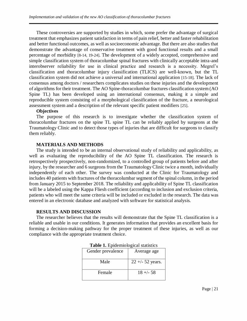
Table 1. Epidemiological statistics
Gender prevalence Average age
Male 22 +/- 52 years.
Female 18 +/- 58

Nikolov Ljupco
Page | 22
The light prevalence of male sex in relation to the female sex. The average age in both groups
shows 52 years for males and 58 for females (Table 1).
Table 2. Mechanism of injury
Direct fall 10 patients
Fall from height 15 patients
Car accident 15 patients
According to the mechanism of injury, fall from height and traffic accidents account for 75% of
the causes of these injuries (Table 2).
Table 3. Neurological status
Intact Deficit
34 patients 6 patients
In 6 patients clinically confirmed the presence of some neurological deficit (Table 3).
Table 4. Radiological investigations of RTG, CT, MRI,
Time of Investigation RTG C. Tomography M. Resonance
<24 hours 40 patients 31 patients 7 patients
<48 hours / 9 patients 15 patients
<72 hours / / 18 patients
Complete radiological investigation, (RTG, CT and MRI), in conditions of our clinic, necessary
for the application of AO Spine classification of thoracolumbar fractures in all 40 patients, was
done during the first 72 hours of the hospitalization (Table 4).
Results of AO Spine classification of thoracolumbar fractures analyzed with Fleiss Kappa
coefficient for intra and inter observing reliability (Table 5).
The agreement, or degree of compliance, can be considered as follows: if a certain number of
people assign numerical values to a certain number of references, then the Kappa coefficient will
give value for how consistent they are. The cap coefficient can be defined as a binary / dichotomous
agreement from the same sample between two observers. The latter represents a measure / value
of an agreement between partners (independent assessors) for categorical (nominal or scale) scales
when there is a minimum of two assessors in which the data from the response are related to the
same observation.
The value of 1 indicates the perfect deal and 0 indicates that there is no contract, or is no better
than what can be obtained by chance.
Implementation and validation of the new AO classification of thoracolumbar fractures
Page | 23
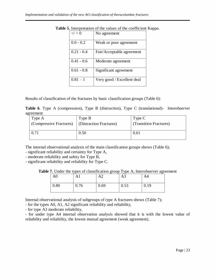
Table 5. Interpretation of the values of the coefficient Kappa.
</ = 0 No agreement
0.0 - 0.2 Weak or poor agreement
0.21 - 0.4 Fair/Acceptable agreement
0.41 - 0.6 Moderate agreement
0.61 - 0.8 Significant agreement
0.81 – 1 Very good / Excellent deal
Results of classification of the fractures by basic classification groups (Table 6):
Table 6. Type A (compression), Type B (distraction), Type C (translational)- Interobserver
agreement
Type A
(Compressive Fractures)
Type B
(Distraction Fractures)
Type C
(Transition Fractures)
0.71 0.50 0.61
The internal observational analysis of the main classification groups shows (Table 6);
- significant reliability and certainty for Type A,
- moderate reliability and safety for Type B,
- significant reliability and reliability for Type C.
Table 7. Under the types of classification group Type A; Interobserver agreement
A0 A1 A2 A3 A4
0.80 0.76 0.69 0.53 0.19
Internal observational analysis of subgroups of type A fractures shows (Table 7);
- for the types A0, A1, A2 significant reliability and reliability,
- for type A3 moderate reliability,
- for under type A4 internal observation analysis showed that it is with the lowest value of
reliability and reliability, the lowest mutual agreement (weak agreement).

Nikolov Ljupco
Page | 24
Table 8. Under the types of classification group Type B; Interobserver agreement
B1 B2 B3
0.61 0.27 0.59
Internal observational analysis of subgroups of type B fractures shows (Table 8);
-for the modes B1 and B3, reliability and reliability,
-for type B2 internal observation analysis showed that it is the lowest value of the coef. Kappa
0,25 with acceptable reliability and security (acceptable agreement).
The comparative internal observation analysis of the subtypes showed the following results;
Table 9. Subtypes Compatibility
A4 B2
0.19 0.25
The A4 and B2 are the lowest values, the lowest mutual agreement (Table 9).
For example, case no. 11 (P.B. 56 yrs., female, fall from height)The question is what type of
fracture is seen on the x-ray.Type A or Type B? Under type A3, A4? Sub type B2 is associated
with A3, A4? (Image 1)
Fig. 1. RTG of thoracolumbar spine
Implementation and validation of the new AO classification of thoracolumbar fractures
Page | 25
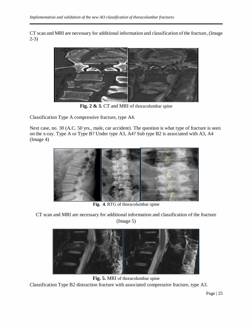
CT scan and MRI are necessary for additional information and classification of the fracture, (Image
2-3)
Fig. 2 & 3. CT and MRI of thoracolumbar spine
Classification Type A compressive fracture, type A4.
Next case, no. 30 (A.C. 50 yrs., male, car accident). The question is what type of fracture is seen
on the x-ray. Type A or Type B? Under type A3, A4? Sub type B2 is associated with A3, A4
(Image 4)
Fig. 4. RTG of thoracolumbar spine
CT scan and MRI are necessary for additional information and classification of the fracture
(Image 5)
Fig. 5. MRI of thoracolumbar spine
Classification Type B2 distraction fracture with associated compressive fracture, type A3.

Nikolov Ljupco
Page | 26
CONCLUSION
Summarized results of an internal observational study of reliability and implementation of the
new AO classification of thoracolumbar fractures; - Kappa coefficient (κ) for all 40 cases is 0.59, which is a moderate reliability.
- Kappa coefficient (κ), for type A injuries, is 0.71, which represents significant reliability and
safety,
- Kappa coefficient (κ), for type B injuries is 0.50, and which represents moderate reliability and
safety,
- Kappa coefficient (κ), for injuries of type C is 0.61, and which represents moderate reliability
and safety,
- Kappa coefficient (κ), for injuries under type A4 is 0,19, and is with the lowest level of agreement
- Internal observational analysis showed the total average value of (κ). for the subtypes of 0.55,
which represents moderate reliability and reproducibility.
Having in itself all the necessary data from the analysis of the morphological characteristics of
the fracture, the neurological status of the injured, specific modifiers for the patient, the results of
this paper show that the new AO classification of thoracolumbar fractures gives a sufficiently large
number of data, making it useful, reliable and safe for use in the daily work of the Traumatology
Clinic.
It generates information that provides an excellent basis for forming a decision-making
pathway for the proper treatment of these injuries, as well as our compliance with the appropriate
treatment choice [27].
REFERENCES
1. Vaccaro AR, Schroeder GD, et al. The surgical algorithm for the AOSpine thoracolumbar
spine injury classification system. Eur Spine J. 2016;25(4):1087–94.
2. Kaul R, Chhabra HS, Vaccaro AR, et al. Reliability assessment of AOSpine thoracolumbar
spine injury classification system and Thoracolumbar Injury Classification and Severity
Score (TLICS) for thoracolumbar spine injuries: results of a multicentre study. Eur Spine
J. 2017;26(5):1470–476.
3. Cheng J, Liu P, Sun D, Qin T, Ma Z, Liu J. Reliability and reproducibility analysis of the
AOSpine thoracolumbar spine injury classification system by Chinese spinal surgeons. Eur
Spine J. 2017;26(5):1477–1482.
4. Schroeder GD, Harrop JS, Vaccaro AR. Thoracolumbar Trauma Classification. Neurosurg
Clin N Am. 2017;28(1):23–29.
5. Спинални повреди (Јордан Савески, 2006).
6. Vaccaro AR, Lehman RA Jr, Hurlbert RJ, et al. A new classification of thoracolumbar
injuries: the importance of injury morphology, the integrity of the posterior ligamentous
complex, and neurologic status. Spine. 2005;30(20):2325–33.

Implementation and validation of the new AO classification of thoracolumbar fractures
Page | 27
7. Bakhsheshian J, Dahdaleh NS, Fakurnejad S, Scheer JK, Smith ZA. Evidence-based
management of traumatic thoracolumbar burst fractures: a systematic review of
nonoperative management. Neurosurg Focus. 2014;37(1):E1.
8. Hitchon PW, Abode-Iyamah K, Dahdaleh NS, et al. Nonoperative Management in
Neurologically Intact Thoracolumbar Burst Fractures: Clinical and Radiographic
Outcomes. Spine (Phila Pa 1976). 2016;41(6):483–9.
9. Thomas KC, Bailey CS, Dvorak MF, Kwon B, Fisher C. Comparison of operative and
nonoperative treatment for thoracolumbar burst fractures in patients without neurological
deficit: a systematic review. J Neurosurg Spine. 2006;4(5):351–8.
10. Avilés C, Flores S, Molina M. Conservative versus operative treatment for thoracolumbar
burst fractures without neurologic deficit. Medwave. 2016;16(Suppl 1):e6383.
11. Milicić A, Jovanović A, Milankov M, Savić D. Operative versus conservative treatment of
the fractures or dislocations of the thoracolumbar spine associated with neurological
deficiency. Srp Arh Celok Lek. 1994;122(1-2):22–4.
12. Fang D, Leong JC, Cheung HC. The treatment of thoracolumbar spinal injuries with paresis
by conservative versus surgical methods. Ann Acad Med Singapore. 1982;11(2):203–6.
13. Yi L1, Jingping B, Gele J, Baoleri X, Taixiang W. Operative versus non-operative
treatment for thoracolumbar burst fractures without neurological deficit. Cochrane
Database Syst Rev. 2006;18(4): CD005079.
14. Bernhardt M, Swartz D, Clothiaux P. Pedicular screw instrumentation for unstable
thoracolumbar fractures. Clinorthop. 1992;284:109–15.
15. Park H, Lee S, Park N, et al. Modified thoracolumbar injury classification and severity
score (TLICS) and its clinical usefulness. Acta Radiol. 2015;57:74–81.
16. Patel C. Evaluation and treatment of thoracolumbar junction trauma. UPOJ 2002;15:7–12.
17. Saunders, Tomycz N, Okonkwo D. Richard H, editor. Diagnosis and management of
thoracic spine fractures. Youmans Neurological Surgery Textbook 6th ed. 2011
18. Rechtine G, Cahill D, Chrin A. Treatment of thoracolumbar trauma: comparison of
complications of operative versus nonoperative treatment. J Spinal Disord. 1999;12:406–
9.
19. Shen W, Liu T, Shen Y. Nonoperative treatment versus posterior fixation for
thoracolumbar junction burst fractures without neurologic deficit. Spine. 2001;26:1038–
45.
20. Denis F. The three column spine and its significance in the classification of acute
thoracolumbar spinal injuries. Spine. 1976;8:817–31.
21. Yi L, Jingping B, Gele J, et al. Operative versus non-operative treatment for thoracolumbar
burst fractures without neurological deficit. Cochrane Database Syst Rev.
2006;18:CD005079.
22. Bakhsheshian J, Dahdaleh N, Fakurnejad S, et al. Evidence-based management of
traumatic thoracolumbar burst fractures: a systematic review of nonoperative management.
Neurosurg Focus. 2014;37:E1.
23. Siebenga J, Leferink V, Segers M, et al. Treatment of traumatic thoracolumbar spine
fractures: a multicenter prospective randomized study of operative versus nonsurgical
treatment. Spine. 2006;31:2881–90.

Nikolov Ljupco
Page | 28
24. Gnanenthiran S, Adie S, Harris I. Nonoperative versus operative treatment for
thoracolumbar burst fractures without neurologic deficit: a meta-analysis. Clin Orthop
Relat Res. 2012;470:567–77.
25. Patel AA, Vaccaro AR, Albert TJ, et al. The adoption of a new classification system: time-
dependent variation in interobserver reliability of the thoracolumbar injury severity score
classification system. Spine. 2007;32:105–10.
26. Jansson KA, Blomqvist P, Svedmark P, et al. Thoracolumbar vertebral fractures in
Sweden: an analysis of 13,496 patients admitted to hospital. Eur J Epidemiol. 2010;25:431-
437.
27. Winklhofer S, Thekkumthala-Sommer M, Schmidt D, et al. Magnetic resonance imaging
frequently changes classification of acute traumatic thoracolumbar spine injuries. Skeletal
Radiol. 2013;42:779-786.

Acta morphol.2019;Vol.16(1):29–34
UDC: 616.314
Page | 29
ORIGINAL ARTICLE
VERTICAL IRREGULARITIES INFLUENCE OVER THE SIZE, FORM AND SHAPE
OF THE SYMPHYSIS
Bogdanovska Biljana1, Pop Stefanova-Trposka M2, Gavrilovic I1, Curcieva-Cuckova G1,
Bogdanovski S3
1Faculty of Dentistry, Department of Orthodontics, Ss Cyril and Methodius University of Skopje,
Skopje, Republic of North Macedonia
2Faculty of Dentistry, European University, Skopje, Republic of North Macedonia 3Faculty of Dentistry, Department of Prosthodontic, Ss Cyril and Methodius”University, Skopje,
Republic of North Macedonia
ABSTRACT
Modern orthodontics is a creation for the best possible balance between occlusal relations,
dental and facial esthetics, result stability and their maintenance as well as teeth restoration.
Regular or irregular vertical development of the facial skeleton is connected to multiple skeletal
groups: nasomaxillary complex, alveolar processes and mandible. According to Beckmann, a
significant correlation exists between the incisal rates and maxillary and mandibular dentoalveolar
height, symphysis size, maxillary and mandibular surface. He concluded that long face cases had
longer mandibular alveolar height, which was connected to a tight form instead of to the increased
symphysis volume. Also, a connection exists between the size of the mandibular symphysis, the
chin and the vertical dimension and morphological and dentoalveolar structure of both jaw
systems. Determination of this connection can be useful in predicting the treatment success in
overbite problems Haskel. The goal of our study is to show the symphysis shape, size and form in
respondent groups. Depending on the vertical incisal rate characteristics - overbite, the respondents
were divided in three groups: first group was consisted of respondents with open bite, meaning the
overbite smaller or equal to -1 mm, the second group was consisted respondents with deep bite,
meaning the overbite is over +4 mm, and the third control group consisted of respondents with
normal overlap, meaning the overbite is more than +1 mm, but lower or equal to +4 mm. Height
of symphysis in respondents with open bite was largest, and smallest in respondents with deep
bite, compared to the control group. Depth of symphysis is the largest in the deep bite group, and
the smallest in the open bite group.
The data obtained for symphysis height and depth can be used to anticipate treatment success
in open and deep bites.
Key words: overbite, deep bite, height of symphysis, depth of symphysis.
INTRODUCTION
Examining the factors that influence facial harmony and disharmony, it is proven that facial
components are inherited regardless of one another, and not as a complex that leads to different
facial configuration creations. The facial configuration and facial expression depend primarily on
the constitutional build of the skeleton, facial bones position and alignment, the upper and lower
jaw position, bite type, soft-tissue components covering the facial base, as well as nose, lip and
chin size.
Bogdanovska et al.
Page | 30
Modern orthodontics goal represents the best possible balance between occlusal relations,
dental and facial esthetics, result stability and their maintenance as well as teeth restoration [1].
Regular or irregular vertical development of the facial skeleton is connected to multiple skeletal
groups: nasomaxillary complex, alveolar processes and mandible. There is connection between the
structure of the front part of the maxilla and mandible and the lower part of the face, so in the case
of open or deep bite, the dentoalveolar development can be insufficient to compensate the
oversized or undersized detachment of the jaw system. According to Beckmann et al. [2, 3] a
significant correlation exists between the incisal rates and maxillary and mandibular dentoalveolar
height, symphysis size, maxillary and mandibular surface. The authors concluded that long face
cases had longer mandibular alveolar height, which was connected to a tight form instead of to the
increased symphysis volume. They also analyzed the contribution of the alveolar process structures
and the basal bone in relation to lower face height and ascertained that longer lower face was
connected to a larger maxillary and mandibular alveolar process area and basal bone, and that a
lower face was connected to a lower maxillary and mandibular frontal alveolar process area and
basal bone. Beckmann et al. [2, 3] conducted measurements of 460 cephalograms of untreated
patients and proved that the cases with short facial structure had a lower area and wider and shorter
symphysis form. Even though cephalograms were two-dimensional, they showed that symphysis
volume was smaller in cases with open bites and larger in cases with deep bites. Open bites could
cause symphysis enlargement and elongation as well as shortening their form [4].According
Haskel [5, 6], a connection exists between the size of the mandibular symphysis, the chin, the
overbite and morphological and dentoalveolar structure of both jaw systems. Sassouni [7] and
Schudy [8] designated two different types of face forms in literature known as: skeletal open bites
or hyperdivergent and skeletal deep bites or hypodivergent face type. Determination of this
connection can be useful in predicting the treatment success in overbite problems [9].
The goal of our study is to show the symphysis shape, size and form in groups with open
and deep bite.
MATERIALS AND METHOD
For the realization of the set goal, examinations were conducted on 90 individuals from both
sexes, aged 13-15, randomly chosen from the Clinic of orthodontics at PHO – Dental Clinical
Centre “St. Pantelejmon” in Skopje.
Selecting the respondents taking part in realizing the set goal was based on the following
criteria: individuals that had not previously underwent orthodontic treatment, with no great
craniofacial disorders and with complete dentition.
In relation to the characteristics of the vertical incisal rate, the respondents were divided in
three groups and classified as:
The first group consisted of respondents with open bite, where the vertical incisal
rate is lower or equal to -1 mm,
The second consisted of respondents with deep bite, where the vertical incisal rate
is over +4 mm, and
The third group consisted of respondents with normal incisal overlap, where the
vertical incisal rate is more than +1 mm, but lower or equal to +4 mm. This group was also the
control group.
Every group consisted of 30 respondents, 15 female and 15 male, that came in the period from
2009-2015.
Vertical irregularities influence over the size, form and shape of the symphysis
Page | 31
In the respondents from the research groups, standardized clinical and diagnostic procedures
were conducted with x-ray cranial imaging in a standardized way in norma lateralis.
The linear parameters used in the research are:
Symphysis height (SH mm). The distance between the points Infradentale and
Menton, (highest point of the alveolar point on the mandible and the point where the shadow of
the mandible base and the shadow of the chin profile meet)
Symphysis depth (SD mm). The distance between the most prominent point of the
chin profile (Pogonion) and the most prominent point of the symphysis posterior wall.
The statistical data analysis was conducted in SPSS for Windows 17,0 program.
For data testing we used Shapiro-Wilk`s W test.
For data depiction descriptive statistics was used.
For comparison of the analyzed parameters between the three analyzed groups, we
used One way Anova, and for inter-group differences we used Tukey test.
For comparison of the analyzed parameters in relation to gender the Student "t" test
was used.
The levels of probability for achieving null hypothesis, concordant to international
standards for bio-medical sciences were 0.05 and 0.01
RESULTS
Average value of the symphysis height parameter SH in the three analyzed groups (open,
deep and normal bite) is 33,9±3,2mm, 25,7±3,8mm and 29,65±2,0mm respectably. The tested
difference in average symphysis height between the three analyzed respondent groups is
significant. (F=35,2 p<0,01). Intergroup difference shows that this parameters have significantly
different average values between the three comparison pairs: open bite to deep and normal bite, as
well as deep bite to normal bite. (Table 1)
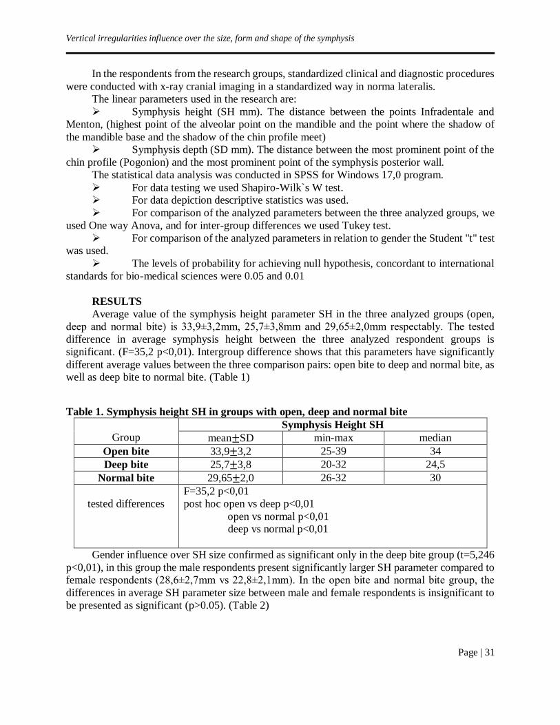
Table 1. Symphysis height SH in groups with open, deep and normal bite
Group
Symphysis Height SH
mean±SD min-max median
Open bite 33,9±3,2 25-39 34
Deep bite 25,7±3,8 20-32 24,5
Normal bite 29,65±2,0 26-32 30
tested differences
F=35,2 p<0,01
post hoc open vs deep p<0,01
open vs normal p<0,01
deep vs normal p<0,01
Gender influence over SH size confirmed as significant only in the deep bite group (t=5,246
p<0,01), in this group the male respondents present significantly larger SH parameter compared to
female respondents (28,6±2,7mm vs 22,8±2,1mm). In the open bite and normal bite group, the
differences in average SH parameter size between male and female respondents is insignificant to
be presented as significant (p>0.05). (Table 2)

Bogdanovska et al.
Page | 32
Table 2. Sex differences for symphysis height SH in groups with open, deep and normal
bite
Group
Sex Symphysis Height SH tested differences
mean±SD min-max median
Open bite Male 35,1±2,4 30-39 35 t=1,794
p>0,05 ns Female 32,7±3,5 25-37 33,5
Deep bite Male 28,6±2,7 23-32 30 t=5,246
p<0,01 sig Female 22,8±2,1 20-27 23
Normal bite Male 30,4±1,5 28-32 30,5 t=1,761 p>0,05 ns Female 28,9±2,2 26-32 28
Statistics analysis shows that for the value F=48,2 (p<0,01), a significant difference exists in
average parameter size for symphysis depth SD between the three analyzed groups. The significant
difference is based on the considerably shorter average parameter length in the open bite group,
compared to the deep bite group (12,75±1,3mm vs 17,1±1,5mm), as well as compared to the
control group (12,75±1,3mm vs 14,3±1,5mm), furthermore a considerably shorter average
parameter length is present in the control group compared to the deep bite group (14,3±1,5mm vs
17,1±1,5mm). (Table 3)
Table 3. Symphysis depth SD in groups with open, deep and normal bite
Group Symphysis Depth SD
mean±SD min-max median
Open bite 12,75±1,3 10-15 13
Deep bite 17,1±1,5 14-19 17
Normal bite 14,3±1,5 12-17 14
tested differences
F=48,2 p<0,01
post hoc open vs deep p<0,01
open vs normal p<0,01
deep vs normal p<0,01
Average symphysis depth in male and female respondents in the open bite groups is 12,8±1,0mm
and 12,7±1,6mm respectably, in the deep bite group it is 17,2mm±1,6mm and 17,05±1,5mm
respectfully, and in the control group it is 14,6mm±1,5mm i 14,6±1,5mm respectfully. Average
symphysis depth parameter size is not significantly dependent on the gender in none of the
analyzed groups. (Table 4)
Table 4. Sex differences for symphysis depth SD in groups with open, deep and normal bite
Group
Sex Symphysis Depth SD tested differences
mean±SD min-max median
Open bite Male 12,8±1,0 10-14 13 t=0,168 p>0,05 ns Female 12,7±1,6 10-15 12,5
Deep bite Male 17,2±1,6 14-19 17 t=0,219
p>0,05 ns Female 17,05±1,5 15-19 17,5
Normal bite Male 14,6±1,5 12-17 14,5 t=0,896
p>0,05 ns Female 14±1,5 12-17 14
Vertical irregularities influence over the size, form and shape of the symphysis
Page | 33
DISCUSSION
The tested difference in average symphysis height between the three analyzed groups is
significant. Our results correspond to the results of Beckmann [2, 3] and Ceylan [10, 11]. In the
open bite and normal bite group, the difference in average SH parameter size is insignificant to be
presented as significant (p>0.05). Gender influence over SH size confirmed as significant only in
the deep bite group (t=5,246 p<0,01), in this group the male respondents presented significantly
larger SH parameter compared to female respondents (28,6±2,7mm vs 22,8±2,1mm).
In studies done by Backmann [2, 3], it was postulated that symphysis size was determined by
a characteristic factor deduced form the factors that control lower face height. The studies clearly
show that symphysis height is connected to the vertical incisal rate. The control factor can be a
genetic factor that determines the overlap.
Descriptive statistics for symphysis depth shows that the average values in the open bite group
is concordant to the Ceylan [10, 11] findings, however the deep bite average values are larger than
the findings in the same one. In all three respondent groups, a statistically significant difference in
gender does not exist.
CONCLUSION
Symphysis height in respondents with open bite was largest, and smallest in respondents with
deep bite, compared to the control group. SH analysis in relation to gender shows no statistically
significant difference in open and normal bite groups, however in the deep bite group is (p<0.01),
meaning that height of symphysis is larger in male respondents compared to female respondents.
Symphysis depth is the largest in the deep bite group, and the smallest in the open bite group.
No differences were noticed in relation to the gender in all three respondent groups (p>0.05).
The data obtained for symphysis height and depth can be used to anticipate treatment success
in open and deep bites.
REFERENCES
1. Proffit WR, Fields H. Contemporary orthodontics. Mosby 5-th ed. 2012.
2. Beckmann S H, Kuitert RB, Prahl-Andersen B, Segner D, Tuinzing DB. Alveolar and
skeletal dimensions associated with overbite. Am J Orthod 1998; 113:443–52.
3. Beckmann SH, Kuitert RB, Tuinzing DB. Alveolar and skeletal dimensions associated with
lower face height. Am J Orthod 1998; 113:498–506.
4. Betzenberger D, Ruf S, Pancherz H. The compensatory mechanism in high-angle
malocclusions: a comparison of subjects in the mixed and permanent dentition. Angle
orthod 1999;69:27–32.
5. Haskell BS. The human chin and its relationship to mandibular morphology. Angle Orthod
1979; 49:153–66.
6. Haskell JA, Bruce S. Haskell BS, Michael E. Spoon ME, Feng C. The relationship of
vertical skeletofacial morphology to oropharingeal airway shape using cone beam
computer tomography: Possible implications for airway restriction. The Angle
Orthodontis. 2014; 84(3):548–554.
7. Sassouni V, Nanda S. Analysis of dentofacial vertical proportions. Am J Orthod. 1964;
50:801–23.
8. Schudy FF. The rotation of the mandible resulting from growth: its implications in
orthodontic treatment. Angle Orthod. 1965;35:36–50.

Bogdanovska et al.
Page | 34
9. Avrum J. Goldberg, R.G. Behrents, Donald R. Oliver and Peter H. Buschang. Facial
divergence and mandibular crowding in the treated subjects. The Angle Orthod. 2013;
83(3):381–388.
10. Ceylan I., Baidas B., Bolukbasi B. Longitudinal cephalometric changes in incisor position,
overjet and overbite between 10 and 14 years of age. Angle Orthod. 2002;72:246–50.
11. Ceylan I.,Eroz B. The effects of overbite on the maxillary and mandibular morphology.
Angle Orthod. 1999;70(2):110–15.

Acta morphol.2019;Vol.16(1):35–41
UDC: 618.18-089.844
Page | 35
CASE REPORT
PERINEO-VAGINAL RECONSTRUCTION WITH IPAP PROPELLER FLAP
A CASE REPORT
Igor Peev1, Zhogovska-Mirchevska E1, Alulovski I2, Krstevska-Dukova I2 1University Clinic for Plastic and Reconstructive surgery,
Medical Faculty, Skopje, Republic of North Macedonia 2University Clinic for Gynecology and Obstetrics,
Medical Faculty, University "St Cyril and Methodius", Skopje, Republic of North Macedonia
ABSTRACT
Background: Perineal defects occur due to infections, trauma or after tumor resection in the
region. There are many options for reconstructions, from spontaneous healing to free flaps.
However, usage of local flaps seems most reasonable and feasible. Since perineum bears several
orifices, this can lead to infection and reconstructive failure. That is why, these defects are really
challenging for plastic surgeons.
Case report: We present a case of reconstruction of vagino-perineal defect after resection of
proliferative vulvar planocellular carcinoma, by using internal pudental artery perforator flap
(iPAP) designed in a propeller fashion.
Conclusion: Propeller perforator flaps are novel reconstructive methods that can be used
successfully for perineal reconstruction, resulting in good functional and aesthetic outcome with
minimal donor site morbidity.
Key words: perineum, reconstruction, propeller, perforator, flap.
INTRODUCTION Perineo-genital defects can occur due to infections, trauma or mainly, after resection of tumors
in this region. There are many options for reconstruction of these defects, from spontaneous
closure, skin grafting, vacuum assisted closure, up to reconstruction with free flaps. Selection
depends on the size of the missing tissue, among others [1].
Perforator flaps are local flaps nourished by a known perforator vessel that is directly
skeletonized intraoperative or preoperatively detected with pencil Doppler, as a freestyle
perforator flap [2]. As we know from Taylor’s anatomic study, there are more than 350 perforators
in a human body [3]. Perforator flaps are safe and dependable methods in plastic surgery that
reconstruct alike tissue. When perforator flap is rotated around the vessel which is a pivot point of
rotation, it becomes propeller perforator flap [4]. The concept was born in 1991, by Hyakusoku,
who gave the term in order to describe an adipose-dermal flap that has been rotated for 90° around
a centrally – based subcutaneous perforator [5]. Hallock in 2006 used an eccentrically based
skeletonized perforator and managed to rotate the flap for 180° around it [6]. A clear definition of
propeller flap was given in 2009 by the advisory panel of the first Tokyo meeting on perforator
and propeller flaps, who defined it as an “island flap that reaches the recipient site through an axial
rotation” [7]. The difference between a propeller flap and other pedicled flaps is that movement
here is “axial”, meaning that the flaps turn around a pivot that is the pedicle in fact.
Peev et al.
Page | 36
According to the Gent consensus on perforator flaps, perforator propeller flaps should be
named after their nutrient vessels [8].
Perineal region has a rich vascularization arriving from branches of femoral and iliac arteries.
It comprises the skin boundaries of the perineum, divided in anterior and posterior part. There are
many perforators coming out from superficial circumflex iliac, superficial inferior epigastric,
superficial external pudendal, deep external pudendal, obturator, perineal and internal pudendal
artery. These perforators can be the basis for flap’s design [9]. Internal pudental artery perforators
(iPAP) are found in dense fatty tissue of ischiorectal fossa, identified as a vascular triangle
connecting three points: ischial tuberosity, the anus and lower vaginal orificium (lower scrotal
raphe). One perforator has a wide vascular territory and flaps with dimensions up to 20 cm length
can be harvested [10].
We present a case where bilateral iPAP propeller flaps were used in reconstruction of vulvo-
perineal defect after excision of massive vulvar carcinoma.
CASE REPORT
A 67 years old lady was referred to gynecologist for treatment of a proliferative neoplasm in
vulvar-perineal region.
Fig. 1. Local appereance of the vulvar-perineal carcinoma
It started to grow as a nodule on the labia majora many years ago. She had denied doctor’s
evaluation due to embarrassment. Lately, patient had difficulties when voiding, defecating and
walking and also, moderate pain was present. Biopsy was made, and planocellular carcinoma was
revealed. Ultrasonography examination of inguinal regions and CT imaging of pelvis, showed
enlarged lymph nodes in the right inquinum, with no progression in the minor pelvis. Colposcopy
examination found an infiltration of vaginal introitus and its distal portion. Treatment plan was
radical carcinoma removal and reconstruction of the defect that will follow. Bilateral groin
dissections were also planned to be done. Those were the reasons why plastic surgeon took place.
Reconstruction comprised bilateral freestyle iPAP propeller flaps.

Perineo-vaginal reconstruction with IPAP propeller flap – a case report
Page | 37
OPERATIVE TECNIQUE:
In general anesthesia, patient was set in a lithotomy position and Foley catheter was put. Using
pencil hand-held Doppler probe, perforators of internal pudental artery were found in the vascular
triangle of ishiorectal fossa. The upper most perforator was marked as a pivot point as the plan
was a propeller flap. The flap was marked in the gluteal fold.
Fig. 2. Preoperative markings for flap harvesting – detected perforators are doted
Gynecologist removed the carcinoma with resection of the distal part of the vagina and whole
perineum down to healthy tissue.
Fig. 3. Defect after resection of the carcinoma
After resection, reconstruction followed. Harvesting started from distally upwards, using 3x
magnification loops. In thick fatty tissue, with no fascia, it is difficult to skeletonize the perforator
totally. That is why a part of the tissue was left around the perforator, but small enough for a 100°
rotation to be made around. The flap was then rotated in vaginal direction and its distal part (now
upper) sutured to the remnant vagina and adjacent tissue.

Peev et al.
Page | 38
Fig. 4. Flaps’ elevation
After one side reconstruction, another iPAP was raised from the opposite side in the same way
and placed similarly as the previous one. In the midline of the perineum, both flaps were sutured
together.
Fig. 5. Final reconstruction with bilateral flaps
Penrose vacuum drains were introduced in donor sides, bilaterally. When reconstruction
finished, radical inquinal lymphadenectomy was made bilaterally, where vacuum drains were set
in as well.
After operation, among other therapy, antibiotics and blood components were administrated.
Low molecular weight heparin was included as patient was advised to rest several days restraining
eating solid food. Flaps had good color, adequate capillary refill and temperature with no vascular
suffering. Flap edema and imbibition were obvious at the beginning, but it settled down after
several days. Drains were removed on day 5 and patient discharged home on day 10. Healing was
uneventful regarding reconstruction. There was seroma collection in the right inquinim with some
lymphorrhea, which stopped after few aspirations.
Pathology sampling showed invasive plano - cellular carcinoma with moderate differentiation,
metastasis in two lymph nodes right, and in one lymph node left. As resection was very close to
tumor margins, patient was sent to radiologist where brachytherapy followed after 3 months post
op. Patient felt good with no progression of the tumor, neither locally nor regionally. Three years
after operations, she is still under follow-up. Reconstruction was sufficient with minimal donor
side morbidity, no restrictions in patient’s daily life and great satisfaction.
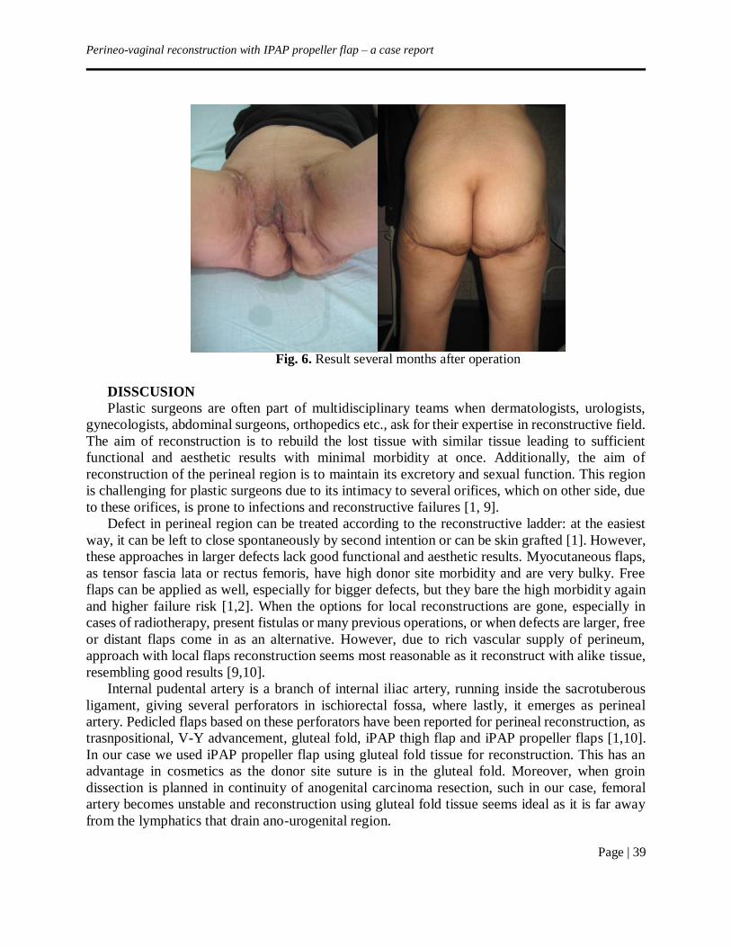
Perineo-vaginal reconstruction with IPAP propeller flap – a case report
Page | 39
Fig. 6. Result several months after operation
DISSCUSION
Plastic surgeons are often part of multidisciplinary teams when dermatologists, urologists,
gynecologists, abdominal surgeons, orthopedics etc., ask for their expertise in reconstructive field.
The aim of reconstruction is to rebuild the lost tissue with similar tissue leading to sufficient
functional and aesthetic results with minimal morbidity at once. Additionally, the aim of
reconstruction of the perineal region is to maintain its excretory and sexual function. This region
is challenging for plastic surgeons due to its intimacy to several orifices, which on other side, due
to these orifices, is prone to infections and reconstructive failures [1, 9].
Defect in perineal region can be treated according to the reconstructive ladder: at the easiest
way, it can be left to close spontaneously by second intention or can be skin grafted [1]. However,
these approaches in larger defects lack good functional and aesthetic results. Myocutaneous flaps,
as tensor fascia lata or rectus femoris, have high donor site morbidity and are very bulky. Free
flaps can be applied as well, especially for bigger defects, but they bare the high morbidity again
and higher failure risk [1,2]. When the options for local reconstructions are gone, especially in
cases of radiotherapy, present fistulas or many previous operations, or when defects are larger, free
or distant flaps come in as an alternative. However, due to rich vascular supply of perineum,
approach with local flaps reconstruction seems most reasonable as it reconstruct with alike tissue,
resembling good results [9,10].
Internal pudental artery is a branch of internal iliac artery, running inside the sacrotuberous
ligament, giving several perforators in ischiorectal fossa, where lastly, it emerges as perineal
artery. Pedicled flaps based on these perforators have been reported for perineal reconstruction, as
trasnpositional, V-Y advancement, gluteal fold, iPAP thigh flap and iPAP propeller flaps [1,10].
In our case we used iPAP propeller flap using gluteal fold tissue for reconstruction. This has an
advantage in cosmetics as the donor site suture is in the gluteal fold. Moreover, when groin
dissection is planned in continuity of anogenital carcinoma resection, such in our case, femoral
artery becomes unstable and reconstruction using gluteal fold tissue seems ideal as it is far away
from the lymphatics that drain ano-urogenital region.
Peev et al.
Page | 40
Additionally, being far away from subsequent irradiation, gluteal fold tissue can be utilized for
reconstruction of radiation –induced injuries. It can reconstruct voluminous tissue defects as in
obese patients, but also thinner flaps can be raised leaving smaller donor site scars. Axial rotation
can be done in many directions allowing coverage of different local defects, for example ischial
defects in pressure ulcers [1,9,10].
Perforators of the arteries can be indirect (myocutaneous, septocutaneous) and direct
perforators such the case of iPA perforators is [11]. Here, they are branched into the fatty tissue,
surrounded only with fat, without adjacent fascia, allowing a wider arc of rotation without much
skeletonization. Actually, total dissection of the perforator is not even recommended because the
perforator is situated only in fatty compartment allowing easy mobilization of the planned flap.
[10] As described by Mardini and Wei, preoperative Doppler signal mapping is sufficient for
perforator detection and with gentle dissection, freestyle perforator flap can be raised on it [12].
We applied the advices, marked the desired perforator detected with pencil Doppler, raised the
flap and axially rotated for 100°, good enough to reconstruct the defect without any suture tension.
It is necessary to detach any tissue resulting in pedicle kinking or twisting as these can lead to flap
ischemia.
Propeller flaps can have 90 – 180° angle of axial rotation without blood supply insufficiency.
They are ideal for small to moderate defects in different body parts: head and neck, upper and
lower extremities, trunk. Advantages for their utilization are: freedom in their design as a freestyle
perforator flaps regarding soft tissue volume needed; they are faster and simpler reconstructive
method comparing to free flaps; possibility to use local tissue that is more similar to the missing
one; easy and fast harvesting when adequate technique is used; the donor site morbidity is minimal.
Having in mind the above and technical refinements achieved lately, indications for perforator
propeller flap reconstructions are constantly increasing [4].
CONCLUSION
Perineo-genital defects are challenging for reconstruction. With rich vascularization in the
region, there are many reconstructive options. However, reconstruction with local similar tissue is
preferred method. Reconstruction with perforator flap is novel approach for local flap usage.
Reconstruction with iPAP flaps can be successful and great alternative for traditional or free flaps.
When being propeller, it can cover large defect, especially when used bilaterally, gaining the
possibility to reconstruct complex wound. On the other site, it is easy and fast to harvest with some
specific technical considerations. This flap has many advantages over traditional flaps such as
better functional, aesthetic results and low donor- site morbidity, among others.
REFERENCES
1. Mughal M, Baker RJ, Muneer A,Mosahebi A. Reconstruction of Perineal Defects. Ann R
Coll Surg Engl. 2013; 95(8):539–544.
2. Kim JT, Kim SW. Perforator Flap Versus Conventional Flap. J Korean Med Sci. 2015;
30(5):514–522.
3. Taylor GI, Palmer JH. The vascular territories (angiosomes) of the body experimental
study and clinical application. Br J Plast Surg. 1987;40:113–141.
4. D'Arpa S., Toia F., Pirrello R., Moschella F. Cordova A. Propeller Flaps: A Review of
Indications, Technique, and Results. BioMed Res Int. 2014;2014:986829.
5. Hyakusoku H, Yamamoto T, Fumiiri M. The Propeller Flap Method. Brit J Plast Surg.
1991;44(1):53–54.
Perineo-vaginal reconstruction with IPAP propeller flap – a case report
Page | 41
6. Hallock GG. The Propeller Flap Version of the Adductor Muscle Perforator Flap for
Coverage of Ischial or Trochanteric Pressure Sores. Ann Plast Surg. 2006; 56(5): 540–542.
7. Pignatti M, Ogawa R, Hallock GG, et al. The “Tokyo” Consensus on Propeller Flaps. Plast
Reconstr Surg. 2011; 127(2): 716–722.
8. Blondeel P.N, van Landuyt KHI, Monstrey SJM, et al. The “Gent” Consensus on
Perforator Flap Terminology: Preliminary Definitions. Plast Reconstr Surg. 2003;112 (5):
1378–1382.
9. Niranjan NS. Perforator Flaps for Perineal Reconstructions. Semin Plast Surg. 2006;
20(2):133–144.
10. Hashimoto I. The Internal Pudendal Artery Perforator Thigh Flap: A New Freestyle
Pedicle Flap for the Ischial Region. Plast Reconstr Surg Glob Open. 2014;2:e142.
11. Blondeel PN, Van Landuyt K, Hamdi M, et al. Perforator Flap Terminology: Update 2002.
Clin Plast Surg. 2003;30:343–346.
12. Wei FC, Mardini S. Free-style Free Flaps. Plast Reconstr Surg. 2004;114: 910–916.

Acta morphol.2019;Vol.16(1):42–52
UDC: 618.34/.38-091.8:618.396
Page | 42
REVIEW ARTICLE
ACUTE CHORIOAMNIONITIS, HISTOPATHOLOGIC CHANGES OF THE
PLACENTA AND PREVALENCE OF MICROBIAL INVASION IN PATIENTS WITH
PRETERM DELIVERY – REVIEW OF LITERATURE
Kochoski Goran1, Spasova R1, Kjaev I1, Karadzova D1, Stojoska Lazarova A1, Janevska V2,
Spasevska L2
1University Clinic for Obstetrics and Gynecology – Skopje, Republic of North Macedonia 2Institute for Pathology, Medical Faculty – Skopje, Republic of North Macedonia
ABSTRACT
Aim: To review to date published results on the topic of histopathological changes of the
placenta, umbilical cord and fetal membranes in patients with preterm delivery and evaluate their
association with microbial invasion of the amniotic cavity and placental disc.
Materials and methods: The PubMed index was searched to identify relevant articles, studies
and abstracts published in English in the past 20 years (1995-2015). The following keywords were
used: placenta, placental pathology, chorionamnionitis, amnionitis, funisitis, pPROM (Preterm
Premature Rupture of the Membranes), intra amniotic infection and intra amniotic inflammation.
Results: The results of the reviewed studies show that histopathologic changes classified as
chorioamnionitis, deciduitis, vilitis and funisitis, can be present even in settings that lack clinical
expression of the syndrome, such as high temperature, tachycardia and leukocytosis. The presence
and intensity of the inflammatory changes are inversely proportional to the gestational age. These
findings are more often present in cases of extreme rather than in cases of moderate to late
prematurity (94.4% in deliveries between 21-24 g.w contrary to 10.7% in deliveries at 33-36 g.w).
At the same time, the detection rate of microbial invasion of the amniotic cavity varies in wide
range from 7.4-74%, depending on the method used for sample collection (microbiological smear,
amniotic fluid culture or PCR).
Conclusion: Histological verification of chorioamnionitis is far more often than the clinical
manifestation of the syndrome in patients with preterm labor. Considering the discrepancy between
pathohistological findings and detection rate of microorganisms ethologically associated with
placental inflammatory changes, the necessity for development of more sensitive techniques for
microbial detection has to be prioritized.
Keywords: placenta, placental pathology, chorionamnionitis, amnionitis, funisitis, PROM,
intra amniotic infection and intra amniotic inflammation.
INTRODUCTION
Preterm delivery is one of the most challenging problems that contemporary perinatology
faces. According to the World Health Organization (WHO) estimations, each year approximately
15 million babies are born prior to the 37th gestational week (g.w) [1]. Complications associated
with preterm delivery are direct cause of mortality in almost 35% out of 3.1 million newborn
deaths. They also represent the second largest cause of under-five mortality.
Acute chorioamnionitis, histopathologic changes of the placenta and prevalence of microbial invasion in patients
with preterm delivery – review of literature
Page | 43
On the other hand, premature newborns are exposed to increased risk of neuro-developmental
disability due to increased risk of cerebral palsy, hearing and sight impairment, behavioral and
cognitive functions impairment (Attention Deficit Hyperactive Disorder) [2].
According to the globally accepted definition of WHO from 1977, preterm delivery is considered
every delivery prior to 37th g.w or 259 days counting from the first day of last menstrual period.
Depending on the gestational age, preterm delivery is divided into three categories: extremely
preterm, less than 28 g.w; very preterm, starting from 28th until 32nd g.w, and moderate to late
preterm, starting from 32nd until 37th g.w. The group of moderate to late preterm is divided into
two sub groups: moderate, from 32nd to 34th g.w, and late, from 34th to 37th week [3].
Preterm delivery is considered a syndrome. There are two main groups of factors responsible. The
first one are those arising from the necessity for emergency or elective termination of pregnancy
due to reasons associated with the condition of the mother or the fetus, so called iatrogenic factors.
These include preeclampsia, imminent eclampsia, eclampsia, HELLP Sy., intrauterine growth
restriction, intrauterine fetal asphyxia or fetal distress, bleeding placenta previa or placental
abruption and preexisting maternal conditions that deteriorate during pregnancy. This group of
indications counts for almost 20% of all preterm deliveries. The second group are premature
preterm rupture of membranes (pPROM) and preterm labor. In around 20% of cases, preterm
delivery is a result of pPROM, but in almost 40% of cases preterm delivery is a result of
spontaneous onset of uterine contractions. Large number of risk factors are listed in literature,
which are responsible for this syndrome, starting with maternal history of preterm delivery in
previous pregnancies, multiple gestation, early or advanced maternal age, short interval between
pregnancies etc. [2, 4]. Among the etiological factors attracting most attention are intrauterine
infections and their association with inflammatory changes of the feto-placental unit. Nearly one
quarter (26%) of premature babies die due to infection [5]. Evaluation of the histopathology of the
placenta, fetal membranes and the umbilical cord in these patients shows that the prevalence of the
acute inflammatory lesions varies from 22-59%, and it is inversely proportionate to the gestational
age [6]. The aim of this review is to summarize data that have been published to this date, on the
prevalence of histopathological changes of the placenta, fetal membranes and the umbilical cord
in patients with preterm delivery and the correlation between these changes and the gestational
age. We also evaluated the association between preterm delivery and microbial invasion of the
amniotic space and the placenta.
MATERIALS AND METHODS
Through PubMed index search, we identified articles and abstracts, written in English,
published in a period of 20 years (1995-2015). We searched for the following key words: placenta,
placental pathology, chorioamnionitis, vilitis, funisitis, preterm delivery, pPROM, intra amniotic
infection and intra amniotic inflammation. Out of the total number of published studies, we
selected those with full free access, review articles, randomized controlled trials, expert reviews,
recommendations and statistical data published by relevant institutions such as WHO and NICE
(National Institute for Health and Care Excellence). In the process of evaluating the quality of
selected studies advantage was given to those studies dealing with the issue in question, i.e.
evaluation of the degree of inflammatory changes of the placenta, umbilical cord and amniotic
membranes, classified according to the criteria of the Amniotic Fluid Infection Nosology
Committee of the Perinatal Section of the Society of Pediatric Pathology [7].
Kochoski et al.
Page | 44
For the analysis of association between the inflammatory changes of the placenta and the
presence of microbial invasion of the placenta and the intra amniotic compartment, we evaluated
studies and articles that used microbiological smears taken from the border between the placental
disc and the amniotic membranes, as well as studies and articles that used amniotic culture and
PCR (Polymerase Chain Reaction) for identification of bacterial rDNA (ribosomal DNA) in the
amniotic fluid, as a method for isolating and identifying microbiological agents.
RESULTS
During the second half of the XX century, the scientific community started to show interest in
the possible association between preterm labor and delivery, and the infections of the feto-
placental unit. During the 1960ies and the 1970ies, Blanc WA, Naeye RL and Fujikura T, started
to publish data from their research which was focused on the association between ascending
vaginal infections, caused by bacteria, fungi and viruses, and histopathological changes of the
placenta defined as placentitis or choriamnionitis. Still, a more serious advance in this field was
made in the last decade of the XX, and at the beginning of the XXI century.
In 1996, Borralho P. published the results from a study performed in 1993. In this study, he
histopathologically evaluated 280 placentas from patients with preterm delivery. In as high as 38%
of analyzed tissue samples, signs of chorioamnionitis were detected. Nevertheless, only 13% of
the patients had clinical signs of chorioamionitis. As the author concluded, the prevalence of the
microbial invasion and the type of the microbiological agents responsible for the expression of the
histopathological changes of the placenta are still unclear [8]. In 1995, Monga M. and Blanco JD,
published results from a systematic review of 15 studies in which amniocentesis for amniotic
culture was performed in patients with preterm delivery. They reported 12.2% success rate in
isolation of microbiological agents, with variations of 0-48%, depending on the characteristics of
the examined population and method used for cultivation. Histopathological analysis of the
placentas in these studies showed that in 60% of the cases there were histopathological signs of
chorioamnionitis. Considering that their analysis showed positive correlation between the positive
amniotic cultures and histopathological changes of the placenta, it was a logical conclusion that
intra uterine infection leads to inflammatory changes of the placenta. Hence, intra amniotic
infection is responsible for the onset of labor and initiation of the process of preterm delivery [9].
Classification of the histopathological changes of the placenta
In 2003, Redline RW and his associates, promoted and validated a complete set of patterns of
placental response that can be identified in cases of infection of the amniotic compartment, hoping
to establish a standard diagnostic frame that would be pragmatic for everyday use. This standard
frame was established based on analysis of 20 cases from the archives of the university clinic in
Cleveland, USA. Six pathologists were included in the study. Each pathologist analyzed 3 tissue
samples from each of the individual cases (umbilical cord, amniotic membranes and one full-
thickness section of the placental disc). After the primary evaluation of the tissue samples, the
same samples were analyzed within the group. In the end, the results were revised based on
modified criteria. Sensitivity of 67-100%, and specificity of 85-100% was reached, depending on
the diagnostic criterion in question [10]. The whole system of classification of placental lesions is
presented in Table 1.
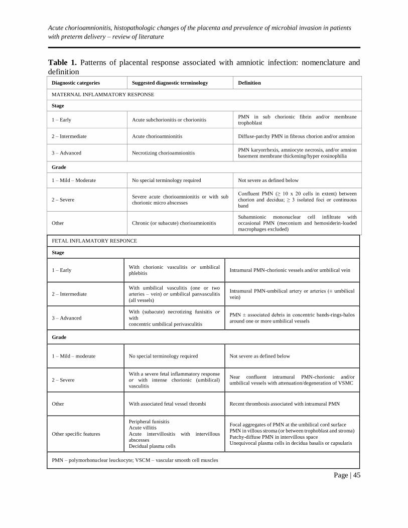
Acute chorioamnionitis, histopathologic changes of the placenta and prevalence of microbial invasion in patients
with preterm delivery – review of literature
Page | 45
Table 1. Patterns of placental response associated with amniotic infection: nomenclature and
definition
Diagnostic categories Suggested diagnostic terminology Definition
MATERNAL INFLAMMATORY RESPONSE
Stage
1 – Early Acute subchorionitis or chorionitis PMN in sub chorionic fibrin and/or membrane
trophoblast
2 – Intermediate Acute chorioamnionitis Diffuse-patchy PMN in fibrous chorion and/or amnion
3 – Advanced Necrotizing chorioamnionitis PMN karyorrhexis, amniocyte necrosis, and/or amnion
basement membrane thickening/hyper eosinophilia
Grade
1 – Mild – Moderate No special terminology required Not severe as defined below
2 – Severe Severe acute chorioamnionitis or with sub
chorionic micro abscesses
Confluent PMN (≥ 10 x 20 cells in extent) between
chorion and decidua; ≥ 3 isolated foci or continuous
band
Other Chronic (or subacute) chorioamnionitis
Subamnionic mononuclear cell infiltrate with
occasional PMN (meconium and hemosiderin-loaded
macrophages excluded)
FETAL INFLAMATORY RESPONCE
Stage
1 – Early With chorionic vasculitis or umbilical
phlebitis Intramural PMN-chorionic vessels and/or umbilical vein
2 – Intermediate
With umbilical vasculitis (one or two
arteries – vein) or umbilical panvasculitis
(all vessels)
Intramural PMN-umbilical artery or arteries (± umbilical
vein)
3 – Advanced
With (subacute) necrotizing funisitis or
with
concentric umbilical perivasculitis
PMN ± associated debris in concentric bands-rings-halos
around one or more umbilical vessels
Grade
1 – Mild – moderate No special terminology required Not severe as defined below
2 – Severe
With a severe fetal inflammatory response
or with intense chorionic (umbilical)
vasculitis
Near confluent intramural PMN-chorionic and/or
umbilical vessels with attenuation/degeneration of VSMC
Other With associated fetal vessel thrombi Recent thrombosis associated with intramural PMN
Other specific features
Peripheral funisitis
Acute villitis
Acute intervillositis with intervillous
abscesses
Decidual plasma cells
Focal aggregates of PMN at the umbilical cord surface
PMN in villous stroma (or between trophoblast and stroma)
Patchy-diffuse PMN in intervillous space
Unequivocal plasma cells in decidua basalis or capsularis
PMN – polymorhonuclear leuckocyte; VSCM – vascular smooth cell muscles
Kochoski et al.
Page | 46
The same author in 2015 published his experts review, redefining the criteria for
histopathological evaluation of the placenta. These criteria were presented at the Amsterdam
international consensus of criteria for diagnosis of placental lesions. According to the author, these
criteria, presented in Table 2, represent an extensive basis for implementing a comprehensive
system or systematized framework that includes all placental lesions [11].
Although it was not in the focus of the group, the following recommendations for tissue
sampling of the placenta were introduced:
To submit at least 4 samples
One of the samples should include 2 cross-section blocks of the umbilical cord and a
roll of extra placental membranes which includes a part of the marginal parenchyma
Other samples should contain full-thickness sections of normal appearing placental
parenchyma provided from within the central two thirds of the placental disc, including
one adjacent to the umbilical cord insertion site
Using Redlines criteria, adopted by the Amniotic Fluid Infection Nosology Committee of the
Perinatal Section of the Society of Pediatric Pathology, Chong JК and his collaborators analyzed
7.505 placentas from singleton pregnancies, delivered after 20th g.w. They used placentas of term
pregnancies as a control. The analysis showed significantly higher presence of inflammatory
changes of the placenta, defined as acute choriamnionitis, in the group delivered between the 21st
and 24th g.w and the trend was descendent in groups with higher gestational age. The prevalence
of the inflammatory changes of the placenta in the first group was up to 94.4%, contrary to 39.6%
in the group at gestational age of 25-28 weeks, and 35.4% in the group at gestational age of 28-32
weeks. Only 10% of patients with so-called late prematurity, 33-36 weeks demonstrated
inflammatory changes of the placenta. Analyzing the placentas of the term patients in the control
group, they concluded that the presence of spontaneous contractions and cervical dilatation > 4 cm
represent an additional poor prognostic factor, if it is to be evaluated in the function of time. The
prevalence of the inflammatory changes of the placenta was 4 times higher in this group, contrary
to the group of patients with no spontaneous onset of uterine contractions (11.6% vs. 4.7%,
p<0.01). It was also presented that the frequency of acute choriamnionitis increased if the length
of the delivery was higher and the cervical dilatation was more than 4 cm (30.4% vs. 11.6%) [6].
They proposed two possible scenarios as an explanation. First, microbial invasion of the amniotic
space in patients with spontaneous onset of labor, when amniotic membranes are intact, is higher
(17% vs. 1.5%) [12]. Second, uterine contraction, by themselves, have an inflammatory basis. The
chorion and amniotic membranes, provided from the patients with evident contractions, display a
high expression of chemokine receptors specific to neutrophils and monocytes, as well as for the
inflammatory proteins specific to macrophages, even though there are no evident signs of
chorioamnionitis. These findings correlate with the reports of increased concentration of
chemokines such as IL-8, cytokines IL-1 and IL-6, chemotactic proteins, as well as growth
associated oncogenes, in the amniotic fluid of term patients with spontaneous onset of contractions,
contrary to those with no contractions.
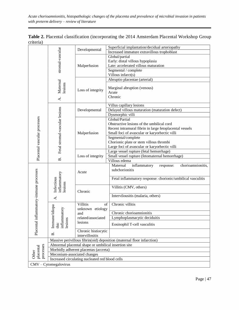
Acute chorioamnionitis, histopathologic changes of the placenta and prevalence of microbial invasion in patients
with preterm delivery – review of literature
Page | 47
Table 2. Placental classification (incorporating the 2014 Amsterdam Placental Workshop Group
criteria) P
lace
nta
l vas
cula
r p
roce
sses
A.
Mat
ernal
st
rom
al-v
ascu
lar
lesi
ons
Developmental Superficial implantation/decidual arteriopathy
Increased immature extravillous trophoblast
Malperfusion
Global/partial Early: distal villous hypoplasia
Late: accelerated villous maturation
Segmental / complete
Villous infarct(s)
Loss of integrity
Abruptio placentae (arterial)
Marginal abruption (venous)
Acute
Chronic
B.
Fet
al s
tro
mal
-vas
cula
r le
sio
ns
Developmental
Villus capillary lesions
Delayed villous maturation (maturation defect)
Dysmorphic villi
Malperfusion
Global/Partial
Obstructive lesions of the umbilical cord
Recent intramural fibrin in large fetoplacental vessels
Small foci of avascular or karyorhectic villi
Segmental/complete
Chorionic plate or stem villous thrombi
Large foci of avascular or karyorhectic villi
Loss of integrity
Large vessel rupture (fetal hemorrhage)
Small vessel rupture (fetomaternal hemorrhage)
Villous edema
Pla
centa
l in
flam
mat
ory
-im
mu
ne
pro
cess
es
A.
Infe
ctio
us
infl
amm
ato
ry
lesi
on
s
Acute
Maternal inflammatory response: chorioamnionitis,
subchorionitis
Fetal inflammatory response: chorionic/umbilical vasculitis
Chronic Villitis (CMV, others)
Intervilousitis (malaria, others)
B.
Imm
une/
idio
pa
thic
infl
amm
atory
lesi
ons
Villitis of
unknown etiology
and
related/associated
lesions
Chronic villitis
Chronic chorioamnionitis
Lymphoplasmacytic deciduitis
Eosinophil T-cell vasculitis
Chronic histiocytic
intervillositis
Oth
er
pla
centa
l
pro
cess
es
Massive perivillous fibrin(oid) deposition (maternal floor infarction)
Abnormal placental shape or umbilical insertion site
Morbidly adherent placentas (accreta)
Meconium-associated changes
Increased circulating nucleated red blood cells
CMV – Cytomegalovirus

Kochoski et al.
Page | 48
Talking about the origin of the neutrophils within the inflammatory substrate in patients with
choriamnionitis, using FISH (Fluorescent in Situ Hybridization) with probes for X and Y-
chromosomes performed in cytospin slides of placentas from male fetuses, it was determined that
almost 90% of neutrophils originate from the mother. In addition, the use of FISH combined with
immunohistochemistry for CD45 positive cells, for identification of leukocytes, demonstrated that
CD45 positive cells in the membranes derive from the mother. On the other hand, inflammation
of the umbilical cord and chorionic vessels of the chorionic plate is of fetal origin, which correlates
with the anatomy of the complex. Neutrophils that migrate from the umbilical blood vessels,
arteries and veins, must originate from fetal circulation in order to penetrate the wall of these blood
vessels [6].
Microbial invasion and its role
Usually, the intra amniotic compartment is considered to be a sterile environment. The pathway
of intra amniotic infection has not been completely understood. Four basic mechanisms are
suggested: ascending path of transport of the microbial agents from the lower genital tract,
haemathogenous dissemination, accidental inoculation of infection during invasive procedures
(amniocentesis, cordocentesis, fetoscopy etc.) and reverse or retrograde advance through the
Fallopian tubes, which is supported by limited number of evidence. The ascending pathway of
infection is thought to be one of the most common mechanisms for intra amniotic infection. There
are two suggested sub mechanisms.
According to the first one, microorganisms pass through the cervical barrier and invade the
decidua, which provides appropriate conditions for their multiplication, and from there they enter
the amniotic space. The second sub mechanism suggests a direct spread of microorganisms into
the amniotic space, as it is in the cases with intact membranes and no pPROM. So far, the data
suggest that inflammatory response starts at the level of decidua in both previously suggested
mechanisms, with the following difference. If the infectious agents advance into the decidua,
maternal inflammatory cells will be concentrated in this compartment, but if there is a direct
advance into the amniotic space, chemotactic gradient will attract the inflammatory cells in the
chorion and further after into the amniotic space [6, 13].
The most serious evidence about the association between the intra amniotic infection and acute
chorioamnionitis originates from the study of Romero R. and his collaborators. The study was
performed on 92 subjects with preterm contractions and intact membranes, the material for
microbiological analysis was provided by percutaneous amniocentesis, and histopathological
evaluation of the placentas was performed 48 hours after the procedure. The results showed
prevalence of the microbial invasion of the amniotic space in 38% of the cases. Acute
chorioamnionitis was histopathologically confirmed in 71.1% of the analyzed tissue samples,
while the diagnosis of acute funisitis was made in 78.7% of cases. The negative predictive value
of the histopathological findings compared to the results of the microbiological analysis of the
amniotic fluid was 87% for the acute chorioamnionitis and 82% for the acute funisitis respectively
[14]. The prevalence of the microbial invasion of the amniotic space is similar between patients
with spontaneous onset of labor at term, compared to those with premature spontaneous onset of
labor and intact membranes (17 vs. 22%). Nevertheless, in preterm neonates there is more frequent
early onset of neonatal sepsis, more intense inflammatory response, and more frequent presence
of funisitis. There are several thesis that have been offered as an explanation to the presented
differences. Microbial invasion of the amniotic space in term patients is of shorter time and it
appears after the initiation of contractions.
Acute chorioamnionitis, histopathologic changes of the placenta and prevalence of microbial invasion in patients
with preterm delivery – review of literature
Page | 49
Such microbial invasion is mainly presented with small inoculum, initiates mild inflammatory
response and it is rarely followed by fetal microbial invasion. On the other hand, in patients with
premature onset of contractions, with intact membranes or pPROM, microbial invasion occurs
prior to initiation of contractions, the process is burdened with higher quantum of microorganisms,
and probably lasts longer providing conditions for more severe inflammatory response [6].
A certain number of evidence suggest that occult intrauterine infection plays a valid role in the
genesis of premature onset of contractions and preterm delivery. Majority of authors agree that the
difficulties to determine the type of microbial agents are associated with the subclinical character
of the infection and with inability to cultivate strains of bacteria responsible for the intrauterine
infection.
Pankuch et al., in 1984 reported a prevalence of microbial invasion of the placentas in women
with preterm delivery as high as 72%, but the efforts to duplicated these results in the following
years, in the same category of patients, with histopathologically confirmed choriamnionitis, were
unsuccessful [15]. Using PCR or amniotic fluid culture on amniotic fluid provided by
amniocentesis form 166 patients, Di Giulio and his associates, reached a rate of detection of 15%,
with notation that the combination of both techniques is more sensitive than the use of amniotic
fluid culture as a sole method of detection (56% vs. 9.6%).
From the samples that were positive, 17 bacterial strains were isolated or detected, 6 of which
by combination of both techniques (Mycoplasma hominis, Ureaplasma, Streptococcus agalactiae,
Lactobaccilus, Prevotella and Fusobacterium nucleatum), 6 were detected by amniotic culture only
(Staphylococcus coagulase neg., Bacillus sp., Peptostreptococcus, Gardnerela vaginalis), while 7
were isolated with PCR (Streptococcus mitis, Delfia acidovorans, Neiseria cinerea, Sneathia
sanguinegens, Leptotrichia amnii, uncultivated Bacroidetes and one unidentified phylum). Further
analysis of the data revealed positive predictive value (PPV) of 100% for the combined approach,
for all categories (preterm delivery prior to 25th g.w, prior to 32nd g.w, prior to 37th g.w and
delivery within one day of the amniocentesis) [16]. Some of the more recent studies, such as the
one from Andrew C. et al. which used material provided from 305 patients with spontaneous
premature contractions and intact membranes, report even lower prevalence for the microbial
invasion of the amniotic space (10.1%) with slightly lower detection rate for used molecular
techniques (rDNA PCR) in comparison to amniotic culture (65% vs.16%). On the other hand,
analyzing the data for the values of IL-6 in the amniotic fluid they found that the level of this
inflammatory marker is high enough to suggest sever to moderate form of infection in 36.1% of
the patients (20.7% with severe inflammation, 15.4% with moderate inflammation). In majority of
these patients, no microbial invasion of the amniotic space was detected [17].
The question arises what initiates the inflammatory response in cases in which intra amniotic
infection has not been proven? Authors theoreticize that one of the possibilities is an infection of
the extra amniotic compartment (decidua, placenta, fetal membranes). Other possibility could be
the inflammatory response to non-infective stimulus such as: trauma, ischemia or abruption.
Interaction between the intra amniotic bacterial colonization and the inflammatory response could
be summarized in 4 stages: homeostasis, initiation, evolution and resolution. Some of the
microorganisms, such as Ureaplasma and some of the genital mycoplasmas, can be detected in
small amounts in utero, in large number of uncomplicated pregnancies. If their presence does not
initiate abundant inflammatory response (homeostasis), such colonization will not influence
neither the mother, nor the fetus.

Kochoski et al.
Page | 50
On the other hand, if the homeostasis is disturbed through uncontrolled proliferation or
invasion with aggressive strains, the result will be intense inflammatory response (initiation). The
authors considered the possibility that the inflammatory response and not microbial invasion is
responsible for the liberation of prostinoids that initiate contractions and lead to cervical ripening,
changes that are clinical characteristics of preterm delivery (evolution). Once the severe intra
amniotic inflammation develops, it will most certainly lead, very promptly, to preterm delivery
(resolution). We still don’t fully understand which factors are responsible for the transition from
one to the next phase, therefore it is still unclear whether clinical interventions in the early stages
of the process could result in different type of resolution i.e. continuation of the pregnancy instead
of unstoppable progression towards preterm delivery.
DISCUSSION
Preterm delivery as a global problem along with its association with long-term risks and
consequences imposes a necessity for multidisciplinary approach in the attempt to deal with the
causes behind its origin. Efforts made to date have not resulted in satisfactory reduction in the rate
of preterm delivery. On contraire, the rate of preterm delivery is rising. Nevertheless, if we exclude
iatrogenic reasons for prematurity, what remains is a large group of patients with intrauterine
inflammation, with or without confirmed infection of the feto-placental unit, as a main
etiopathogenic factor.
In the last 20 years, large number of authors tried to explain the problem of
inflammation/infection of the intrauterine compartment of the gravid uterus, and to establish
consequential relationship between infection and histopathological changes that develop in
different parts of the feto-placental unit: decidua, placental disc, fetal membranes, umbilical cord
and amniotic fluid. Results from these studies impose consistent conclusion that the inflammatory
changes of the placenta and the umbilical cord, defined as acute chorioamnionitis and funisitis,
appear more often in conditions of spontaneous onset of labor. It seems that spontaneous onset of
uterine contractions strongly correlates with already present inflammatory changes, regardless of
the integrity of the fetal membranes, intact or not. To complicate the present situation even more,
there is a confirmed correlation between the inflammatory changes of the feto-placental unit and
the gestation age. The smaller the gestational age, the higher and more severe the inflammatory
changes of the placenta and the umbilical cord. From this point of view, we should concentrate on
those cases with extreme and early prematurity (< 28 g.w) in which the prevalence of
histopathologically proven chorioamnionitis is high (> 90%), and the rate of mortality and
prevalence of late complications related to prematurity is also high.
Studies that have been carried out to date failed to provide answer to the question of correlation
between the inflammatory changes of the placenta and the umbilical cord, and microbial invasion
of the intrauterine compartment. Although the role of microorganisms, especially bacteria, is
undoubted, in only 10-15%, maximum 38% of cases (depending on the study) bacterial presence
can be detected in the samples taken from the placenta, umbilical cord and amniotic fluid. When
microbiological studies are performed, advantage is given to the combination of amniotic fluid
culture and molecular techniques for isolation of bacterial rDNA (PCR).

Acute chorioamnionitis, histopathologic changes of the placenta and prevalence of microbial invasion in patients
with preterm delivery – review of literature
Page | 51
CONCLUSION
Histopathologically verified chorioamnionitis is a more frequent finding in patients with
preterm delivery if compared to clinical manifestation of the clinical syndrome presented through
its pathognomic signs (fever, painful tender uterus, leukocytosis, maternal and fetal tachycardia).
The presence of chorioamnionitis is associated with increased risk for perinatal mortality and
morbidity (neonatal sepsis, bronchopulmonary dysplasia and cerebral palsy). Even though the
association between the histopathological changes of the feto-placental unit and premature onset
of contractions is well established and proven, there is still a mist around the question, whether the
inflammatory process is a trigger or a consequence of the process of the preterm delivery that has
already started.
Considering the historically accepted empiric standing on the role of genital infections in
etiology of preterm delivery, further analysis are needed to determine the reasons behind the
relatively small percentage of isolations of microorganisms in the analyzed samples.
REFERENCES 1. WHO Media center. “Preterm birth” Fact sheet No. 363; November 2015.
2. Blencowe H, Cousens S, Chou D, Oestergaard M. Born too soon: The global epidemiology
of 15 million preterm births. Reprod Health. 2013;10(Suppl 1):S2.
3. World Health Organization. Born too soon: the global action report on preterm birth. WHO Cataloguing-in-Publication Data 2012.
4. National Institute for Health and Care Excellence. Preterm labor and birth, NICE
guidelines [NG25], November 2015.
5. Lawn JE, Cousens S, Zupan J. 4 million neonatal deaths: when? Where? Why? Lancet. 2005;365 (9462):891–900.
6. Kim CJ, Romero R, Chaemsaithong P, Chaiyasit N, Yoon BH, Kim YM. Acute
chorioamnionitis and funisitis: definition, pathologic features, and clinical significance. Am J Obstet Gynecol. 2015; 213(4 Suppl):S29–52.
7. Redline RW. Amniotic Infection Syndrome: Nosology and Reproducibility of Placental
Reaction Patterns. Pediatr Dev Pathol. 2003;6(5):435–48.
8. Borrhalo P, Cunha F, Pinto M, da Silva AT, Meirinho M. Perinatal morbidity and mortality
related to gestational infection. The histological identification of chorioamnionitis and its
incidence in the population studied. Acta Med Port. 1996;9(10-12):319–23.
9. Monga M, Blanco JD. Intrauterine infection and preterm labor. Infect Dis Obstet Gynecol. 1995;3:37–44.
10. Redline RW, et al. Amniotic infection syndrome: Nosology and reproducibility of placental
reaction patterns. Pediatr Dev Pathol. 2003;6(5):435–48.
11. Redline RW. Classification of placentla lesions. Am J Obstet Gynecol. 2015;213(4 Suppl):S21–8.
12. Lee SM, Lee KA, Kim SM, Park CW, Yoon BH. The risk of intra-amniotic infection,
inflamation and chorionamnionitis in term pregnant women with intact membranes and labor. Placenta. 2011;32:516–21.
Kochoski et al.
Page | 52
13. Kim SM, Romero R, Park JW, Oh KJ, Jun JK, Yoon BH. The relationship between
intensity of intra-amniotic inflammation and the presence and severity of acute histologic
chorioamnionitis in preterm gestation. J Matern Fetal Neonatal Med. 2015;28(13):1500–9. 14. Romero R, Salafia CM, Athanassiadis AP, et al. The relationship between acute
inflammatory lesion of the preterm placenta and amniotic fluid microbiology. Am J Obstet
Gynecol. 1992;166:1382–8.
15. Pankuch GA, Appelbaum PC, Lorenz RP, Botti JJ, Schachter J, Naeye RL. Placental
microbiology and histology and pathogenesis of chorionamnionitis. Obstet Gynecol.
1984;64(6):802–6.
16. Di Giullio D, Romero R, Amogan HP, et al. Micobial prevalence, diversity an abundance
in amniotic fluid during preterm labor: molecular and culture-based investigation. PLoS
One. 2008;3(8):e3056.
17. Combs CA, Gravett M, Garite TJ, et al. Amniotic fluid infection, inflammation and
colonization in preterm labor with intact membranes. Am J Obstet Gynecol.
2014;210(2):125.e1–125.e15.
Acta morphol.2019;Vol.16(1):53–62
UDC: 601.4:575.22]:179
Page | 53
REVIEW ARTICLE
ETHICAL CONSIDERATIONS TOWARDS INDIVIDUAL RESULTS
DELIVERY AND THE CONSEQUENCES OF INCIDENTAL FINDINGS IN THE
WHOLE GENOME AND EXOME SEQUENCING
Kostovski Marko1, Simeonovski V2 1Institute of Medical Histology and Embryology, Faculty of Medicine, Ss Cyril and Methodius University
of Skopje, Skopje, Republic of North Macedonia 2University Clinic of Dermatology, Skopje, Republic of North Macedonia
ABSTRACT
There are more and more genome and exome sequencing studies every day in which subjects
with different demographics are included, especially age differences, from infants to adults.
Precisely today’s advanced techniques represent serious tools that provide not only one simple
answer, but more complex plural answers. A special issue within this dilemma remains the
incidental findings. It is clear that the true challenge of an investigation is solving the particular
problem, disease, mutation (mystery), but it is even more challenging when an unexpected finding
is discovered. In this article we review the different attitudes towards manipulation with individual
results and consequently with incidental molecular findings, through presentation of some ethical
dilemmas that circulate the scientific community.
INTRODUCTION
Ever since 1977, with the breakthrough of the technique for fast DNA sequencing by Frederick
Sanger [1], through the introduction of the polymerase chain reaction- PCR, 33 years ago [2], and
the beginning of one of the most prominent human undertakings – The Human Genome Project,
in 1990, a new era in the field of medicine has begun [3]. Sequencing i.e. the discovery of the
human genome remains to be one of the few great endeavors of the human kind and serves as a
historical hallmark of science, rivalled even to the moon landing. The Human Genome Project,
which sequenced the whole genome of the human species, took 13 years to complete and it cost 3
billion dollars [3].
Today, we live in the era of Next Generation Sequencing (NGS), that, in comparison to the 13
years’ time of the first generation sequencing methods [4], has the ability to sequence a lot more
genomes for a vastly shorter period of time – 18,000 human genomes per year or 45 genomes per
day, and only for a price of less than 2000 dollars [5]. The most important methods of sequencing
nowadays are genome sequencing and transcriptomes, as well as the newly popular epigenomic
studies [6]. The great spectrum of challenges in the personal genomics, have unlocked the
possibility for research of the individual genome testing. Hence, great opportunities of this
prospect have arisen and would lead to a bright scientific future [7], but at the same time, this
deeper view into oneself have provoked a number of ethical dilemmas [8]. Namely, there are more
and more genome and exome sequencing studies every day in which subjects with different
demographics are included, especially age differences, from infants to adults.
Kostovski et al.
Page | 54
Precisely today’s advanced techniques represent serious tools that provide not only one simple
answer, but more complex plural answers. The increased activity of people on the social platforms,
the curiosity to learn more about one self’s genetic code, as well as the boosted benefit for the
clinicians with the personalized genome results, opens the door to the delivery and importance of
individual results in the genomic studies [9].
In this context, a new ethical dilemma arises, of how to operate with the said results.
Specifically, is it completely regulated which particular results should be presented to the patient?
Who is the one who should inform the subjects (patients) about the results with a thorough
explanation? Do the results match the high criteria of good clinical practice? How is the principle
of “not to know” implemented without the loss of patient autonomy?
There are an increasing number of supporters of the claim that the return of the complete
individual results to the patient would have vast moral implications and would be very important
in the genome/exome studies [10,11]. A special issue within this dilemma remain the incidental
findings. It is clear that the true challenge of an investigation is solving the particular problem,
disease, mutation (mystery), but it is even more challenging when an unexpected finding is
discovered. Henceforth, the ethical dilemma of the incidental findings- should they be searched
for and are they relevant or even are we obliged to individually present them to the patients? Or,
should we stick to the particular analyzed gene?
Incidental findings, are defined as findings that may or may not be detected adjacent to the
primary aim of the enquiry and accordingly, have potential clinical or reproductive significance
[12,13]. Thus, it is interesting to point out the predicament: what if incidental findings became part
of the algorithms and forms of informed consent? Will they still be considered “incidental”?
Consequently, a recent study encourages a new term for these findings, naming them secondary
variants, in order to bypass the flaws of the former taxonomy [14]. It is to be noted that the number
of identified incidental findings in subjects is not modest in the least, on the contrary, which
implicates a need of their proper regulation [15]. Studies show that there is no legal regulative for
delivery of relevant individual results from the increased number of genomic studies to the subjects
yet, which only intensifies the significance of the ethical questions that arise from this predicament
[16]. There are more arguments in favor of giving access to the incidentalomas to the patient, since
they are of great significance for the individuals [17,18, 19, 20]. Accordingly, even the studies that
do not support the exposition of incident findings and go by the saying “not to transform the
research setting into an extension of the clinic”, are not strictly categorical, in terms of accepting
the need of these results with specific relevance in particular settings and individual approach [21].
Some instances have regulated this current ethical issue by commencing directive guidelines [22,
23].
Informed consent is a convenient tool that could be used to regulate the ethical dilemmas
connected to individual results. That is to say, when one signs the consent, one could have the
possibility to regulate the management of incidental findings in terms of his further knowledge
about the outcomes that derive from the study [17, 23, 24, 25].
THE DIVISION IN CLINICAL AND RESEARCH DOMAIN
The debate around returning secondary findings in research is rather complicated by the fact
that the discrepancy between clinical and research domains is becoming blurred as sequencing
studies are being translated into clinical practice. Although under a research protocol, subjects’
initial contact was the clinician who recruited them in a healthcare-providing facility.
Ethical considerations towards individual results delivery and the consequences of incidental findings in the whole
genome and exome sequencing
Page | 55
Clinical standards of ethics must be therefore ensured by the clinician researchers, which
becomes a dispute in pure research context where the same standards of care do not normally exist.
Berkman et al. (2014) argue that the lack of consensus for delivery of these results comes from
the nature of the field, which is perceived as a transitional one, and the collapse of this
differentiation is expected and sensible [26]. Researchers, however, should be aware of their
ethical duties especially in the era of advanced genomic technology implementing in various
clinical trials. Therefore, higher clinical and ethical aspects of responsibilities of researchers are to
be achieved in the future.
On the other hand, Hallowell et al. (2014) noted, that discriminating between these two
undertakings is essential as those involved have different prime commitments: Researchers tend
to improve scientific aspects of their research, while when it comes to the clinicians, patient duty
remains their imperative [27]. Translational research is more present nowadays, where new
technology advances tend to be implemented in clinical context. As Wolf (2012) argue [28],
bridging researchers and clinicians through intensified multidisciplinary approach together with
the pivotal role of clinician- researchers in recent research, the dilemma around secondary variants
(incidental findings) should be addressed through the context of the translational research process
[29]. Interestingly, a study conducted by Kaphingst et al. concluded that the subjects themselves
do not have a preference of who will deliver the results and put less emphasis on the title of the
person who delivers them, but were rather more concerned about the manner of delivery [30].
Contradicting Wolf are more than few studies which surveyed the opinions of research participants
on ‘who should return incidental findings’. It was clearly instructive that the most respondents
wanted such findings returned by participant’s physician or primary health care giver. In support
of this point is the fact that researchers may not be adequately trained to return such findings and
burdening them with this duty may distract them from achieving their primary study goal [31].
Considering the abovementioned arguments for the research clinical divide, the authors of this
article lean more towards the viewpoint that suggests blurring the boundary between clinical and
scientific. Thus, we will continue in the similar narrative.
INDIVIDUAL FINDINGS
Reviewed in this article, are the different attitudes towards manipulation with individual results
and consequently with incidental findings, through presentation of some ethical dilemmas that
circulate the scientific community. One of the main doctrines of ethics is “benefacere” – doing
good. It is undoubtedly a question of why this principle could not be equally projected in the
research studies as well. Therefore, all scientists are bound by this norm and should always disclose
the individual results to the subjects included in the research. Nevertheless, there are arguments in
the literature supporting a different approach of the “benefacere” principle in doctors versus
scientists. It is believed that the “doing good” norm directly depends with the role that the scientist
has in the doctor-patient relationship i.e. whether the scientist has contact with the patient (is the
patient’s doctor), or not.
While doctors have a clear responsibility for doing good for their patients, this obligation is
still a grey area in terms of scientific work, especially in research projects where scientists have
no personal contact with the subjects. Hence, this could become a burden to the researcher and an
issue when it comes to future accountability [16, 32].
Kostovski et al.
Page | 56
Furthermore, another interesting ethical question is fabricated- what is the responsibility of the
researches who are doing science on data collected in the biological banks, that is to say, scientists
who are not prime collectors of material and had never had any previous contact with the subjects
(referred to as secondary researchers)? In general, their duties depend on the course of the main
study and how will the delivery of individual results affect its realization. From this point of view,
the principle of “benefacere” could be satisfied by publication of the collective results and
conclusions of the study only, without the responsibility of delivering individual results. On the
other hand, it is reported that where individual results could have a great impact on the individual
receiving them, and do not compromise the main study, their individual delivery can be a norm for
the secondary researcher [33].Therefore, new guidelines for the biological banks are constituted.
Namely, it is emphasized that the clinically relevant results could be a product of the prime
investigators, biobanks as well as the secondary investigators, and consequently, they should be
offered to the subjects by all of the abovementioned instances [34]. Nonetheless, the views of the
right of the subjects to have the availability to end results of the study is unanimous, in terms of
obligating the scientists to point out the results that may have direct harmful effect of the
individual’s health or may help prevent potential conditions [4, 34, 35]. Owing to all the previously
mentioned ethical dilemmas, the National Heart, Lung and Blood Institute (NHLBI) has
established ethical guidelines, which are regularly reviewed in accordance with the progress and
growth of the genomic studies [23,24]. An interesting study from Beskow et al. (2010) shows the
need of research contextualization for these guidelines. In other words, they propose that the option
for delivery of individual results should be predicted and planned as early as possible in the study,
or it should even be incorporated and precisely formed in the informed consent form for the
selected clinical or scientific study [36]. Likewise, no less of a factor for this issue should be
considered the great availability of genomic sequencing today. People are more and more
interested in their codes, which is an additional reason why these ethical dilemmas should be
defined most precisely.
But then what is the right way to deliver these results? Who is the one who should announce
them? The rapid progress of genetic research has driven the scientists to a blind end when it comes
to deciding what is the right direction in which they should advance in order to preserve the ethics
in the delivery of individual results [23]. The most controversial and important dilemma is the one,
whether the obtained results from a single study are as equally relevant as the results achieved in
clinical trials. From the aspect of the primary goal of a study, that is generating overall results, the
investigators are not compelled to deliver the results with prior verification with the standards of
clinical diagnostics. Therefore, it is suggested that the results be referred to as investigatory
(scientific) only, with an addendum that they be additionally evaluated, confirmed and monitored
[32]. Albeit the vast number of studies, there is still no ample agreement on how these results
should be delivered to the subjects. Regulations also flail in the part of who should be responsible
for the delivery- does the clinician have the necessary competencies from genetics to be able to
interpret and act upon the said results?
Or is the researcher trained in delivering the information appropriately to a person he has never
met before? Opinions regarding this dilemma vary between clinical geneticists, genetic counselors,
but also even family doctors.
What is considered, is the need for enriched education of the family doctors about genetics and
a greater involvement of geneticists and clinical genetics specialists, in order to provide clinical
care for patients with established relevant findings [37].

Ethical considerations towards individual results delivery and the consequences of incidental findings in the whole
genome and exome sequencing
Page | 57
One study points out that doctors who do not have training in clinical genetics, have in general
lesser knowledge about the discipline, which is seen as an additional problem for the
miscommunication between doctors and patients [38]. On the other hand scientists do not have
experience to communicate “bad news” of additional incidental findings. Moreover, the
excitement about the unexpected findings and eagerness to publish might be a barrier to the use of
these incidental findings in an ethical way. In order for the genomic research to really make a
difference in the clinical practice, it is necessary to foremost educate a competent personnel
between the medical workers, for proper interpretation and analysis of the results [39]. Context
wise, another study concludes that the scientists working genomics do not have major experience
in announcing results, but nevertheless are increasingly motivated to deliver [18]. A different study
shows that the patients emphasize the importance of the nature of the results- their accuracy and
possibility for prevention/treatment directly correlates to patients’ willingness for receiving them
(40).
Interestingly, the offer for individual results to be delivered at the end of the study, increases
the eagerness of the subjects to participate in the studies [41]. Hence, a preparation of a study with
a formal study protocol, explaining the subjects the scope of the project and management with
individual results, is advised [37]. Thereafter, the notion of creating a relevant and precise
informed consent form presents a vital concept in regulation of this important question connected
to delivery of individual results [42,43]. In this form, the subjects could individually select which,
and if at all, results they would like to obtain. This approach to the matter is said to be very
important for preserving the principle of autonomy [44]. In contrast, for the incidental findings,
the patients do not yet have the possibility to choose whether or not they want to be presented with
the data. In this context, this issue should be further discussed and regulated [45]. One study
concluded that 70% of the genomic studies either did not have an individual results segment in the
informed consent or even explicitly stated that there is no room for delivery of said results [46].
Given the traditional postulates of research, personal interest for benefit of the genomic studies
should not be a motivation for the subjects to advance in the study [47]. Furthermore, in terms of
relevance of the results with momentarily knowledge about a certain condition i.e. if the result has
clinical significance, it is advised only the clinically relevant results to be included in the final
medical report. But, as clinically nonsignificant results are equally relevant, it is considered to
design guidelines for management and create a system of individual records with a possibility to
upgrade the data for the individual clinical significance of any future investigations [37].
Although, in general, there are uniformed guidelines for managing the individual results
covering their delivery in accordance to study protocols, still the scientific community is not in
unison about the ethical aspects of this problem [46, 32]. It is almost unbelievable that a consensus
about this question will be reached, but nonetheless future discussions about deciphering this
dilemma should be strongly encouraged [48].
INCIDENTAL FINDINGS
Incidental findings have lots of common traits with the individual primary results, but in the
core they differ fundamentally. Namely, they demand additional investigation outside the premises
of the study’s aims and inquire supplementary expert interpretation that the prime investigators
involved might lack [13].
Kostovski et al.
Page | 58
Still, it is implicated that with the advancement of the technology and implementation of such
research results in clinical practice, it will be prudent to study and assess these findings.
Incidental findings should be potentially analyzed when a benefit of the patient and burden to
the study balance is established, all the while bearing in mind the exclusivity of the results to the
researchers (genomic researches are mostly done for scientific reasons instead of clinical practice,
thus giving the investigator the sole exclusivity of the findings for certain conditions which he
might choose not to publicize) [49]. Yet, with the progress of technology the access to genome
sequencing is immensely increasing.
The American College of Medical Genetics and Genomics (ACMG) has established a list of
minimum requirements as part of the algorithm for clinical conceptualization of genomic studies.
It consists of all the clinically relevant genes (defined incidentalomes of clinical significance)
which need to be routinely investigated in every genomic research [50]. Analogously, Townesend
et al report similar recommendations for ethical delivery of incident findings [51]. But, it should
be pondered that the patients often have unrealistic expectations of the genomic research,
anticipating to receive the entire array of deviations, making the incidental findings altogether
confusing [52]. Naturally, this represents a hurdle because it gets in question the autonomy of the
patients and their right “not to know” a certain information. Actually, the subject who gets involved
in the study for a primary indication, ends up with a whole set of results that could potentially have
a negative effect [51]. Nonetheless, personal preferences of the patients are not included in the
ACMG algorithm, with the justification that at this moment the principles of “doing good” and
“not doing harm” outweigh the one of autonomy and individual predilections of patients in clinical
trials [50]. Still, even in investigative (scientific) context, the subjects do not have the opportunity
to sound their opinions whether or not they want to receive the incidental findings. One study
presents scarce recommendations about upgrading the informed consent form [53], but after all,
lots of questions around the obligation of the researchers to pursue incidental/secondary variants
remain unanswered. Albeit all, at the present moment, there is no regulative that obligates the
researchers to track and analyze incidental findings [49]. From the researcher’s standpoint, detailed
and precise delivery policy of the study, balance between the scope of the study and the patients’
rights and a strong ethical principle of “primum non nocere”, is advised to be always established
beforehand [54]. A study on the subjects preferences about the delivery of individual and incidental
results about familial risk of cancer, concludes that the major percent of patients preferred to
receive both kinds of results [55]. Likewise, another research in the field of exome sequencing,
once again concludes that the patients agreed on delivery of both variants of incidental/secondary
findings [56]. Further study, analyzed the attitude of parents towards the secondary variants of the
clinical genome sequencing in their children [57]. Namely, with the help of these incidental
findings, prevention against many conditions would be accessible and the branch of medicine that
would benefit the most from these genomic researches would be, unsurprisingly, oncology [15].
It should be taken in consideration that throughout these studies a number of potentially
significant results could be attained, so the scientific community should aim to overcome these
dilemmas in order to provide access to the patients for the clinically significant results [58].
Furthermore, because the genome sequencing is not part of the routine practice, some results
from these studies could be unique, and therefore researches should undoubtedly be burdened with
the responsibility to deliver the incidental findings [57]. A recent study encloses the principles and
guidelines that should be followed when delivering individual and incidental findings [59].
Ethical considerations towards individual results delivery and the consequences of incidental findings in the whole
genome and exome sequencing
Page | 59
CONCLUSION
The predicament about incidental findings represents a real ethical challenge, for which this
essay is insufficient in terms of scope to process and emphasize all the aspects of ethical regulation
and principles which burden the dilemma of incidental findings. New technologies have introduced
new ethical dillemmas about the communication of the molecular results if incidental findings
occur. Close collaboration between clinical geneticists and molecular researchers in the
development of new tools such as appropriate informed consents and guidelines for information is
warranted.
REFERENCES
1. Sanger F, Nicklen S, Coulson AR. DNA sequencing with chain-terminating inhibitors.
Proc Natl Acad Sci U S A. 1977;74(12):5463–5467.
2. Bartlett JM, Stirling D. A short history of the polymerase chain reaction. Methods Mol
Biol. 2003;226:3–6.
3. Leroy Hood, Lee Rowen. The Human Genome Project: big science transforms biology and
medicine. Genome Med. 2013;5(9):79.
4. Heather JM, Chain B. The sequence of sequencers: The history of sequencing DNA.
Genomics. 2016;107(1):1–8
5. Reuter JA, Damek Spacek D, and Snyder MP. High-Throughput Sequencing
Technologies. Mol Cell. 2015;58(4):586–597.
6. Mardis ER. Next-generation DNA sequencing methods. Annu Rev Genomics Hum
Genet. 2008;9:387–402.
7. Ansorge WJ. Next-generation DNA sequencing techniques. N
Biotechnol. 2009;25(4):195–203.
8. FuldaKG, Lykens K. Ethical issues in predictive genetic testing: a public health
perspective. J Med Ethics. 2006; 32(3):143–147.
9. Wallace SE, Kent A. Population biobanks and returning individual research results:
mission impossible or new directions?. Hum Genet. 2011;130(3):393–401.
10. Clayton EW, McGuire AL. The legal risks of returning results of genomics research. Genet
Med. 2012;14(4):473-7.
11. Knoppers BM, Joly Y, Simard J, Durocher F. The emergence of an ethical duty to disclose
genetic research results: international perspectives. Eur J Hum Genet. 2006;14(11):1170–
8.
12. Kohane IS, Hsing M, Kong SW. Taxonomizing, sizing, and overcoming the
incidentalome. Genet Med. 2012;14(4):399–404.
13. Wolf SM. Introduction: The Challenge of Incidental Findings. J Law Med Ethics.
2008;36(2):216–218.
14. Christenhusz GM, Devriendt K, Dierickx K. Secondary variants – in defense of a more
fitting term in the incidental findings debate. Eur J Hum Genet. 2013;21(12):1331–1334.
15. Johnston JJ, Rubinstein WS, Facio FM, et al. Secondary Variants in Individuals
Undergoing Exome Sequencing: Screening of 572 Individuals Identifies High-Penetrance
Mutations in Cancer-Susceptibility Genes. Am J Hum Genet. 2012;91(1):97–108.
16. Clayton EW, McGuire AL. The legal risks of returning results of genomics research. Genet
Med. 2012;14(4):473–477.
Kostovski et al.
Page | 60
17. Greely HT. The uneasy ethical and legal underpinnings of large-scale genomic biobanks.
Annu Rev Genomics Hum Genet. 2007;8:343–64.
18. Ramoni RB, McGuire AL, Robinson JO, Morley DS, Plon SE, Joffe S. Experiences and
Attitudes of Genome Investigators Regarding Return of Individual Genetic Test
Results. Genet Med. 2013;15(11):882–887.
19. Angrist M. You never call, you never write: why return of “omic” results to research
participants is both a good idea and a moral imperative. Personalized medicine.
2011;8(6):651–657.
20. Roberts JS, Shalowitz DI, Christensen KD et al. Returning individual research results:
development of a cancer genetics education and risk communication protocol. J Empir Res
Hum Res Ethics. 2010;5(3):17–30.
21. Burke W, Evans BJ, Jarvik GP. Return of results: ethical and legal distinctions between
research and clinical care. Am J Med Genet C Semin Med Genet. 2014;166C(1):105–11.
22. Knoppers BM, Deschênes M, Zawati MH, Tassé AM. Population studies: return of
research results and incidental findings Policy Statement. Eur J Hum
Genet. 2013;21(3):245–7.
23. Fabsitz RR, McGuire A, Sharp RR, et al. Ethical and Practical Guidelines for Reporting
Genetic Research Results To Study Participants: Updated Guidelines from an NHLBI
Working Group. Circulation Cardiovascular genetics. 2010;3(6):574–580.
24. Bookman EB, Langehorne AA, Eckfeldt JH, et al. Reporting Genetic Results in Research
Studies: Summary and Recommendations of an NHLBI Working Group. Am J Med Genet.
2006;140(10):1033–1040.
25. McGuire AL, Caulfield T, Cho MK. Research ethics and the challenge of whole-genome
sequencing. Nat Rev Genet. 2008;9(2):152–156.
26. Berkman BE, Hull SC, Eckstein L. The unintended implications of blurring the line
between research and clinical care in a genomic age. Per Med. 2014;11:285–95.
27. Hallowell N, Hall A, Alberg C, et al. Revealing the results of whole-genome sequencing
and whole-exome sequencing in research and clinical investigations: some ethical issues.
J Med Ethics. 2014;41:317–21.
28. Wolf SM. The role of law in the debate over return of research results and incidental
findings: the challenge of developing law for translational science. Minn J Law Sci
Technol. 2012;13.435–449.
29. Mackley MP, Capps B. Expect the unexpected: screening for secondary findings in clinical
genomics research. Br Med Bull. 2017;122(1):109–122.
30. Kaphingst KA, Ivanovich J, Elrick A, et al. How, who, and when: preferences for delivery
of genome sequencing results among women diagnosed with breast cancer at a young
age. Mol Genet Genomic Med. 2016;4(6):684–695.
31. Ewuoso C. A Systematic Review of the Management of Incidental Findings in Genomic
Research. BEOnline. 2016;3(1):1–21.
32. Jarvik GP, Amendola LM, Berg JS, et al. Return of Genomic Results to Research
Participants: The Floor, the Ceiling, and the Choices In Between. Am J Hum Genet.
2014;94(6):818–826.
33. Ossorio P. Taking aims seriously: repository research and limits on the duty to return
individual research findings. Genet Med 2012;14(4):461–466.

Ethical considerations towards individual results delivery and the consequences of incidental findings in the whole
genome and exome sequencing
Page | 61
34. Wolf SM, Crock BN, Van Ness B, et al. Managing Incidental Findings and Research
Results in Genomic Research Involving Biobanks & Archived Datasets. Genet Med.
2012;14(4):361–384.
35. Roberts JS, Shalowitz DI, Christensen KD, et al. Returning Individual Research Results:
Development of a Cancer Genetics Education and Risk Communication
Protocol. JERHRE. 2010;5(3):17–30.
36. Beskow LM, Burke W. Offering Individual Genetic Research Results: Context Matters. Sci
Transl Med. 2010;2(38):38cm20.
37. McGuire AL, Caulfield T, Cho MK. Research ethics and the challenge of whole-genome
sequencing. Nat Rev Genet. 2008;9(2):152–156.
38. Baars MJ, Henneman L, Ten Kate LP. Deficiency of knowledge of genetics and genetic
tests among general practitioners, gynecologists, and pediatricians: a global problem.
Genet Med. 2005;7(9):605–10.
39. Guttmacher AE, Porteous ME, McInerney JD. Educating health-care professionals about
genetics and genomics. Nat Rev Genet. 2007;8(2):151–7.
40. Murphy J, Scott J, Kaufman D, Geller G, LeRoy L, Hudson K. Public Expectations for
Return of Results from Large-cohort Genetic Research. AJOB. 2008;8(11):36–43.
41. Kaufman D, Murphy J, Scott J, Hudson K. Subjects matter: a survey of public opinions
about a large genetic cohort study. Genet Med. 2008;10(11):831–9.
42. Henderson GE, Wolf SM, Kuczynski KJ, et al. The Challenge of Informed Consent and
Return of Results in Translational Genomics: Empirical Analysis and Recommendations. J
Law Med Ethics. 2014;42(3):344–355.
43. Shalowitz DI, Miller FG. Communicating the Results of Clinical Research to Participants:
Attitudes, Practices, and Future Directions. PLoS Medicine. 2008;5(5):e91.
44. Burke W, Evans BJ, Jarvik GP. return of results: ethical and legal distinctions between
research and clinical care. American journal of medical genetics Part C, Seminars in
medical genetics. 2014;0(1):105–111.
45. Berkman BE, Hull SC. The “Right Not to Know” in the Genomic Era: Time to Break From
Tradition? AJOB. 2014;14(3):28–31.
46. Pereira S, Oliver Robinson J, McGuire AL. Return of individual genomic research results:
what do consent forms tell participants? Eur J Hum Genet. 2016;24(11):1524–1529.
47. Dressler LG, Smolek S, Ponsaran R, et al. IRB perspectives on the return of individual
results from genomic research. Genet Med. 2012;14(2):215–222.
48. McBride KA, Hallowell N, Tattersall MHN, et al. Timing and context: important
considerations in the return of genetic results to research participants. J Comm Genet.
2016;7(1):11–20.
49. Gliwa C, Berkman BE. Do Researchers Have an Obligation to Actively Look for Genetic
Incidental Findings? AJOB. 2013;13(2):32–42.
50. Green RC, Berg JS, Grody WW, et al. ACMG Recommendations for Reporting of
Incidental Findings in Clinical Exome and Genome Sequencing. Genet Med.
2013;15(7):565–574.
51. Townsend A, Rousseau F, Friedman J, Adam S, Lohn Z, Birch P. Autonomy and the
patient’s right “not to know” in clinical whole-genomic sequencing. Eur J Hum Genet.
2014;22(1):6.
Kostovski et al.
Page | 62
52. Christenhusz GM, Devriendt K, Dierickx K. Secondary variants – in defense of a more
fitting term in the incidental findings debate. Eur J Hum Genet. 2013;21(12):1331–1334.
53. Simon CM, Williams JK, Shinkunas L, Brandt D, Daack-Hirsch S, Driessnack M.
Informed Consent and Genomic Incidental Findings: IRB Chair Perspectives. JERHRE.
2011;6(4):53–67.
54. Souzeau E, Burdon KP, Mackey DA, et al. Ethical Considerations for the Return of
Incidental Findings in Ophthalmic Genomic Research. Translational Vision Science &
Technology. 2016;5(1):3.
55. Loud JT, Bremer RC, Mai PL, et al. Research participant interest in primary, secondary
and incidental genomic findings: Intentions to receive genomic research results. Genet
Med. 2016;18(12):1218–1225.
56. Shahmirzadi L, Chao EC, Palmaer E, Parra MC, Tang S, Gonzalez KDF. Patient decisions
for disclosure of secondary findings among the first 200 individuals undergoing clinical
diagnostic exome sequencing. Genet Med. 2014;16(5):395–399.
57. Christenhusz GM, Devriendt K, Van Esch H, Dierickx K. Focus group discussions on
secondary variants and next-generation sequencing technologies. Eur J Med
Genet. 2015;58(4):249–57.
58. Biesecker LG. Secondary variants and human subjects research. Genet
Med. 2013;15(2):157.
59. Sénécal K, Rahimzadeh V, Knoppers BM, Fernandez CV, Avard D, Sinnett D. Statement
of principles on the return of research results and incidental findings in paediatric research:
a multi-site consultative process. Genome. 2015;58(12):541–8.
Acta morphol.2019;Vol.16(1):63–76
UDC: 616.248-074:546.172.6]-053.2
Page | 63
REVIEW ARTICLE
UTILITY OF EXHALED NITRIC OXIDE IN PEDIATRIC PRACTICE
REVIEW OF LITERATURE
Arnaudova-Danevska Ivana1, Balkanov T2, Boshkovska K1, Jakjovska T1, Popova G1,
Cakalarovska I1 1Institute for Respiratory Diseases in Children - Kozle, Skopje, Republic of North Macedonia
2Institute of Preclinical and Clinical Pharmacology with Toxicology- Skopje
ABSTRACT
The field of interest for measurement of exhaled nitric oxide (NO) and nasal NO is significantly
evolving over the last 25 years, with over 1000 publications published in that area. Inflammation
of the airways is a central process in asthma and other lung disorders, but the monitoring of the
inflammation has not been included in the current recommendations. The exhaled air contains
volatile media such as nitric monoxide, carbon monoxide, ethane, pentane and non-volatile
substances in the liquid phase in the exhalation, as a condensate (hydrogen peroxide). It is
increasingly confirmed that the measurement of exhaled mediators in general, and especially NO,
is a new way to monitor certain aspects of asthma, COPD and interstitial lung disease, which
cannot be estimated with other methods, like lung function. In asthma, exhaled NO is
recommended to be used as a marker for diagnosis, for monitoring the response of anti-
inflammatory drugs, confirming the safety of therapy and predicting asthma exacerbation.
Measurements of FeNO are easily performed, they are reproducible and technically less expensive
than the analysis of induced sputum. In symptomatic patients, high FeNO levels (> 50 ppb), refer
to significant eosinophilia in the airways, which will most likely respond to treatment with ICS.
The current data provides support for the diagnostic use of FeNO in children with symptoms of
asthma. For patients with chronic and/or severe asthma, FeNO levels are useful for determining
whether eosinophilic inflammation of the airways is active or not. Both high (> 50ppb) and low
(<25 ppb) levels of FeNO can be used to for predicting the outcome in patients with a definitive
history of asthma who are currently in remission and who have stopped treatment with ICS.
Key words: asthma, children, exhaled nitric oxide, recommendations.
The field of interest for measurement of exhaled nitric oxide (NO) and nasal NO is significantly
evolving over the last 25 years, with over 1000 publications published in that area. Interest is
growing because the measurement of airways inflammation can contribute in the management of
lung diseases and the same measurement may be included in the clinical practice [1].
Evaluation of the airway inflammation
Inflammation of the airways is a central process in asthma and other lung disorders, but the
monitoring of the inflammation has not been included in the current recommendations [2].Taking
a direct sample of the airway cells and the mediators is invasive technique, as well as analysis of
induced sputum or bronchoscopy with lavage and biopsy. The exhaled air contains volatile media
such as nitric monoxide, carbon monoxide, ethane, pentane and non-volatile substances in the
liquid phase in the exhalation, as a condensate (hydrogen peroxide). Non-invasive measurements
of these exhaled mediators are appropriate for patient monitoring.
Arnaudova Danevska et al.
Page | 64
Exhaled NO
The presence of NO in the exhaled air in both animals and humans for the first time was
described in 1991. It has been shown that correlates with other indicators of moderate asthma
(eosinophils in induced sputum) and bronchial reactivity in non-steroid-treated patients. Generally,
the exhaled NO does not correlate with the lung function parameters in asthma. It is increasingly
confirmed that the measurement of exhaled mediators in general, and especially NO, is a new way
to monitor certain aspects of asthma, COPD and interstitial lung disease, which cannot be
estimated with other methods, like lung function. In asthma, exhaled NO is recommended to be
used as a marker for diagnosis [3], monitoring the response of anti-inflammatory drugs, to confirm
the safety of therapy [4] and to predict asthma exacerbation [5-7]. It is believed that the adaptation
of anti-inflammatory drugs led by the monitoring of non-invasive markers as eosinophils in the
sputum and the exhaled NO can provide good control of the asthma.
Nasal NO
Concentrations of nasal NO are relatively higher according to the lower respiratory tract in
humans, with the highest level in the paranasal sinuses. Patients with ciliary dyskinesia and with
cystic fibrosis have extremely low NO values and accordingly, the nasal NO may be a useful
clinical test for early diagnosis.
Recommendations for a standardized procedure for the measurement of exhaled NO in
adults
Basic principles for clinical use of NO measurement
In Europe, clinical testing began in the late 1990s, while American food and drug administration
recommended the first NO analyzer for clinical monitoring of anti-inflammatory therapy in asthma
in 2003, which was produced by Aerokrin AB from Stockholm, Sweden. Online measurements
include measurement of the fraction of exhaled nitric oxide (FeNO) in real time, whereas offline
measurements refer the collection of the exhale air in an appropriate time for a delayed analysis
[8]. Measurement of FeNO, as a clinical agent, requires standardized measurement techniques that
are obtained from previously processed data from different adult groups.
Standardization of terminology and measuring units for exhaled NO
Online measurement. FeNO is expressed in parts per billion (ppb), which is equivalent to
nanoliters per liter. The output of NO is the level of the exhaled NO, which is designated as volume
of NO. It is obtained when the concentration of NO in nanoliters per liter is multiplied with the
level of expiratory flow per minute corrected by BTPS (body temperature, pressure, saturated).
VNO (nl/min) = NO (nl/l) x expiratory flow (l/min)
Offline measurements. FeNO refers to NO concentration in the exhaled air from the vital capacity.
If the exhalation is carried out under a constant flow (in liters per second) and it should be written
as a subscript.
Basic principles for the measurement of exhaled NO
Source of exhalated NO. It is currently thought that NO is produced in the upper and lower
respiratory tract and diffuses into the lumen according to the concentration gradient. Alveolar NO
is probably very low because of the large hemoglobin uptake in the pulmonary capillary blood.

Utility of exhaled nitric oxide in pediatric practice – Review of literature
Page | 65
Although the level of gastric NO is high, it is considered that there is no effect on the level of
exhaled NO due to the closure of the upper and lower esophageal sphincter.
Contamination with nasal NO. The nasal NO can be accumulated in relatively high concentrations
in the low respiratory tract. But techniques that provide a lower respiratory tract NO should prevent
the contamination of the sample with nasal NO.
Ambient NO Because environmental NO can reach high levels relative to those in exhaled breath,
standardized techniques must prevent the contamination of biological samples with ambient NO.
Dependence on the level of expiratory flow. This flow dependence is a feature of the diffusion-
based processes and relates to the transfer of NO from the wall to the lumen of the airways and
can be easily explained. The fast flow minimizes the time of transit of the alveolar gas in the
airways and consequently reduces the amount of transferred NO. The output level of NO as well
is higher in the flow rate, analogous to the loss of respiratory heat.
Air retention. The air retention results in the accumulation of NO in the nose, the lower airways
and probably in the oropharyngs, which leads to a NO peak in the NO exhalation profiles relative
to the time. Accordingly, air retention is not used in standard techniques [1].
Other patient-related factors that affect the values of the exhaled NO
Age/Sex: Exhaled NO levels increase with the age. As there are contradictory data for the effects
of the sex, menstrual cycle and pregnancy, these characteristics should be taken in account at the
time of the measurement [9-12].
Respiratory maneuvers: Spirometry reduces the level of exhaled NO and accordingly, NO analysis
should be performed prior to spirometry [13, 14].
Airway caliber: It turned out that the level of FeNO may vary in relation to the degree of the
airway obstruction or after bronchodilator, probably due to the mechanical effect of the NO output.
Because of this, it is recommended to note the time of administration of the bronchodilator and
some measurements of the caliber of the airways, like FEV1 [13].
Food and liquids: Patients should be restrained from food and drink before analyzing NO. There
is an increase in FeNO after ingestion of nitrates or food containing nitrates. Drinking water and
caffeine intake may lead to transient change of the levels of FeNO. It is likely that the mouthwash
can also reduce the effect of foods containing nitrates. Patient should avoid to eat or drink one hour
before the measurement and should be asked about the food recently consumed. The alcohol intake
reduces FeNO [15].
Daily rhythm: It is not yet certain how FeNO values vary during the day and therefore it is
recommended that serial measurements of NO should be done in the same time of day and the time
should be recorded [16].
Smoking reduces FeNO levels. However, smokers with asthma have elevated levels of FeNO.
Patients should not smoke one hour before the measurement and the history of short or long active
or passive smoking should be recorded [17].
Viral infections of the upper and lower respiratory tract lead to elevated levels of FeNO in asthma.
HIV infection is associated with reduction of the levels.
Other factors: The change in the blood flow in the lungs has no effect in humans, but hypoxia
reduces the exhaled NO. Positive end-expiratory pressure application increases the level of FeNO
in animals, while the pressure of the airways in humans does not influence the level of the exhaled
NO. There are studies about the impact of exercise, with different conclusions and it is therefore
considered to avoid physical effort one hour before the measurement.
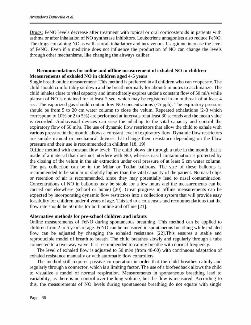
Arnaudova Danevska et al.
Page | 66
Drugs: FeNO levels decrease after treatment with topical or oral corticosteroids in patients with
asthma or after inhalation of NO synthetase inhibitors. Leukotriene antagonists also reduce FeNO.
The drugs containing NO as well as oral, inhallatory and intravenous L-arginine increase the level
of FeNO. Even if a medicine does not influence the production of NO can change the levels
through other mechanisms, like changing the airways caliber.
Recommendations for online and offline measurement of exhaled NO in children
Measurements of exhaled NO in children aged 4-5 years
Single breath online measurement: This method is preferred in all children who can cooperate. The
child should comfortably sit down and be breath normally for about 5 minutes to acclimatize. The
child inhales close to vital capacity and immediately expires under a constant flow of 50 ml/s while
plateau of NO is obtained for at least 2 sec. which may be registered in an outbreak of at least 4
sec. The vaporized gas should contain low NO concentrations (<5 ppb). The expiratory pressure
should be from 5 to 20 cm water column to close the velum. Repeated exhalations (2-3 which
correspond to 10% or 2 to 5%) are performed at intervals of at least 30 seconds and the mean value
is recorded. Audiovisual devices can ease the inhaling to the vital capacity and control the
expiratory flow of 50 ml/s. The use of dynamic flow restrictors that allow the child to exhale with
various pressure in the mouth, allows a constant level of expiratory flow. Dynamic flow restrictors
are simple manual or mechanical devices that change their resistance depending on the blow
pressure and their use is recommended in children [18, 19].
Offline method with constant flow level: The child blows air through a tube in the mouth that is
made of a material that does not interfere with NO, whereas nasal contamination is protected by
the closing of the velum in the air extraction under oral pressure of at least 5 cm water column.
The gas collection can be in the Milar or Tedlar balloons. The size of these balloons is
recommended to be similar or slightly higher than the vital capacity of the patient. No nasal clips
or retention of air is recommended, since they may potentially lead to nasal contamination.
Concentrations of NO in balloons may be stable for a few hours and the measurements can be
carried out elsewhere (school or home) [20]. Great progress in offline measurements can be
expected by incorporating dynamic flow restrictors into a collection system that will provide easy
feasibility for children under 4 years of age. This led to a consensus and recommendations that the
flow rate should be 50 ml/s for both online and offline [21].
Alternative methods for pre-school children and infants
Online measurements of FeNO during spontaneous breathing. This method can be applied to
children from 2 to 5 years of age. FeNO can be measured in spontaneous breathing while exhaled
flow can be adjusted by changing the exhaled resistance [22].This ensures a stable and
reproducible model of breath to breath. The child breathes slowly and regularly through a tube
connected to a two-way valve. It is recommended to calmly breathe with normal frequency.
The level of exhaled flow is adjusted to 50 ml/s (from 40-60) with continuous adaptation of
exhaled resistance manually or with automatic flow controllers.
The method still requires passive co-operation in order that the child breathes calmly and
regularly through a connector, which is a limiting factor. The use of a biofeedback allows the child
to visualize a model of normal respiration. Measurements in spontaneous breathing lead to
variability, as there is no control over the lung volume, but the flow is measured. According to
this, the measurements of NO levels during spontaneous breathing do not equate with single
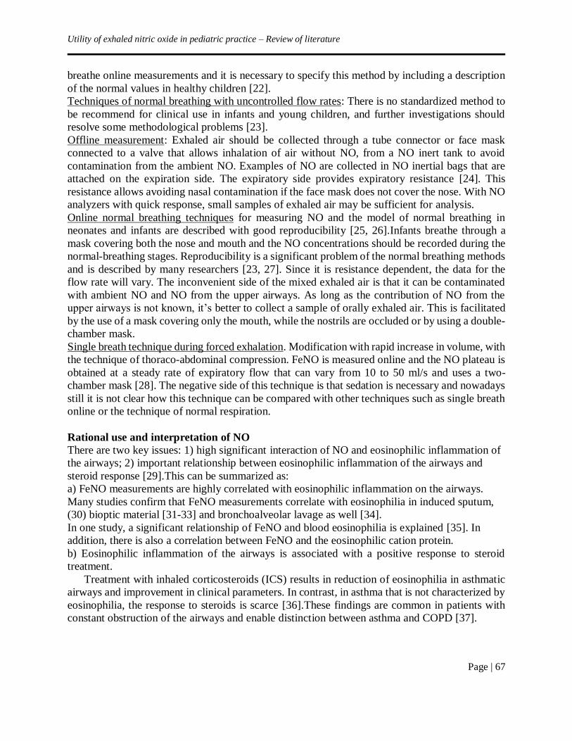
Utility of exhaled nitric oxide in pediatric practice – Review of literature
Page | 67
breathe online measurements and it is necessary to specify this method by including a description
of the normal values in healthy children [22].
Techniques of normal breathing with uncontrolled flow rates: There is no standardized method to
be recommend for clinical use in infants and young children, and further investigations should
resolve some methodological problems [23].
Offline measurement: Exhaled air should be collected through a tube connector or face mask
connected to a valve that allows inhalation of air without NO, from a NO inert tank to avoid
contamination from the ambient NO. Examples of NO are collected in NO inertial bags that are
attached on the expiration side. The expiratory side provides expiratory resistance [24]. This
resistance allows avoiding nasal contamination if the face mask does not cover the nose. With NO
analyzers with quick response, small samples of exhaled air may be sufficient for analysis.
Online normal breathing techniques for measuring NO and the model of normal breathing in
neonates and infants are described with good reproducibility [25, 26].Infants breathe through a
mask covering both the nose and mouth and the NO concentrations should be recorded during the
normal-breathing stages. Reproducibility is a significant problem of the normal breathing methods
and is described by many researchers [23, 27]. Since it is resistance dependent, the data for the
flow rate will vary. The inconvenient side of the mixed exhaled air is that it can be contaminated
with ambient NO and NO from the upper airways. As long as the contribution of NO from the
upper airways is not known, it’s better to collect a sample of orally exhaled air. This is facilitated
by the use of a mask covering only the mouth, while the nostrils are occluded or by using a double-
chamber mask.
Single breath technique during forced exhalation. Modification with rapid increase in volume, with
the technique of thoraco-abdominal compression. FeNO is measured online and the NO plateau is
obtained at a steady rate of expiratory flow that can vary from 10 to 50 ml/s and uses a two-
chamber mask [28]. The negative side of this technique is that sedation is necessary and nowadays
still it is not clear how this technique can be compared with other techniques such as single breath
online or the technique of normal respiration.
Rational use and interpretation of NO
There are two key issues: 1) high significant interaction of NO and eosinophilic inflammation of
the airways; 2) important relationship between eosinophilic inflammation of the airways and
steroid response [29].This can be summarized as:
a) FeNO measurements are highly correlated with eosinophilic inflammation on the airways.
Many studies confirm that FeNO measurements correlate with eosinophilia in induced sputum,
(30) bioptic material [31-33] and bronchoalveolar lavage as well [34].
In one study, a significant relationship of FeNO and blood eosinophilia is explained [35]. In
addition, there is also a correlation between FeNO and the eosinophilic cation protein.
b) Eosinophilic inflammation of the airways is associated with a positive response to steroid
treatment.
Treatment with inhaled corticosteroids (ICS) results in reduction of eosinophilia in asthmatic
airways and improvement in clinical parameters. In contrast, in asthma that is not characterized by
eosinophilia, the response to steroids is scarce [36].These findings are common in patients with
constant obstruction of the airways and enable distinction between asthma and COPD [37].

Arnaudova Danevska et al.
Page | 68
According to this, determination of the character of inflammation of the airways (eosinophilia)
is important in the initial management of patients with chronic respiratory symptoms in order to
identify those who would benefit from the steroid treatment.
c) Increased levels of FeNO predict positive steroid response in patients with non-specific
respiratory symptoms. The clinical benefit of increasing steroid treatment in patients with asthma
is higher in patients with increased levels of NO [38]. Some studies have shown that in determining
the outcome of treatment with inhaled Fluticasone, the level of FeNO as predictor is superior than
spirometry, bronchodilator test and bronchial hyperactivity. It is important that this study identifies
the optimal value for the steroid response to 47 ppb. This has been proved in studies in children
and adults [39, 40].
d) The use of ICS in asthma results in decreased levels of NO and according to that, the link
between ICS and FeNO is dose-dependent and has a significant correlation between changes in
FeNO and changes in eosinophilia in induced sputum and ICS therapy [41].
These data provide a fundamental confirmation that FeNO measurements play an important role
in the evaluation and treatment of patients with airway diseases. FeNO can be used as a surrogate
marker for diseases of the airways that are characterized by eosinophilia, such as atopic asthma,
asthma like coughing and eosinophilic bronchitis. Because of the close relationship between the
steroid response and eosinophilia in the airways, FeNO measurements play important role in
predicting and monitoring the response of the treatment with ICS [29].
Diagnosing airway diseases
Asthma: FeNO measurements are useful in distinguishing asthma from non-asthma [42]. The
test should be used as diagnostic tool when chronic symptoms with a duration of 6 weeks and more
are present, as the virus infection can lead to a false positive result. The predictive value is almost
identical with the cell count in induced sputum. The combination of the FeNO > 33 ppb and
abnormal spirometry (FEV1 <80% PV) provides great sensitivity (94%) and a specificity of 93%
for diagnosing asthma. Normal values do not exclude diagnosis of asthma. This confirms the
heterogeneity of asthma phenotype and FeNO measurements provide only one aspect of asthma
syndrome [43, 44].
Non-specific respiratory symptoms: FeNO measurements have a widespread role in the
examination of patients with undiagnosed chronic respiratory symptoms. Children with recurrent
wheezing bronchitis, cystic fibrosis, congenital abnormalities of the airways and primary ciliary
dyskinesia should also be considered. For the eosinophilic bronchitis and Cough variant asthma,
which are characterized by eosinophilic inflammation of the airways and elevated FeNO levels,
the positive effect of corticosteroid treatment is more likely. For other diagnoses, like vocal cord
dysfunction which is presented as asthma, many clinicians often give empirical steroid treatment
with very small benefit.
Pre-school children: Taking into account that spirometry and sputum induction cannot be
easily performed in pre-school children, non-invasive measurements of the airway inflammation
are potentially very useful.
The single breathing technique for measuring FeNO at this age is not suitable, so other
alternatives that vary from modification of standard online techniques to offline spontaneous
breathing method with flow control are developed [45-51]. Generally, these techniques are less
sensitive to discriminating between asthmatic and non-asthma patients [53, 53].

Utility of exhaled nitric oxide in pediatric practice – Review of literature
Page | 69
In acute wheezing episode FeNO level is significantly higher in those with recurrent wheezing
versus healthy controls, whereas in children with first wheezing episode FeNO levels do not differ
from normal children. Also, montelucast reduces the FeNO values of small children with early
onset of asthma [54, 55].
Atopy: There is a strong correlation between FeNO levels with total and specific IgE [56,
57].This suggests that asymptomatic atopic children may have moderate inflammation of the
airways leading to a raised FeNO level [58].
Chronic obstructive pulmonary disease (COPD):FeNO levels are non-consistent in patients
with COPD. This may be the result of smoking or the heterogeneity of the airways inflammation
[59, 60].
Cystic fibrosis (CF): In patients with CF, it has not been found that FeNO measurements can
be clinically useful. First, there is a reduced expression of nitric oxide synthetase in patients with
CF and second, elevated levels of nitrites are found in the breathing condensate of patients with
cystic fibrosis [61].
Primary Ciliary Dyskinesia (PCD): FeNO levels are significantly lower in these patients than
in healthy individuals. The nasal NO is extremely low in patients with PCD of all ages and
completely separates affected from unaffected individuals. The measurement of nasal NO can
probably become a screening test. The diagnostic sensitivity and specificity of nasal NO for PCD
ranges from 89 to 100%, and the specificity is from 97 to 100% [62, 63].This is due to the reduced
activity of NO synthetase and the mucus that affects the diffusion of NO from sinuses in the nasal
cavity or from epithelial cells to the lumen of the airways and makes the NO elimination more
difficult. Even young infants with PCD, have low nasal NO [64, 65]. Probably NO is also involved
in the stimulation of the ciliary motility. The nasal NO may play a role in non-specific immunity,
which includes a direct toxic effect on the microorganisms [66].
Lung transplantation: FeNO levels are increased in post-transplant patients with an unstable
lung function [67].
FeNO measurement for chronic asthma management
Prediction of exacerbations: The prognostic value of FeNO measurements for asthma
exacerbation seems to be limited. In a small study with steroidal reduction protocol, changes in
sputum eosinophils were superior comparing with FeNO for prediction of exacerbation. In the
second study, measurements of bronchial hyperactivity, sputum eosinophilia and FeNO
measurements ranged from a sensitivity of 21% (sputum eosinophils> 4%) to 65% in FeNO greater
than 10 ppb at a flow rate of 250 ml/s. Although positive predictive values ranged from 80 to 88%.
Therefore, it is preferable to examine more parameters than just one. The increase in FeNO of 60%
is considered to be optimal, but this has a 50% sensitivity with a positive predictive value of 83%.
These studies used a protocol to exclude steroids in order to imitate clinical exacerbations and so
they are not suitable [68, 69].
Predicting the outcome of ICS reduction in stable asthma: The main issue is when airway
inflammatory markers can be used to predict a successful reduction or exclusion of ICS treatment.
Studies showed that number of eosinophils in sputum was significantly more predictive than FeNO
measurements in asthma control over a period of 6 months and 16 weeks.
From the results of the many studies it can be concluded that the number of eosinophils in the
sputum (more than 1%) probably provides superior prognostic safety when it is necessary to
determine whether a patient needs to continue treatment with ICS.

Arnaudova Danevska et al.
Page | 70
In situations where induced sputum can not be obtained (some centers and small children), the
high FeNO level (> 50 ppb) probably predicts asthma relapse and a low FeNO level <20 ppb in
children (<25 in adults) predicts asthma stability if the measurement is obtained at least 4 weeks
after interruption or reduction in ICS in asymptomatic patient [70-73].
Increasing ICS doses: Correlation between airway inflammation and/or symptoms and/or
pulmonary function is very poor. So, the use of these treatment should be considered as the second
best [29].
Measurement and interpretation
Normal Values and Clinically Important Changes
In the past, studies showed that the upper limit is age dependent and ranges from 15,7 ppb for the
age of 4 years to 22,5 ppb for adolescents. The reason for this age-dependence is still unknown,
but it can be result of an increase of the surface area in the airways which is age-related, age-
depended induction of NO synthetase as a result of recurrent immunological stimulation or
progressive reduction of the constant exhalation level that is relatively high in children. So, that’s
why the upper limit for healthy adults is 33 ppb, while for school children is 25 ppb [74].
Measurement of FeNO level when asthma is stable may be the basic refrence point for a single
patient. It is considered that the mean value of FeNO change that occurs between stability and
"loss of control" after stopping ICS treatment is 16,9 ppb (or mean 24,9) [41].
Interpretation
Based on the currently available data, two algorithms for interpreting the FeNO results in everyday
practice had been developed. First, as a diagnostic tool and second for further management of
asthma (Shown on the tables):
1) Diagnostic use is pretty clear (Table 1).
Table 1. FeNO as a diagnostic tool FeNO
(ppb)
Level Eosinophil
inflammation
Children
5-25 Low (< 20 in less than 12 years, <25 in
more than 12 years)
Probably not Obstructive bronchitis, GER,
ENT disturbances, CF, PCD
(FeNO<5 ppb check nasal NO),
congenital airway anomalies,
other immune deficiencies
30-45 Intermediate Present but moderate Interpretation based on the clinic
presentation
50-65 High Significant Atopic asthma, if FEV1 <80%
PV, diagnosis for asthma is highly probable. Eosinophilic
bronchitis, a positive response to
steroid treatment.
2) In order to monitor patients correctly, increased levels of FeNO in symptomatic patients reffer
to uncontrolled eosinophilic inflammation. This is very often result of a weak compliance with
anti-inflammatory therapy or a poor inhalation technique than inadequate dosing of ICS. Low
levels of FeNO implicate the absence of eosinophilic inflammation of the airways. When we
assume that the result is not credible, as long as the patient has symptoms or has atopic asthma,
this may be due to the use of cigarets which reduces the level of FeNO up to 60%.
Utility of exhaled nitric oxide in pediatric practice – Review of literature
Page | 71
It is commonly referred to nonatopic (probably neutrophilic) asthma, gastroesophageal reflux
(GER), rhinosinusitis with postnasal drip or left heart failure (Table 2).
Table 2. FeNO results for management of asthma FeNO
(ppb)
Level Eosinophil
inflammation
Children
5-25 Low Probably not If there are symptoms revision of the diagnosis. Considering
Obstructive bronchitis, congenital airway anomalies, CF, PCD.
If there are no symptoms and uses ICS: good compliance with
treatment, reduction of the dose of ICS or, in case of a low dose,
exclusion of ICS
30-45 Intermediate Present but
moderate
If there are symptoms: infection as a cause of worsening, high
exposure to allergens, adding therapy other than ICS (for
example long-acting β agonist). Consider increasing ICS doses,
inadequate treatment with ICS, check compliance, check the
inhalation technique and consider a metered dose inhaler or
spacer if the patient uses a dry powder inhaler
If there are no symptoms: no changes in the ICS dose
50-65 High Significant If there are symptoms of inadequate treatment with ICS: check
the compliance, check the inhaler technique, inadequate ICS
dose, high exposure to allergens, exacerbation or relapse,
consider the metered dose inhaler or spacer if the patient uses an
inhaler with a dry powder
If there are no symptoms: no changes in the ICS dose.
The data shown in the tables is for guidance only. Further studies may lead to certain changes.
Patients with various clinical phenotypes may have different referent values of FeNO. Since
asthma is stable, FeNO levels may remain high. Prove that "normalization" of FeNO leads to a
clinical benefit is not confirmed. It may be preferable to have individual "FeNO typing" on the
refrent values [29].
CONCLUSION
FeNO measurement offers a step ahead in the assessment of the airway diseases. As the
"inflamometer," FeNO provides information about the nature of airway inflammation.
Measurements of FeNO are easily performed, they are reproducible and technically less expensive
than the analysis of induced sputum. FeNO results require careful consideration with the clinical
aspect. In symptomatic patients, high FeNO levels (> 50 ppb), refer to significant eosinophilia in
the airways, which will most likely respond to treatment with ICS.
The current data provides support for the diagnostic use of FeNO in children with symptoms
of asthma. Whether or not it needs to be used to predict the response of steroids or to guide the
need for ICS in small children with recurrent wheezing, is still not clear. For patients with chronic
and/or severe asthma, FeNO levels are useful for determining whether eosinophilic inflammation
of the airways is active or not. Both high (> 50ppb) and low (<25 ppb) levels of FeNO can be used
to for predicting the outcome in patients with a definitive history of asthma who are currently in
remission and who have stopped treatment with ICS. Depending of symptoms, at the same time,
high and low levels of FeNO offer assistance to clinicians in adjustment of the dose of ICS.

Arnaudova Danevska et al.
Page | 72
REFERENCES
1. ATS/ERS Recommendations for Standardized Procedures for the Online and Offline
Measurement of Exhaled Lower Respiratory Nitric Oxide and Nasal Nitric Oxide, 2005
2. Green RH, Brightling CE, Wardlaw AJ, Pavord ID. Asthma exacerbations and sputum
eosinophil counts. Lancet. 2003;361:1303. 3. Smith AD, Cowan JO, Filsell S, McLachlan C, Monti-Sheehan G, Jackson P, Taylor DR.
Diagnosing asthma: comparisons between exhaled nitric oxide measurements and
conventional tests. Am J RespirCrit Care Med. 2004;169:473–478.
4. Beck-Ripp J, Griese M, Arenz S, Koring C, Pasqualoni B, Bufler P. Changes of exhaled
nitric oxide during steroid treatment of childhood asthma. EurRespir J. 2002;19:1015–
1019.
5. Jones SL, Kittelson J, Cowan JO, Flannery EM, Hancox RJ, McLachlan CR, Taylor DR.
The predictive value of exhaled nitric oxide measurements in assessing changes in asthma
control. Am J RespirCrit Care Med. 2001;164:738–743.
6. Harkins MS, Fiato KL, Iwamoto GK. Exhaled nitric oxide predicts asthma exacerbation. J
Asthma. 2004;41:471–476.
7. Pijnenburg MW, Hofhuis W, Hop WC, De Jongste JC. Exhaled nitric oxide predicts asthma
relapse in children with clinical asthma remission. Thorax. 2005;60:215–218.
8. Deykin A, Massaro AF, Drazen JM, Israel E. Exhaled nitric oxide as a diagnostic test for
asthma: online versus offline techniques and effect of flow rate. Am J Respir Crit Care
Med. 2002;165:1597–1601.
9. Franklin PJ, Stick SM, LeSouef PN, Ayres JG, Turner SW. Measuring exhaled nitric oxide
levels in adults: the importance of atopy and airway responsiveness. Chest.
2004;126:1540–1545.
10. Grasemann H, Van’s Gravesande KS, Buscher R, Drazen JM, Ratjen F. Effects of sex and
of gene variants in constitutive nitric oxide synthases on exhaled nitric oxide. Am J
RespirCrit Care Med. 2003; 167(8):1113–1116.
11. Oguzulgen IK, Turktas H, Erbas D. Airway inflammation in premenstrual asthma. J
Asthma. 2002;39:517–522.
12. Johannesson M, Ludviksdottir D, Janson C. Lung function changes in relation to menstrual
cycle in females with cystic fibrosis. Respir Med. 2000;94:1043–1046.
13. Kissoon N, Duckworth LJ, Blake KV, Murphy SP, Lima JJ. Effect of beta2-agonist
treatment and spirometry on exhaled nitric oxide in healthy children and children with
asthma. PediatrPulmonol. 2002;34: 203–208.
14. Deykin A, Massaro AF, Coulston E, Drazen JM, Israel E. Exhaled nitric oxide following
repeated spirometry or repeated plethysmography in healthy individuals. Am J RespirCrit
Care Med. 2000;161:1237–1240
15. Bruce C, Yates DH, Thomas PS. Caffeine decreases exhaled nitric oxide. Thorax.
2002;57:361–363.
16. Mattes J, van’s Gravesande KS, Moeller C, Moseler M, Brandis M, Kuehr J. Circadian
variation of exhaled nitric oxide and urinary eosinophil protein X in asthmatic and healthy
children. Pediatr Res. 2002;51(2):190–194.
17. Horvath I, Donnelly LE, Kiss A, Balint B, Kharitonov SA, Barnes PJ. Exhaled nitric oxide
and hydrogen peroxide concentrations in asthmatic smokers. Respiration (Herrlisheim).
2004;71:463–468.
Utility of exhaled nitric oxide in pediatric practice – Review of literature
Page | 73
18. Pedroletti C, Zetterquist W, Nordvall L, Alving K. Evaluation of exhaled nitric oxide in
schoolchildren at different exhalation flow rates. Pediatr Res. 2002;52:393–398.
19. Kharitonov SA, Gonio F, Kelly C, Meah S, Barnes PJ. Reproducibility of exhaled nitric
oxide measurements in healthy and asthmatic adults and children. Eur Respir J.
2003;21:433–438.
20. Jobsis Q, Schellekens SL, Kroesbergen A, Hop WC, de Jongste JC. Off-line sampling of
exhaled air for nitric oxide measurement in children: methodological aspects. Eur Respir
J. 2001;17:898–903.
21. Baraldi E, de Jongste JC. Measurement of exhaled nitric oxide in children Eur Respir J.
2002;20(1):223-37.
22. Buchvald F, Bisgaard H. FeNO measured at fixed exhalation flow rate during controlled
tidal breathing in children from the age of 2 yr. Am J Respir Crit Care Med. 2001;163:699–
704.
23. Franklin PJ, Turner SW, Mutch RC, Stick SM. Measuring exhaled nitric oxide in infants
during tidal breathing: methodological issues. PediatrPulmonol. 2004;37:24–30.
24. Avital A, Uwyyed K, Berkman N, Godfrey S, Bar-Yishay E, Springer C. Exhaled nitric
oxide and asthma in young children. PediatrPulmonol. 2001;32:308–313.
25. Ratjen F, Kavuk I, Gartig S, Wiesemann HG, Grasemann H. Airway nitric oxide in infants
with acute wheezy bronchitis. Pediatr Allergy Immunol. 2000;11:230–235.
26. Hall GL, Reinmann B, Wildhaber JH, Frey U. Tidal exhaled nitric oxide in healthy,
unsedated newborn infants with prenatal tobacco exposure. J Appl Physiol. 2002;92:59–
66
27. Franklin PJ, Turner SW, Mutch RC, Stick SM. Comparison of singlebreath and tidal
breathing exhaled nitric oxide levels in infants. Eur Respir J. 2004;23:369–372.
28. Martinez TM, Weist AD, Williams T, Clem C, Silkoff P, Tepper RS. Assessment of
exhaled nitric oxide kinetics in healthy infants. J Appl Physiol. 2003;94:2384–2390.
29. Taylor DR, Pijnenburg MW, Smith AD, Jongste JCD. Exhaled nitric oxide measurements:
clinical application and interpretation. Thorax. 2006;61:817–827.
30. Berlyne GS, Parameswaran K, Kamada D, et al. A comparison of exhaled nitric oxide and
induced sputum as markers of airway inflammation. J Allergy Clin Immunol.
2000;106:638–44.
31. Brightling CE, Symon FA, Birring SS, et al. Comparison of airway immunopathology of
eosinophilic bronchitis and asthma. Thorax. 2003;58:528–32.
32. Payne DN, Adcock IM, Wilson NM, et al. Relationship between exhaled nitric oxide and
mucosal eosinophilic inflammation in children with difficult asthma, after treatment with
oral prednisolone. Am J Respir Crit Care Med. 2001;164:1376–81.
33. van den Toorn LM, Overbeek SE, de Jongste JC, et al. Airway inflammation is present
during clinical remission of atopic asthma. Am J RespirCrit Care Med. 2001;164:2107–13.
34. Warke TJ, Fitch PS, Brown V, et al. Exhaled nitric oxide correlates with airway eosinophils
in childhood asthma. Thorax. 2002;57:383–7.
35. Strunk RC, Szefler SJ, Phillips BR, et al. Relationship of exhaled nitric oxide to clinical
and inflammatory markers of persistent asthma in children. J Allergy Clin Immunol.
2003;112:883–92.

Arnaudova Danevska et al.
Page | 74
36. Green RH, Brightling CE, Woltmann G, et al. Analysis of induced sputum in adults with
asthma: identification of subgroup with isolated sputum neutrophilia and poor response to
inhaled corticosteroids. Thorax. 2002;57:875–9.
37. Brightling CE, Monteiro W, Ward R, et al. Sputum eosinophilia and short-term response
to prednisolone in chronic obstructive pulmonary disease: a randomised controlled trial.
Lancet. 2000;356:1480–5.
38. Little SA, Chalmers GW, MacLeod KJ, et al. Non-invasive markers of airway
inflammation as predictors of oral steroid responsiveness in asthma. Thorax. 2000;55:232–
4.
39. Smith AD, Cowan JO, Brassett KP, et al. Exhaled nitric oxide: a predictor of steroid
response. Am J Respir Crit Care Med. 2005;172:453–9.
40. Szefler SJ, Phillips BR, Martinez FD, et al. Characterization of within-subject responses to
fluticasone and montelukast in childhood asthma. J Allergy Clin Immunol. 2005;115:233–
42.
41. Jones SL, Kittelson J, Cowan JO, et al. The predictive value of exhaled nitric oxide
measurements in assessing changes in asthma control. Am J Respir Crit Care Med.
2001;164:738–43.
42. Deykin A , Massaro AF, Drazen JM, et al. Exhaled nitric oxide as a diagnostic test for
asthma: online versus offline techniques and effect of flow rate. Am J Respir Crit Care
Med. 2002;165:1597–601.
43. Smith AD, Cowan JO, Filsell S, et al. Diagnosing asthma: comparisons between exhaled
nitric oxide measurements and conventional tests. Am J Respir Crit Care Med.
2004;169:473–8.
44. Smith AD, Taylor DR. Is exhaled nitric oxide measurement a useful clinical test in
asthma? CurrOpin Allergy Clin Immunol. 2005;5:49–56.
45. Artlich A , Jonsson B, Bhiladvala M, et al. Single breath analysis of endogenous nitric
oxide in the newborn. Biol Neonate. 2001;79:21–6.
46. BaraldiE ,Scollo M, Zaramella C, et al. A simple flow-driven method for online
measurement of exhaled NO starting at the age of 4 to 5 years. Am J RespirCrit Care Med.
2000;162:1828–32.
47. Daniel PF, Klug B, Valerius NH. Measurement of exhaled nitric oxide in young children
during tidal breathing through afacemask. Pediatr Allergy Immunol. 2005;16:248–53.
48. Buchvald F , Bisgaard H. FeNO measured at fixed exhalation flow rate during controlled
tidal breathing in children from the age of 2 yr. Am J Respir Crit Care Med. 2001;163:699–
704.
49. JobsisQ ,Schellekens SL, Kroesbergen A, et al. Off-line sampling of exhaled air for nitric
oxide measurement in children: methodological aspects. EurRespir J. 2001;17:898–903.
50. ЈobsisQ ,Raatgeep HC, Hop WC, et al. Controlled low flow off line sampling of exhaled
nitric oxide in children. Thorax. 2001;56:285–9.
51. Ratjen F , Kavuk I, Gartig S, et al. Airway nitric oxide in infants with acute wheezy
bronchitis. Pediatr Allergy Immunol2000;11:230–5.
52. BaraldiE , de Jongste JC. Measurement of exhaled nitric oxide in children. Eur Respir J.
2002;20:223–37.
53. ATS/ERS. Recommendations for standardized procedures for the online and offline
measurement of exhaled lower respiratory nitric oxide and nasal nitric oxide, 2005. Am J
RespirCrit Care Med. 2005;171:912–30.
Utility of exhaled nitric oxide in pediatric practice – Review of literature
Page | 75
54. Straub DA, Moeller A, Minocchieri S, et al. The effect of montelukast on lung function
and exhaled nitric oxide in infants with early childhood asthma. EurRespir J. 2005;25:289–
94.
55. Straub DA, Minocchieri S, Moeller A, et al. The effect of montelukast on exhaled nitric
oxide and lung function in asthmatic children 2 to 5 years old. Chest. 2005;127:509–14.
56. van Amsterdam JG, Janssen NA, de Meer G, et al. The relationship between exhaled nitric
oxide and allergic sensitization in a random sample of school children. ClinExp Allergy.
2003;33:187–91.
57. Saito J, Inoue K, Sugawara A, et al. Exhaled nitric oxide as a marker of airway
inflammation for an epidemiologic study in schoolchildren. J Allergy Clin Immunol.
2004;114:512–6.
58. van den Toorn LM, Prins JB, de Jongste JC, et al. Benefit from anti-inflammatory treatment
during clinical remission of atopic asthma. Respir Med. 2005;99:779–87.
59. CliniE , Bianchi L, Vitacca M, et al. Exhaled nitric oxide and exercise in stable COPD
patients. Chest. 2000;117:702–7.
60. Delen FM, Sippel JM, Osborne ML, et al. Increased exhaled nitric oxide in chronic
bronchitis: comparison with asthma and COPD. Chest. 2000;117:695–701.
61. Downey D ,Elborn JS. Nitric oxide, iNOS, and inflammation in cystic fibrosis. J Pathol.
2000;190:115–6.
62. NarangI ,Ersu R, Wilson NM, et al. Nitric oxide in chronic airway inflammation in
children: diagnostic use and pathophysiological significance. Thorax. 2002;57:586–9.
63. Corbelli R , Bringolf-Isler B, Amacher A, et al. Nasal nitric oxide measurements to screen
children for primary ciliary dyskinesia. Chest. 2004;126:1054–9.
64. CsomaZ , Bush A, Wilson NM, et al. Nitric oxide metabolites are not reduced in exhaled
breath condensate of patients with primary ciliary dyskinesia. Chest. 2003;124:633–8.
65. BaraldiE , Pasquale MF, Cangiotti AM, et al. Nasal nitric oxide is low early in life: case
study of two infants with primary ciliary dyskinesia. EurRespir J. 2004;24:881–3.
66. Sanders SP, Proud D, Permutt S, et al. Role of nasal nitric oxide in the resolution of
experimental rhinovirus infection. J Allergy Clin Immunol. 2004;113:697–702.
67. GabbayE , Walters EH, Orsida B, et al. Post-lung transplant bronchiolitis obliterans
syndrome (BOS) is characterized by increased exhaled nitric oxide levels and epithelial
inducible nitric oxide synthase. Am J Respir Crit Care Med. 2000;162:2182–7.
68. JatakanonA , Lim S, Barnes PJ. Changes in sputum eosinophils predict loss of asthma
control. Am J RespirCrit Care Med. 2000;161:64–72.
69. Harkins MS, Fiato KL, Iwamoto GK. Exhaled nitric oxide predicts asthma exacerbation.J
Asthma. 2004;41:471–6.
70. Leuppi JD, Salome CM, Jenkins CR, et al. Predictive markers of asthma exacerbation
during stepwise dose reduction of inhaled corticosteroids. Am J RespirCrit Care Med.
2001;163:406–12.
71. DeykinA , Lazarus SC, Fahy JV, et al. Sputum eosinophil counts predict asthma control
after discontinuation of inhaled corticosteroids. J Allergy Clin Immunol. 2005;115:720–7.
72. ZacharasiewiczA , Wilson N, Lex C, et al. Clinical use of noninvasive measurements of
airway inflammation in steroid reduction in children. Am J RespirCrit Care Med.
2005;171:1077–82.

Arnaudova Danevska et al.
Page | 76
73. Pijnenburg MW, Hofhuis W, Hop WC, et al. Exhaled nitric oxide predicts asthma relapse
in children with clinical asthma remission. Thorax. 2005;60:215–8.
74. BuchvaldF ,Baraldi E, Carraro S, et al. Measurements of exhaled nitric oxide in healthy
subjects age 4 to 17 years. J Allergy Clin Immunol. 2005;115:1130–6.
INSTRUCTIONS FOR AUTHORS
Page | 77
These guidelines are in accordance with the “Uniform Requirements for Manuscripts
Submitted to Biomedical Journals”. (Complete document available at www.icmje.org )
Manuscripts are accepted for processing if neither the article nor any essential part, tables or
figures, has been or will be published or submitted elsewhere before presenting in Acta
Morphologica. This restriction does not apply to abstracts or press reports related to scientific
meetings. The Editors will consider both invited and uninvited review articles. Authors should
detail how their work differs from existing reviews on subject in cover letter.
Manuscripts/General Guidelines
The manuscript should conform the guidelines set forth in the “Uniform Requirements for
Manuscripts Submitted to Biomedical Journals”, 5th edition, New Engl J Med 1997; 336 (4):
309–315.
Manuscript must contain no more than 5000 words. A cover letter signed by all authors should
identify the person (post address, telephone number, and e-mail address) responsible for
negotiations. Each author must sign a statement attesting that he or she fulfills the authorship
criteria of the Uniform Requirements. Each author must significantly contribute to the
submitted work.
Form of Manuscript
Three copies of each manuscript, along with a disk (see “Instructions for Electronic Manuscript
Submission”), must be submitted in English, in double-spaced typewritten form with a 5-cm
(2-inch) left margin. (Do not use “erasable” bond.) The text should be written in following
sequence: Introduction, Methods, Results, Discussion, Acknowledgement, References, Tables,
Illustrations and Figure Legends, Structured Abstract with key words and Condensed Abstract.
Page 1 should bear an article title, name(s) of the author(s) and institution where the work was
done and a person whom proofs and reprint request should be sent, with complete address
(including postal codes), telephone number and email address (address for correspondence).
Tables should be typed neatly, each on a separate sheet, with title above and any notes below.
All abbreviations should be explained. Do not provide duplicite information in tables and
figures.
Illustrations should be submitted as clear glossy prints (two duplicate sets may be
photocopied), with lettering large enough to be legible if reduced. The maximal final size of
any figure in the printed journal will be 20 by 28 cm (8.25x11inch). On the back of each figure,
the name of author and the figure number should be writen, with the top indicated by an arrow.
Each figure should have a separate, fully explicit legend; all parts of the figure and all
abbreviations and symbols should be clearly defined. Figure legends should be typed on
separate pages; figure numbers must follow their reference in text.
Drug names. Generic names should be used; trade names may be given in parentheses in the
first mention, and generic names should be used thereafter.
Abbreviations. The list of abbreviations given in “Uniform Requirements for Manuscripts
Submitted to Biomedical Journals” (section References) should be followed. For additional
abbreviations, consult the CBE Style Manual (available from the Council of Biology Editors,
9650 Rockville Pike, Bethesda, Maryland 20814, U.S.A.) or other standard sources.

INSTRUCTIONS FOR AUTHORS
Page | 78
References
The journal complies with the reference style given in “Uniform Requirements for Manuscripts
Submitted to Biomedical Journals”. References should be cited in text by number and
numbered in order they are cited. The reference should by written in double-spaced form at the
end of the text, following the sample formats given below. For the abbreviations of journal
names, refer to the List of Journals Indexed in Index Medicus (available from the
Superintendent of Documents, U.S. Government Printing Office, Washington, D.C. 20402,
U.S.A., DHEW Publication No. NIH 83-267; ISSN 0093-3821). Provide all names of authors
when fewer than seven: when seven or more, list the first three and add et al. Provide article
titles and inclusive pages. The author is responsible for the accuracy of reference data.
Article: 1. Greenblatt DJ, Abernethy DR, Shader Jr RI. Pharmacokinetic aspects of drug therapy in the
elderly (commentary).Ther Drug Monit. 1986;8(6):249–55.
Book: 2.Mitchell JR, Horning MG, (editors). Drug metabolism and drug toxicity. 2nd ed. New York:
Raven Press; 1984.
Chapter of book: 3. Kutt H, Pippenberg CE et al. Plasma clearance of nor-methsuximide in a uremic patient.
223-226. In: Levy RH, Pitlick WH, Meijer J, (editorss). Metabolism of antiepileptic drugs.
New York: Raven Press; 1984. pp-1-25.
Internet:
Greenblatt DJ, Abernethy DR, Shader Jr RI. Pharmacokinetic aspects of drug therapy in the
elderly (commentary). Ther Drug Monit 1986; 8 (6): 249-55. Available
from:http://www.med.monash.edu.au/medical
Structured Abstract A structured abstract should be provided on a separate page with no more than 250 words,
presenting essential data in five paragraphs introduced by separate headings in following order:
Objectives, Background, Methods, Results, Conclusion. Complete sentences should be used.
All data in the structured abstract must be present also in the submitted text or tables. Three to
five key words should be added. Terms from Index Medicus should be used.
Condensed Abstract (for table of contents)
A condensed abstract of no more than 50 words should be provided for the expanded table of
contents, stressing clinical implications. Do not include data which are not present in the text
or tables.
Proofs
Proof must be returned within 3 days; late return may cause a delay in publication. Please check
text, tables, legends, and references carefully.

INSTRUCTIONS FOR AUTHORS
Page | 79
Instructions for Electronic Manuscript Submission The preferred manuscript submission is via journal’s e-mail address:
[email protected]. Each submitted manuscript must be clearly labeled with the
name of the first author and article title.
The manuscript submitted on a disk must be in the final corrected version and must agree with the
final accepted version of the submitted paper manuscript. The submitted disk should contain only
the final version of the manuscript. Delete all other material from the disk. Please follow the
general instructions on style/arrangement and, in particular, the reference style as given in
“Instruction to Authors”.
Note, that while the paper version of the manuscript must be presented in the traditional double
spaced format, the electronic version will be typeset and should not contain extraneous formatting
instructions. Do not use tabs or extra space at the beginning of a paragraph or for list entries. Do
not indent runover lines in references. Turn off line spacing. Do not specify page breaks, page
numbers, or headers. Do not specify typeface.
Take care to enter “one” (1) and lower case “el” (1), as well as “zero” (0) and capital “oh” (O)
correctly.
Please note the following conventions on dashes: Use a single hyphen with space before it for a
minus sign, use a double hyphen (with space before and after) to indicate a “long dash” in text,
and a triple hyphen (with no extra space) to indicate a range of numbers (e.g. “23–45”).
Non-standard characters (Greek letters, mathematical symbols, etc.) should be coded consistently
throughout the text. Please make a list and provide a listing of the used codes.
Authors agree to execute copyright transfer forms as requested. Authors should express all
measurements in conventional units, with Systéme International (SI) units given in parentheses
throughout the text. Conventional units should be used in figures and tables, with conversion
factors given in legends or footnotes.
In electronic manuscript submission text editor Word 6 or higher is recommended (editor T602
is possible). Text should be aligned left (not justified), without hyphenation, without bullets,
numbering and underlines, without extra hard returns at the end of line (only at the end of
paragraphs). One type of Word paragraph should be used throughout the text. Word graphic
experiments should not be used.
Word tables: do not use vertical lines, unless it is necessary. Provide tables as a separate file (do
not place in text).
Excel graphs: provide as Excel file.
Word graphs: provide as a separate Word file (do not place in text!)
Table and graph legends should be provided separately at the end of the text.

INSTRUCTIONS FOR AUTHORS
Page | 80
Graphs should be processed for black and white print. Graphs printed on laser or ink printers
could not serve as templates– always provide original electronic files!
Figures: provide original or scan. Scan to 600-800 dpi! – set to B/W or line art.
Figures – black and white photos – provide high-quality original or scan to 350 dpi
Figures – color photos — provide high-quality original or scan to 350 dpi
Figures scanned to 72 or 96 dpi are not suitable for print!
On principle, do not place scans in text. Always provide original figures in tif or jpg format (with
minimal compression). Placing scan in Word text causes a loss of quality!
Figure legends should be provided as a separate text file.
Do not place figures in PowerPoint – this application is meant for presentations and it is not
possible to use it as a template for print!
Figures from digital camera should not be placed in text. Provide them in tif or jpg format
(with minimal compression)! Transciption of Macedonian Cyrillic Alphabet into English Latin On
the basis of ISO Recomandation R-9-1968 International List of Periodical Title Abbreviations
(1970).
Transciption of Macedonian Cyrillic Alphabet into English Latin
On the basis of ISO Recomandation R-9-1968 International List of Periodical Title Abbreviations (1970)
AN EXCLUSIVE STATEMENT
Page | 81