Embed Size (px)
Citation preview

Muscular System
“The Machines of the Body”

Muscle Introduction
Muscles make up the bulk of the body –about one-third of its weight
Their ability to contract not only enables the body to move, but also provides the force that pushes substances, such as blood and food, through the body.
Without the muscular system, none of the other organ systems would be able to function.

Functions of Muscular System
External and internal body movement
Maintaining posture
Stabilizing joints
Generation of heat (shivering)
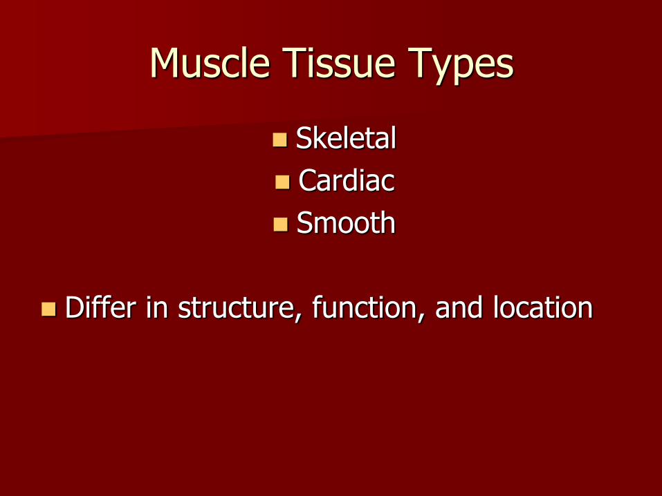
Muscle Tissue Types
Skeletal
Cardiac
Smooth
Differ in structure, function, and location

How are the types the same?
All muscle cells are elongated – called muscle fibers (thousands depending on muscle)
Ability of muscle to shorted depends on two type of myofilaments
Terminology – – Myo or mys = muscle
– Sarco = flesh

Skeletal Muscle
Body Location = attached to bone or to skin
Appearance = single, long, cylindrical, multinucleated, striated
Control = voluntary
Speed of Contraction = slow to fast

Cardiac Muscle
Body Location = walls of heart
Appearance = branching chains, uninucleated, striated
Control = involuntary
Speed of Contraction = slow

Smooth Muscle
Body Location = walls of hollow organs
Appearance = single, tapered at each end, uninucleated, not striated
Control = involuntary
Speed of Contraction = very slow

Different parts of a muscular organ
belly: bulging part of a muscle
origin (head): the less moveable attachment (there can be more than one origin)
insertion: the moveable attachment

Types of Body Movements Flexion = decrease in joint angle and brings two
bones closer together
Extension = increase in joint angle and brings two bones farther apart
Pronation = moving from upward facing or anterior to downward facing or posterior
Supination = moving from posterior position to anterior position – Like your holding a cup of soup

Abduction = moving a limb away from midline of body
Adduction = moving a limb toward the midline of body
Circumduction = combination of flexion, extension, adduction, and abduction
Dorsiflexion = movement of ankle bringing the toes up toward the shin
– Standing on your heels
Plantarflexion = movement of ankle causing the toes to point down
– Standing on your toes

Types of Muscles
Muscles can’t push, they can only pull as they contract
Movement is the result of pairs or teams of muscles working together

Types of Muscles
1) Prime Movers – when several muscles are contracting at once, it the muscle that has the major responsibility for causing the movement
2) Antagonists – muscles that oppose or reverse a movement
3) Synergists – help prime movers by making same movement or reduce other unnecessary movements
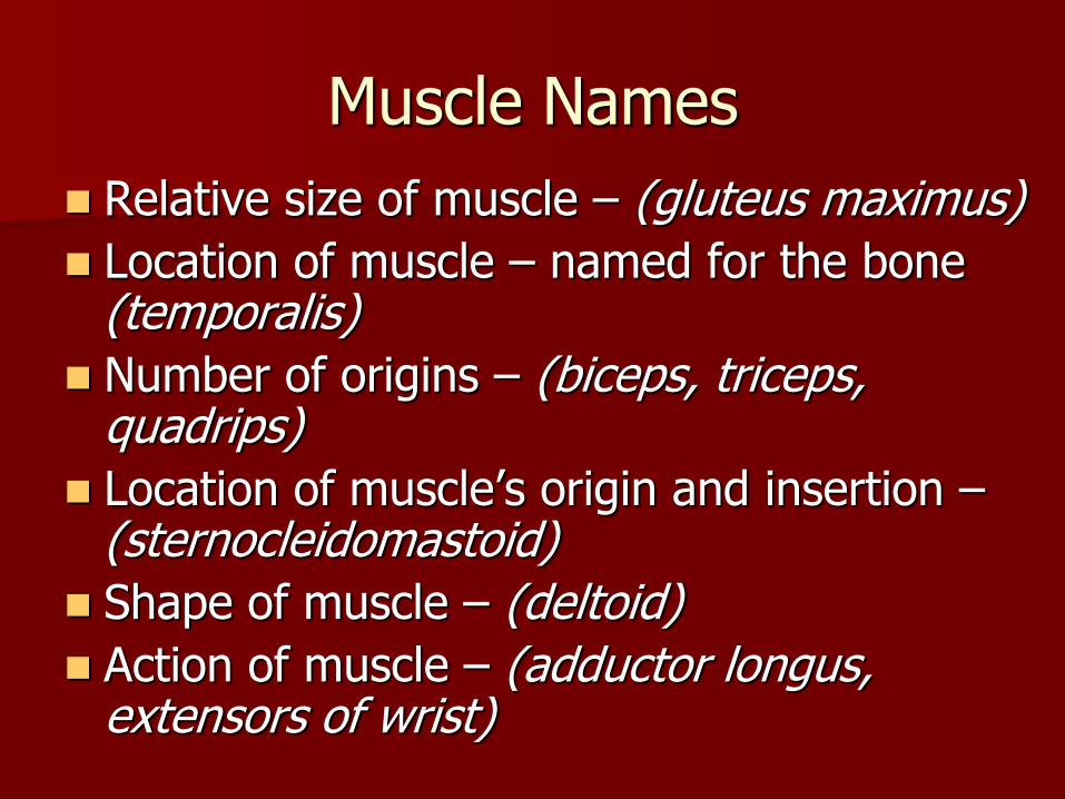
Muscle Names
Relative size of muscle – (gluteus maximus)
Location of muscle – named for the bone (temporalis)
Number of origins – (biceps, triceps, quadrips)
Location of muscle’s origin and insertion – (sternocleidomastoid)
Shape of muscle – (deltoid)
Action of muscle – (adductor longus, extensors of wrist)

Muscle Activity
With a partner, demonstrate the following movements at different joints in the body.
Write down what muscles are responsible for each movement

Neck: flexion and extension
Shoulder: adduction and abduction
Elbow: flexion and extension
Wrist: adduction and abduction
Knee: flexion and extension
Ankle: dorsiflexion and plantar flexion

General Skeletal Muscle Structure
Muscle tissue = Muscle fibers, as well as large amounts of connective tissue, blood vessels, and nerves
Connective tissue covers and supports each muscle fiber and reinforces the muscle as a whole

Health of muscle depends on a sufficient nerve and blood supply. Each skeletal muscle has a nerve ending that controls its activity.
Active muscles use a lot of energy and require a continuous supply of oxygen and nutrients
– supplied by arteries
– muscles produce large amounts of metabolic waste that must be removed by veins

Organization of Skeletal Muscle
Fascia – fibrous connective tissue under the hypodermis that surrounds functional groups of the muscle
Single muscles are surrounded by tough, dense connective epimysium - which extends and merges with the tendon (epi = upon) (myo = muscle)
Tendon attaches to periosteum (covers bone)
Epimysium surrounds many fascicles (bundles)

A single fascicle is surrounded by perimysium (collagenic) (peri = around).
A fascicle contains endomysium (areolar tissue) and muscle fibers (muscle cells).
Endomysium surrounds muscle fibers (cells)
Muscle fibers are long cylindrical cells containing myofilaments
Myofilaments are proteins which are part of the functional contractile unit of skeletal muscle, known as the sarcomere


Microscopic Anatomy of Muscle

Parts of Muscle Fiber Many Nuclei Cell membrane = sarcolemma Myofibrils Cytoplasm Mitochondria Sarcoplasmic reticulum Transverse tubules – T tubes
Myofibrils contain light and dark bands depending
on where the actin and myosin are located
Myofibrils = chains of tiny contractile units called sarcomeres


TWO Types of Protein Filaments
Thick ones and Thin ones:
THICK FILAMENTS are made up of a PROTEIN called MYOSIN
THIN FILAMENTS are made of a PROTEIN called ACTIN

Myosin and Actin Filaments are arranged to form overlapping patterns
Theses are responsible for the Light and Dark Bands that can be seen in Skeletal Muscle (Striated Appearance)

Sarcomere – tiny contractile units
The structural and functional unit of skeletal muscle
Actin is surrounded by the T-and-T system (troponin and tropomyosin)
Myosin has extensions called heads, which can attach at actin binding sites reference points of a sarcomere

Zones of Sarcomere
Z-line: the terminating end of a sarcomere (middle of one I-band)
I-band (light): contains actin only
A-band (dark): contains actin and myosin
H-zone: contains myosin only

Muscle Physiology

Muscles are able to contract because of irritability and
contractility (ability to shorten)
Steps to Muscle Contraction
1) Electrical impulse starts in spinal cord, travels down axon of motor neuron to the axonal terminal – caused by depolarization, Na in
2) Electrical impulse reaches the neuromuscular junction and the neurotransmitter Acetylcholine (ACh) is released from the vesicles of the neuron into the synaptic cleft

3) Acetylcholine binds to a protein and temporarily causes the sarcolemma to be permeable to Na+ and Na+ rushes into the muscle fiber
4) Upset in Na+ levels generates an action potential that doesn’t stop until it travels the length of the cell membrane

Sliding Filament Theory
5) Meanwhile, the action potential causes Ca+ to be released from the sarcoplasmic reticulum and binds to the T and T system of actin
6) Ca+ causes the proteins to move, which exposes the binding sites for the heads of the myosin

7) Myosin heads attach to actin and pivot which shortens the sarcomere. Heads release and reattach causing more shortening. This process requires ATP.
8) Myosin never completely lets go of the actin, which causes the actin not to lengthen in between pivots of the myosin heads.

9) Ca+ and electrical impulse will cause the shortening of the muscle fibers and contract the muscle.

Muscle Relaxation
Acetylcholine is broken down by enzymes as soon as action potential has passed. Single electrical impulse causes only one contraction.
Sarcoplasmic reticulum begins to pump Ca+ back into its sacs
As Ca+ is stripped from the T and T system on the actin, the proteins get returned to their original positions
Myosin heads can’t bind to sites on actin anymore
Thick and thin filaments are no longer attached, and slide past one another and back to original resting length

Graded Response
“All or None” Law – muscle cell will contract to its fullest when adequately stimulated
– Never partially contracts
Graded Response – degrees of shortening
1) Changing speed of muscle contraction
2) Changing number of muscle cells being stimulated
Muscle Response to Increasingly Rapid Stimulation
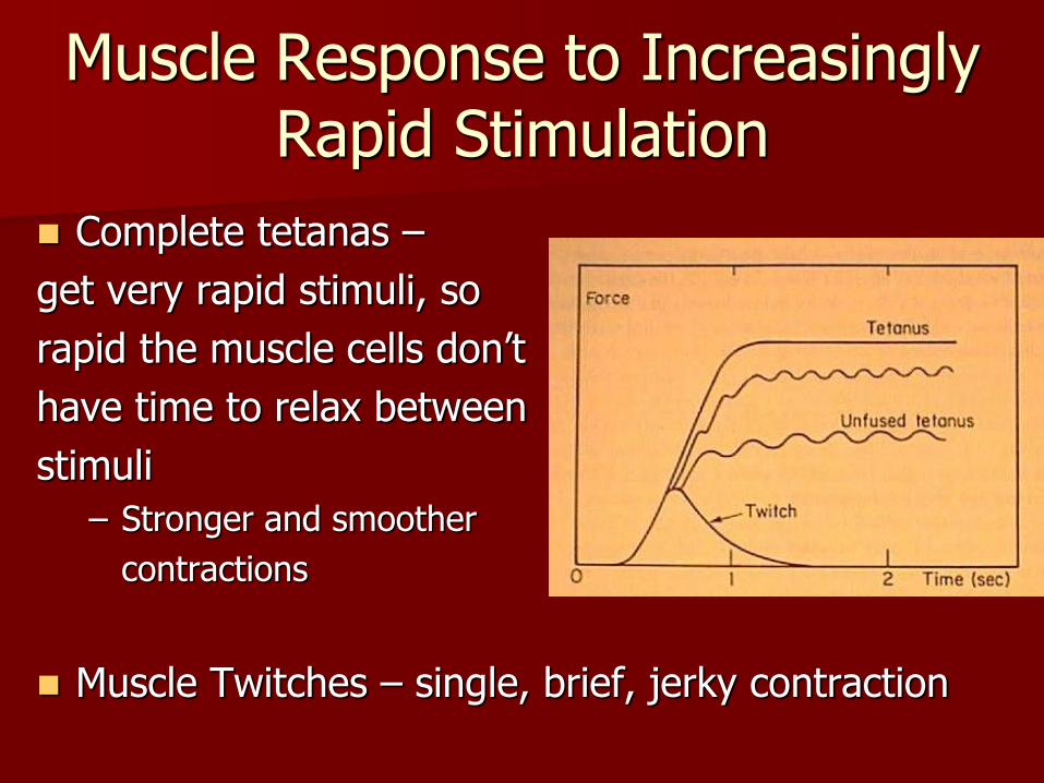
Complete tetanas –
get very rapid stimuli, so
rapid the muscle cells don’t
have time to relax between
stimuli
– Stronger and smoother
contractions
Muscle Twitches – single, brief, jerky contraction

Muscle Response to Stronger Stimuli
When a few muscle cells are stimulated = contraction is slight
When all muscle cells are stimulated = contraction is strong
“The same hand that soothes can deliver a stinging slap”

Energy for Muscle Contractions
Need ATP to cause contractions – body only stores 4 to 6 seconds worth
So, out body has to regenerate the ATP
Our body does this by three pathways:

1)Direct phosphorylation of ADP by creatine phosphate
- CP + ADP Creatine + ATP
- Creatine Monohydrate- natural supplement that helps your body make more ATP which delays the development of lactic acid as you workout

2) Aerobic Respiration – needs O2
- Glucose + O2 H2O + CO2 + ATP
- Glycolysis Krebs Cycle Electron Transport Chain Makes 38 ATP
– first pathway the body uses, but it is slow

3) Anaerobic glycolysis and lactic acid formation – does not need O2
- Glycolysis No O2 Lactic Acid Makes
2 ATP
– working muscles requiring more nutrients that the body has to offer
– produces less ATP per glucose, but can last for 20 to 30 sec of strenuous activity
– drawbacks: use up glucose and accumulates lactic acid (muscle fatigue and soreness)