Embed Size (px)
Citation preview
Introduction
A number of cymothoid isopods from the Red Sea and theSuez Canal were described by Kossmann (1880), Stebbing(1910), Monod (1933a,b & 1937), Trilles (1972b, 1975a,b,1976 & 1979), Avdeev (1978), Bruce & Harrison-Nelson(1988). They include: Cymothoa eremita (Brünnich);Cymothoa selari Avdeev ; Cymothoa borbonica Schioedte& Meinert; Ceratothoa oxyrrhynchaena (Koelbel);Ceratothoa imbricata (Fabricius); Ceratothoa venusta(Avdeev); Irona nanoides Stebbing, transferred to Livoneca(see Bruce, 1986) and then, to Elthusa (see Bruce, 1990);
Irona vatia Schioedte & Meinert a junior synonym ofMothocya plagulophora (Haller), as suggested by Bruce,1986; Irona cypselurus Avdeev, transferred to Mothocyaand accepted as a junior synonym of Mothocya melanosticta(Schioedte & Meinert) (see Bruce, 1986); Anilocraleptosoma Bleeker; Nerocila bivittata (Risso) and Nerocilasigani Bowman & Tareen. A teratological specimen ofNerocila that could not be identified was reported by Monodas Nerocila sp. (see Monod, 1937) and by Trilles asNerocila sp. 2 (see Trilles, 1994).
Only a few cymothoid isopods have been reported fromthe coast of Israel (Trilles & Paperna, 1980; Bruce &Harrison-Nelson, 1988), and only two species, Cterissaapogonae Trilles & Paperna and Nerocila sigani Bowman& Tareen, have been positively identified from the Gulf of
Cah. Biol. Mar. (1999) 40 : 1-14
Two new species and a new record of Cymothoid Isopods from the Red Sea
Jean-Paul TRILLES1, Angelo COLORNI2 and Daniel GOLANI3
1 Laboratoire d'Ecophysiologie des Invertébrés, Université de Montpellier II, Sciences et Techniques, Place E. Bataillon - 34095 Montpellier Cedex 05
Fax : (33) 4 67 14 30 31 - e-mail : [email protected] Israel Oceanographic and Limnological Research, National Center for Mariculture,
P.O.B. 1212, Eilat 88112, Israel3 Department of Evolution, Systematics and Ecology,
Hebrew University of Jerusalem, Jerusalem 91904, Israel
Reçu le 12 janvier 1998 ; accepté après révision le 12 mai 1998Received 12 January 1998 ; accepted in revised form 12 May 1998
Abstract: Two new species of cymothoid isopods, Ceratothoa marisrubri sp. nov. and Livoneca papernea sp. nov., aredescribed from the Red Sea coast of Israel and the Sinai peninsula. A third isopod, Nerocila sigani, is reported again fromthe Gulf of Eilat. A part of the specimens live as parasites on fish caught in recent years in the Gulf of Eilat; another partwere recovered from the National Fish Collection of the Hebrew University of Jerusalem.
Résumé : Deux espèces nouvelles et une nouvelle signalisation d’Isopodes Cymothoidés de la Mer Rouge. Deux espècesnouvelles d’Isopodes Cymothoidae, Ceratothoa marisrubri sp. nov. et Livoneca papernea sp. nov., sont décrites des côtesIsraéliennes de la Mer Rouge et de la péninsule du Sinaï. Une troisième espèce, Nerocila sigani, est à nouveau signalée dansle Golfe d’Eilat. Une partie des spécimens étudiés parasitaient des poissons pêchés récemment dans le Golfe d’Eilat ; lesautres ont été récoltés sur des poissons conservés dans la collection nationale de l’Université Hébraïque de Jérusalem.
Keywords : Crustacea, Cymothoidae, Red Sea, Nerocila, Ceratothoa, Livoneca.
Eilat. Trilles & Paperna (1980) also reported three otherspecimens, two belonging to the genus Livoneca and one toNerocila (Livoneca sp. 1, Livoneca sp. 2 and Nerocila sp. 1= Nerocila sp. 16 Trilles, 1994).
This article describes three species of cymothoid isopodsfrom the Red Sea coast of Israel and the Sinai peninsula. Apart of the specimens live as parasites on fish caught inrecent years (1995, 1996) in the Gulf of Eilat and anotherpart were recovered from fish preserved in the National FishCollection of the Hebrew University of Jerusalem.Ceratothoa marisrubri sp. nov. and Livoneca papernea sp.nov., are new species. A bio-ecological study on Livonecasp., now identified as Livoneca papernea sp. nov., has beenpublished (Colorni & al., 1997). The third species wasidentified as Nerocila sigani Bowman & Tareen, 1983, asenior synonym to Nerocila arres according to Bruce &Harrison-Nelson, 1988. A description of the mouthparts,omitted by previous investigators, is given and the range ofthe hosts is extended
All specimens are deposited in the Hebrew University ofJerusalem (HUJ) collection.
Ceratothoa Dana, 1852.Restricted synonymy
Ceratothoa Dana, 1852; Bruce & Bowman 1989; Trilles 1994.
Oniscus Fabricius, 1775.Cymothoa Fabricius, 1793.
Canolira Risso, 1826.Codonophilus Haswell, 1881.
Meinertia stebbing, 1893.
In a recent work on species from the mouths of flying fishesand Halfbeaks (Beloniformes), Bruce & Bowman (1989),discussing the validity of the genus Ceratothoa, suggest aprovisional diagnosis of a female Ceratothoa and proposethe synonymy as well with Cteatessa Schioedte & Meinert(a junior synonym of Ceratothoa), Rhexana Schioedte &Meinert and Rhexanella Stebbing. For the moment, thissuggestion is only accepted as valid for the synonymyRhexana = Rhexanella (Trilles, 1972b, 1994).
Ceratothoa marisrubri sp. nov.Figs. 1, 2, and 3
Specimens : one holotype and eleven paratypes, depositedin the HUJ collection.Eight specimens were collected from the oral cavity of thedeep water red mullet Upeneus subvittatus (Temminck &Schlegel) (Mullidae) from different sites. 30 November1989, Eilat Red Sea, female, holotype, TL (total length)25 mm (Huj.Isop 1); 11 November 1989, Eilat, female TL18 mm (Huj.Isop 2); 18 January 1990, Red Sea, female andmale TL 25 mm and 12 mm respectively (Huj.Isop 3); 20
April 1990, Eilat, female and male TL 22 mm and 11mmrespectively (Huj.Isop 4); 26-27 May 1990, Red Sea, femaleTL 30 mm (Huj.Isop 5); 28 July 1990, Eilat, female TL26 mm (Huj.Isop 6).
Two specimens on the blueskin seabream Polysteganuscoeruleopunctatus (Klunzinger) (Sparidae): 19 June 1990,Eilat Red Sea, female TL 28 mm and one male TL 12 mm(Huj.Isop 7).
Two specimens on the king soldierbream Argyropsspinifer (Forsskål) (Sparidae): 18 March 1992, Eilat RedSea, female TL 14 mm and male TL 9 mm (Huj. Isop.8).
Etymology: from the type locality.
DescriptionFemale* (Figure 1a, b; Figure 2a-i,u; Figure 3a-g).The body is symmetrical, ovoid, and convex. In females, thebody length varies from 14 to 30 mm. The first fourpereonites are longer than the following three. The seventhpereonite is very curved medially, especially at its distaledge, and almost entirely covers pleonite 1 and large part ofpleonite 2, whereas the last three pleonites are completelyvisible. The pleotelson is wider than it is long; its distaledge, which is barely surpassed by the uropods, has a medialdepression.
The triangular cephalon is well-developed andsurrounded laterally by the latero-anterior extensions ofpereonite 1. These reach the middle of the head. The eyesare relatively well-developed.
The antennules (Fig. 2a) and the antennae (Fig. 2b) areboth composed of seven segments. The only recognizableornamentation is a short, setal bristle on the latero-anterioredge of the fifth segment of antennae.
The mandibular palp (Fig. 2c) has three segments and agroup of three very short setal bristles at the distal end of thelast segment. Two other similar setae protrude from theanterior internal angle of the second segment.
The maxillules (Fig. 2d) have three to four stout, distalspines. The maxillae (Fig. 2e) have smaller spines, groupedinto two bundles : five spines on the coxal endite and fourspines on the basal endite.
The maxillipeds (Fig. 2f, g) have three blunt extensionsat their distal end.
The pereopods (Fig. 3) sequentially increase in size, fromthe first to the last pair. A very well-developed carina,similar to that of Ceratothoa oxyrrhynchaena, is present onthe P7 basipodite.
The pleopods (Fig. 2g, h) are typical and the uropodendites are of the same length (Fig. 2u).
2 CYMOTHOID ISOPODS FROM THE RED SEA
* Cymothoid Isopods are protandrous hermaphrodites.These fish parasites pass through successive male andfemale sexual stages (Trilles, 1969).

J.-P. TRILLES, A. COLORNI, D. GOLANI 3
Figure 1. Ceratothoa marisrubri sp. nov.(a) and (b) female, dorsal and lateral views (bars = 4 mm).(c) male, dorsal view (bar = 4 mm).(d-p) appendages of the male. (d) antennule; (e) antenna; (f) mandibular palp; (g) detail of the mandibular palp; (h) maxillule; (i) dis-
tal end of the maxillule; (j) maxilla; (k) detail of the distal end of the maxilla; (l) maxilliped; (m) detail of the maxilliped; (n) pleopod 1;(o) pleopod 2; (p) uropod.
Scale bars: 1= 500 µm for n, o and p; 2= 500 µm for d and e; 3= 300 µm for f, h, j and l; 4= 100 µm for g, i, k and m.Figure 1. Ceratothoa marisrubri sp. nov.(a) et (b) femelle, vue dorsale et vue latérale (échelles = 4 mm).(c) mâle, vue dorsale (échelle = 4 mm).(d-p) appendices du mâle. (d) antennule; (e) antenne ; (f) palpe mandibulaire; (g) détail du palpe mandibulaire; (h) maxillule; (i) détail
de l’extrémité distale de la maxillule ; (j) maxille; (k) détail de l’extrémité distale de la maxille; (l) maxillipède; (m) détail du maxillipè-de; (n) pléopode 1; (o) pléopode 2; (p) uropodes.
Échelles : 1 = 500 µm pour n, o et p ; 2 = 500 µm pour d et e ; 3 = 300 µm pour f, h, ,j, e, l ; 4 = 100 µm pour g, i, k et m.
4 CYMOTHOID ISOPODS FROM THE RED SEA
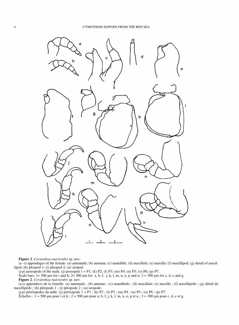
Figure 2. Ceratothoa marisrubri sp. nov.(a –i) appendages of the female. (a) antennule; (b) antenna; (c) mandible; (d) maxillule; (e) maxilla; (f) maxilliped; (g) detail of maxil-
liped; (h) pleopod 1; (i) pleopod 2; (u) uropod.(j-p) pereopods of the male. (j) pereopod 1 = P1; (k) P2; (l) P3; (m) P4; (n) P5; (o) P6; (p) P7.Scale bars: 1= 500 µm for i and h; 2= 500 µm for a, b, f, j, k, l, m, n, o, p and u; 3 = 300 µm for c, d, e and g.Figure 2. Ceratothoa marisrubri sp. nov.(a-i) appendices de la femelle. (a) antennule ; (b) antenne ; (c) mandibule ; (d) maxillule; (e) maxille ; (f) maxillipède ; (g) détail du
maxillipède ; (h) pléopode 1 ; (i) pléopode 2 ; (u) uropode.(j-p) péréiopodes du mâle :(j) péréiopode 1 = P1 ; (k) P2 ; (l) P3 ; (m) P4 ; (n) P5 ; (o) P6 ; (p) P7.Échelles : 1 = 500 µm pour i et h ; 2 = 500 µm pour a, b, f, j, k, l, m, n, o, p et u ; 3 = 300 µm pour c, d, e et g.

Male (Figure 1c-p).The body is elliptical and quite globular. The length variesfrom 9 to 12 mm. The largest width is at the level of the fifthpereonite. Dorsally, all thoracic segments are about thesame length, except the first one, which is longer, and thelast one, which is shorter. The first segment distinctlyprotrudes anteriorly, at the level of its two latero-anteriorangles, and the two resulting extensions reach the medianlevel of the cephalon (Fig. 1c). The pleotelson is muchwider than it is long and the posterior edge is broadlyrounded.
The cephalon is well-developed, with an anterior, ovoid-shaped edge.
The antennules (Fig. 1 d) and the antennae (Fig. 1e) haveseven segments with no ornamentation, except for three,short, distal, setal bristles on the last segment of A1, and asimilar setal bristle on the antero-posterior edge of thefourth segment of A2.
The mandibular palp (Fig. 1 f, g) is composed of threesegments, with a row of six to seven setal bristles increasing insize on the internal distal edge of the last segment and onesmaller, supplementary setal bristle on the penultimate segment.
J.-P. TRILLES, A. COLORNI, D. GOLANI 5
Figure 3. Ceratothoa marisrubri sp. nov.Pereopods of the female. (a) pereopod 1(P1); (b) P2; (c) P3; (d) P4; (e) P5; (f) P6;(g) P7. Scale bar: 1 mm.Figure 3. Ceratothoa marisrubri sp. nov.Péréiopodes de la femelle. (a) péréiopode 1(P1) ; (b) P2 ; (c) P3 ; (d) P4 ; (e) P5 ; (f) P6 ; (g) P7. Échelle = 1 mm.

The maxillules (Fig. 1 h, i) carry a distal group of 4slightly hooked spines; five smaller spines are also visibleon the maxillae (Fig. 1 j, k), three on the basal endite andtwo (plus a short setal bristle) on the coxal endite.
The maxillipeds (Fig. 1 l) are composed of threesegments. The last one has two spines relatively hooked atits distal end.
The size of the pereopods increases in length and widthfrom the first to the last pereopod. They do not display anyornamentation. However, the seventh pair of pereopods, asin the female, have a very high carina visible at the level ofthe basipodite.
The pleopod structure is typical. There are no hookingretinacula on the basipodites. The appendix masculina isvisible on Pl2 (Fig. 1 o); its length distinctly surpasses thedistal edge of the corresponding endopodite.
The two uropod endites are the same length and do notextend past the posterior edge of the pleotelson (Fig. 1 p).
Taxonomic remarks: Some morphological features of theadult female manifestly places this parasite in the genusCeratothoa, especially : (1) cephalon more-or-lessimmersed in pereonite I, with anterolateral angles extendedand encompassing cephalon; (2) antennules with basalarticle flattened, broad, in contact; (3) pereopod I-III moreslender than V-VII; (4) V-VII basipodites with prominentposterior expansion more-or-less manifestly developped;(5) pleonite I (or pleonite I and II) much narrower thanposterior pleonites and immersed in pereon.
The well-developed carina on the P7 basipodite of thefemale indicates that it is closely related to Ceratothoaoxyrrhynchaena Koelbel, 1878; however it differs from thisspecies in the more globular shape of the female, similar tothat of C. steindachneri Koelbel, 1878, which however hasa distinct dorsal hollow on the cephalon which is lacking inC. marisrubri.
Furthermore, C. marisrubri differs from C.oxyrrhynchaena by the presence of: 1) a distinctive setationor spinulation on the mouthpart; 2) a very well-developedcarina on the basipodite of P7 in both the female and themale, whereas in C. oxyrrhynchaena this feature is presentonly in the female (Trilles, 1972a).
Pullus II, which could have provided further distinctivecharacteristics, was unavailable for comparison.
Finally, it appears that, like other Ceratothoa species, C. marisrubri is non-specific in its host selection. However,more material is needed to confirm this assumption. Thecomplete geographic range of the species also remains to bedetermined.
Livoneca Leach, 1818
The validity, limits and synonymy of the genus Livoneca,compared with others genera such as Elthusa Schioedte &
Meinert, 1884 and Mothocya Costa (in Hope, 1851), havebeen recently discussed by Bruce (1986, 1990).
Livoneca papernea sp. nov.Figs. 4, 5, 6, and 7
Livoneca sp. nov. Colorni, Trilles, Golani, 1997.Informations on the biology of this parasite was recentlyreported (Colorni & al., 1997). Its provisional taxonomy iscompleted here.Specimens : one holotype and eight paratypes, deposited inthe HUJ collection.The specimens were collected in shallow water on thesilverside Atherinomorus lacunosus (Forster) (Atherinidae):22 December 1994, female (with 194 pulli I), Holotype, TL13 mm (Huj Isop. 9); 22 December 1994, male, TL 10 mm(Huj.Isop 9); 22 February 1996, female (with eggs) TL9 mm and male TL 8 mm (Huj. Isop 10); 22 February 1996,female (with eggs) TL 12 mm (Huj. Isop 11); 22 February1996, male TL 10 mm (Huj. Isop 12); 22 April 1996, female(non gravid) TL 10 mm and a male TL 8 mm (Huj. Isop 13);22 February 1996, female (non gravid) TL 8 mm (Huj. Isop14).Etymology: this new species is named after Prof. IlanPaperna, a world renowned parasitologist from the HebrewUniversity of Jerusalem, to honor his extensive contributionto ichthyoparasitology.
DescriptionFemale (Figure 4a, b; Figure 5a-o; Figure 7h-m).
The body is symmetrical and dorso-ventrally flattened(Fig. 4 a, b). The length in females varies from 8 to16.5 mm. A very blunt dorsal carina marks the middle of thebody wall. The first pereonite is the longest; the followingthree are about the same length while the size of the lastthree progressively decreases. All pleonites are visible asnone is covered by the pereon. Posteriorly, the pleotelsonhas a regular, rounded shape; the uropods clearly extendpast its posterior edge.
The cephalon is well-developed, triangulate, and encasedin the first pereonite. Its anterior end is slightly extended andtruncate. The eyes are conspicuous.
The antennules (Fig. 5 a) have eight segments; thesecond to the eighth, carry each one, two or three short setalbristles, located at either the latero-anterior end, or at thedistal end. The antennae (Fig. 5 b) have nine segments; thelast segment has a tuft of very short setal bristles at its distalend.
The mandibular palp has three segments, with noparticular ornamentation (Fig. 5 c). The maxillules (Fig. 5 d,e) end with four distal slightly hooked spines. The maxillae(Fig. 5 f, g) also carry four spines, two on each endite.
The three-segmented maxillipeds have two spines on thedistal segment (Fig. 5 h)
6 CYMOTHOID ISOPODS FROM THE RED SEA

The length of the pereopods is constant, except the first pair.The first three (Fig. 5 i, j, k), (in particular P2), are the mostmassive and the last four are the narrowest (Fig. 5 l, m, n, o).
The pleopods are typical, without hooking retinacula.
Their size is similar for Pl1, Pl2, Pl3, more reduced for Pl4and especially reduced for Pl5 (Fig. 7 h-l). The uropodeexopodite and endopodite are approximately the samelength and shape (Fig. 7 m).
J.-P. TRILLES, A. COLORNI, D. GOLANI 7
Figure 4. Livoneca papernea sp. nov.(a) and (b) female, dorsal and lateral views (arrow: dorsal carina profile); (c) young male, dorsal view; (d) and (e) adult male, dorsal
and lateral views; (f) pullus I with only six pairs of pereopods.Scale bars: 2 mm for a, b, c, d, e and 1 mm for f.Figure 4. Livoneca papernea sp. nov.(a) et (b) femelle, vue dorsale et vue latérale (flèche : profil de la carène dorsale) ; (c) jeune, mâle vue dorsale ; (d) et (e) mâle adulte,
vue dorsale et vue latérale ; (f) pullus I avec seulement six paires de péréiopodes.Échelles = 2 mm pour a, b, c, d, e et 1 mm pour f.

Male (Figure 4c-e; Figure 6a-p; Figure 7a-g).They are usually symmetrical, sometimes slightly right-
or left-bent, conforming to the host branchial cavity theyoccupy. The body length varies from 7.5 to 10.2 mm and iswidest at the third and fourth pereonite (Fig. 4 c-e).
The cephalon is well-developed; as in the female, theanterior end is clearly extended and truncate. The eyes arewell-developed and occupy a large part of the dorsal surfaceof the adult head.
The antennules (Fig. 6 a) are stocky and composed of
8 CYMOTHOID ISOPODS FROM THE RED SEA
Figure 5. Livoneca papernea sp. nov.(a - o) appendages of the female. (a) antennules; (b) antennae; (c) mandible; (d) maxillule; (e) detail of the maxillule; (f) maxilla; (g)
detail of the maxilla; (h) maxilliped; (i) pereopod 1(P1); (j) P2; (k) P3; (l) P4; (m) P5; (n) P6; (o) P7.Scale bars: 1= 500 µm for i, j, k, l, m, n and o; 2= 300 µm for a, b, c, d, f and h; 3= 100 µm for e and g.Figure 5. Livoneca papernea sp. nov.(a - o) appendices de la femelle. (a) antennule ; (b) antenne ; (c) mandibule ; (d) maxillule ; (e) détail de la maxillule ; (f) maxille ; (g)
détail de la maxille ; (h) maxillipède ; (i) péréiopode 1(P1) ; (j) P2 ; (k) : P3 ; (l) P4 ; (m) P5 ; (n) P6 ; (o) P7Échelles : 1 = 500 µm pour i, j, k, l, m, n et o ; 2 = 300 µm pour a, b, c, d, f, et h ; 3 = 100 µm pour e et g.

J.-P. TRILLES, A. COLORNI, D. GOLANI 9
Figure 6. Livoneca papernea sp. nov.Appendages of the male. (a) antennules; (b) antennae; (c) mandibular palp; (d) maxillule; (e) detail of the maxillule; (f) maxilla; (g)
detail of the maxilla; (h) maxilliped; (i) distal end of maxilliped; (j) pereopod 1(P1); (k) P2; (l) P3; (m) P4; (n) P5; (o) P6; (p) P7. Scale bars 1= 500 µm for j, k, l, m, n, o and p; 2= 300 µm for a and b; 3= 300 µm for c, d, f and h; 4= 100 µm for e, g and i.Figure 6. Livoneca papernea sp. nov.Appendices du mâle. (a) antennule ; (b) antenne ; (c) palpe mandibulaire ; (d) maxillule; (e) détail de la maxillule ; (f) maxille ; (g)
détail de la maxille ; (h) maxillipède ; i : extrémité distale du maxillipède ; (j) péréiopode 1 (P1) ; (k) P2 ; (l) P3 ; (m) P4 ; (n) P5 ; (o) P6 ;(p) P7.
Échelles : 1 = 500 µm pour j, k, l, m, n, o et p ; 2 = 300 µm pour a et b ; 3 = 300 µm pour c, d, f et h ; 4 = 100 µm pour e, g et i.
eight segments of almost the same length, some of which(three to eight) carry setal bristles on the anterior and/orposterior side.
The antennae (Fig. 6 b), more slender, have eightsegments, of similar length in the first four and slightlydecreasing in the following four ; the second and third
10 CYMOTHOID ISOPODS FROM THE RED SEA
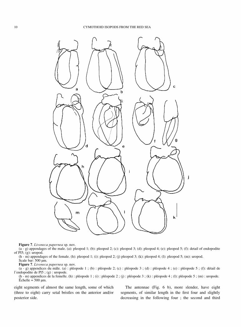
Figure 7. Livoneca papernea sp. nov.(a - g) appendages of the male. (a): pleopod 1; (b): pleopod 2; (c): pleopod 3; (d): pleopod 4; (e): pleopod 5; (f): detail of endopodite
of Pl5; (g): uropod.(h - m) appendages of the female. (h): pleopod 1; (i): pleopod 2; (j) pleopod 3; (k): pleopod 4; (l): pleopod 5; (m): uropod.Scale bar: 500 µm.Figure 7. Livoneca papernea sp. nov.(a - g) appendices du mâle. (a) : pléopode 1 ; (b) : pléopode 2; (c) : pléopode 3 ; (d) : pléopode 4 ; (e) : pléopode 5 ; (f): détail de
l’endopodite de Pl5 ; (g) : uropode.(h - m) appendices de la femelle. (h) : pléopode 1 ; (i) : pléopode 2 ; (j) : pléopode 3 ; (k) : pléopode 4 ; (l): pléopode 5 ; (m) : uropode.Échelle = 500 µm.
segments have long setal bristles on the anterior edge, whilethe fifth, sixth, seventh and eighth segments have one tothree very short spines at the antero-distal angle.
The mandibular palp has three segments; the two distalsegments have a row of setal bristles (Fig. 6 c). The setaelength is the same in the second segment, whereas in thethird segment their size increases gradually, except for thelast bristle which is considerably longer. The maxillules(Fig. 6 d, e) typically end with four distal spines, onlyslightly hooked. The maxillae (Fig. 6 f, g) have two spineson the basal endite and one on the coxal endite. Themaxillipeds (Fig. 6 h) are three-segmented and have twoclearly hooked spines at the distal end of the third segment.
In the young male (Fig. 4 c), the first six pereonites areabout the same size: the seventh is definitely shorter. In theadult male, the first pereonite is the longest; the followingtwo are about the same length and slightly shorter than thefirst one; the last four are shortest and their size graduallyand progressively decreases (Fig. 4 d, e).
The pereopods sharply increase from the first to the third(Fig. 6 i, j, k); they all have very well-developed dactyles,with a conspicuous row of indentations on the anteriorsurface ; a setal bristle is also visible at the postero-anteriorangle of meropodite of P2 ; P3 has a setal bristle with twoshort additional spines on the anterior edge of the propodite.The P4, P5, P6 and P7 are devoid of ornamentation and aremore slender than the preceding ones; their dactylopoditesare less developed and do not have indentations (Fig. 6 m-p).
The pleopods are the same size, except for Pl1 which aresmaller than the others (Fig. 7 a). The basipodites of thesymmetrical pleopods have short setal bristles and/or sparsehooking retinacula. The Pl2 carry an appendix masculina,slightly shorter than the related endopodite (Fig. 7 b). Pl1 toPl4 are like a typical pleopod, whereas the endopodites ofPl5 show a leaf-like growth which increases the surface ofcontact with the environment (Fig. 7 e, f). The uropodexopods and endopods are relatively larger than in thefemale (Fig. 7 g).
Taxonomic remarksThe generic placement of this species is difficult and
some characters, halfway between those of Livoneca andElthusa recently clarified by Bruce (1990), suggest that thespecies could be assigned to a new genus. The morphologyof the brood pouch and pleopods of the female is notconform to the morphology of the genus Livoneca describedby Bruce (1990) and it is closely related to Elthusa.However, the absence of the oostegial lobe at the level of themaxillipeds of the ovigerous female, the pleonites notimmersed in the pereon, the mandible palp of the femalewithout setae, the male pleopods with folding on theendopod, clearly differentiate this species from Elthusa.
Several features rule out its assignment to the closelyrelated genera Enispa Schioedte & Meinert, 1884 andNorileca Bruce, 1990: the posterior edge of the cephalon,not deeply divided into three lobes, and the five to sevencoxal plates, not any longer than the respective segments,distinguish Livoneca papernea from the genus Enispa; themandibular palp is neither flattened nor wide, and thereforealso different from the genus Norileca.
At present, until further studies, it is better to maintainthis species in the genus Livoneca, than regard it as incertaesedis.
This isopod does not correspond to any known species ofthe genera Livoneca and Elthusa. The most important,distinctive features are the absence of dissymmetry, thefairly parallel lateral edges, the median carina in the femaleand the presence in the male of lobes on the endopodites ofPl5 which are lacking in the female.
These specimens and those described by Colorni & al.(1997) parasitized the Red Sea silverside Atherinomoruslacunosus. They were not found in any other species of fish,including Hypoatherina temminckii (Bleeker), another RedSea silverside that shares the shallow coastline habitat withA. lacunosus (Colorni & al., 1997). Thus Livonecapapernea appears to have a monospecific host range.
Its geographic distribution is probably restricted to theRed Sea. A. lacunosus is a lessepsian immigrant into theMediterranean Sea (Ben Tuvia, 1966; Ben-Tuvia & Golani,1993), but L. papernea has not been detected in fish caughtin the Mediterranean Sea and preserved in the collection ofthe Hebrew University. This suggests that, unlike its host,the isopod failed to survive in the new habitat (Colorni &al., 1997).
Females always occupied the oral cavity of their host,firmly holding onto the tongue. All gravid female, carryingeither eggs or pulli (Figure 4f) in the marsupium, werecoupled with a male settled in either branchial cavity.
Nerocila Leach, 1818Restricted synonymy
Nerocila Leach, 1818:351Cymothoa Risso, 1816:143.Anilocra Risso, 1826 :124.
Ichthyophilus Latreille, 1829:133.Lironeca Van Beneden, 1871:32.
Pterisopodus Boone, 1918:596-598.Rosca Stebbing, 1923:10.
The synonymyzation with Emphylia Koelbel, 1879 andNerocila (Emphylia) Miers, 1880 is also proposed by Bruce1987, who recognized two species groups within Nerocila:the Nerocila group and the Emphylia group, the lastincluding particularly Nerocila sigani Bowman & Tareen,1983.
J.-P. TRILLES, A. COLORNI, D. GOLANI 11

Nerocila sigani Bowman and Tareen, 1983Fig. 8 a-l
Nerocila (Nerocila) sigani Bowman & Tareen, 1983:12, Fig. 9.
Nerocila sigani.- Bruce, 1987:406; Bruce & Harrison-Nelson, 1988: 597-598, Figs 6G, H, J.Nerocila (Nerocila) arres Bowman & Tareen,
1983: 7, 8, 12-17, Figs 10-12.Nerocila arres.- Trilles, 1994:82.
Specimens: one specimen from the caudal fin of the redmullet Upeneus subvittatus (Temminck & Schlegel)(Mullidae):11 November 1989, Eilat, Female TL 23 µm(Huj.Isop 15; National Fish Collection of the HebrewUniversity of Jerusalem).
IdentificationPreviously described in Kuwait (Arabian Gulf) on the basisof a single female specimen and differenciated from
Nerocila arres by Bowman & Tareen (1983), this species isconsidered by Bruce & Harrinson-Nelson, 1988, as thesenior synonym of Nerocila arres. Nerocila sigani is alsoclosely related to Nerocila trivittata Bleeker, 1856 (=Nerocila serra Schioedte & Meinert, 1881). However, itclearly differs from N. trivittata in the shape and the size ofthe latero-posterior extensions of the various pereonites(Fig. 8 a, b), in particular the 6th and the 7th, in the moremassive shape of the uropod endopodite (a detail oftaxonomic importance according to Bowman & Tareen,1983), and in the larger size of the indentations visible on itsexternal edge (Fig. 8 c). Similar indentations are alsopresent on both edges of the endopodites in Nerocilamonodi Hale, 1940 (Bruce, 1987). The mouthparts of N. sigani, are described for the first time (Fig. 8 d-k), forfurther detailed comparaisons and discussions. Themandibular palp is triarticulated, with two setal bristle onthe last segment (Fig. 8 d, e). The maxillules (Fig. 8 f, g)
12 CYMOTHOID ISOPODS FROM THE RED SEA
Figure 8. Nerocila sigani (female).(a, b): dorsal and lateral views (bar = 4 mm).(c - l) appendages. (c): uropods; (d): mandi-
bular palp; (e): distal end of the mandibularpalp; (f): maxillule; (g): distal end of the maxil-lule; (h): maxilla; (i): distal end of the maxilla;(j): maxilliped; (k): distal end of maxilliped;(l): Pereopod (1) with dactylus nodules.
Scale bars: 1 = 500 µm for c; 2 = 500 µm forl; 3 = 500 µm for j; 4 = 300 µm for d, f, h, andk; 5 = 100 µm for e, g and i.
Figure 8. Nerocila sigani (femelle).(a, b) : Vue dorsale et vue latérale.
Échelles = 4 mm.(c – l) appendices (c) : uropodes ; (d) : palpe
de la mandibule ; (e) : extrémité distale dupalpe mandibulaire ; (f) : maxillule ; (g) : extré-mité distale de la maxillule ; (h) : maxille ; (i) :extrémité distale de la maxille ; (j) : maxillipè-de ; (k): extrémité distale du maxillipède ; (l) :péréiopode 1 P1), dactylopodite avec nodules.
Échelles : 1 = 500 µm pour c ; 2 = 500 µmpour l ; 3 = 500 µm pour j ; 4= 300 µm pour d,f, h et k ; 5= 100 µm pour e, g et i.

carry a distal group of 4 slightly hooked spines; two smallerspines are visible on the basal endite of the maxillae (Fig. 8 h, i). The maxillipeds are tri-articulated and have threespines at the distal end of the last segment (Fig. 8 j, k).
Geographic distributionThis species is now known from the Western Indian Oceanand the Northern Indo-Pacific (Bruce & Harrisson-Nelson,1988).
HostsThe five female specimens reported by Bowman & Tareen(1983) belong to the collection of the United States NationalMuseum, Smithsonian Institution, Washington. Four werecollected in 1977 from Epinephelus tauvina (Forskål)(Epinephelidae), Acanthopagrus latus (Houttuyn)(Sparidae), Nemipterus japonicus (Bloch) (Nemipteridae)and Siganus oramin (Bloch & Scheider) (Siganidae),whereas the fifth was collected in 1982 from Nemipterustolu (Valenciennes) (Nemipteridae).
Bruce & Harrison-Nelson (1988) recorded this speciesfrom: Sciaenia dussumieri (Valenciennes), Argyrosomahololepidotus (Lacépède), A. macrocephalus (Tang), A. nibe (Jordan & Thompson) (Sciaenidae), Parastromateusniger (Bloch) (Carangidae) and Pomadasys sp.(Pomadasydae).
The present material has been collected from a red mulletUpeneus subvittatus (Temminck & Schlegel) (Mullidae).This extends the range of the hosts. Like many of the othersNerocila spp., this species does not seem to be demandingin its host selection.
References
Avdeev V.V. 1978. Parasitic Isopods of the family Cymothoidae(Crustacea, Flabellifera) from the Red Sea. Marine BiologyVladivostok, 4: 30-35, Figs. 1-3 (in Russian).
Ben-Tuvia A. 1966. Red Sea fishes recently found in theMediterranean. Copeia 2: 254-275.
Ben-Tuvia A. & Golani D. 1993. Some observations on the bio-logy of Atherinid fishes from the Mediterranean and Red Seacoasts of Israel. In : Pour qui la Méditerranée au 21e siècle ? LeSystème Littoral Méditerranéen, Actes du ColloqueScientifique. Maison de l’Environnement, Montpellier, France,22-23 Avril 1993, pp 58-63.
Bowman T.E. & Tareen I.V. 1983. Cymothoidae from fishes ofKuwait (Arabian Gulf) (Crustacea, Isopoda). SmithsonianContribution to Zoology, N° 382: 1-30, Figs. 1-20.
Bruce N.L. 1986. Revision of the Isopod Crustacean genusMothocya Costa, in Hope, 1851 (Cymothoidae: Flabellifera),parasitic on marine fishes. Journal of Natural History, 20:1089-1192.
Bruce N.L. 1987. Australian species of Nerocila Leach, 1818 andCreniola n. gen. (Isopoda: Cymothoidae), Crustacean Parasitesof Marine Fishes. Records of the Australian Museum,39: 355-412, Figs. 1-35.
Bruce N.L. 1990. The genera Catoessa, Elthusa, Enispa,Ichthyoxenus, Idusa, Livoneca and Norileca n. gen. (Isopoda,Cymothoidae), Crustacean Parasites of Marine Fishes, withdescriptions of Eastern Australian Species. Records of theAustralian Museum, 42: 247-300, Figs. 1-31.
Bruce N.L. & Bowman T.E. 1989. Species of the parasitic iso-pod genera Ceratothoa and Glossobius (Crustacea:Cymothoidae) from the mouths of flying fishes and halfbeaks(Beloniformes). Smithsonian Contributions to Zoology 489: I-III, 1-28.
Bruce N.L. & Harrison-Nelson B. 1988. New records of fishparasitic marine Isopod Crustaceans (Cymothoidae, subfamilyAnilocrinae) from the Indo-West Pacific Proceedings of theBiological Society of Washington, 101 (3): 585-602.
Colorni A. Trilles J-P. & Golani D. 1997. Livoneca sp.(Flabellifera: Cymothoidae), an isopod parasite in the oral andbranchial cavities of the Red Sea silverside Atherinomoruslacunosus (Perciformes, Atherinidae). Diseases of AquaticOrganisms 31 (1): 65-71.
Kossman R. 1880. Malacostraca (2. Theil, Anomoura, Macrura,Schizopoda, Stomatopoda, Isopoda, Laemodipoda,Amphipoda). Zoologische Ergebnisse einer im Auftrage der K.Acad. d. Wissensch. zu Berlin ausge - Fübrten Reise in dieKüstengebiete des Rothen meeres., Zweite Hälfte, ersteLieferung : 67-140, pl. IV-XV.
Monod Th. 1933a. Résumé analytique du mémoire de ThéodoreMonod sur les Isopodes (inclus Tanaïdacea). In : MissionRobert Ph. Dollfus en Egypte. Bulletin de l’Institut d’Egypte,XV, session 1932-1933: 151-157.
Monod Th. 1933b. Tanaidacea et Isopoda. In : Mission Robert Ph.Dollfus en Egypte. Mémoires de l’Institut d’Egypte, XXI: 161-264, Figs. 1-80.
Monod Th. 1937. Crustacés. In : Mission A. Gruvel dans le Canalde Suez. Mémoires de l’Institut d’Egypte, XXXIV: 12-19.
Stebbing T.R.R. 1910. Reports on the Marine Biology of theSudanese Red Sea. The Journal of the Linnean Society,Zoology, XXXI, 1907-1915: 215-230, pl. 21-23.
Trilles J.-P. 1969. Recherches sur les Isopodes Cymothoidae descôtes françaises. Aperçu général et comparatif sur le bionomieet la sexualité de ces Crustacés. Bull. Soc. Zool. Fr., 94 (3): 433-445.
Trilles J.-P. 1972a. Les Cymothoidae (Isopoda, Flabellifera) descôtes françaises (Systématique, faunistique, Ecologie et réparti-tion géographique). I. Les Ceratothoinae Schioedte et Meinert1883. Bulletin du Museum National d’Histoire Naturelle, 3e
sér., n° 91, sept-oct. 1972, Zool. 70: 1191-1229, pl. I, II, III.Trilles J.-P. 1972b. Les Cymothoidae (Isopoda, Flabellifera) du
Muséum National d’Histoire Naturelle de Paris. Etude critiqueaccompagné de précisions en particulier sur la répartition géo-graphique et l’écologie des différentes espèces représentées. I.Les Ceratothoinae Schioedte et Meinert, 1883. Bulletin duMuséum National d’Histoire Naturelle, 3e sér., N° 91, Sept.-Oct. 1972, Zool. 70: 1231-1268, pl. I-II.
Trilles J.-P. 1975a. Les Cymothoidae (Isopoda, Flabellifera) descollections du Muséum National d’Histoire Naturelle de Paris.II. Les Anilocridae Schioedte et Meinert, 1881. GenresAnilocra Leach, 1818, et Nerocila Leach, 1818. Bulletin duMuséum National d’Histoire Naturelle, 3e sér., N° 290, Zool.200: 303-346, pl. I-II.
J.-P. TRILLES, A. COLORNI, D. GOLANI 13

Trilles J.-P. 1975b. Les Cymothoidae (Isopoda, Flabellifera) descôtes Françaises. II. Les Anilocridae Schioedte et Meinert,1881. Genres Anilocra Leach, 1818, et Nerocila Leach, 1818.Bulletin du Muséum National d’Histoire Naturelle, 3e sér., N° 290, Zool. 200: 347-378, pl. I.
Trilles J.-P. 1976. Les Cymothoidae (Isopoda, Flabellifera) descollections du Muséum National d’Histoire Naturelle de Paris.IV. Les Lironecinae Schioedte et Meinert, 1884. Bulletin duMuséum National d’Histoire Naturelle, 3e sér., N° 390, Zool.272: 773-800, pl. I-II.
Trilles J.-P. 1979. Les Cymothoidae (Isopoda, Flabellifera; para-
sites de poissons) du Rijksmuseum van Natuurlijke Histoire deLeiden. II. Afrique, Amérique et régions Indo-Ouest-Pacifique.Zoologische Mededelingen, Leiden, 54(17): 245-275, pl. I-II,Figs. 1-2.
Trilles J.-P. 1994. Les Cymothoidae (Crustacea, Isopoda) duMonde (Prodrome pour une faune). Studia Marina, 21/22 (1-2), 1991: 5-288.
Trilles J.-P. & Paperna I. 1980. Sur quelques Crustacés Isopodes(Corallanidae, Lironecinae et Anilocridae) d’Israël. Bulletin duMuséum National d’Histoire Naturelle, 4e sér., 2, section A, N° 2 : 445-454, Figs. 1-40, pl. I.
14 CYMOTHOID ISOPODS FROM THE RED SEA


![Magic and Witchcraft [1852]](https://img.dokumen.tips/doc/110x75/55cf94c1550346f57ba42a66/magic-and-witchcraft-1852.jpg)