Embed Size (px)
Citation preview

42]. There are two major classification schemes for vas-cular tumors. That of Enzinger et al. [12] relies onpathological criteria and includes clinical and radiolog-ical features when appropriate. On the other hand, theclassification of Mulliken and Glowacki [42] is based onendothelial growth characteristics and distinguisheshemangiomas from vascular malformations. The latterclassification shows good correlation with the clinicalpicture and imaging findings.
Hemangiomas are characterized by a phase of prolif-eration and a stationary period, followed by involution.Vascular malformations are no real tumors and can bedivided into low- or high-flow lesions [65].
Cutaneous and subcutaneous lesions are usually easily diagnosed and present no significant diagnosticproblems. On the other hand, hemangiomas or vascularmalformations that arise in deep soft tissue must be dif-ferentiated from malignant neoplasms. Detailed assess-ment by medical imaging is necessary for adequateplanning of surgery.
On magnetic resonance imaging (MRI) benign vas-cular lesions have a characteristic configuration, gener-ally allowing a correct diagnosis. MRI is superior toother imaging techniques in defining the extent of theselesions, which is important since some types may in-volve large segments of the body.
Generally the classification of soft tissue vascularanomalies based on endothelial growth characteristics[66] shows good correlation with MRI appearance ofthese lesions [37]. Since this classification has been use-ful clinically [54], recognition of these characteristicMRI features is essential for improving therapeutic out-come in these patients.
Vascular lesions of intermediate malignancy and ma-lignant vascular tumors are far more rare. Hemangio-endothelioma is a neoplasm of endothelial cells that canbe benign or malignant. Angiosarcoma is an aggressivetumor with high local recurrence rate and risk of dis-tant metastases. Imaging findings have only beensparse, probably due to the tendency of these lesions toinvolve skin and superficial tissue, in contrast to othersoft tissue sarcomas. MRI is used for staging rather thanfor characterization of these lesions.
16.1 Introduction
Tumors and tumor-like conditions of the vascular sys-tem are divided into three categories according to theirdegree of malignancy: benign vascular lesions, lesionsof intermediate malignancy, and malignant vascular tu-mors. The vast majority of the lesions belong to the be-nign group. These are found predominantly in youngerchildren and adolescents. They may involve either theskin and subcutis or the deep soft tissues. Classificationof these lesions is still the source of much controversyand is based on clinical appearance, pathology, embry-ology, and endothelial growth characteristics [12, 41,
Chapter
Tumors and Tumor-like Lesionsof Blood VesselsF. Ramon
16
16.1 Introduction . . . . . . . . . . . . . . . . . . . . . . . . 263
16.2 Definition and Classification . . . . . . . . . . . . . . . 26416.2.1 Benign Vascular Tumors . . . . . . . . . . . . . 26416.2.1.1 Classification of Mulliken . . . . . . . . . . . . 26416.2.1.2 Classification of Enzinger . . . . . . . . . . . . 26416.2.1.3 WHO Classification . . . . . . . . . . . . . . . . 26516.2.2 Vascular Tumors of Borderline
or Intermediate Malignancy . . . . . . . . . . . 26516.2.3 Malignant Vascular Tumors . . . . . . . . . . . 26516.2.4 Glomus Tumor . . . . . . . . . . . . . . . . . . 26616.2.5 Hemangiopericytoma . . . . . . . . . . . . . . 266
16.3 Incidence and Clinical Behavior . . . . . . . . . . . . . 26616.3.1 Benign Vascular Tumors . . . . . . . . . . . . . 26616.3.2 Angiomatous Syndromes . . . . . . . . . . . . 26716.3.3 Hemangioendothelioma . . . . . . . . . . . . . 26716.3.4 Angiosarcomas . . . . . . . . . . . . . . . . . . 26816.3.5 Glomus Tumor . . . . . . . . . . . . . . . . . . 26816.3.6 Hemangiopericytoma . . . . . . . . . . . . . . 268
16.4 Imaging . . . . . . . . . . . . . . . . . . . . . . . . . . . 26816.4.1 Imaging Studies Other than MRI . . . . . . . . 26816.4.2 Imaging Findings on MRI . . . . . . . . . . . . 27016.4.3 Imaging Strategy . . . . . . . . . . . . . . . . . 280
References . . . . . . . . . . . . . . . . . . . . . . . . . . . . 281
Contents
16_DeSchepper_Tumors_and 15.09.2005 13:27 Uhr Seite 263

16.2 Definition and Classification
16.2.1 Benign Vascular Tumors
In the past it was debated whether vascular tumors aredevelopmental malformations or true tumors. In thenineteenth century vascular lesions were thought to be’produced by the longing of the mother, for particularthings, or her aversion to them’. Expressions such as ne-vus materneus or stigma metrocelis were in reference tothe mother. Because vascular tumors sometimes closelyresemble normal vessels, it is difficult to distinguishclearly between neoplasm and malformation on histo-logical examination [41].
The term hemangioma has frequently been used in-correctly, although accurate nomenclature is of utmostimportance for correct diagnosis and treatment of theselesions [66].
16.2.1.1 Classification of Mulliken
Some authors have suggested that the clinical presenta-tion provides the necessary perspective for classifyingvascular lesions. Mulliken et al. presented a usefulscheme for separating cutaneous vascular lesions basedon endothelial growth characteristics [42]. Their stud-ies reveal two major types of vascular lesions. One ex-hibits a rapid growth phase followed by a period of sta-bilization and finally involution; these show a female
predominance and are usually not present at birth. Be-cause of cellular proliferation and the mass effect, theseare called (infantile) hemangiomas (Table 16.1).
The majority of hemangiomas do not need treat-ment.
On the other hand, many vascular lesions show nocellular proliferation; they usually are not present atbirth but grow with the child and have no involutionphase. These lesions are called vascular malformations,and they are divided into capillary, venous, arterial andlymphatic types depending on the predominant vesseltype [42].
The classification scheme was updated during the1992 meeting of the International Society for the Studyof Vascular Anomalies (ISSVA) [75, 82] (Table 16.2).
The endothelial lining of the vascular malformationgroup is not proliferative. Although they are stable froma cellular point of view, they can be clinically devastat-ing, specially when there is arteriovenous shunting.
16.2.1.2 Classification of Enzinger
Enzinger et al., however, do not rely on clinical presenta-tion for classification of benign vascular lesions [12].Some congenital lesions do not become apparent untiladult life, depending on their location and growth. Inview of this limitation no attempt is made to separatemalformations from benign neoplasms. All lesions arecalled hemangiomas, and hemangioma is defined as ’abenign but nonreactive process in which there is an in-crease in the number of normal or abnormal-appearingvessels [12]. Hemangiomas may be either of two types:those localized in one area and those involving largesegments of the body. The histological classification ofvascular tumors proposed by Enzinger et al. [12] is asfollows (Table 16.3).
Localized hemangiomas are the more common. Theyhave been classified according to clinical, embryologic,or pathological criteria but no system is entirely satis-factory.
F. Ramon264
Table 16.1. Differentiating features of hemangiomas and vascularmalformations (modified from [65])
Hemangiomas Vascular malformations
Exhibit cellular proliferation Comprised of dysplastic vessels
Small or absent at birth Present at birth
Rapid growth during infancy Growth proportional to child
Involution during childhood No regression
Table 16.2. ISSVA classification of vascular anomalies (modified from [76, 83])
Vascular malformations
Vascular tumor Simple Combined
Hemangioma
Proliferative phase Capillary malformation Arteriovenous fistula, arteriovenous malformation, capillary-venous malformation, capillary-lymphatic-venous malformation (Klippel Trénaunay syndrome)
Involutive phase Lymphatic malformation Lymphatic-venous malformation, capillary-arteriovenous malformation(Parkes-Weber syndrome), capillary-lymphatic-arteriovenous malformation
Other tumors Venous malformation
A more pertinent issue considering imaging and therapy is classifying vascular malformations as either low-flow or high-flow lesions [84]
16_DeSchepper_Tumors_and 15.09.2005 13:27 Uhr Seite 264

reliably distinguish vascular from lymphatic endotheli-um. Therefore, they classify lymphangioma as a vascu-lar tumor.
16.2.2 Vascular Tumors of Borderline or Intermediate Malignancy
The term hemangioendothelioma is currently used forlesions which are histologically intermediate in appear-ance between hemangiomas and angiosarcomas.
According to Enzinger, the three types are epithelioidhemangioendothelioma, spindle cell hemangioendo-thelioma and malignant endovascular papillary hem-angioendothelioma. Only the epithelioid subgroup oc-curs in deep soft tissues, most commonly of the extrem-ities. The other types develop preferentially in thedermis or subcutaneous tissues [13].
The WHO classification differentiates locally aggres-sive tumors of intermediate malignancy and rarelymetastasizing types. Kaposiform hemangioendothe-lioma is characterized by a ‘Kaposi sarcoma like’ fascic-ular spindle cell growth pattern. It most commonlyoccurs in the retroperitoneum or the skin.
16.2.3 Malignant Vascular Tumors
Angiosarcomas are tumors that can vary from highlydifferentiated, resembling hemangioma, to those whoseanaplasia makes it difficult to distinguish from carcino-mas. They are characterized by irregular anastomosingvascular channels lined by atypical endothelial cells.
They are usually located superficially but may involvedeep structures, such as skeletal muscle. Depending onthe predominant vessel type, the former are grouped ascapillary, cavernous, venous or arteriovenous. Otherforms are more rare.
Hemangiomas involving the deep soft tissues aregrouped as intramuscular, synovial or intraneuraltypes. Intramuscular hemangioma usually shows anovergrowth of adipose tissue, giving the impression ofangiolipoma. Unlike their cutaneous variants, it is notalways possible to make a clear distinction between dif-ferent types of intramuscular hemangiomas. A classifi-cation into small-vessel, large-vessel and mixed typeswith different clinical behavior was proposed by Allento avoid misinterpretation since some of the small-ves-sel type lesions show alarming histological features re-sembling those of a sarcoma [1].
Angiomatosis is a rare condition in which large seg-ments of the body are involved by proliferating vessels.Mostly the extremities are affected. A characteristic fea-ture of this condition is the large amount of mature fatthat accompanies the proliferating vessels [12].
The distinction between intramuscular hemangiomaand angiomatosis is made better by clinical than patho-logical criteria.
16.2.1.3 WHO Classification
The classification of the WHO (Table 16.4) closelyresembles that of Enzinger. In this classification, no dis-tinction is made between benign vascular neoplasms orvascular malformations. Similarly, it is impossible to
Chapter 16 Tumors and Tumor-like Lesions of Blood Vessels 265
Table 16.3. Histological classification of vascular tumors (modi-fied from [12])
Benign vascular tumorsLocalized Hemangioma
Capillary HemangiomaCavernous HemangiomaVenous hemangiomaArteriovenous hemangiomaEpithelioid hemangiomaHemangioma of the granulation tissue typeDeep soft tissue hemangioma
Angiomatosis
Vascular tumors of intermediate malignancyEpithelioid hemangioendotheliomaSpindle cell hemangioendotheliomaMalignant endovascular papillary hemangioendothelioma
Malignant vascular tumorsAngiosarcomaKaposi’s sarcoma
Table 16.4. WHO classification of vascular tumors (modified from[68])
BenignHemangiomas of
Subcutis/deep soft tissueCapillaryCavernousArteriovenousVenousIntramuscularSynovial
Epithelioid hemangiomaAngiomatosisLymphangioma
Intermediate (locally aggressive)Kaposiform hemangioendothelioma
Intermediate (rarely metastasizing)Retiform hemangioendotheliomaPapillary intralymphatic angioendotheliomaComposite hemangioendotheliomaKaposi sarcoma
MalignantEpithelioid hemangioendotheliomaAngiosarcoma of soft tissue
16_DeSchepper_Tumors_and 15.09.2005 13:27 Uhr Seite 265

The terms hemangiosarcoma and lymphangiosarcomaare no longer appropriate [14, 45]. The lesions occurpreferentially in the skin or soft tissues of the scalp andface. Kaposi’s sarcomas have spindle cell areas contain-ing vascular channels.
The disease is associated with the Human HerpesVirus (HHV-8) infection. It presents as cutaneous le-sions in the form of multiple patches. According to theWHO classification Kaposi sarcoma is of intermediatemalignancy and epithelioid hemangioendothelioma ismalignant.
16.2.4 Glomus Tumor
The glomus tumor is a neoplasm consisting of cellswhich closely resemble smooth muscle cells of the nor-mal glomus body. The glomus body is an arteriovenousanastomosis that has an important role in thermoregu-lation. It is located in the subungual region, digits, andpalms. The lesion shows both muscle fibers and epithe-lial-appearing glomus cells [15].
16.2.5 Hemangiopericytoma
Hemangiopericytoma is an uncommon tumor, first de-scribed by Stout and Murray, and is composed mainly ofpericytes [8, 15, 26, 28, 33, 44, 52, 55, 63].According to themost recent WHO classification [67], hemangiopericy-toma will be discussed in Chap. 13 (Tumors of FibrousTissue).
16.3 Incidence and Clinical Behavior
16.3.1 Benign Vascular Tumors
To avoid confusion we follow here a combination of theclassification of Enzinger and the WHO [12, 67].
The differentiation of Mulliken and Glowacki be-tween hemangiomas and vascular malformations is on-ly considered when specifically mentioned.
Although hemangiomas are uncommon tumors, theymake up about 7% of all benign soft tissue tumors.Most of the lesions, however, are located in the skin orsubcutaneous tissues, while only a minority is deeplyseated [12].
Capillary hemangiomas form the largest group. Ex-cept for cherry angioma they occur predominantly inchildhood. Juvenile hemangioma is an immature formof capillary hemangioma with a characteristic clinicalevolution. They occur mostly in superficial tissue ofhead and neck region and arise a few weeks after birth.They grow slowly to reach a maximal size at six monthsof age and then regress. Some lesions may pose cosmet-
ic problems or threaten vital structures. They are treat-ed by systemic steroid therapy and subsequent surgery.Acquired tufted angioma, verrucous hemangioma, andsenile angioma are clinical variants of the same subtypeof hemangioma.
Cavernous hemangiomas are less frequent but shareage and anatomical distribution with the capillaryhemangiomas. However, they are usually larger and lesscircumscribed and may be locally destructive. Themajority require surgical resection.
Superficial arteriovenous and venous types are lesscommon. They are distinguished from the capillary andcavernous counterparts by the presence of thick-walledvessels. Superficially located arteriovenous heman-giomas cause no clinical problems. On the other hand,shunting of blood causing cardiac overload, pain andhypertrophy of the involved extremity are problemsassociated with deep lesions and require an adequatetherapeutic approach.
Epithelioid hemangioma and granulation type hem-angioma are unusual vascular tumors.
Epithelioid hemangioma consists of well formed butimmature vessels and a prominent inflammatory com-ponent. They occur predominantly at the forehead andthe digits, and present as a nodular mass which gives theimpression of an epidermal cyst.
Compared to superficial hemangiomas, deep-seatedlesions are quite uncommon, with a reported frequencyof 0.8% of all lesions [44]. However, they deserve specif-ic mention because of their different clinical presenta-tion. Because they present with pain or swelling, it is dif-ficult to make a diagnosis based on physical examina-tion. Some 80–90% of intramuscular hemangiomasoccur in the first three decades of life. There is no femalepredilection, as for superficial hemangiomas. The ma-jority of lesions are located in the lower extremity, espe-cially in the muscles of the thigh. Pain is reported to bemore common in tumors involving long, narrow mus-cles, probably through stretching of the muscle fibers.
Lesions have often been present for many years and itis therefore likely that many examples are congenital.
Physical deformity, cardiac decompensation, or de-struction of adjacent vital structures may require surgi-cal intervention. Therapy is aimed at complete excision,with or without prior embolization [10, 63]. Intramus-cular hemangiomas are benign, but with a high inci-dence of local recurrence.
Synovial hemangioma and hemangioma of peripher-al nerve are rare. The synovial type almost always in-volves the knee joint, presenting with pain, swelling and joint effusion. The most common site is the supra-patellar pouch. Because of repetitive bleeding into the joint, a radiographic appearance identical to that ofhemophilic arthropathy is seen [12, 28]. Only a smallproportion of lesions are correctly diagnosed beforesurgery (Fig. 16.1).
F. Ramon266
16_DeSchepper_Tumors_and 15.09.2005 13:27 Uhr Seite 266

Kasabach-Merritt syndrome was first reported in1954 [25]. The cardinal features of this syndrome in-clude an enlarging hemangioma, thrombocytopeniaand microangiopathic hemolytic anemia with acute orchronic consumptive coagulopathy. The etiology of thecoagulopathy is still unknown. Mortality is estimated atapproximately 21%. The major cause of death is bleed-ing. Many therapeutic modalities have been applied,aiming at a twofold objective: to control the bleedingand to reduce the size of the lesion [11, 25, 32, 38]. Maf-fucci’s syndrome is a rare dysplasia characterized bymultiple hemangiomas and enchondromas. The vascu-lar tumors are usually noted at birth and are of the cav-ernous type. According to Silverman et al. [54], the le-sions that are termed hemangiomas are in fact vascularmalformations. The cartilaginous lesions typically de-velop after the vascular tumors. The bones are short-ened and have multiple exostoses and enchondromas[6, 12] (Fig. 16.2). Malignant sarcomatous degenerationis noted in about 21% of patients.
Klippel-Trénaunay-Weber syndrome consists of cu-taneous hemangioma, bone and soft tissue hypertrophyand varicose veins. The syndrome is usually unilateraland involves the lower extremity [68].
Proteus syndrome is a complex hamartomatous dis-order defined by local overgrowth (macrodactyly), sub-cutaneous tumors and various bone, cutaneous and/orvascular anomalies. Vascular anomalies are commonand are distributed at random sites of the body. Theclinical presentation is highly variable [69, 70].
Angiomatosis is a rare condition in which large seg-ments of the body are involved by proliferating vessels[71]. Principally the extremities are affected. A charac-teristic feature of this condition is the large amount ofmature fat that accompanies the proliferating vessels[12]. The disorder probably starts during intrauterinelife when the limb buds form.
Approximately two-thirds of cases develop within thetwo first decades of life and nearly all are apparent byage 40 years.
It usually presents as a swelling, induration, or discol-oration of the affected area with or without limb hyper-trophy. Mortality is high because of space occupyingeffects and consumption coagulopathy [12, 28].
16.3.3 Hemangioendothelioma
Epithelioid hemangioendothelioma of the soft tissuesdoes not occur preferentially in children. All age groupsare affected, and there is no sex predilection. Mostlesions are solitary. Overall prognosis of this tumor isquite favorable. Only a small proportion does metasta-size and cause death. Therapy includes wide local exci-sion without adjuvant chemotherapy or radiotherapy.Regional lymph nodes should be evaluated [30, 41].
16.3.2 Angiomatous Syndromes
Angiomatous syndromes include Kasabach-Merrittsyndrome, Maffucci’s syndrome, Klippel-Trénaunay-Weber syndrome, Osler-Weber-Rendu disease, Gorhamdisease, Proteus syndrome and angiomatosis.
Chapter 16 Tumors and Tumor-like Lesions of Blood Vessels 267
Fig. 16.1Ia, b. Synovial hemangioma of the knee. a Sagittal T1-weighted MR image. b Axial T2-weighted MR image. A multilobu-lar inhomogeneous lesion involving both the suprapatellar bursaand the extra-articular fatty tissue and muscles is seen. The lesionis isointense to muscle on T1-weighted images and a large drain-ing or feeding vessel is seen in the popliteal fossa.Axial T2-weight-ed images show a lesion of high signal intensity. There is no jointeffusion
a
b
16_DeSchepper_Tumors_and 15.09.2005 13:27 Uhr Seite 267

16.3.4 Angiosarcomas
Angiosarcomas of the deep tissue are one of the rarestforms of soft tissue neoplasms, accounting for less than1% of all sarcomas. These tumors are evenly distributedthroughout all decades and show predilection for thelower extremity and the abdominal cavity, in contrast tothe cutaneous forms which are found mostly in headand neck. Chronic lymphedema is the most widelyrecognized predisposing factor in angiosarcomas.Angiosarcomas are often characterized as enlarging,painful masses lasting for several weeks and are occa-sionally associated with hemorrhage, anemia or coagu-lopathy. Epithelioid angiosarcoma is the most frequent-ly observed pattern, but the morphological spectrum iswide [36]. Malignant degeneration in a preexisting be-nign lesion is probably an unusual event [15].
The paucity of data precludes statements concerningthe optimal mode of therapy [5, 45].
16.3.5 Glomus Tumor
Glomus tumors are uncommon, with an equal frequen-cy of occurrence in both sexes. The most common loca-tion is the subungual tissue at the tip of the finger. For
this location however, there is a striking female pre-dominance. Glomus tumors are also seen at the palm,wrist, forearm, and foot. Multiple lesions may be pre-sent, especially during childhood. Glomus tumors causea radiating pain which is elicited by a change in temper-ature. Clinical examination usually reveals a character-istic blue-red nodule. Therapy aims at complete exci-sion, still leaving a recurrence rate of 10% [15].
16.3.6 Hemangiopericytoma
As earlier mentioned, hemangiopericytoma (HPC) willbe discussed in Chap. 13, which is in line with the mostrecent WHO classification (2002).
16.4 Imaging
16.4.1 Imaging Studies Other than MRI
Although findings on plain radiography are frequentlynormal, they are sometimes helpful in diagnosing softtissue hemangiomas. Plain radiographs reveal the pres-ence of a lesion by displacement or loss of the normaltissue planes. The presence of phleboliths is a specificsign, but unfortunately of low sensitivity.
F. Ramon268
Fig. 16.2Ia, b. An 18-year-old woman with Maffuci’s syndrome.a Plain radiograph of the left hand. b Plain radiograph (lateralview) of the left wrist. Multiple, expansile, well-defined and pre-dominantly lytic lesions within the phalanges and metacarpals ofthe left hand are shown. The second digit has previously been am-
putated. Phleboliths are seen within a soft tissue swelling at the volar aspect of the wrist. The combination of multiple enchondro-mas and soft tissue hemangiomas is characteristic for Maffuci’ssyndrome
a b
16_DeSchepper_Tumors_and 15.09.2005 13:27 Uhr Seite 268

the proximity of the lesion to bone, whereas medullarychanges correspond to size of the hemangioma [73, 74].
Plain film studies are superior to MRI in demonstrat-ing the type and extent of reaction [36]. On the otherhand, information about size and extent of the lesion islimited [2, 29, 43, 44, 50, 57].
On computed tomography (CT) benign angiomatouslesions show a mottled low density pattern, partiallydue to the mixture of fatty, fibrous, and vascular tissueelements. The slow flow and pooling of blood some-times cause the presence of punctate and curvilinearstructures. CT has a greater sensitivity in detectingassociated phleboliths (Figs. 16.3 and 16.4) and alsoclearly shows the relation to adjacent structures in theaxial plane. CT may also be useful for exclusion of othersoft tissue lesions such as lipomas, which are character-ized by a homogeneous low-density attenuation [2, 21,22, 51].
On angiography the appearance of benign vasculartumors is variable, ranging from small poolings of con-trast material over coarse and fine hypervascularity to the presence of large tortuous blood vessels. Angio-graphy has long been the method of choice for definingthe lesions extent, vascularity and feeding vessels, allow-ing distinction from other well-vascularized masses[39, 40].
Angiography of venous malformations, although notindicated, shows contrast pooling in dilated vessels. Inarteriovenous malformation, feeding and draining ves-sels as well as large arteriovenous shunts can be demon-strated [65].
However, underestimation of the extent, false-nega-tive results and inability to define the relationship to ad-jacent neural structures or fascial planes, have beenreported [31, 32]. Moreover, differentiation betweenhemangiomas and other soft tissue tumors is often notpossible.Angiography is, however, still helpful in identi-
Phleboliths are found in venous malformations andcorrespond to calcifications in thrombosis [72].
Lesions that are in close proximity to bone may causebone erosion or periosteal reaction (Fig. 16.3). Twentypercent of hemangiomas of deep soft tissues cause adja-cent bony changes.
The osseous changes can be categorized as periosteal,cortical or medullary. All three categories correlate with
Chapter 16 Tumors and Tumor-like Lesions of Blood Vessels 269
Fig. 16.3Ia, b. A 35-year-old man with painless soft tissue mass atthe left hand. a Radiograph of the left hand. b Coronal spin echoT1-weighted MR image. Plain radiography shows an increaseddensity of the soft tissues at the left thenar. Multiple rounded cal-cifications of varying size are present, corresponding to phlebo-liths. This finding is characteristic for intramuscular hemangio-mas. There is an erosion of the ulnar sided cortex at the base ofmetacarpal I (a). On the MR image the muscles of the thenar areslightly hyperintense due to infiltration by the hemangioma. Therounded spots of low signal intensity correspond to the phlebo-liths. Pressure erosion of metacarpal I is shown without change inthe adjacent bone marrow (b)
a
b
Fig. 16.4. A 47-year-old woman with a swelling of the left calf.Unenhanced CT slice shows an inhomogeneous aspect of the later-al head of the gastrocnemius muscle. The muscle is enlarged andhas dispersed internal areas of decreased attenuation. A fewrounded phleboliths are seen. There is no involvement of theunderlying soleus muscle. Histological examination revealed thepresence of a hemangioma. Areas of decreased attenuation corre-spond to intralesional fat
16_DeSchepper_Tumors_and 15.09.2005 13:27 Uhr Seite 269
fication of feeding and draining vessels for planningembolic therapy or preoperative embolization [44, 50].
Excellent palliation has been reported with em-bolotherapy of symptomatic arteriovenous malforma-tions [11, 64]. The pain is thought to be secondary toperineural congestion.
Subselective embolization using ethanol, coils andparticles usually relieves pain [65].
Ultrasound is a very sensitive technique for detectingsoft tissue abnormalities, as well as for demonstratinghemangiomas. If phleboliths are present, they are seenas an echogenic focus with acoustic shadowing. Heman-giomas usually present as oval masses with smoothmargins. They are heterogeneous and generally hypere-choic. However, there is no ultrasound appearance spe-cific to muscular hemangiomas [7, 58].
Reports on the use of color Doppler are sparse andusually do not deal with vascular lesions in particular.In some hemangiomas no Doppler signal is noted be-cause of low flow. On the other hand, a characteristiclow resistance flow pattern can be noted in high flowvascular malformations [75].
Doppler sonography can also be used for follow-up ofAVM after therapy [65].
High vessel density and high peak arterial Dopplershift can be used to distinguish hemangiomas fromother soft tissue masses [9].
However, literature findings are contradictory [75].Paltiel et al. have even tried to distinguish hemangiomafrom vascular malformation on the basis of mean venouspeak velocity and mean resistive index [9, 37, 47, 59].
Only one report in the literature mentions the role ofpositron emission tomography in the evaluation ofhemangioma. PET is promising for differentiating be-nign hemangiomas from other soft tissue tumors [76].
On plain radiographs a glomus tumor is seen as a softtissue mass usually at the dorsal surface of the finger. Acharacteristic bone erosion, possibly with sclerotic mar-gin is sometimes noticed [15, 44]. Ultrasound reveals ahypoechoic lesion but is not sensitive [17].
It is not possible to differentiate between heman-gioendothelioma and angiosarcoma on the basis ofplain radiography [16].
Reports on CT and ultrasound findings in malignantvascular tumors are sparse and usually non-specific [16,18]. CT show a heterogeneous mass which becomessharply delineated owing to marked contrast enhance-ment. Angiosarcoma following chronic lymphedema ischaracterized by fibrous thickening, increased fat atten-uation, and fluid collections surrounding the muscles[27, 50].
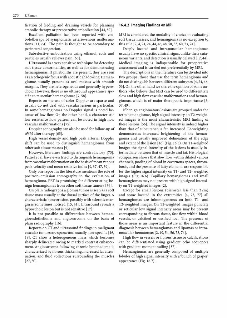
16.4.2 Imaging Findings on MRI
MRI is considered the modality of choice in evaluatingsoft tissue masses, and hemangioma is no exception tothis rule [2, 4, 21, 24, 44, 46, 48, 50, 53, 60, 73, 74].
Deeply located and intramuscular hemangiomasusually have no specific clinical signs, unlike their cuta-neous variants, and detection is usually delayed [12, 44].Medical imaging is indispensable for preoperativeassessment and is carried out preferentially by MRI.
The descriptions in the literature can be divided intotwo groups: those that use the term hemangioma anddo not distinguish between different subtypes [4, 24, 46,56]. On the other hand we share the opinion of some au-thors who believe that MRI can be used to differentiateslow and high flow vascular malformations and heman-giomas, which is of major therapeutic importance [3,37, 49].
If benign angiomatous lesions are grouped under theterm hemangiomas, high signal intensity on T2-weight-ed images is the most characteristic MRI finding ofthese lesions [56]. The signal intensity is indeed higherthan that of subcutaneous fat. Increased T2-weightingdemonstrates increased brightening of the heman-gioma and usually improved delineation of the edgeand extent of the lesion [46] (Fig. 16.5). On T1-weightedimages the signal intensity of the lesions is usually in-termediate between that of muscle and fat. Histologicalcomparison shows that slow flow within dilated venouschannels, pooling of blood in cavernous spaces, throm-bosis, and the presence of fatty elements are responsiblefor the higher signal intensity on T1- and T2- weightedimages (Fig. 16.6). Capillary hemangiomas and smallhemangiomas may not present with high signal intensi-ty on T1-weighted images [2].
Except for small lesions (diameter less than 2 cm)and some located in the extremities [4, 73, 77] allhemangiomas are inhomogeneous on both T1- and T2-weighted images. On T2-weighted images punctateor reticular low signal intensity areas may be presentcorresponding to fibrous tissue, fast flow within bloodvessels, or calcified or ossified foci. The presence ofthose areas is an important feature in the differentialdiagnosis between hemangiomas and lipomas or intra-muscular hematomas [2, 49, 54, 56, 73, 74].
High flow in vessels or fibrous tissue or calcificationscan be differentiated using gradient echo sequenceswith gradient-moment nulling [37].
Hemangiomas are generally composed of multiplelobules of high signal intensity with a ‘bunch of grapes’appearance (Fig. 16.7).
F. Ramon270
16_DeSchepper_Tumors_and 15.09.2005 13:27 Uhr Seite 270

Chapter 16 Tumors and Tumor-like Lesions of Blood Vessels 271
Fig. 16.5Ia, b. High signal intensity of heman-gioma on T2 weighted images: a subcuta-neous hemangioma of the wrist. Coronal T2-weighted image with fat suppressionshows a multilobular mass which involvesthe subcutaneous fat. The lesion is hyperin-tense and inhomogeneous due to internalseptations; b capillary hemangioma ofthe neck in a two-years old boy. Axial T2-weighted image shows an inhomogeneousseptate mass which is sharply delineated and is hyperintense to subcutaneous fat.There is no invasion of the underlying muscle
a b
Fig. 16.6Ia–c. Signal intensity of hemangioma on T1-weighted im-ages: a,b synovial hemangioma of the left elbow. The lesion is lo-cated in the posterior recess of the joint and is hyperintense com-pared to the triceps muscle. High signal intensity is probably
caused by stagnant blood; c the signal intensity of the flexor digi-torum muscles is minimally increased due to the presence of intra-muscular hemangioma. Note also the presence of peripheral fattycomponents
a
bc
16_DeSchepper_Tumors_and 15.09.2005 13:27 Uhr Seite 271

This is probably due to cavernous or cystic vascularspaces containing stagnant blood [4]. It is also withinthese spaces that fluid-fluid levels can be notes. Thesefluid-fluid levels are mostly seen in cavernous heman-giomas and are caused by hemorrhage. On T2-weightedimages a high signal intensity ’supernatant’ correspond-ing to serous fluid overlies a low signal intensity lowerlayer. Fluid-fluid levels are not appreciated as easily onT1-weighted images, but they are nevertheless present.On T1-weighted images the signal intensity of the higher layer is low, and that of the lower layer is high(Fig. 16.8).
As noted above, hemangiomas are generally inhomo-geneous on T1-weighted image. The lesions are pre-dominantly isointense with muscle, with internal ser-piginous high signal intensity strands. These strandscorrespond to enlarged vessels and generally are orient-
F. Ramon272
Fig. 16.7. Coronal T1-weighted image of the right thigh shows anintramuscular mass which involves the hamstring muscles. Thelesion is composed of multiple nodular components which make itresemble ‘a bunch of grapes’
Fig. 16.8. a Axial T1-weighted MR image. b Axial T1-weighted MRimage after gadolinium contrast injection. c Sagittal T2-weightedMR image. A fluid–fluid level is seen on axial T1-weighted images.The supernate is slightly hypointense compared with the lowerlayer due to its serous composition (a). The fluid–fluid level be-comes clearer after administration of gadolinium due to the sedi-mentation of gadolinium to the lower part of the lesion, the samephenomenon as is noted within the bladder after intravenous ad-ministration of gadolinium chelates (b). On T2-weighted imagesthere is increased contrast between the hyperintense serous upperlayer and the lower layer, which is hypointense due to the sedimen-tation of erythrocytes (c)
a b
c
16_DeSchepper_Tumors_and 15.09.2005 13:28 Uhr Seite 272

ed parallel to the muscle fibers [21, 56]. In some heman-giomas peripheral high signal intensity areas are notedon T1-weighted images, corresponding to fat within thelesions (Fig. 16.9).
A frequent finding is hypertrophy or atrophy of themuscle or the subcutaneous fat involved by the heman-giomas (Fig. 16.10). Hypertrophy of the limb in an-giomatosis is also known.
The classification of Mulliken differentiates heman-giomas, venous malformations and arteriovenous mal-formations.
Slow flow vascular malformations include venous,capillary, cavernous and mixed types. The appearanceon MRI depends on the composition. Venous portionsshow large spaces with internal linear or serpentinestructures of low or high signal intensity depending onthe pulse sequence and blood flow velocity [78, 79].
In addition they are oriented along the long axis ofthe limb, follow neurovascular distributions, and aresometimes multifocal (Fig. 16.11). Usually there is anenlargement of the neighboring subcutaneous fat. Thecombination of all findings suggests a congenital tissuedysplasia.
On the other hand, high flow arteriovenous malfor-mations have signal voids on all pulse sequences. Al-though the lesions can be associated with surroundingedema or fibrofatty stroma, no focal soft tissue mass isfound [37, 49] (Fig. 16.12).
Chapter 16 Tumors and Tumor-like Lesions of Blood Vessels 273
Fig. 16.9. Intramuscular hemangioma in a 40-years-old man.On sagittal T1-weighted images, the signal intensity of the lesion isinhomogeneous. There are central nodular components that areisointense to muscle. The peripheral parts are mainly composed offatty tissue and are hyperintense to muscle
Fig. 16.10Ia, b. A 55-year-old woman complaining of interscapularpain. a CT after iodinated contrast injection. b Axial spin echo T1-weighted MR image. There is a marked hypertrophy of the righttrapezoid muscle compared to the contralateral side. Central andperipheral enhancing areas are noted, corresponding to enlarged
vessels (a). In addition to the hypertrophy, signal intensity of theright trapezoid muscle is inhomogeneous and increased, nearlyisointense to subcutaneous fat. The increase is caused by slow flowin enlarged veins (b). The presence of enlarged veins and the hy-pertrophy of the muscle are characteristic for hemangioma
a b
16_DeSchepper_Tumors_and 15.09.2005 13:28 Uhr Seite 273

F. Ramon274
Fig. 16.11Ia, b. Venous hemangiomas: a axial T2-weighted imagewith fat saturation shows the involvement of multiple compart-ments of the knee region by the lesion. There are hyperintensenodular and serpentine areas in the subcutis, the semimembra-nous muscle and within the joint, in the anterior fat pad. High sig-
nal intensity is caused by slow flowing blood in dilated venouschannels; b coronal inversion recovery T2-weighted image in an-other patient shows pooled blood within saccular dilated bloodvessels. There is extension along the long axis of the involved leg
a b
Fig. 16.12Ia–c. High-flow arteriovenous malformation in the flex-or muscles of the forearm. a Sagittal T1-weighted MR image. b Ax-ial T2-weighted MR image. c Axial T1-weighted MR image aftergadolinium contrast injection. Multiple serpiginous areas of signalvoid are present within the flexor muscles of the fingers. Thesemuscles are nearly isointense on T1-weighted images (a). On T2-
weighted images the involved muscles are slightly hyperintense,with internal areas of signal void. High signal intensity areas cor-respond to the matrix of vascular malformation, while signal voidis caused by high flow in dilated vessels (b).After administration ofGd contrast, there is only enhancement of the solid part of themass (c). (From H. Van Moer, with permission)
a
b
c
16_DeSchepper_Tumors_and 15.09.2005 13:28 Uhr Seite 274

Conventional MR imaging generally does not allowto making a distinction between different types ofvenous malformations.
However,Verstraete et al. maintain that dynamic con-trast-enhanced MRI and ‘first pass’ images enable thedifferentiation between cavernous and capillary he-mangiomas and between low flow and high flow vascu-lar malformations [62] (Fig. 16.15; see Chap. 6).
Low slope values are usually found in cavernoustypes due to low perfusion, while capillary types showhigh perfusion and high slope values. The first-pass im-ages show good correlation with angiographic findings.
More recent studies confirm that dynamic contrastenhanced MRI provides images that are comparable toconventional angiography. In addition time-intensitycurves allow to distinguishing venous malformationsfrom mixed capillary-venous and arteriovenous mal-formations [80, 81].
Infantile hemangiomas display the features de-scribed above: a lobulated mass that is hyperintense onT2-WI and isointense to muscle on T1-WI. They have prominent feeding or draining vessels which areidentified as central or peripheral high flow channels(Fig. 16.13).
T1-weighted images after the administration ofgadolinium show a moderate to strong enhancement ofthe vascular lesions. The pattern of enhancement varieswith the interval between imaging and the time of injec-tion, depending on the rate of inflow of contrast into theblood-filled spaces [56]. Degree of enhancement varieswith flow velocities of blood in the vessels of the he-mangioma. Therefore more pronounced enhancementoccurs in the low signal intensity parts on native scans,and the lesions become more homogeneous. The con-trast-to-noise ratio between hemangiomas and sur-rounding tissues, however, remains lower than on T2-weighted images (Fig. 16.14).
Chapter 16 Tumors and Tumor-like Lesions of Blood Vessels 275
Fig. 16.13Ia–d. Hemangiomas of infancy. a, b Axial T1- and T2-weighted MR images in a 2-year-old girl. c, d Axial T1- and T2-weighted MR images in a 17-month-old girl. A preauricular massthat is isointense on the T1-weighted image and hyperintense onthe T2-weighted image is seen (a, b). Enlarged feeding vessels areseen within the subcutis anterior to the lesion and peripherally
within the lesion. The same findings are noted on axial T1- and T2-weighted images of the neck in the other patient (c, d). A horizon-tal snakelike area of signal void is seen on both images. The pres-ence of enlarged feeding or draining vessels is typical for heman-giomas of infancy and is not seen in venous malformations
a b
c d
16_DeSchepper_Tumors_and 15.09.2005 13:28 Uhr Seite 275

Teo et al. retrospectively reviewed the records ofpatients with soft tissue hemangiomas and comparedthem with those having malignant masses. No singleMR feature was diagnostic, but analysis of morphology,and signal intensity on native and enhanced images al-lowed differentiation. The combination of high signalintensity on T2-weighted images, high contrast en-hancement, and lobulated, septate morphology wastypical for hemangioma [60].
Synovial hemangiomas present with similar featuresas intramuscular types, except for the high frequency ofpressure erosions of adjacent cortical bone and the lowsignal intensity synovial lining [20, 33, 56] (Figs. 16.2and 16.16). The appearance of angiomatosis on MRI isidentical to that of solitary angiomatous lesions. MRI isideally suited for defining the extent of soft tissueinvolvement [40] (Fig. 16.17).
On MRI a glomus tumor is seen as a homogeneoushyperintense lesion on T2-weighted images. Definitionof extent is superior to that on other techniques [59].
MRI characteristics of hemangioendothelioma andangiosarcoma may be non specific (Fig. 16.18). Someauthors mention the presence of prominent serpentinevessels suggestive for the diagnosis.
F. Ramon276
Fig. 16.14Ia–c. A 27-year-old man presenting with a painful swell-ing of the right calf. a Axial T1-weighted MR image. b Axial T1-weighted MR image after gadolinium contrast injection. c AxialT2-weighted MR image. An ill-defined, inhomogeneous masswithin the lateral head of the gastrocnemius muscle is seen. Themass has a hyperintense medial border. The lateral part is nearlyisointense to muscle (a). After contrast administration homogen-ization of signal intensity takes place due to a higher degree of en-hancement of the low signal intensity parts on native T1-weightedimages (b). The exact extent of the lesion, however, is still betterappreciated on T2-weighted images (c)
a
b
c
16_DeSchepper_Tumors_and 15.09.2005 13:28 Uhr Seite 276

Chapter 16 Tumors and Tumor-like Lesions of Blood Vessels 277
Fig. 13.15Ia–f. Differentiation of high-flow from low-flow heman-gioma with dynamic MRI. a Axial T2-weighted image in a 34-year-old man. b Axial T2-weighted MR image in a 19-year-old girl.c, d Time–intensity curves. e, f First-pass MR images (turbo-FLASH; TR/TE/TI/flip angle: 9ms/4ms/200ms/8°). Axial T2-weighted MR images show a soft tissue lesion of the right handwith low, intermediate, or high signal intensity, corresponding to an arteriovenous hemangioma (a) and to a cavernous heman-
gioma within the vastus lateralis muscle which displays interme-diate to high signal intensity (b). On the time–intensity curve, thearteriovenous hemangioma has a high first-pass enhancementthat parallels the arterial curve, indicating high perfusion, where-as the cavernous hemangioma has a slow perfusion (c, d). On first-pass images the arteriovenous hemangioma appears as bright asthe feeding artery (e, arrowhead), whereas the cavernous heman-gioma appears dark, due to slow perfusion (f, arrowheads)
a
b
c
d
e f
16_DeSchepper_Tumors_and 15.09.2005 13:28 Uhr Seite 277

F. Ramon278
Fig. 16.16Ia–d. Synovial hemangioma. a Sagittal T1-weighted MRimage. b Sagittal T2-weighted MR image. c Axial T1-weighted MRimage. d Axial T2-weighted MR image. A well-defined homogene-ous lesion with lobulated contours is seen within the suprapatellarbursa. The lesion is isointense to muscle on T1-weighted images(a, c) and hyperintense to fat on T2-weighted images (b, d) with
small linear and punctate areas of low signal intensity. The axialslices reveal erosion of the anterior cortex of the distal femur withadjacent changes within the medullary fat, probably correspond-ing to bone marrow edema. (Reprinted from [33], with permis-sion)
a b
c d
16_DeSchepper_Tumors_and 15.09.2005 13:28 Uhr Seite 278

Chapter 16 Tumors and Tumor-like Lesions of Blood Vessels 279
Fig. 16.17. Angiomatosis of the right lower limb. Axial T1-weight-ed image after gadolinium contrast injection shows the extensionof the angiomatous lesion involving the subcutaneous tissues andall muscles with exception of the peroneal muscles, which displaynormal signal intensity. Note also the obvious hypertrophy of theright lower limb
Fig. 13.18Ia–e. A 35-year-old woman with retroperitoneal bleeding.a Axial T1-weighted MR image. b Axial proton density weighted MRimage. c Sagittal T1-weighted MR image after gadolinium contrastinjection. d Axial T2-weighted MR image. e CT. Axial MR imagesdemonstrate the presence of a retroperitoneal mass which encirclesboth the aorta and the inferior caval vein. The lesion is hypointenseon the T1-weighted images (a) and has an intermediate signal inten-sity on the proton density weighted image (b). After administrationof gadolinium there is a minimal enhancement of the lesion (c). Thediagnosis of angiosarcoma is made after open biopsy. On the sagittalimages inhomogeneous signal intensity of the intravertebral bonemarrow is noticed (c). On the T2-weighted image multiple high sig-nal intensity spots are seen within the vertebral bodies (d), whichcorrespond to lytic areas on CT (e). Finally, a hyperintense liver le-sion is seen on the T2-weighted image, which proved to be a heman-gioma.MRI findings in angiosarcoma are only sparsely reported andare nonspecific.In this patient there is the coincidence of angiosarco-ma, liver hemangioma, and diffuse skeletal hemangiomatosis
a b
c
d
e
16_DeSchepper_Tumors_and 15.09.2005 13:28 Uhr Seite 279
16.4.3 Imaging Strategy
Cutaneous and subcutaneous vascular tumors usuallypresent with characteristic clinical features. For this rea-son diagnosis usually poses no problems. Surgical exci-sion can be performed without preoperative radiologi-cal assessment [44]. However, if extension to or involve-ment of the underlying tissues is suspected, MRI has adefinite role in the evaluation.
Because patients with deep-seated vascular tumorspresent with a mass or pain, it is difficult to make a diag-nosis based on clinical examination. Nonspecific com-plaints lead to delayed detection and misdiagnosis. Amajority of patients undergo surgery without a definiteevaluation of the extent of the lesion or preoperative tis-sue diagnosis. A high recurrence rate due to incompleteresection is reported.
The choice of imaging techniques is dictated by theavailability of equipment and patient-related considera-tions. When a deep hemangioma is suspected, imagingshould start with plain radiography. Characteristicphleboliths and bone erosion can be depicted. Plain ra-diographs are ideally followed by MRI. MRI can providecharacteristic features such as high signal intensity onT2-weighted images and curvilinear or serpentine in-homogeneities on all pulse sequences. Patterns of signalintensity and morphology may allow a presumptive diagnosis of hemangioma even in patients in whomvascular lesions are not suspected. The use of T1-weighted sequences is essential in evaluating heman-giomas since most other lesions have low signal intensi-ty on T1-weighted images.
However, one must be aware of certain pitfalls.Myxoid tumors can show the same reticular high andlow signal intensity and the infiltrative appearance ofangiomatosis.
Other intramuscular tumors complicated by intrale-sional hemorrhage or intramuscular hematoma havethe same signal intensity as hemangiomas but lack thepresence of enlarged vessels. Tumors that infiltrate thesubcutaneous fat or are primarily fatty may also resem-ble hemangiomas [19].
Differentiation of hemangiomas and low flow or highflow vascular malformations is also preferentially doneby MRI. Donnely et al. use a combination of axial T1-weighted, fat sat T2-weighted and gradient echo MR im-ages with the addition of coronal or sagittal spin echoT2 weighted images. Differentiation of the lesions is es-sential for providing appropriate monitoring and thera-py. Other authors believe that the combination of con-ventional MR characteristics with dynamic contrast en-hanced features can be used for this purpose [78, 80, 81].
Several reports have shown the superiority of MRIover CT and angiography in delineating the extent ofvascular lesions. The multiplanar format permits acomplete assessment of size, location, extent and topo-
graphic relationship. Since about one third of all lesions,especially venous malformations, are multifocal, this isbest achieved by slices oriented parallel to the limb axis.The precision of CT in defining the craniocaudal extentor the extent of lesions without a fatty margin ismarkedly inferior, due to similar attenuation of heman-gioma and muscle. The better differentiation betweenvascular lesions and the surrounding tissues on T2-weighted images allows a precise definition of the sizeand the extent [23, 35].
MRI is also superior to evaluate the involvement ofvital structures such as neurovascular bundles. Such in-formation is vital to planning surgery or imaging-guid-ed procedures [65].
The extent of venous angiomas is best demonstratedon short inversion time inversion recovery images.Slow-flowing blood is responsible for marked hyperin-tensity on these short inversion time inversion recoveryimages, permitting excellent depiction of topography[53]. T1-weighted images may not be useful in cases ofsubcutaneous hemangiomas because it is difficult todifferentiate normal subcutaneous fat from that belong-ing to the tumor. The only major limitation of MRIcompared with plain radiography and CT is the lower sensitivity to identify phleboliths. Large phle-boliths can be seen as areas of low signal intensity onT1- and T2- weighted images. However, this is a non-specific finding, since this cannot be differentiated fromfibrous tissue.
Concerning vascular tumors of intermediate malig-nancy and malignant vascular tumors, no specific liter-ature findings concerning imaging strategy have beenpublished. However, we recommend that imaging startswith plain radiography to demonstrate the presence of calcifications or ossifications. Plain radiographs arefollowed by MRI because of its superiority in staging of soft tumors [5, 64]. The administration of gadoliniumcontrast is indispensable since it is essential for thedemonstration of intratumoral necrosis, which is a veryspecific sign indicating malignancy [61]. It is also essen-tial for postoperative assessment in differentiation ofpostoperative fibrosis and tumor recurrence.
Things to remember:1. The majority of vascular tumors are benign and
are located in the skin or subcutis. They are classi-fied as hemangiomas, which show cellular prolif-eration, and vascular malformations, which repre-sent a dysplasia rather than a tumor.
2. Hemangiomas can involve large segments of thebody, when they are a part of angiomatous syn-dromes or angiomatosis. However, they are usual-ly small and clinically insignificant.
F. Ramon280
16_DeSchepper_Tumors_and 15.09.2005 13:28 Uhr Seite 280

17. Fornage BD (1988) Glomus tumor in the fingers: diagnosiswith US. Radiology 167:183–185
18. Grant EG, Growal S, Sarosi TE, Birts FT, Holm HH, SchellingerD (1982) Sonographic findings in four cases of hemangioperi-cytoma. Radiology 154:547–551
19. Greenspan A, MC Gahan JP, Vogelsang P, Szabo RM (1992)Imaging strategies in the evaluation of soft-tissue heman-giomas of the extremities: correlation of the findings of plainradiography, angiography, CT, MR imaging, and ultrasonogra-phy in 12 histologically proven cases. Skeletal Radiol 21:11–18
20. Greenspan A, Azouz EM, Matheus J II, Decarie JC (1995) Syn-ovial hemangioma: imaging features in eight histologicallyproven cases, review of the literature, and differential diagno-sis. Skeletal Radiol 24:583–590
21. Hawnaur JM, Whitehouse RW, Jenkins JP, Isherwood I (1990)Musculoskeletal hemangiomas: comparison of MR imagingwith CT. Skeletal Radiol 21:261–268
22. Hill JH, Mafee MF, Chow JM, Applebaum EL (1985) Dynamiccomputerized tomography in the assessment of hemangioma.Am J Otolaryngol 6:26–31
23. Jabra AA, Taylor GA (1993) MR imaging evaluation of superfi-cial soft tissue lesions in children. Pediatr Radiol 26:546–551
24. Kaplan PA, Williams SM (1987) Mucocutaneous and peripher-al soft tissue hemangiomas: MR imaging. Radiology 163:163–166
25. Kasabach HH, Merritt KK (1954) Capillary hemangioma withextensive purpura: report of a case. Am J Dis Child 59:1063–1070
26. Kato N, Kato S, Ueno H (1990) Hemangiopericytoma: charac-teristic features observed by magnetic resonance imaging andangiography. J Dermatol 17:701–706
27. Kazeroni E, Hessler C (1991) CT appearance of angiosarcomaassociated with chronic lymphedema. AJR Am J Roentgenol156:554–555
28. Kneeland JB, Middleton WD, Matlaub HS (1987) High resolu-tion MR imaging of glomus tumor. J Comput Assist Tomogr11:551–552
29. Kransdorf MJ, Jelinek JS, Moser RP (1993) Imaging of soft tis-sue tumors. Radiol Clin North Am 31:552–559
30. Lai FM, Allen PW, Yuen PM, Leung PC (1991) Locally metasta-sizing vascular tumor: spindle cell, epithelioid, or unclassifiedhemangioendothelioma. Am J Clin Pathol 96:660–663
31. Larsen EC, Zinkham WH, Eggleston JC, Zitelli BJ (1987)Kasabach-Merritt syndrome: therapeutic considerations. Pe-diatrics 79:971–980
32. Llauger J, Monill JM, Palmer J, Clotet M (1995) Synoval heman-gioma of the knee: MR imaging findings in two cases. SkeletalRadiol 24: 579–581
33. Levin D, Gordon D, McSweeney J (1976) Arteriography ofperipheral hemangiomas. Radiology 121:626–632
34. Lorigan JG, David CL, Evans HL, Wallace S (1989) The clinicaland radiologic manifestations of hemangiopericytoma. AJRAm J Roentgenol 155:545–549
35. McCarville MB, Kaste SC, Pappo AS (1999) Soft tissue malig-nancies in infancy. AJR Am J Roentgenol 173:973–977
36. Meis-Kindblom JM, Kindblom LG (1998) Angiosarcoma ofsoft tissue: a study of 80 cases. Am J Surg Pathol 22:683–697
37. Meyer JS, Hoffer FA, Barnes PD, Mulliken JB (1991) Biologicalclassification of Soft-Tissue vascular anomalies: MR correla-tion. AJR Am J Roentgenol 157:559–564
38. Milikow E, Ash T (1970) Hemangiomatosis, localized growthdisturbance and intravascular coagulation disorder present-ing with an unusual arthritis resembling hemophilia. Radio-logy 97:547–548
39. Mitty HA (1993) Musculoskeletal neoplasms: role of angio-graphy in diagnosis and intervention. Semin Interv Radiol 10:313–317
40. Montgomery SP, Guillot AP, Barth RA (1990) MR imaging ofdisseminated neonatal hemangiomatosis: case report. PediatrRadiol 21:214–215
41. Mulliken JB, Glowacki J (1982) Hemangiomas and vascularmalformations in infants and children: a classification basedon endothelial characteristics. Plast Reconstr Surg 69: 541–552
3. When a deep hemangioma is suspected, plainradiography is performed to demonstrate phle-boliths, followed by MRI; T1-weighted imagesshow characteristic high signal intensity, and T2-weighted images show high signal intensity withcurvilinear or serpentine inhomogeneities.
4. Diagnosis of vascular malformations is done pref-erentially by MRI. MRI can be used to differenti-ate slow and high flow vascular malformationsand hemangiomas using both conventional anddynamic contrast enhanced sequences.
References
1. Allen PW, Enzinger FM (1982) Hemangioma of skeletal mus-cle: an analysis of 89 cases. Cancer 31:8–26
2. Buetow PC, Kransdorf MJ, Moser RP, Jelinek JS, Berrey BH (1990) Radiologic appearance of intramuscular hem-angioma with emphasis on MR imaging. AJR Am J Roent-genol 154:653–567
3. Cohen EK, Kressel HY, Perosio T, Lawrence Burk D, DalinkaMK, Kanal E, Schiebler ML, Fallon MD (1988) MR imaging ofsoft tissue hemangiomas: correlation with pathologic find-ings. AJR Am J Roentgenol 154:1079–1081
4. Cohen J, Weinreb J, Redman H (1986) Arteriovenous malfor-mations of the extremities: MR imaging. Radiology 158:555–559
5. Coldwell DM, Baron RL, Charnsangavej C (1989) Angiosarco-ma: diagnosis and clinical course. Acta Radiol 32:631–631
6. Collins PS, Han W,Williams LR, Rich N, Lee JF,Villavicencio JL(1992) Maffucci’s syndrome [hemangiomatosis osteolytica]: areport of four cases. J Vasc Surg 16: 544–551
7. Derchi LE, Balconi G, De Flavii L, Oliva A, Rosso F (1989) Sono-graphic appearances of hemangiomas of skeletal muscle.J Ultrasound Med 8:263–267
8. De Schepper A, Ramon F, Degryse H (1992) Statistical analysisof MR parameters predicting malignancy in 155 soft tissuetumors. Rofo Fortschr Geb Rontgenstr Neuen Bildgeb Ver-fahr 155:587–593
9. Dubois J, Patriquin HB, Garel L, Powell J, Filiatrault D, DavidM, Grignon A (1998) Soft tissue hemangiomas in children andinfants: diagnosis using doppler ultrasonography. AJR Am JRoentgenol 171:247–252
10. Ehara S, Sone M, Tamakowa Y, Nishida J, Abe M, Hachiya J(1994) Fluid-fluid levels in cavernous hemangioma of soft tis-sue. Skeletal Radiol 26:107–109
11. Enjolras O, Riche MC, Merland JJ, Escande JP (1990) Manage-ment of alarming hemangiomas in infancy: a review of 26 cas-es. Pediatrics 85: 551–558
12. Enzinger FM, Weiss SW (1995) Benign tumors and tumorlikelesions of blood vessels. In: Enzinger FM, Weiss SW (eds) Softtissue tumors, 3 rd edn. Mosby, St Louis, pp 579–626
13. Enzinger FM, Weiss SW (1995) Hemangioendothelioma: vas-cular tumors of intermediate malignancy. In: Enzinger FM,Weiss SW (eds) Soft tissue tumors, 3 rd edn. Mosby, St Louis,pp 631–654
14. Enzinger FM, Weiss SW (1995) Malignant vascular tumors. In:Enzinger FM, Weiss SW (eds) Soft tissue tumors, 3 rd edn.Mosby, St Louis, pp 655–677
15. Enzinger FM, Weiss SW (1995) Perivascular tumors. In:Enzinger FM, Weiss SW (eds) Soft tissue tumors, 3 rd edn.Mosby, St Louis, pp 701–755
16. Flickinger FW, Corey-Wright J (1994) Angiosarcoma of theextremity: preoperative evaluation with CT, MR imaging,ultrasonography and angiography. South Med J 87: 926–931
Chapter 16 Tumors and Tumor-like Lesions of Blood Vessels 281
16_DeSchepper_Tumors_and 15.09.2005 13:28 Uhr Seite 281

42. Mulliken JB, Zetter BR, Folkman J (1982) In vitro characteris-tics of endothelium from hemangiomas and vascular malfor-mations. Surgery 92:548–553
43. Munk PL, Helms CA (1999) Deep soft tissue hemangiomas.Skeletal Radiol 28:57–58
44. Murphey MD, Fairbairn KJ, Parman LM, Baxter KG, Pasa B,Smith WS (1995) Musculoskeletal angiomatous lesions: radio-logic–pathologic correlation. Radiographics 15:893–917
45. Naka N, Ohsawa M, Tomita Y, Kanno H, Uchida A, Aozasa K(1995) Angiosarcoma in Japan: a review of 99 cases. Cancer75:989–996
46. Nelson MC, Stull MA, Teitelbaum GP, Patt RH, Lack EE, Bogu-mill GP, Freedman MR (1990) Magnetic resonance imaging ofperipheral soft tissue hemangiomas. Skeletal Radiol 21:542–557
47. Paltiel HJ, Burrows PE, Kozakewich HP, Zurakowski D, Mul-liken JB (2000) Soft tissue vascular anomalies: utility of US fordiagnosis. Radiology 214:747–754
48. Peiss VJ, Füzesi L, Bohndorf K, Neuerburg J, Urhahn R, Gün-ther RW (1993) MR-Morphologie von Hemangiomen undLymphangiomen der peripheren Weichteile – Korrelation mitAngiographie und Histologie. Rofo Fortschr Geb RontgenstrNeuen Bildgeb Verfahr 158:543–550
49. Rak KM, Yakes WF, Ray RL, Dreisbach WF, Parker SH, LuethkeJM, Stavros AT, Slater DD, Burke BJ (1992) MR Imaging ofsymptomatic peripheral vascular malformations. AJR Am JRoentgenol 159:107–112
50. Ramon F, Degryse H, De Schepper A (1992) Vascular soft tissuetumors: medical imaging. J Belge Radiol 75:323–330
51. Rauch RF, Silverman PM, Korobkin M, Dunnick NR, Moore AV,Wertman D, Martinez S (1984) Computed tomography ofbenign angiomatous lesions of the extremities. J ComputAssist Tomogr 8:1154–1155
52. Rodriguez-Galindo C, Ramsey K, Jenkins JJ, Poquette CA,Kaste SC, Merchant TE, Rao BN, Pratt CB, Pappo AS (2000)Hemangiopericytoma in children and infants. Cancer 88:198–204
53. Saks AM, Paterson FC, Irvine AT,Ayers BA, Burnand KG (1995)Improved MR venography: use of fast short inversion time in-version recovery technique in evaluation of venous angiomas.Radiology 214: 908–911
54. Silverman RA (1991) Hemangiomas and vascular malforma-tions. Pediatr Dermatol 54:811–854
55. Stout AP, Murray MR (1942) Hemangiopericytoma: a vasculartumor featuring Zimmerman’s pericytes. Am Surg 116:26–30
56. Suh JS, Hwang G, Hahn SB (1994) Soft tissue hemangiomas:MR manifestations in 26 patients. Skeletal Radiol 26:621–626
57. Sung MS, Kang HS, Lee HG (1998) Regional bone changes indeep soft tissue hemangiomas: radiographic and MR features.Skeletal Radiol 27:205–210
58. Taylor GA, Perlman EJ, Scherer LR, Gearhart JP, Leventhal BG,Wiley J (1991) Vascularity of tumors in children: evaluationwith color-doppler imaging. AJR Am J Roentgenol 157:1267–1311
59. Taylor KJW, Ramos I, Carter D, Morse SS, Snower D, Fortune K(1988) Correlation of Doppler US tumor signals with neovas-cular morphologic features. Radiology 166:57–62
60. Teo EHJ, Strause PJ, Hernandez RJ (2000) MR Imaging differ-entiation of Soft Tissue Hemangiomas from malignant soft tis-sue masses. AJR Am J Roentgenol 174:1623–1628
61. Tung GA, Davis LM (1993) The role of magnetic resonanceimaging in the evaluation of the soft tissue mass. Crit RevDiagn Imaging 54:269–329
62. Verstraete KL, De Deene Y, Roels H, Dierick A, Uyttendaele D,Kunnen M (1994) Dynamic contrast-enhanced MR Imaging ofbenign and malignant musculoskeletal lesions: parametric‘first-pass’ images depict tissue vascularization and perfusion.Radiology 212:854–855
63. Yagmai I (1978) Angiographic features of soft tissue andosseous hemangiopericytomas. Radiology 155:655–659
64. Yakes WF, Luethke JM, Parker SH, Stavros AT, Rak KM, HopperKD, Dreisbach JN, Griffin DJ, Seibert CE, Carter TE, GuillibandJD (1990) Ethanol embolization of vascular malformations.Radiographics 10:787–796
65. Donnelly LF,Adams DM, Bissett GS III (2000) Vascular malfor-mations and hemangiomas: a practical approach in a multidis-ciplinary clinic. Am J Roentgenol 174:597–608
66. Vilanova JC, Barcelo J, Villalon M (2004) MR and MR angiog-raphy characterization of soft tissue vascular malformations.Curr Probl Diagn Radiol 33:161–170
67. Christopher D, Unni K, Mertens F (2002) Vascular tumours.WHO Classification of tumors. Pathology and genetics: tu-mors of soft tissue and bone. IARC, Lyon, France, pp 155–177
68. Ziyeh S, Spreer J, Rossler J, Strecker R, Hochmuth A, Schu-macher M, Klisch J (2004) Parkes Weber or Klippel- Trénaunaysyndrome? Non invasive diagnosis with MR projection an-giography. Eur Radiol 14(11):2025–2029
69. Hoeger PH, Martinez A, Maerker J, Harper JI (2004) Vascularanomalies in Proteus syndrome. Clin Exp Dermat 29:222–230
70. Vanhoenacker FM, De Beuckeleer LH, Deprettere A, De MoorA, De Schepper AM (2000) Proteus syndrome: MRI character-istics of plantar cerebriform hyperplasia. Skeletal Radiol29(2):101–103
71. Vanhoenacker FM, De Schepper AM, De Raeve H, Berneman Z(2003) Cystic angiomatosis with splenic involvement: unusualMRI findings. Eur Radiol 13:Suppl 4:L35–39
72. Bisdorff A, Jomaah N, Bousson V, Capot R, Laredo JD, EnjolrasO (2004) Imagerie des anomalies vasculaires des partiesmolles. In: Laredo JD, Tomeni B, Malghem J et al. (eds) Con-duite a tenir devant une image osseuse ou des parties mollesd’allure tumorale. Sauramps Medical, Montpellier, pp 417–427
73. Ly JQ, Sanders TG (2003) Hemangioma of the chest wall.Radiology 229:726–729
74. Ly JQ, Sanders TG, Mulloy JP, Soares GM, Beall DP, Parsons TW,Slabaugh MA (2003) Osseous change adjacent to soft tissue he-mangiomas of the extremities: correlation with lesion size andproximity to bone. Am J Roentgenol 180:1695–1700
75. Dubois J, Garel L, David M, Powell J (2002) Vascular soft tissuetumors in infancy: distinguishing features on doppler sonog-raphy. Am J Roentgenol 178:1541–1545
76. Hatayama K, Watanabe H, Ahmed AR, Yanagawa T, ShinozakiT, Oriuchi N, Aoki J, Takeuchi K, Endo K, Takagishi K (2003)Evaluation of hemangioma by positron emission tomography:role in a multimodality approach. J Comput Assist Tomogr27(1):70–77
77. Theumann NH, Bittoun J, Goettmann S, Le Viet D, Chevrot A,Drapé JL (2001) Hemangiomas of the fingers: MR imagingevaluation. Radiology 218:841–847
78. Vilanova JC, Barcelo J, Smirniotopoulos JG, Pérez-Andrés R,Villalon M, Miro J, Martin F, Capellades J, Ros PR (2004)Hemangioma from head to toe: MR imaging with pathologiccorrelation. Radiographics 24:367–385
79. Kern S, Niemeyer C, Darge K, Merz C, Laubenberger J, Uhl M(2000) Differentiation of vascular birthmarks by MR imaging.An investigation of hemangiomas, venous and lymphatic mal-formations. Acta Radiol 41(5):453–457
80. Herborn CU, Goyen M, Lauenstein TC, Debatin JF, Ruehm SG,Kröger K (2003) Comprehensive time-resolved MRI of periph-eral vascular malformations. Am J Roentgenol 181:729–735
81. Van Rijswijk CS, van der Linden E, van der Woude HJ, vanBaalen JM, Bloem JL (2002) Value of dynamic contrast-en-hanced MR imaging in diagnosing and classifying peripheralvascular malformations. Am J Roentgenol 178:1181–1187
82. Dubois J, Soulez G, Oliva VL, Berthiaume MJ, Lapierre C,Therasse E (2001) Soft-tissue venous malformations in adultpatients: imaging and therapeutic issues. Radiographics 21:1519–1531
83. Fishman SJ, Mulliken JB (1993) Hemangiomas and vascularmalformations of infancy and childhood. Pediatr Clin NorthAm 40(6):1177–1200
F. Ramon282
16_DeSchepper_Tumors_and 15.09.2005 13:28 Uhr Seite 282
![Pancreatic Cytopathology Cystic Lesions Cytol… · Cystic Lesions Cystic Lesions Of The Pancreas [Practical Issues] ... 1-2% of all pancreatic tumors LMP epithelial tumor of uncertain](https://img.dokumen.tips/doc/110x75/5f6d9c61a7374f61f46d815c/pancreatic-cytopathology-cystic-lesions-cytol-cystic-lesions-cystic-lesions-of.jpg)