Embed Size (px)
Citation preview

Immunol Res 1991;10:141-155 �9 1991 S. Karger AG, Basel
0257-277XI91/0102-014152.75/0
Tumor Necrosis Factor Regulation of Major Histocompatibility Complex Gene Expression
David R. Johnson, Jordan S. Pober
Department of Pathology, Brigham and Women's Hospital and Harvard Medical School, Boston, Mass., USA
Introduction
Two qualitatively different activity types of tumor necrosis factor (TNF) have been extensively characterized: (1) TNF is di- rectly cytotoxic, independent of protein or mRNA synthesis, to some oncogenically transformed cells [1], and (2) TNF regulates the expression of proteins in normal and transformed cells, including the highly poly- morphic, immunoregulatory molecules en- coded within the major histocompatibility complex (MHC). The immunological impor- tance of the MHC arises from the fact that specific helper or killer T cells only recognize antigen efficiently when it is bound by one of two classes of MHC-encoded cell surface proteins: class-I molecules, which are ex- pressed on nearly all cell types, or class-II molecules, which are normally found only on B cells, macrophages and certain other cells. Although both class-I and class-II molecules appear to fold into similar peptide-binding structures, these molecules show important functional differences. Class-I molecules preferentially bind peptide antigens from en- dogenously synthesized proteins, e.g. viraI proteins, that are subsequently displayed on the cell surface to be recognized by cytotoxic
CD8 + T cells, which kill virus-infected cells. In contrast, class-II MHC molecules prefer- entially bind peptides from exogenous pro- teins and present antigen most efficiently to helper CD4 § T cells. The quantitative ex- pression of class-I and class-II MHC mole- cules may be important in determining the efficiency of T-cell stimulation. Here we re- view the specific actions of TNF upon MHC molecule expression and discuss the mecha- nisms of TNF-mediated gene regulation.
TNF Effects on M H C Molecule Expression
TNF increases the cell surface expression of class-I molecules, 2- to 4-fold in 24 h [2]. This increase in surface expression reflects the increase in total cellular class-I protein, which is preceded by a rise in the steady-state levels of mRNA encoding both subunits of the class-I ctl3 heterodimer. In endothelial cells we have found that the increase in mRNA results from increased transcription, with no evidence of increased mRNA stabil- ity [3]. Since peptides bind most readily to class I as the nascent a and 13 chains fold to form the antigen-binding structure [4], this

142 Johnson/Pober
increased rate of MHC protein biosynthesis might be more relevant than the overall level of surface expression in determining the effi- ciency of antigen presentation.
TNF is usually found together with other cytokines at sites of inflammation. In partic- ular, interferon-13 (IFN-[3) and TNF are syn- thesized by virus-infected cells [5] and IFN-y and TNF are released by some activated T cells [6]. Individually, these interferons also induce MHC class-I expression (8- to 10- fold). However, TNF combines with either IFN to produce a much greater than additive (i.e. synergistic) increase in MHC class-I ex- pression (>20-fold). In contrast, the two IFN types combine to produce a less than additive induction of class I. Although these observations were initially made in cultured vascular endothelial cells, which are of par- ticular interest because endothelium might play a role in recruiting specific T cells from the bloodstream into inflammatory sites, these observations have since been extended to several other cell types [7] (and HeLa, unpublished observation).
Class-II regulation by TNF is controver- sial, perhaps reflecting differences among the cell types in which it has been studied. TNF does not induce de novo expression of class-II mRNA in human endothelial cells [2, 8] (although contradictory results have been reported in rat endothelial cells [9], in human vascular smooth muscle cells [10]), or in normal murine macrophage, although induction occurs in malignant murine mac- rophage [11]. In several studies TNF inhib- ited the induction of class-II molecules by IFNq, in human endothelial cells [12-I4], although the effect is small in comparison to that of IFN-13, which markedly inhibits IFN- 7 induction of class-II mRNA [12, 13]. In contrast, TNF has been reported to enhance
class-II expression on virus- or IFNq,-in- duced astrocytes [ 15, 16], activated T-cell- or IFN-y-induced Schwann cells [17], and IFN- y-induced monocytes [18]. In human mono- cyte cell lines TNF increases constitutive class-II expression in THP-t but does not induce de novo expression in U937 [11]. Similarly, class II is induced in human pan- creatic [3 cells by combined IFN-y and TNF but not by either cytokine alone [19], al- though it should be noted that this combina- tion markedly impairs 13-cell function [20] and has been implicated in insulin-depen- dent diabetes [21]. Therefore, TNF may reg- ulate on-going class-II synthesis but it is usually unable to initiate transcription of the class-II gene.
In summary, the inflammatory cytokines TNF, IFN-[3 and IFN-y, each induce class-I MHC expression; however, TNF combines with either IFN type to increase synergisti- cally the transcription of MHC class-I genes. In general TNF does not induce class-II mol- ecule expression de novo and its interaction with IFNs may be cell-type dependent.
TNF Receptor Signal Transduction
All TNF effects are believed to be ini- tiated by the binding of TNF to cell surface receptors. The signal generated by TNF binding is transduced by activation of one or more second messenger pathways. Second messenger pathways may be complex, with single agents activating multiple, cell-spe- cific enzyme systems. Therefore, to unam- biguously identify a second messenger path- way as the mediator of a cytokine signal, three criteria must be met: (1) the cytokine must activate the second messenger pathway in the cell of interest; (2) activators of the

TNF Regulation of MHC Gene Expression 143
second messenger must mimic the cytokine, and (3) inhibitors of the second messenger must inhibit the cytokine. To date, these cri- teria have not been met for the TNF regula- tion of MHC genes. Several second messen- gers are activated by TNF in a variety of experimental systems. We will review the interaction of TNF with its receptor and dis- cuss TNF-act ivated second messengers as they may relate to the induction of transcrip- tion factors that regulate MHC genes.
TNF Receptor Biochemistry The detailed structure of the TNF recep-
tor (TNF-R) is a yet unknown. Human endo- thelial cells [22] and HeLa cells [23] have a single class of high affinity receptors (K~ about 10-1~ although a second class of lower affinity receptors may exist, or co-exist (SK-CO-1) [24] on myeloid cells [25]. The TNF-R affinity has been shown to change with cell density in cultured bovine endothe- lial cells [26]. Biochemical analyses suggest that the two classes of TNF-Rs are each com- posed of at least two, possibly distinct [25], TNF-binding subunits that combine to form one or more TNF-binding sites and that TNF binding stabilizes this receptor com- plex [25, 27].
Lymphotoxin (LT), also known as TNF- ~, is a close functional homologue of TNF that competes with TNF for binding to TNF- Rs. TNF and LT have essentially identical activities and potencies on MHC genes [12, 28], although potencies may differ for some activities [24].
Interleukin-1 (IL-1) does not stimulate MHC gene expression [28], although IL-1 and TNF share many other activities [29]. Therefore, second messenger pathways acti- vated by both TNF and IL-1 are less likely to be directly involved in MHC regulation.
However, IL-1 may regulate the response of MHC genes to other cytokines; IL-let has been reported to antagonize TN F synergy with IFN-7 inducing class-II expression on rat endothelium [30].
TNF Receptor Regulation The cytotoxic, antiviral [31] and MHC-
inducing [12] activities of T N F are enhanced by IFN-3,, which was thought to result from an IFN-7-induced increase in TNF-receptor expression on the cell surface [32]. However, this view is challenged by the observation that in some cells IFN-ct and IFN-[3 also potentiate TN F cytotoxicity though they do not increase TNF-R expression [33]. Simi- larly, synergy between IFNs and TNF in inducing MHC class-I expression by endo- thelial cells does not appear to require in- creased TNF-R expression, since IFN-y does not significantly alter the number of TNF receptors or their affinity on these cells [3].
TNF-R expression is increased up to 7- fold in some human tumor cells and normal peripheral blood monocytes by direct activa- tors of the cAMP-dependent protein ki- naseA (PKA), dibutyric-cAMP and 8-bro- too-cAMP, and by inhibitors of phospho- diesterase, which stimulate PKA indirectly by elevating cytosolic cAMP levels. This PKA activity is blocked on these cells by activators of protein kinase C (PKC), which act by reducing the affinity of the TNF-R [34]. TNF-R expression is also downregu- lated by PKC activators on human fibroblast [35] and cell lines [36, 37]. In contrast, PKC activators reverse TNF-R downregulation induced by IFN-et or IFN-y on primary or transformed murine macrophages, which is surprising since IFN-y has been shown to stimulate PKC activity in the macrophage cell lines THP-I and U937 [38] (and endo-

144 Johnson/Pober
thelial cells [39]). These reports of changes in TNF-R number have been interpreted as im- plicating PKC and PKA in regulating TNF signal transduction in several cell types. However, this interpretation is open to ques- tion since both growth- and MHC-regulating activities of TNF appear to be independent of TNF-R modulation.
Autocrine and Paracrine Mediators of TNF Activity In human endothelial cells, TNF-me-
diated increases in class-I mRNA levels is protein synthesis-dependent [2], consistent with the possibility that TNF induces a sec- ond cytokine which mediates or augments the TNF effect. TNF induces a transient accumulation of TNF mRNA, which is en- hanced by IFNq,, in murine peritoneal mac- rophages [40]. TNF has also been reported to induce IL-1 and IL-6 in endothelial cells [22]. However, these cytokines do not in- crease class-I MHC expression. Autocrine secretion of TNF by IFN-7-treated human monocytes has been reported to enhance class-II expression [18]. In contrast, exoge- nous TNF inhibits IFN-y-induced class-II expression by endothelial cells [ 12]. The con- stitutive and TNF-induced expression of classI in human fibroblasts has been re- ported to be mediated by IFN-~3 [41], al- though this mechanism is probably not oper- ating in human endothelial cells or in HeLa cells since (1) neutralizing antibodies di- rected against IFNq3 (or IFN-a) do not in- hibit TNF induction of class I on endothe- lial cells (unpublished observations), and (2) maximal IFN-13-induced class-I expres- sion is markedly further enhanced by exoge- nous TNF [12]. Moreover, TNF does not induce measurable IFN-13 mRNA or activity in these cells.
TNF-stimulated growth of confluent hu- man FS-4 fibroblasts is enhanced by the ad- dition of the prostglandin inhibitors, indo- methacin or acetylsalicylic acid (aspirin), suggesting that prostaglandins induced by TNF antagonize its mitogenic activity [42, 43]. Whether prostaglandins affect the ex- pression of MHC molecules by these cells is unclear, although prostaglandins have been shown to regulate MHC expression in mu- rine macrophages [44].
Intracelhdar Second Messenger Coupling The best described intracellular second
messenger systems are PKC (activated by Ca 2+ and diacyl glycerol or arachidonic acid), tyrosine kinase (often receptor-associ- ated and directly activated by ligand bind- ing) and calmodulin-dependent kinase (acti- vated by Ca 2§ and calmodulin). There is no evidence that TNF affects intracellular cal- cium or can be mimicked by calcium iono- phore in endothelial cells (unpublished ob- servations), although it should be noted that a calcium ionophore has been shown to mimic TNF in inducing a transcription fac- tor in human fibroblasts [45]. In several sys- tems TNF is mimicked by activators of PKC, observations that initially suggested that TNF binding is transduced by PKC acti- vation. In endothelial cells both TNF and phorbol-i 2-myristate-13-acetate (PMA), an activator of PKC, induce transient expres- sion (4 h maximum) of the adhesion mole- cule ELAM-1, although protracted PMA stimulation results in PMA unresponsive- ness while leaving the TNF response largely intact [28]. Similarly, in human gingival fi- broblasts, TNF and PMA induce phosphory- lation and reduction in number of epidermal growth factor receptors; the activity of PMA is blocked by the PKC inhibitor staurospo-

TNF Regulation of MHC Gene Expression 145
fine [46], but TNF is not [47]. Similarly, we have found that in human endothelium, staurosporine also blocks PMA-induced but not TNF-induced expression of ELAM-I (unpublished observations).
TNF and IL- 1 cause a rapid accumulation of intracellular cAMP in human FS-4 fibro- blasts, which peaks in 5 rain and returns to basal levels by 15 min [48]. Extracts of TNF- treated cells cause the phosphorylation of added histone proteins under conditions that are chosen to minimize PKC activity and phosphorylation is blocked by the PKA inhibitor H-8. The addition of an activator of PKA, forskolin, induces a similar rise in cAMP and activation of histone kinase in these cells. Furthermore, TNF, IL-1 and forskolin each strongly induce the accumula- tion of IL-6 mRNA by 1 h. In contrast to the abrogation of agonist-induced cAMP eleva- tion by H-8, however, only the forskolin induction of IL-6 is blocked by H-8; the induction of IL-6 by TNF or IL- 1 is only par- tially reduced. These results implicate PKA in mediating TNF (and IL- 1 ) effects but sug- gest the involvement of additional second messenger systems.
TNF stimulates the rapid (30 s) phos- phorylation of serine and tyrosine residues of a protein and myeloid HL-60 cells [49]. Tyrosine kinase activity is correlated with mitogenic activity in products of many cellu- lar oncogenes. Both TNF and epidermal growth factor (EGF) stimulate growth in confluent diploid human FS-4 fibroblasts [50]. In human carcinoma cell lines TNF transiently stimulates EGF-R tyrosine ki- nase activity 3-fold within 10 min, which correlates with increased phosphorylation of the EGF-R at sites similar to those modified by EGF but distinct from those modified by PMA [42]. IL-1 also induces phosphoryla-
tion of the EGF-R, coincident with reduc- tion in affinity for EGF, by a PKC-indepen- dent mechanism [47]. Growth conditions may directly affect constitutive and TNF- induced MHC expression by altering the lev- els of inhibitory or activating protooncogene products. For example, addition of serum or growth factor to endothelial cell cultures in- duces both c-fos, which promotes class-I ex- pression, and c-myc, which inhibits class-I expression [51].
The coupling of ligand binding to second messenger activation is often mediated by guanosine triphosphate (GTP)-binding pro- teins (G proteins) and often involves the action of phospholipases. TNF increases the binding of GTPTs, an unhydrolyzable ana- log, to several cellular G proteins, and stim- ulates a pertussis toxin-sensitive GTPase ac- tivity nearly 4-fold in the promyelocytic leu- kemia cell line HE-60 and murine L929 fibroblasts [52]. One of the pertussis toxin- binding G proteins was identified as Gi, which shortens activation by virtue of its high GTPase activity. Furthermore, the TNF-induced increase in permeability of en- dothelial cell monolayers is blocked by per- tussis toxin [53], suggesting a G-protein in- termediary in this TNF activity. However, IL-1 also acts through pertussis toxin-sensi- tive G proteins [54] and cAMP [55], though a novel mechanism has been proposed in T cells wherein IL-1 stimulates PKC activation by causing hydrolysis of phosphotidylcho- line, without demonstrable hydrolysis of phosphatidylinositol [56]. Similarly, TNF acts independent of phosphatidylinositol hy- drolysis by phospholipase C or phospholi- pase D, albeit through a pertussis toxin-in- sensitive, dibutyryl cAMP-inhibited path- way in the respiratory burst response of hu- man neutrophils [57]. Since IL-1 does not
146 Johnson/Pober
induce MHC class-I expression, these simi- larities in G-protein activation by TNF and IL-1 suggest that this is not a major pathway of TNF-regulated MHC class-I expression. On the other hand, IL-1 and TNF have sim- ilar activities on some cells in regulating class-II expression, so G proteins may me- diate this TNF activity.
Phospholipase has been implicated in the cytotoxic and mitogenic activities of TNF on dividing or quiescent cultured cells [58]. Both activities are inhibited by quinacrine, an inhibitor of phospholipase, and mim- icked by melittin, an activator of phospholi- pase-A2. Whether this phospholipase is di- rectly involved in regulating MHC genes is unknown. However, one of the products of phospholipid hydrolysis by phospholipase- A2, arachidonic acid, can be further metabo- lized to yield prostaglandins, which are able to directly regulate MHC expression by some cell types [44].
Transcriptional Activation of MHC Genes by TNF
MHC Genes The MHC occupies approximately 3,500
kb on chromosome 6 in man and chromo- some 17 in mice. Within the MHC, class-Ict chain genes, called HLA-A, B, C in man, and class-II genes, called HLA-DR, DP, DQ in man, are found grouped by class and, in class-II genes, a and [3 genes for each mole- cule are proximal. The class-II3 chain, also called 132-microglobulin, is monomorphic and encoded on a separate chromosome in both man and mice. The Y-flanking region of class-I genes contains homologous cis-act- ing DNA sequences, which allow the coordi- nate transcriptional regulation of these
genes. Similarly, class-II genes share regula- tory elements that allow coordinate expres- sion of the class-II genes. However, these ele- ments differ between class-I and class-II genes, consistent with their distinct patterns of expression.
Protein Factors/DNA Elements Mediating TNF Effects TNF activates at least two families of
transcription factors: AP-1 and NF-~B. In human fibroblasts TNF causes transcrip- tional activation of e-jun and c-los [59], the genes encoding the major subunits of AP-1, which in turn promotes the transcription of genes containing a so-called TPA (12-0- tetradecanoylphorbol-13-acetate, PMA) re- sponse element (TRE) in their 5'-noncoding regions (GTGAGTA/cA) [60-62]. These pro- tooncogenes regulate their own transcription through TREs and their transcription is also regulated through serum response elements (SREs) and cAMP response elements (CREs) [63]. In T cells TNF and PMA activate the transcription factor NK-KB, which promotes transcription of genes containing a 5'-KB ele- ment (ACGGGGACTTTCCG) [64]. NF-~B is found ubiquitously associated with an in- hibitory subunit, I-KB, in the cytoplasm of unstimulated cells. TNF- or PMA-activated NF-~:B dissociates from the inhibitory sub- unit, perhaps as a result of the phosphoryla- tion of I-~:B, and is then translocated to the nucleus where, in T cells, it promotes ~:B- mediated transcription of IL-2 and IL-2 re- ceptor genes [65, 66], although a require- ment for additional regulatory elements in TNF-induced IL-2 receptor expression has been suggested [67]. However, in T cells stimulated with IL-1 and antigen the tran- scription ofjun andros is also activated, sug- gesting a role for AP-1 in mediating IL-2
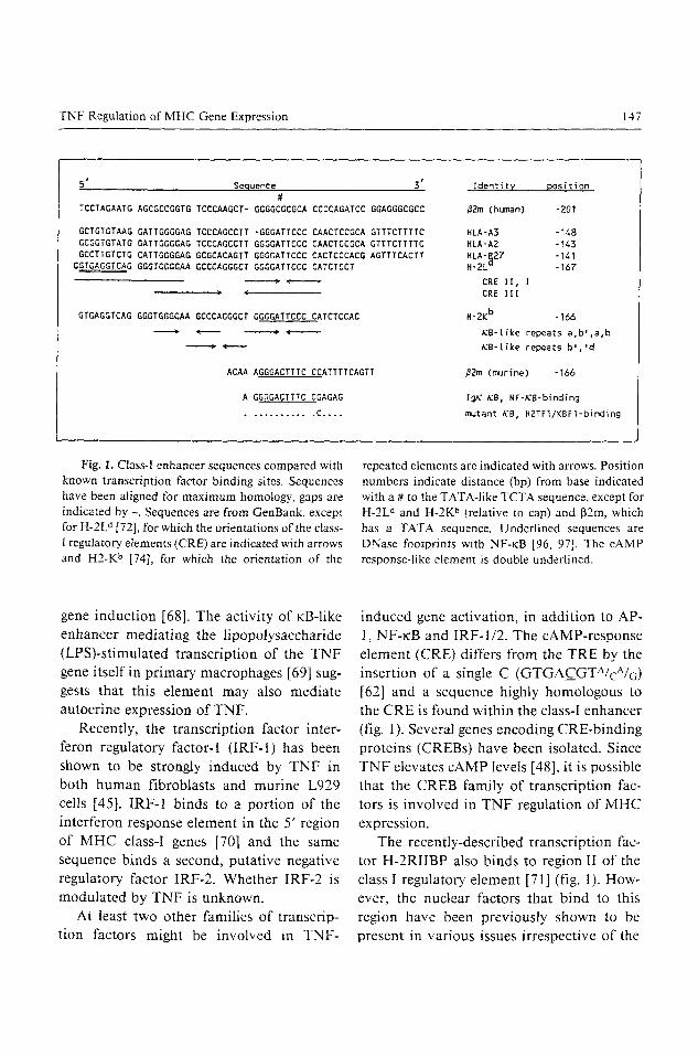
TNF Regulation of MHC Gene Expression 147
5' Sequence 3' Identity position #
TCCTAGAATG AGCGCCGGTG TCCCAAGCT- GGGGCGCGCA CCCCAGATCC GGAGGGCGCC ~2m (human) -201
GCTGTGTAAG GATTGGGGAG TCCCAGCCT/ -GGGATTCCC CAACTCCGCA GTTTCTTTTC HLA-A3 -148 GCGGTGTATG GATTGGGGAG TCCCAGCCTT GGGGATTCCC CAACTCCGCA GTTTCTTTTC HLA*A2 -143 GCCTTGTCTG CATTGGGGAG GCGCACAGTT GGGGATTCCC CACTCCCACG AGTTTCACTT HLA'~27 "141
GGTGAGGTCAG GGGTGGGGAA GCCCAGGGCT GGGGATTCCC CATCTCCT H-2L- -167
�9 �9 �9 CRE I I , I ( CRE I I I
H-2K b -166 GTGAGGTCAG GGGTGGGG~ GCCCAGGGCT GGGGATTCCC CATCTCCAC �9 ( ) ~ - -
ACAA AGGGACTTTC CCATITTCAGTT
A GGGGACTTTC CGAGAG
............ C ....
KB-(ike repeats a , b ' . a , b
gB-Like repeats b J . ' d
~2m (murine) -166
Ig~ ~8, NF-KB-binding
mutant K8, H2TF1/KBF1-binding
Fig. 1. Class-I enhancer sequences compared with known transcription factor binding sites. Sequences have been aligned for maximum homology, gaps are indicated by -. Sequences are from GenBank, except for H-2L d [72], for which the orientations of the class- I regulatory, elements (CRE) are indicated with arrows and H2-K b [74], for which the orientation of the
repeated elements are indicated with arrows. Position numbers indicate distance (bp) from base indicated with a # to the TATA-like TCTA sequence, except for H-2L d and H-2K b (relative to cap) and 132m, which has a TATA sequence. Underlined sequences are DNase footprints with NF-~B [96, 97]. The cAMP response-like element is double underlined.
gene induction [68]. The activity of KB-like
enhancer mediating the lipopolysaccharide (LPS)-stimulated transcription of the TNF gene itself in primary macrophages [69] sug- gests that this element may also mediate autocrine expression of TNF.
Recently, the transcription factor inter- feron regulatory factor-1 (IRF-I) has been
shown to be strongly induced by TNF in both human fibroblasts and murine L929 cells [45]. IRF-1 binds to a portion of the
interferon response element in the 5' region of MHC class-I genes [70] and the same sequence binds a second, putative negative regulatory factor IRF-2. Whether IRF-2 is modulated by TNF is unknown.
At least two other families of transcrip- tion factors might be involved in TNF-
induced gene activation, in addition to AP- 1, NF-~:B and IRF-1/2. The cAMP-response
element (CRE) differs from the TRE by the insertion of a single C (GTGAC__GTA/cA/6) [62] and a sequence highly homologous to the CRE is found within the class-I enhancer
(fig. 1). Several genes encoding CRE-binding proteins (CREBs) have been isolated. Since TNF elevates cAMP levels [48], it is possible
that the CREB family of transcription fac- tors is involved in TNF regulation of MHC expression.
The recently-described transcription fac- tor H-2RIIBP also binds to region II of the class I regulatory element [71] (fig. 1). How- ever, the nuclear factors that bind to this region have been previously shown to be
present in various issues irrespective of the

148 Johnson/Pober
A 10 20 30 40 50 60 70 80 90
I i i i i i i
AAG~TTA~T~TcT~A~AAAcT~cATG~GATGAT~TTT~TC~TA~AAGAGT~CA~GT~AcA~TAA~GA~T~G~A~T~A~GA~TC~AGTT~A~ 100
i i i | I i i
GGACAGAGATTACGGGATAA~AGGAGAGGGACGGGG~CCATGCCGAGGG~TTCTCCCTTGTTTCTCAGACAG~TCTTGGGCCAAGACTCAGGG 200 i i i r i i i
AGACATTGAGACAGAGC•CTTGGCA•AGAAGCAGAGGGGTCAGGGCGAAGTCCAGGGCCC•AGGCGTTGGCTCTCAGGGTCT•AGGCC••GAAGGCGGTG 300
i I i i i i
TATGGATTGGGGAGTCCCAGCCTTGGGGATTCCCCAAcTCCGCAGTTTCTTTTCTCCCTCTCCCAACCTATGTAGGGTCCTTCTTCCTGGATACTCACGA 400
I I I I I | I
CG~GGAC~CAGTTC~C}CCATTGGGTGT~GGGTTT~CAGAGAAG~AAT~AGTGT~GT~GCGGTCG~GGTT~TAAAGTCCGEACGCAC~CA~EGGGA 500
CTCAGATTCTCCCCAGACGCCGAGGA~GCCGTCATGGCGCCCCGAA
B C
TGAGTCA consensus TRE [60] ~ H-2Kb [70] ,..,..T HLA-A2 190" ...... HLA-A2 122" .... A.. 207 ..... C 415
Fig. 2. A Analysis of the 5' flanking region of the human class-I MHC gene HLA-A2. Sequence is from GenBank, locus HUMMHA2, accession number K02883. CAAT box and variant TATA box (TTCTAAA) are underlined, translation initiation co- don is indicated with an arrow. TPA response ele- ments (TRE) homologies are underlined, interferon
regulatory factor-l-binding site homologies are boxed. The NF-KB/H2TFI/KBFI-binding site ho- mology is indicated by a dotted line over the sequence [76]. B. TRE homologies are listed and compared with the consensus TRE [60]. C. IRF-l-binding site homologies are listed and compared with that of the murine class-I binding site [70].
class-I expression [72] and little is known about the regulation of this transcription fac- tor.
Class-I Regulation The 5' regions of MHC class-I a and [3
chain genes contain a sequence homologous
to a ~:B site and several sequences with ho- mologies to the TRE (fig. 2). In addition, a CRE is located within the class-I regulatory.
element (CRE) region I [72]. Whether these sites mediate TNF activation ofclass-I genes remains unclear.
Several groups have noted an enhancer activity in a region 5' of class-I genes [72]. Zachow and Orr [73] have shown that the
amount of enhancer binding activity corre-
lates with the level of constitutive class-I expression in human leukemic T-cell lines.
Israel et al. [74] have suggested that TNF induces transcription of the murine class-I gene (H-2K b) by activating an NK-~:B-like
transcription factor, which displaces con- stitutive binding proteins (AP-2 and
KBF1/H2TF1) from two imperfect ~B sites and stimulates transcription. They show that either site is unresponsive while multimers
of either site are TNF-responsive. However, the human class-I ct chain genes HLA-A2, A3
and B27, which have only a single full repeat, and human [32-microglobulin, which has only a partial element 5' of the gene, respond
to TNF [2, 3] (fig. I). In addition, in func- tional studies with transfected reporter genes

TNF Regulation of MHC Gene Expression 149
these investigators showed that a promoter construct lacking most of the principal ~cB site responds even better to TNF than does a promoter with the entire KB site, weakening the argument that this nB site mediates the T N F response of class-I genes.
A recently described transcription factor, PRDII-BF1, binds the ~cB-like sequence in the murine class-I gene H-2K b, and displays novel characteristics [75]. Unlike other ~cB- binding factors, PRDII-BF1 is under tran- scriptional regulation and is induced only slowly, over a period of hours, by serum or virus stimulation of human MG63 cells. These are the characteristics that would be expected of a transcription factor mediating TNF regulation of HLA class-I genes since they are induced slowly and since cyclohex- imide, a protein-synthesis inhibitor, has pre- viously been shown to block TNF induction, implicating a protein factor synthesized de novo [2]. Moreover, the authors report that serum stimulation increases HLA class-I mRNA with kinetics that are slightly slower than those of PRDII-BF1. A similar or iden- tical factor has been identified in endothelial cells [Tucker Collins, Brigham and Women's Hospital, personal commun.]. Therefore this factor is an attractive candidate for a role in mediating TNF induction of class-I genes. It should be noted that PRDII-BF1 is much larger (predicted 250-290 kD) than either NF-KB (45-50 kD and an associated 65-kD polypeptide) [76] or KBF1/H2TF1 (120 kD), so it is unlikely to be the factor observed by Israel et al. [74].
Class-H Regulation The cell-type-specific regulation of class-
II mRNA expression is reflected in the vari- ety of nuclear factors that bind to the pro- moter regions of class-II genes. For example.
the 5' region of the HLA-DRct gene contains a l l0-bp sequence that binds different fac- tors in different cell types, such as B cells, T cells, murine macrophages, and IFN-v- treated or untreated HeLa cells [77, 78]. One common factor, RF-X, appears to be re- quired for IFN-y inducibility in fibroblasts, and a second, NF-S, binds to a CRE-like ele- ment [79]. Little is known about protein fac- tors or sequence elements that may mediate positive and negative T N F regulation of class-II expression. Although TN F strongly induces IRF-1, no binding site is apparent in the l l0-bp sequence mentioned above. Whether a cryptic site exists in this region, or can be found elsewhere, remains to be seen. A second attractive mediator for TN F regulation of class II is mXBP/CRE-BP2. It is unknown whether this factor is regulated by TNF, but some regulatory effects are likely through TNF regulation of PKA ac- tivity.
Transcription Factor Modification Protein kinases can control the activity of
transcription factors. For example, PKC is believed to phosphorylate I•B, resulting in activated NF-~cB [64]. Subunits of the tran- scription factor AP-1 (c-Fos,c-Jun) are also regulated by phosphorylation [80, 81]. It is unclear whether Jun-B or Jun-D, which are recently described alternative, regulatory forms ofc -Jun [82, 83], are also regulated by phosphorylation. CREB is phosphorylated by PICA [84] and is also predicted to have PKC and casein kinase-II phosphorylation sites [85]. In addition, CREB [85] and CRE- BP2 [86], like Jun and Fos, contain leucine- zipper domains, which mediate hetero- and homo-dimerizat ion of these transcription factors. The presence of PKA, PKC and ca- sein kinase-II phosphorylation sites on
150 Johnson/Pober
CREB suggests that this transcription factor could be modified by different activated sec- ond messengers systems.
Second Messenger~Transcription Factor Cross-Talk In one cell type the response of a single
gene to either IFN- 7 or IFN-a can be blocked by different second messenger antagonists [87]. This suggests that different receptors will be found that activate distinct second messengers. However, a single transcription factor subunit (c-Jun) can combine with dif- ferent subunits (mXBP/CRE-BP2 or c-Fos) to form complexes mediating responses to cAMP or PMA [86]. In addition, c-Fos but not v-Fos represses c-fos transcription through a serum response element (SRE) [88], perhaps because v-Fos is not phosphor- ylated under the same conditions as c-Fos [80]. In contrast, the c-fos SRE mediates transcriptional stimulation by an activated G protein, c-Ha-ras, or by an inhibitor of phosphodiesterase, dibutyl cAMP, both of which may stimulate PKA [63].
Taken together, these results suggest a mechanism by which cross-talk between sec- ond messenger systems might occur: particu- lar agonist-activated second messenger en- zyme(s) may modify transcription factors that are used in common by several different transcription complexes. Transcription fac- tors may undergo phosphorylation-regulated dimerization (or form higher multimers) me- diated by interaction domains, such as leu- cine zippers. These complexes may be them- selved modified by phosphorylation and un- dergo allosteric changes such as those ob- served in the phosphorylation of muscle phosphorylase [89]. The initial pattern of gene expression induced by TNF would be a function of the second messenger systems
activated and the pre-existing transcription factors that are substrates of the activated second messenger enzyme systems. Subse- quent gene expression is likely to be complex due to alterations in second messenger en- zyme activities over time and interactions with previously modified transcription fac- tors.
Post-Transcriptional Regulation TN F stimulates phosphorylation of a cap-
binding protein (p28) in vivo and in vitro, suggesting a role in post-transcriptional regu- lation [90]. However, MHC class-I expres- sion is regulated exclusively at the transcrip- tional level in endothelial cells [3]. Class II is also regulated at the transcriptional level in most cells, but post-transcription regulation is reported in some cells. TN F is itself regu- lated in response to LPS at the transcrip- tional level [69], in the stability of T N F mRNA, and at the secretory level [9 i].
Oncogenes The adenovirus E IA protein is a strong,
general repressor of many different kinds of enhancers, including the ~B element in the murine class-I gene H-2K b enhancer, the AP- 1 binding element in polyomavirus and hu- man metallothionein enhancers and the multiple enhancer elements of SV40 [92]. This is one of the ways in which adenovirus reduces the expression of class I on the cell surface. TNF overcomes this repression in adenovirus-transformed mouse fibroblast cell lines and synergizes with IFN-7 in this activity [93]. IFN-y has been shown to in- crease mRNA for the class I c~ and 13 chains in such cells. It is probable, therefore, that these cytokines are circumventing E1A re- pression at the level of class-I transcription. The cytokine stimulation of class I appears

TNF Regulation of MHC Gene Expression 151
to be independent of the anti-tumor activi-
ties of IFN-y and TNF since Ad l2 E1A
mRNA and protein continues to accumulate in treated ceils.
Conclusion
In this review we have correlated early
events in TNF signal transduction, such as second messenger activation, with later tran- scriptional activation of MHC genes. This
description remains incomplete at several levels. However, we have suggested guide- lines for defining activated second messen- gers involved in mediating TNF effects in-
tracellularly, and we have indicated some of the ways in which transcription factors may be modified by TNF-activated second mes- senger enzymes to regulate gene expression. An exact model for TNF-activated gene ex- pression can be formulated only when more
is understood about the various transcrip- tion factors and sequence elements that are involved. However, from what is already
known about these mediators it can be anti- cipated that transcriptional regulation by TNF will initially involve enzymatic modifi- cation of pre-existing transcription factors
that are then capable of forming active tran- scription complexes. Subsequent regulation will probably involve competition for tran-
scription factor subunits between transcrip- tion complexes and allosteroic modifications of the complexes themselves.
The importance of TNF in regulating
MHC molecule expression is underscored by the observation that TNF is most potent in vivo at causing the regression of immuno- genic tumors and is less effective in immu- nosuppressed mice [94]. In addition, a role for TNF in regulating MHC expression in
vivo is suggested by the observation that TNF enhances the T-cell dependent immune
response in mice [95]. However, TNF has many additional immunoregulatory func-
tions. The induction by TNF of cell adhe-
sion molecules, such as ELAM-1, ICAM-1, INCAM-1 10/VCAM-1, and cytokines, such as IL-1, IL-6, IL-8 and MCP-1, is also clearly
of great importance in the context of the immune response. Nevertheless, because of
the central role of MHC molecules in cell and antigen recognition by T cells, the affect of TNF on MHC expression is a major com-
ponent of these immunoregulatory activi-
ties.
References
1 Chen M, Holskin B, Strickler J, et al: Induction by E 1A oncogene expression of cellular susceptibility to lysis by TNF. Nature 1987;330:581-583.
2 Collins T, Lapierre LA, Fiers W, et al: Recombi- nant human tumor necrosis factor increases mRNA levels and surface expression of HLA-A,B antigens in vascular endothelial cells and dermal fibroblasts in vitro. Proc Natl Acad Sci USA 1986; 83:446-450.
3 Johnson DR, Pober JS: TNF and IFN-gamma synergistically increase transcription of HLA heavy and light chain genes in vascular endothe- lium. Proc Natl Acad Sci USA 1990;87:5183- 5187. Cox JH, Yewdell JW, Eisenlohr LC, et al: Antigen presentation requires transport of MHC class t molecules from the endoplasmic reticulum. Science 1990;247:715-718. Goldfield A, Maniatis T: Coordinate viral induc- tion of tumor necrosis factor alpha and interferon beta in human B cells and monocytes. Proc Natl Acad Sci USA 1989;86:1490-1494. Cherwinski H, Schumacher J, Brown K, et al: Two types of mouse helper T cell clone. III. Further differences in lymphokine synthesis between Thl and Th2 clones. J Exp Med 1987;166:1229- 1244. Takemura Y, Koide Y, Kawabata M, et al: Syner-

152 Johnson/Pober
gistic enhancement of class I major histocompati- bility complex antigen expression in K562 cells induced by recombinant human interferon- gamma and tumor necrosis factor in combination. Exp Hematol 1989; 17:795-799.
8 Pober JS, Gimbrone MA Jr, Cotran RS, et al: Ia expression by vascular endothelium is inducible by activated T cells and by human gamma-inter- feron. J Exp Med 1983;157:1339-1353.
9 Male D, Pryce G: Synergy between interferons and monokines in MHC induction on brain endo- thelium. Immunol Lett 1988; 17:267-271.
10 Warner S, Friedman G, Libby P: Regulation of major histocompatibility gene expression in hu- man vascular smooth muscle cells. Arteriosclero- sis 1989;9:279-288.
l 1 Hofmann M, Weinberg J: Tumor necrosis factor- alpha induces increased hydrogen peroxide pro- duction and Fc receptor expression, but not in- creased Ia antigen expression by peritoneal mac- rophages. J Leukocyte Biol 1987;42:704-707.
12 Lapierre L, Fiers W, Pober JS: Three distinct classes of regulator5, cytokines control endothelial cell MHC antigen expression. Interactions with IFN-gamma differentiate the effect of TNF and LT from those of IFN-alpha and IFN-beta. J Exp Med 1988;167:794-804.
13 Leeuwenberg JF, Van Damme J, Meager T, et al: Effects of tumor necrosis factor on the interferon- gamma-induced major histocompatibility com- plex class II antigen expression by human endo- thelial cells. Eur J Immunol 1988;18:1469-1472.
14 Wedgwood J, Hatam L, Bonagura V: Effect of interferon-gamma and tumor necrosis factor on the expression of class I and class II major histo- compatibility complex molecules by cultured hu- man umbilical vein endothelial cells. Cell Immu- nol 1988;111:1-9.
15 Benveniste E, Sparacio S, Bethea J: Tumor necro- sis factor-alpha enhances interferon-gamma-me- diated class II antigen expression on astrocytes. J Neuroimmunol 1989;25:209-219.
16 Massa P, Schimpl A, Wecker E, et al: Tumor necrosis factor amplifies measles virus-mediated Ia induction on astrocytes. Proc Natl Acad Sci USA 1987;84:7242-7245.
17 Kingston A, Bergsteinsdottir K, Jessen K, et al: Schwann cells co-cultured with stimulated T cells and antigen express major histocompatibility complex (MHC) class 1I determinants without in-
terferon-gamma pretreatment: Synergistic effects of interferon-gamma and tumor necrosis factor on MHC class II induction. Eur J Immunol 1989;19: 177-183.
18 Arenzana-Seisdedos F, Mogensen S, Vuillier F, et al: Autocrine secretion of tumor necrosis factor under the influence of interferon-gamma ampli- fies HLA-DR gene induction in human mono- cytes. Proc Natl Acad Sci USA 1988;85:6087- 6091.
19 Campbell I, Oxbrow L, West J, et al: Regulation of MHC protein expression in pancreatic beta-cells by interferon-gamma and tumor necrosis factor- alpha. Mol Endocrinol 1988;2:101-107.
20 Campbell I, Harrison L: Viruses and cytokines: Evidence for multiple role in pancreatic beta cell destruction in type 1 insulin-dependent diabetes mellitus. J Cell Biochem 1989;40:57-66.
21 Campbell I, lscaro A, Harrison L: IFN-gamma and tumor necrosis factor-alpha. Cytotoxicity to murine islets of Langerhans. J Immunol 1988; 141:2325-2329.
22 Nawroth P, Bank I, Handley D, et al: Tumor necrosis factor/cachectin interacts with endothe- lial cell receptors to induce release of interleu- kin 1. J Exp Med 1986;163:1363-1375.
23 Baglioni C, McCandless S, Tavernier J, et al: Binding of human tumor necrosis factor to high affinity receptors on HeLa and lymphoblastoid cells sensitive to growth inhibition. J Biol Chem 1985;260:13395-13397.
24 Browning J, Ribolini A: Studies on the differing effects of tumor necrosis factor and lymphotoxin on the growth of several human tumor lines. J Immunol 1989;143:1859-1867.
25 Hohmann H, Remy R, Brockhaus M, et al: Two different cell types have different major receptors for human tumor necrosis factor (TNF alpha). J Bio| Chem 1989;264:14927-14934.
26 Gerlach H, Lieberman H. Bach R, et al: Enhanced responsiveness of endothelium in the grow- ing/motile state to tumor necrosis factor/cachec- tin. J Exp Med 1989;170:913-931.
27 Stauber G, Aiyer R, Aggarwal B: Human tumor necrosis factor-alpha receptor. Purification by im- munoaffinity chromatography and initial charac- terization. J Biol Chem 1988;263:19098-19104.
28 Pober J, Lapierre L, Stolpen A, et al: Activation of cultured human endothelial cells by recombinant lymphotoxin: Comparison with tumor necrosis

TNF Regulation of MHC Gene Expression 153
factor and interleukin 1 species. J Immunol 1987; 138:33 l 9-3324.
29 Dinarello C, Mier J: Current concepts: Lympho- kines. N Engl J Med I987;317:940-945.
30 Leszczynski D: Interleukin-I alpha inhibits the effects of gamma-interferon and tumor necrosis factor alpha on the expression of the major histo- compatibility antigens by the rat endothelium. Am J Pathol 1990;136:229-237.
31 Wong G, Goeddel D: Tumor necrosis factors al- pha and beta inhibit virus replication and syner- gize with interferons. Nature 1986;323:819-822.
32 Aggarwal B, Eessalu T, Hass P: Characterization of receptors for human tumor necrosis factor and their regulation by gamma-interferon. Nature 1985;318:665-667.
33 Aggarwal B, Eessalu T: Induction of receptors for tumor necrosis factor-alpha by interferons is not a major mechanism for their cytotoxic response. J Biol Chem t 987;262:10000-10007.
34 Scheurich P, Kobrich G, Pfizenmaier K: Antago- nistic control of tumor necrosis factor receptors by protein kinases A and C. Enhancement of TNF receptor synthesis by protein kinase A and trans- modulation of receptors by protein kinase C. J Exp Med 1989;170:947-958.
35 Holtmann H, Wallach D: Down regulation of the receptors for tumor necrosis factor by interleu- kin 1 and 4-beta-phorbol- 12-myristate- 13-acetate. J Immunol 1987;139:1161-1167.
36 Aggarwal B, Eessalu T: Effect of phorbol esters on down-regulation and redistribution of cell surface receptors for tumor necrosis factor. J Biol Chem 1987;262:16450- 16455.
37 Unglaub R, Maxeiner B, Thoma B, et al: Down- regulation of tumor necrosis factor (TNF) sensi- tivity via modulation of TNF binding capacity by protein kinase C activators. J Exp Med 1987;166: 1788-1797.
38 Fan X-D, Goldberg M, Bloom B: IFN-gamma- induced transcriptional activation is mediated by protein kinase C. Proc Natl Acad Sci USA 1989; 85:5122-5125.
39 Mattila P, H~iyry P, Renkonen R: Protein ki- nase C is crucial in signal transduction during interferon-gamma induction in endothelial cells. Transplant Proc 1990;22:130-130.
40 Kindler V, Sappino A-P, Grau G, et al: The induc- ing role of tumor necrosis factor in the develop-
ment of bactericidal granulomas during BCG in- fection. Cell 1989;56:731-740.
41 Leeuwenberg J, Van Damme J, Jeunhomme G, et al: Interferon beta t, an intermediate in the tumor necrosis factor alpha-induced increased MHC class I expression and an autocrine regulator of the constitutive MHC class I expression. J Exp Med 1987;166:1180-1185.
42 Donato N, Gallick G, Steck P, et al: Tumor necro- sis factor modulates epidermal growth factor re- ceptor phosphorylation and kinase activity in hu- man tumor cells. Correlation with cytotoxicity. J Biol Chem 1989;264:20474-20481.
43 Hori T, Kashiyama S, Hayakawa M, et al: Possi- ble role of prostaglandins as negative regulators in growth stimulation by tumor necrosis factor and epidermal growth factor in human fibroblasts. J Cell Physiol 1989;141:275-280.
44 Snyder DS, Beller DI, Unanue ER: Prostaglandins modulate macrophage Ia expression. Nature 1982;299:163-165.
45 Fujita T, Reis LFL, Watanabe N, et al: Induction of the transcription factor IRF-1 and interferon-J3 mRNAs by cytokines and activators of second- messenger pathways. Proc Natl Acad Sci USA 1989;86:9936-9940.
46 Bird T, Saklatvala J: Down-modulation of epider- mal growth factor receptor affinity in fibroblasts treated with interleukin 1 or tumor necrosis factor is associated with phosphorylation at a site other that threonine 654. J Biol Chem 1990;IX265: 235-240.
47 Bird T, Saklatvala J: IL-I and TNF transmodulate epidermal growth factor receptors by a protein kinase C-independent mechanism. J Immunol 1989;142:126-133.
48 Zhang Y, Lin J-X, Yip Y, et al: Enhancement of cAMP levels and of protein kinase activity by tumor necrosis factor and interleukin l in human fibroblasts: Role in the induction of IL-6. Proc Natl Acad Sci USA 1988;85:6802-6805.
49 Evans J, Mire-Sluis A, Hoffbrand A, et al: Binding of G-CSF, GM-CSF, tumor necrosis factor-alpha, and gamma-interferon to cell surface receptors on human myeloid leukemia cells triggers rapid tyro- sine and serine phosphorylation of a 75-Kd pro- tein. Blood 1990;75:88-95.
50 Hori T, Kashiyama S, Hayakawa M, et al: Tumor necrosis factor is cytotoxic to human fibroblast in

I54 Johnson/Pober
the presence of exogenous arachidonic acid. Exp Cell Res 1989; 185:41-49.
51 Lampugnani MG, Polentarutti N, Pedenovi M, et al: c-fos and c-myc expression in human endothe- lial cells as a function of different culture condi- tions. Exp Cell Res 1990;186:381-384.
52 Imamura K, Sherman M, Spriggs D, et al: Effect of tumor necrosis factor on GTP binding and GTPase activity in HL-60 and L929 cells. J Biol Chem 1988;263:10247-10253.
53 Brett J, Gerlach H, Nawroth P, et al: Tumor necrosis factor/cachectin increases permeability of endothelial cell monolayers by a mechanism involving regulatory G proteins. J Exp Med 1989; 169:1977-1991.
54 Chedid M, Shirakawa F, Naylor P, et al: Signal transduetion pathway for IL-l. Involvement of a pertussis toxin-sensitive GTP-binding protein in the activation of adenylate cyclase. J Immunol 1989;142:4301-4306.
55 Shirakawa F, Yamashita U, Chedid M, et al: Cy- clic AMP - an intracellular second messenger for interleukin 1. Proc Natl Acad Sci USA 1988;85: 8201-8205.
56 Rosoff P, Savage N, Dinarello C: Interleukin-I stimulates diacylglycerol production in T lympho- cytes by a novel mechanism. Cell 1988;54:73-81.
57 Laudanna C, Miron S, Berton G, et al: Tumor necrosis factor-alpha/cachectin activates the O2- generating system in human neutrophils indepen- dently of the hydrolysis of phosphoinositides and the release of arachidonic acid. Biochem Biophys Res Commun 1990; 166:308-315.
58 Palombella V, Vilcek J: Mitogenic and cytotoxic actions of tumor necrosis factor in BALB/c 3T3 cells. Role of phospholipase activation. J Biol Chem 1989;264:18128-18136.
59 Brenner DA, O'Hara M, Angel P, et al: Prolonged activation of jun and collagenase genes by tumor necrosis factor-a. Nature 1989;337:661-663.
60 Lee W, Mitchell P, Tjian R: Purified transcription factor AP-I interacts with TPA-inducible en- hancer elements. Cell 1987;49:741-752.
61 Rauscher F, Sambucetti L, Curran T, et al: Com- mon DNA binding site for Fos protein complexes and transcription factor AP-1. Cell 1988;52:471- 48O.
62 Ziff E: Transcription factors: a new family gathers as the cAMP response site. Trends in Genetics 1990;6:69-72.
63 Fukumoto Y, Kaibuchi K, Oku N, et al: Activa- tion of the c-los serum-response element by the activated c-Ha-ras protein in a manner indepen- dent of protein kinase C and cAMP-dependent protein kinase. J Biol Chem 1990;265:774-780.
64 Baeuerle PA, Baltimore D, Ikappa B: A specific inhibitor of the NF-kappaB transcription factor. Science 1988;242:540-546.
65 Hoyos B, Balard DW, Bohnlein E, et al: Kappa B-specific DNA binding proteins: Role in the reg- ulation of human interleukin-2 gene expression. Science 1989;244:457-460.
66 Cross SL, Halden NF, Lenardo M J, et al: Func- tionally distinct NF-kappaB binding sites in the immunoglobulin kappa and IL-2 receptor alpha chain genes. Science 1989;244:466-469.
67 Freimuth W, Depper J, Nabel G: Regulation of the IL-2 receptor alpha-gene. Interaction of a kappa B binding protein with cell-specific tran- scription factors. J Immunol 1989;143:3064- 3068.
68 Muegge K, Williams T, Kant J, et al: Interleukin-I costimulatory activity on the interleukin-2 pro- moter via AP-I. Science 1989;246:249-251.
69 Shakhov A, Collart M, Vassalli P, et al: Kappa- B-type enhancers are involved in lipopolysaccha- ride-mediated transcriptional activation of the tu- mor necrosis factor alpha gene in primary macro- phages. J Exp Med 1990;171:35-47.
70 Miyamoto M, Fujita T, Kimura Y, et al: Regu- lated expression of a gene encoding a nuclear fac- tor, IRF-I, that specifically binds to IFN-beta gene regulatory elements. Cell 1988;54:903-913.
71 Hamada K, Gleason S, Levi B, et al: H-2RIIBP, a member of the nuclear hormone receptor super- family that binds to both the regulatory element of major histocompatibility class I genes and the es- trogen response element. Proc Natl Acad Sci USA 1989;86:8289-8293.
72 Burke P, Hirschfeld S, Shirayoshi Y, et al: Devel- opmental and tissue-specific expression of nuclear proteins that bind the regulatory element of the major histocompatibility complex class I gene. J Exp Med 1989;169:1309-1321.
73 Zachow K, Orr H: Regulation of HLA class I tran- scription in T cells. J Immunol 1989:143:3385- 3389.
74 Israel A, Le Bail O, Hatat D, et al: TNF stimulates expression of mouse MHC class I genes by induc- ing an NFnB-like enhancer binding activity which

TNF Regulation of MHC Gene Expression I55
displaces constitutive factors. EMBO J 1989;8: 3793-3800.
75 Fan C-M, Maniatis T: A DNA-binding protein containing two widely separated zinc finger motifs that recognize the same DNA sequence. Genes Dev 1990:4:29-42.
76 Lenardo M J, Baltimore D: NF-kappaB: A pleio- tropic mediator of inducible and tissue-specific gene control. Cell 1958;58:227-229.
77 Dedrick RL, Jones PP: Sequence elements re- quired for activity of a murine major histocom- patibility complex class II promoter bind com- mon and cell-type-specific nuclear factors. Mol Cell Biol 1990;10:593-604.
78 Tsang SY, Nakanishi M, Peterlin BM: Mutational analysis of the DRA promoter: Cis-acting se- quences and trans-acting factors. Mol Cell Biol 1990;10:711-719.
79 Kobr M, Reith W, Herrero-Sanchez C, et al: Two DNA-binding proteins discriminate between the promoters of different members of the major his- tocompatibility complex class II multigene fami- ly. Mol Cell Biol 1990;10:965-971.
80 Barber JR, Verma IM: Modification of Fos pro- teins: phosphorylation of c-Fos, but not v-Fos, is stimulated by 12-tetradecanoyl-phorbol-13-ace- tare and serum. Mol Cell Biol 1987;7:2201- 2211.
81 Berk A J, Schmidt MC: How do transcription fac- tors work. Genes Dev 1990;4:15 I-155.
82 Chiu R. Angel P, Karin M: Jun-B differs in its biological properties from, and is a negative regu- lator of, c-Jun. Cell 1989;59:979-986.
83 Schutte J, Viallet J, Nau M, et al: Jun-B inhibits and c-fos stimulates the transforming and trans- activating activities of c-jun. Cell 1989;59:987- 997.
84 Yamamoto KK, Gonzalez GA, Menzel P, et al: Characterization of a bipartite activator domain in transcription factor CREB. Cell 1990;60:611- 617.
85 Gonzalez G, Yamamoto K, Fischer W, et al: A cluster of phosphorylation sites on the cyclic AMP-regulated nuclear factor CREB predicted by its sequence. Nature 1989;337:749-752.
86 Ivashkiv LB, Liou H-C, Kara CJ, et al: mXBP/CRE-BP2 and c-Jun form a complex which binds to the cyclic AMP, but not to the 12- o-tetradecanoylphorbot-13-acetate, response ele- ment. Mol Cell Biol 1990;t0:i609-1621.
87 Lew D J, Decker T, Darnell JE Jr: Alpha interferon and gamma interferon stimulate transcription of a single gene through different signal transduction pathways. Mol Cell Biol 1989;9:5404-5411.
88 Lucibello F, Lowag C, Neuberg M, et al: Trans- repression of the mouse c-fos promoter: A novel mechanism of Fos-mediated trans-regulation. Cell 1989;59:999-1007.
89 Perutz M: Control by phosphorylation. Nature 1988;336:202-203.
90 Marino M, Pfeffer L, Guidon P Jr, et al: Tumor necrosis factor induces phosphorylation o fa 28kD mRNA cap-binding protein in human cervical carcinoma ceils. Proc Natl Acad Sci USA 1989;86: 8417-8421.
91 Beutler B, Cerami A: The biology of cachec- t in/TNF - a primary mediator of the host re- sponse. Annu Rev Immunol 1989;7:625-655.
92 Rochette-Egly C, Fromental C, Chambon P: Gen- eral repression of enhanson activity by the adeno- virus-2 EIA proteins. Genes Dev 1990;4:137- 150.
93 Eager K, Pfizenmaier K, Ricchiardi R: Modula- tion of MHC class I genes in Adl2 transformed cells. IFN-gamma increases class I expression by a mechanism that circumvents E1A induced-repres- sion and TNF enhances the effect of IFN-gamma. Oncogene 1988;4:39-44.
94 Havell E, Fiers W, North R: The ant i tumor func- tion of tumor necrosis factor. J Exp Med 1988; 167:1067-1085.
95 Ghiara P, Boraschi D, Nencioni L, et al: Enhance- ment of in vivo immune response by tumor necro- sis factor. J Immunol 1987;139:3676-3679.
96 Mauxion F, Sen R: Alteration of a single nucleo- tide allow efficient binding of H2TFI/KBFI to the immunoglobulin kappa enhancer motif. Mol Cell Biol 1989;9:3548-3552.
97 Yano O, Kanellopoulos J, Kieran M, et al: Purifi- cation of KBF1, a common factor binding to both H-2 and beta2-microglobulin enhancers. EMBO J 1987;6:3317-3324.
Dr. David R. Johnson Department of Pathology Brigham and Women's Hospital and Harvard Medical School Boston, MA 02115 (USA)