Embed Size (px)
Citation preview

CLINICAL ARTICLEJ Neurosurg Pediatr 21:449–455, 2018
Lumbar disc herniation (LDH) is a well-known cause of low-back pain and lower-extremity radiculopathy in adults.13 However, LDH is rare in the pediatric
population, with children accounting for only 5% of LDH cases.15 While the pediatric pathophysiology is not fully
understood, certain risk factors, including trauma, devel-opmental anatomical variants, and genetics, may predis-pose certain patients to symptomatic LDH.6,14,15 Moreover, unlike the degenerative discs frequently seen in adults, herniated pediatric discs tend to be highly elastic and
ABBREVIATIONS BMI = body mass index; EBL = estimated blood loss; LDH = lumbar disc herniation; LOS = length of stay; MIS = minimally invasive surgery; ODI = Oswestry Disability Index.SUBMITTED August 13, 2017. ACCEPTED November 16, 2017.INCLUDE WHEN CITING Published online February 23, 2018; DOI: 10.3171/2017.11.PEDS17293.
Tubular approach to minimally invasive microdiscectomy for pediatric lumbar disc herniationJulio D. Montejo, BA,1 Joaquin Q. Camara-Quintana, MD,1 Daniel Duran, MD,1 Jeannine M. Rockefeller, MSN, APRN,1 Sierra B. Conine,1 Alyssa M. Blaise,1 Kristopher T. Kahle, MD, PhD,1–3 and Michael L. DiLuna, MD1,2
Departments of 1Neurosurgery, 2Pediatrics, and 3Cellular & Molecular Physiology and Centers for Mendelian Genomics, Yale School of Medicine, New Haven, Connecticut
OBJECTIVE Lumbar disc herniation (LDH) in the pediatric population is rare and exhibits unique characteristics com-pared with adult LDH. There are limited data regarding the safety and efficacy of minimally invasive surgery (MIS) using tubular retractors in pediatric patients with LDH. Here, the outcomes of MIS tubular microdiscectomy for the treatment of pediatric LDH are evaluated.METHODS Twelve consecutive pediatric patients with LDH were treated with MIS tubular microdiscectomy at the authors’ institution between July 2011 and October 2015. Data were gathered from retrospective chart review and from mail or electronic questionnaires. The Macnab criteria and the Oswestry Disability Index (ODI) were used for outcome measurements.RESULTS The mean age at surgery was 17 ± 1.6 years (range 13–19 years). Seven patients were female (58%). Prior to surgical intervention, 100% of patients underwent conservative treatment, and 50% had epidural steroid injections. Preoperative low-back and leg pain, positive straight leg raise, and myotomal leg weakness were noted in 100%, 83%, and 67% of patients, respectively. The median duration of symptoms prior to surgery was 9 months (range 1–36 months). The LDH level was L5–S1 in 75% of patients and L4–5 in 25%. The mean ± SD operative time was 90 ± 21 minutes, the estimated blood loss was ≤ 25 ml in 92% of patients (maximum 50 ml), and no intraoperative or postoperative com-plications were noted at 30 days. The median hospital length of stay was 1 day (range 0–3 days). The median follow-up duration was 2.2 years (range 0–5.8 years). One patient experienced reherniation at 18 months after the initial operation and required a second same-level MIS tubular microdiscectomy to achieve resolution of symptoms. Of the 11 patients seen for follow-up, 10 patients (91%) reported excellent or good satisfaction according to the Macnab criteria at the last follow-up. Only 1 patient reported a fair level of satisfaction by using the same criteria. Seven patients completed an ODI evaluation at the last follow-up. For these 7 patients, the mean ODI low-back pain score was 19.7% (SEM 2.8%).CONCLUSIONS To the authors’ knowledge, this is the longest outcomes study and the largest series of pediatric pa-tients with LDH who were treated with MIS microdiscectomy using tubular retractors. These data suggest that MIS tubu-lar microdiscectomy is safe and efficacious for pediatric LDH. Larger prospective cohort studies with longer follow-up are needed to better evaluate the long-term efficacy of MIS tubular microdiscectomy versus other open and MIS techniques for the treatment of pediatric LDH.https://thejns.org/doi/abs/10.3171/2017.11.PEDS17293KEY WORDS adolescents; lumbar disc herniation; minimally invasive surgery; microdiscectomy; spine
J Neurosurg Pediatr Volume 21 • May 2018 449©AANS 2018, except where prohibited by US copyright law
Unauthenticated | Downloaded 02/16/22 06:43 PM UTC

J. D. Montejo et al.
J Neurosurg Pediatr Volume 21 • May 2018450
nondegenerative in nature, thereby predisposing pediatric patients to poorer resorption and poorer response to con-servative management.8,15
The approach to adult and pediatric LDH has histori-cally included open laminectomy with discectomy, which is an approach that dates back to the mid-20th century.17 More recently, minimally invasive surgery (MIS) tech-niques have grown in favor because of their ability to preserve important muscle attachments and avoid exces-sive bone removal, which may compromise spinal stabil-ity.5 These concepts are especially important in children and adolescents because they undergo significant growth with a continuously changing skeletal structure. MIS ap-proaches for pediatric LDH presently include keyhole laminotomy with the use of a microscope to perform mi-crodiscectomy16 and percutaneous endoscopic microdisc-ectomy.9,12,18,19 Short-term follow-up studies of microdisc-ectomy for pediatric LDH have shown comparable results to those of traditional open discectomy, thus validating MIS techniques in pediatric patients.16 However, few stud-ies have looked at the safety and efficacy of using a tubular approach for MIS microdiscectomy as a treatment option for pediatric LDH.16
The purpose of this study was 2-fold: 1) to characterize the unique clinical features seen in pediatric patients with LDH who require surgery, and 2) to evaluate and validate the perioperative, postoperative, short-term, and longer-term outcomes of MIS tubular microdiscectomy for the treatment of pediatric LDH.
MethodsPatients
The inclusion criteria for this case series were 1) pedi-atric patients who were 19 years or younger; 2) diagnosis of LDH based on physical findings and MRI examination; 3) surgical treatment with MIS tubular microdiscectomy; and 4) date of surgery between July 2011 and October 2015. There were 12 consecutive pediatric patients who underwent surgical treatment performed by the senior au-thor (M.L.D.) and met all inclusion criteria. None of these 12 patients had a history of spinal oncology, prior spinal infection, acute fracture to the spine due to trauma, known isthmic spondylolisthesis, or lumbar instability on imag-ing. Every operation took place in a pediatric operating room. Patients admitted to the hospital following surgery stayed on a pediatric ward. The institutional review board at Yale University School of Medicine approved the study.
Preoperative EvaluationThe electronic medical records were reviewed indepen-
dently by 3 authors (J.D.M., S.B.C., and J.Q.C.-D.) to de-termine all chart review data as documented by the treat-ing neurosurgeon. Demographic variables included age at surgery and sex. Risk factors included body mass index (BMI), participation in athletic activities, precipitating trauma, comorbidities, and tobacco use. Presurgical treat-ment variables included the use of nonsteroidal antiin-flammatory drugs, opioid pain medication, physical thera-py, chiropractor therapy, epidural steroid injection, as well as the number of emergency department visits due to low-
back pain and radicular pain prior to surgical intervention. Variables at presentation included duration of symptoms prior to surgery, presence of radicular low-back pain or leg pain, and bladder or bowel dysfunction. Variables at physi-cal examination included positive straight leg raise, myo-tomal lower-extremity weakness, and dermatomal sensory loss. All patients underwent diagnostic preoperative MRI evaluation of the lumbar spine, as well CT or plain ra-diography when indicated. MRI findings were reviewed as documented by the treating neurosurgeon and radiolo-gist. Fig. 1 illustrates the representative LDH findings on MRI of typical cases included in this cohort. Every patient had at least 4 months of conservative therapy before being considered for surgery unless the presence of neurologi-cal deficits, as exemplified by myotomal lower-extremity weakness, justified more urgent surgical intervention.
Surgical TechniqueWritten informed consent was obtained from all pa-
tients or their guardians. All patients were placed under general anesthesia during surgery. No patient required a Foley catheter. Patients were placed prone on a Jackson table with a Wilson frame. A < 15-mm paramedian inci-sion about 1 cm off the midline was made, and a tubular retractor system (most commonly METRx [Medtronic], but MARS3 V [Globus Medical] and Luxor [Stryker] were also used depending on availability) was docked against the medial facet and inferior lateral edge of the lamina at the level and side of the herniated disc. The position and angle of the tubular retractor system was confirmed with
FIG. 1. Representative radiological images of pediatric LDH. A: Pre-operative sagittal (upper) and axial (lower) T2-weighted magnetic reso-nance images of a pediatric case of LDH at L4–5. B: LDH at L5–S1. Note the significant narrowing of the spinal canal that is highlighted by the reduction of the cerebrospinal fluid signal at the level of herniation.
Unauthenticated | Downloaded 02/16/22 06:43 PM UTC
J Neurosurg Pediatr Volume 21 • May 2018 451
J. D. Montejo et al.
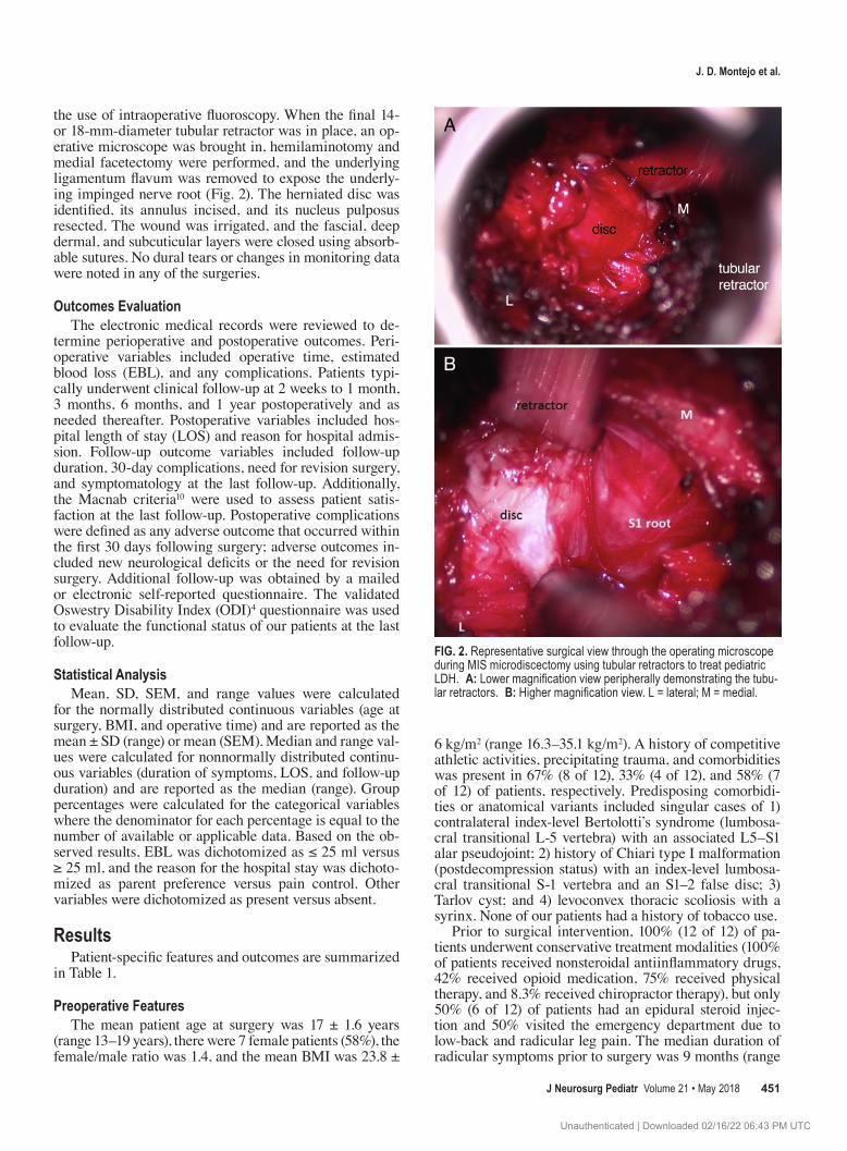
the use of intraoperative fluoroscopy. When the final 14- or 18-mm-diameter tubular retractor was in place, an op-erative microscope was brought in, hemilaminotomy and medial facetectomy were performed, and the underlying ligamentum flavum was removed to expose the underly-ing impinged nerve root (Fig. 2). The herniated disc was identified, its annulus incised, and its nucleus pulposus resected. The wound was irrigated, and the fascial, deep dermal, and subcuticular layers were closed using absorb-able sutures. No dural tears or changes in monitoring data were noted in any of the surgeries.
Outcomes EvaluationThe electronic medical records were reviewed to de-
termine perioperative and postoperative outcomes. Peri-operative variables included operative time, estimated blood loss (EBL), and any complications. Patients typi-cally underwent clinical follow-up at 2 weeks to 1 month, 3 months, 6 months, and 1 year postoperatively and as needed thereafter. Postoperative variables included hos-pital length of stay (LOS) and reason for hospital admis-sion. Follow-up outcome variables included follow-up duration, 30-day complications, need for revision surgery, and symptomatology at the last follow-up. Additionally, the Macnab criteria10 were used to assess patient satis-faction at the last follow-up. Postoperative complications were defined as any adverse outcome that occurred within the first 30 days following surgery; adverse outcomes in-cluded new neurological deficits or the need for revision surgery. Additional follow-up was obtained by a mailed or electronic self-reported questionnaire. The validated Oswestry Disability Index (ODI)4 questionnaire was used to evaluate the functional status of our patients at the last follow-up.
Statistical AnalysisMean, SD, SEM, and range values were calculated
for the normally distributed continuous variables (age at surgery, BMI, and operative time) and are reported as the mean ± SD (range) or mean (SEM). Median and range val-ues were calculated for nonnormally distributed continu-ous variables (duration of symptoms, LOS, and follow-up duration) and are reported as the median (range). Group percentages were calculated for the categorical variables where the denominator for each percentage is equal to the number of available or applicable data. Based on the ob-served results, EBL was dichotomized as ≤ 25 ml versus ≥ 25 ml, and the reason for the hospital stay was dichoto-mized as parent preference versus pain control. Other variables were dichotomized as present versus absent.
ResultsPatient-specific features and outcomes are summarized
in Table 1.
Preoperative FeaturesThe mean patient age at surgery was 17 ± 1.6 years
(range 13–19 years), there were 7 female patients (58%), the female/male ratio was 1.4, and the mean BMI was 23.8 ±
6 kg/m2 (range 16.3–35.1 kg/m2). A history of competitive athletic activities, precipitating trauma, and comorbidities was present in 67% (8 of 12), 33% (4 of 12), and 58% (7 of 12) of patients, respectively. Predisposing comorbidi-ties or anatomical variants included singular cases of 1) contralateral index-level Bertolotti’s syndrome (lumbosa-cral transitional L-5 vertebra) with an associated L5–S1 alar pseudojoint; 2) history of Chiari type I malformation (postdecompression status) with an index-level lumbosa-cral transitional S-1 vertebra and an S1–2 false disc; 3) Tarlov cyst; and 4) levoconvex thoracic scoliosis with a syrinx. None of our patients had a history of tobacco use.
Prior to surgical intervention, 100% (12 of 12) of pa-tients underwent conservative treatment modalities (100% of patients received nonsteroidal antiinflammatory drugs, 42% received opioid medication, 75% received physical therapy, and 8.3% received chiropractor therapy), but only 50% (6 of 12) of patients had an epidural steroid injec-tion and 50% visited the emergency department due to low-back and radicular leg pain. The median duration of radicular symptoms prior to surgery was 9 months (range
FIG. 2. Representative surgical view through the operating microscope during MIS microdiscectomy using tubular retractors to treat pediatric LDH. A: Lower magnification view peripherally demonstrating the tubu-lar retractors. B: Higher magnification view. L = lateral; M = medial.
Unauthenticated | Downloaded 02/16/22 06:43 PM UTC

J. D. Montejo et al.
J Neurosurg Pediatr Volume 21 • May 2018452
1–36 months). Preoperative signs and symptoms of radicu-lar low-back and leg pain, positive straight leg raise, and myotomal leg weakness were notable in 100% (12 of 12), 83% (10 of 12), and 67% (8 of 12) of patients, respectively. However, dermatomal sensory loss was notable in only 17% (2 of 12) of patients, and none experienced bladder or bowel dysfunction. The level of LDH was L5–S1 in 75% (9 of 12) and L4–5 in 25% (3 of 12) of patients, respective-ly, and 58% (7 of 12) of patients had LDH on the left side.
Perioperative and Postoperative OutcomesThe mean operative time was 90 ± 21 minutes (range
46–124 minutes), and EBL was ≤ 25 ml in 92% (11 of 12) of patients (maximum 50 ml in 1 patient). There were no intraoperative or postoperative complications at 30 days. Postoperatively, the median LOS was 1 day (range 0–3 days), with 25% (3 of 12) of patients discharged on the day of surgery. The primary reason for a 1-night hospital admission was parent preference to ensure continued ade-quate pain control and voiding status. The primary reason for more than 1 night of admission was inadequate pain control, with 1 patient additionally developing a urinary tract infection in the absence of an indwelling catheter for which appropriate antibiotics were started while the pa-tient was still in the hospital.
Longer-Term OutcomesThe overall median follow-up time for our cohort was
2.2 years (range 0–5.8 years). Eleven of 12 patients (92%) underwent follow-up. Of those 11 patients, 1 patient (9.1%) experienced a recurrence of radicular symptoms at 18 months after the initial operation and required a second same-level MIS tubular microdiscectomy, which success-fully resolved the patient’s symptoms. At the last follow-up, symptoms had completely resolved or significantly
improved in 91% (10 of 11) of our patients, while 1 (9.1%) patient did not significantly improve. Correspondingly, the Macnab criteria at the last follow-up revealed that 91% (10 of 11) of patients had excellent or good patient satisfaction, while 1 patient (9.1%) had fair satisfaction.
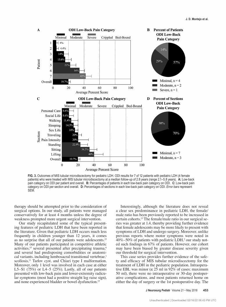
Seven of 11 patients (64%) completed an ODI question-naire at a median follow-up of 2.8 years (range 2.1–5.8 years). According to the ODI results, 57% of our patients (4 of 7) had minimal features of low-back pain, while 2 patients (29%) had mildly moderate features and another patient (14%) had mildly severe features due to adjacent-level degenerative disease. Detailed ODI section analysis revealed that our cohort had minimal features of low-back pain in 7 sections (personal care, social life, walking, sleeping, sex life, traveling, and pain intensity) and moder-ate features in 3 sections (standing, sitting, and lifting). On average, our cohort demonstrated a low-back pain score on ODI of 19.7% (SEM 2.8%). The ODI results are sum-marized in Fig. 3 and detailed in Table 2.
DiscussionFew studies have investigated outcomes after MIS tu-
bular microdiscectomy for pediatric LDH. Despite being introduced by Foley and Smith in 1997 for adults,5 the first reported case series on the short-term outcomes of MIS tubular microdiscectomy for pediatric LDH was not performed until 14 years later in 2011 by Thomas et al.,16 which included only 6 patients. To the best of our knowl-edge, there have been no further reports, thereby making our study the longest outcomes study and the largest.
Pediatric LDH is rare, and although this patient popula-tion tends to respond less to conservative management,8,15 many patients do respond and are never seen by a spine specialist.2 Thus, it is generally agreed that in the absence of neurological deficits or intractable pain, conservative
TABLE 1. Summary of 12 consecutive patients with pediatric LDH treated with MIS tubular microdiscectomy
Age (yrs), Sex
Pain Duration
(mos)
Myotomal Leg
Weakness* Level
Intraop EBL (ml)†
LOS (days), Reason
Follow-Up (yrs)
30-Day Complications Pain & Weakness Outcome
Macnab Criteria
Low-Back Pain ODI Category
17, F 9 4 DF L5–S1 Minimal 1, PP 5.8 No Weakness resolved & pain improved Good Moderate18, M 2 4 DF, 3 EHL L5–S1 Minimal 0 0 NA Lost to follow-up NA NA16, F 12 None L5–S1 Minimal 1, PP 2.0 No Pain resolved (required redo op) Excellent NA19, F 6 4 TA L4–5 Minimal 1, PP 3.3 No Weakness & pain resolved Excellent Minimal18, M 11 4 DF/PF &
4 EHLL5–S1 Minimal 1, PP 3.0 No Weakness resolved & persistent pain Fair Severe
17, M 12 4 TA/EHL L5–S1 Minimal 0 2.8 No Weakness & pain resolved Excellent Minimal15, F 36 4 TA/EHL L4–5 Minimal 3, PC 0.5 No Weakness resolved & pain improved Good NA18, F 4 None L5–S1 Minimal 0 0.1 No Pain resolved Excellent NA13, M 1 4 TA & 3 EHL L4–5 Minimal 1, PP 2.6 No Weakness & pain resolved Excellent Minimal16, F 6 None L5–S1 Minimal 1, PP 2.1 No Weakness & pain resolved Excellent Minimal18, F 18 None L5–S1 Minimal 2, PC 2.2 No Pain improved Good Moderate17, M 9 4 DF & 3 EHL L5–S1 50 1, PP 0.2 No Weakness resolved & pain improved Good NA
DF = dorsiflexion; EHL = extensor hallucis longus; NA = not available; PC = pain control; PF = plantar flexion; PP = parent preference; TA = tibialis anterior.* Muscle group strength was measured on an ascending scale from 0 (completely flaccid) to 5 (normal strength). † Minimal EBL was defined as ≤ 25 ml.
Unauthenticated | Downloaded 02/16/22 06:43 PM UTC
J Neurosurg Pediatr Volume 21 • May 2018 453
J. D. Montejo et al.
therapy should be attempted prior to the consideration of surgical options. In our study, all patients were managed conservatively for at least 4 months unless the degree of weakness prompted more urgent surgical intervention.
Our study recapitulated some of the typical present-ing features of pediatric LDH that have been reported in the literature. Given that pediatric LDH occurs much less frequently in children younger than 12 years, it comes as no surprise that all of our patients were adolescents.15 Many of our patients participated in competitive athletic activities,15 several presented after precipitating trauma,6 and several had predisposing comorbidities or anatomi-cal variants, including lumbosacral transitional vertebrae,1 scoliosis,11 Tarlov cyst, and Chiari type I malformation. Moreover, only 1 level was involved in each case at either L5–S1 (75%) or L4–5 (25%). Lastly, all of our patients presented with low-back pain and lower-extremity radicu-lar symptoms (most had a positive straight leg raise sign), and none experienced bladder or bowel dysfunction.15
Interestingly, although the literature does not reveal a clear sex predominance in pediatric LDH, the female/male ratio has been previously reported to be increased in certain cohorts.15 The female/male ratio in our surgical se-ries was greater at 1.4, thereby providing further evidence that female adolescents may be more likely to present with symptoms of LDH and undergo surgery. Moreover, unlike previous reports where motor symptoms were noted in 40%–50% of patients with pediatric LDH,2 our study not-ed such findings in 67% of patients. However, our cohort may have been biased by greater disease severity given our threshold for surgical intervention.
This case series provides further evidence of the safe-ty and efficacy of MIS tubular microdiscectomy for the treatment of LDH in the pediatric population. Intraopera-tive EBL was minor (≤ 25 ml in 92% of cases; maximum 50 ml), there were no intraoperative or 30-day postoper-ative complications, and most patients returned home on either the day of surgery or the 1st postoperative day. The
FIG. 3. Outcomes of MIS tubular microdiscectomy for pediatric LDH. ODI results for 7 of 12 patients with pediatric LDH (4 female patients) who were treated with MIS tubular microdiscectomy at a median follow-up of 2.8 years (range 2.1–5.8 years). A: Low-back pain category on ODI per patient and overall. B: Percentages of patients in each low-back pain category on ODI. C: Low-back pain category on ODI per section and overall. D: Percentages of sections in each low-back pain category on ODI. Error bars represent SEM.
Unauthenticated | Downloaded 02/16/22 06:43 PM UTC

J. D. Montejo et al.
J Neurosurg Pediatr Volume 21 • May 2018454
majority of patients who remained in the hospital over-night postoperatively did so due to parent preference to ensure continued adequate pain control and voiding status. Prior studies of other MIS techniques have reported short-term complications that could similarly be encountered in patients treated using the tubular approach. These com-plications include wound hematoma and delayed wound healing, which are reported in up to 4% and 3% of cases, respectively, as well as rare surgical wound infections or discitis.15 However, our patients did not experience any short-term complications.
We monitored the majority of our cohort (92%) for me-dian 2.2 years and maximum 5.8 years postoperatively. Per the Macnab criteria, 91% of patients had excellent or good patient satisfaction at the last follow-up. Furthermore, on average, our cohort demonstrated a low-back pain score on ODI of 19.7% (SEM 2.8%) at the last follow-up, thereby indicating borderline minimal or moderate low-back pain interference in overall quality of life. Our findings further substantiate the aforementioned improvements in reduced low-back pain and radicular leg pain and weakness af-ter using MIS tubular microdiscectomy to treat pediatric LDH.
Long-term complications reported by studies of other MIS techniques include disc space narrowing, foraminal stenosis, adjacent-disc degeneration, and a variable risk of reherniation that requires reoperation (range 0%–24%).3,7
In our cohort, 1 patient (9.1%) experienced lumbar disc reherniation at 18 months after the initial operation and required reoperation with an MIS tubular approach. Fur-thermore, while most patients had minimal or only mildly moderate features of low-back pain on ODI, the ODI re-sponses of 1 patient were 2% over the threshold for severe features and necessitated further improvement in all ele-ments of pediatric LDH, including conservative manage-ment, preoperative evaluation, and postoperative manage-ment.
Our study has several limitations. The significant limi-tations are the retrospective nature of this study and the cohort size, which predispose the study to selection and sampling bias. To address potential bias, we included consecutive patients in our study. Importantly, our study represents the results of a single neurosurgeon at a single institution. Given the rarity of pediatric LDH in patients who are younger than 13 years, our study consisted of pe-diatric teenagers (13 years or older), and thus we cannot make conclusions about the safety or efficacy of the tubu-lar approach for MIS microdiscectomy in patients who are younger than 13 years. Lastly, the preoperative ODI scores or those at the intermediate follow-up intervals were not available for comparison against the ODI scores at the last follow-up, thereby precluding a more complete assessment of the postsurgical trend in improvement relative to pre-surgical baseline.
TABLE 2. ODI results for 7 of 12 patients with pediatric LDH treated with MIS tubular microdiscectomy
Variable ValueSum Total
Sum Max Percentage SEM
Low-Back Pain ODI Category*
Patient characteristics Age at surgery, yrs 17 19 18 17 13 16 18 Sex F F M M M F F Follow-up, yrs 5.8 3.3 3 2.8 2.6 2.1 2.2ODI section† Pain intensity 4 0 3 0 0 0 0 7 35 20 13.1 Min Personal care 2 0 1 0 0 0 0 3 35 8.6 5.9 Min Lifting 2 2 3 0 3 0 4 14 35 40 11.5 Mod Walking 0 0 3 0 0 0 1 4 35 11.4 8.6 Min Sitting 2 2 4 0 2 1 1 12 35 34.3 9.5 Mod Standing 2 0 3 1 0 0 2 8 35 22.9 9.2 Mod Sleeping 1 2 1 0 0 0 1 5 35 14.3 5.7 Min Sex life 1 0 1 1 NA NA 1 4 25 16 4 Min Social life 2 0 1 0 0 0 0 3 35 8.6 5.9 Min Traveling 1 1 1 0 2 1 1 7 35 20 4.4 MinODI score Total sum 17 7 21 2 7 2 11 67 Max sum 50 50 50 50 45 45 50 340 Percentage 34 14 42 4 15.6 4.4 22 19.7 SEM 6.7 6 7.6 2.7 8 2.9 7.6 2.8 Low-back pain ODI category* Mod Min Sev Min Min Min Mod Min
Max = maximum; min = minimum; mod = moderate; sev = severe.* Per ODI definitions: minimal = 0%–20%; moderate = 21%–40%; severe = 41%–60%; crippled = 61%–80%; and bed-bound = 81%–100%.† Self-scored from 0 (best quality of life) to 5 (worst quality of life).
Unauthenticated | Downloaded 02/16/22 06:43 PM UTC

J Neurosurg Pediatr Volume 21 • May 2018 455
J. D. Montejo et al.
Further observational studies, and ideally randomized clinical trials involving larger pediatric LDH cohorts with longer follow-up durations, are needed to validate and ex-pand our findings and to investigate relative efficacy across different types of MIS techniques. Additional long-term outcomes after MIS tubular microdiscectomy that are in need of investigation include the incidence of iatrogenic pars defects, adjacent-segment changes, and the need for lumbar spine fusion.
ConclusionsTo our knowledge, this is the longest outcomes study
and the largest series of pediatric patients with LDH who were treated with MIS microdiscectomy using tubular re-tractors. Our data suggest that MIS tubular microdiscecto-my is safe and efficacious for pediatric LDH. Larger pro-spective cohort studies with longer follow-up durations are needed to better evaluate the long-term efficacy of MIS tubular microdiscectomy versus other open and MIS tech-niques for the treatment of pediatric LDH.
AcknowledgmentsWe thank our patients and their families for the opportunity to
participate in their care.
References 1. Ahn SS, Chin DK, Kim SH, Kim DW, Lee BH, Ku MG: The
clinical significance of lumbosacral transitional vertebrae on the surgical outcomes of lumbar discectomy: a retrospective cohort study of young adults. World Neurosurg 99:745–750, 2017
2. Cahill KS, Dunn I, Gunnarsson T, Proctor MR: Lumbar mi-crodiscectomy in pediatric patients: a large single-institution series. J Neurosurg Spine 12:165–170, 2010
3. Durham SR, Sun PP, Sutton LN: Surgically treated lumbar disc disease in the pediatric population: an outcome study. J Neurosurg 92 (1 Suppl):1–6, 2000
4. Fairbank JC, Pynsent PB: The Oswestry Disability Index. Spine (Phila Pa 1976) 25:2940–2952, 2000
5. Foley KT, Smith MM: Microendoscopic discectomy. Tech Neurosurg 3:301–307, 1997
6. Haidar R, Ghanem I, Saad S, Uthman I: Lumbar disc hernia-tion in young children. Acta Paediatr 99:19–23, 2010
7. Ishihara H, Matsui H, Hirano N, Tsuji H: Lumbar interver-tebral disc herniation in children less than 16 years of age. Long-term follow-up study of surgically managed cases. Spine (Phila Pa 1976) 22:2044–2049, 1997
8. Lavelle WF, Bianco A, Mason R, Betz RR, Albanese SA: Pediatric disk herniation. J Am Acad Orthop Surg 19:649–656, 2011
9. Lee DY, Ahn Y, Lee SH: Percutaneous endoscopic lumbar discectomy for adolescent lumbar disc herniation: surgi-cal outcomes in 46 consecutive patients. Mt Sinai J Med 73:864–870, 2006
10. Macnab I: Negative disc exploration. An analysis of the
causes of nerve-root involvement in sixty-eight patients. J Bone Joint Surg Am 53:891–903, 1971
11. Matsui H, Ohmori K, Kanamori M, Ishihara H, Tsuji H: Significance of sciatic scoliotic list in operated patients with lumbar disc herniation. Spine (Phila Pa 1976) 23:338–342, 1998
12. Mayer HM, Mellerowicz H, Dihlmann SW: Endoscopic disc-ectomy in pediatric and juvenile lumbar disc herniations. J Pediatr Orthop B 5:39–43, 1996
13. Mixter WJ, Barr JS: Rupture of the intervertebral disc with involvement of the spinal canal. N Engl J Med 211:210–215, 1934
14. Patel AA, Spiker WR, Daubs M, Brodke D, Cannon-Albright LA: Evidence for an inherited predisposition to lumbar disc disease. J Bone Joint Surg Am 93:225–229, 2011
15. Slotkin JR, Mislow JMK, Day AL, Proctor MR: Pediatric disk disease. Neurosurg Clin N Am 18:659–667, 2007
16. Thomas JG, Hwang SW, Whitehead WE, Curry DJ, Luerssen TG, Jea A: Minimally invasive lumbar microdiscectomy in pediatric patients: a series of 6 patients. J Neurosurg Pedi-atr 7:616–619, 2011
17. Wahren H: Herniated nucleus pulposus in a child of twelve years. Acta Orthop Scand 16:40–42, 1945
18. Wang X, Zeng J, Nie H, Chen G, Li Z, Jiang H, et al: Percu-taneous endoscopic interlaminar discectomy for pediatric lumbar disc herniation. Childs Nerv Syst 30:897–902, 2014
19. Zheng C, Wu F, Cai L: Transforaminal percutaneous endo-scopic discectomy in the treatment of far-lateral lumbar disc herniations in children. Int Orthop 40:1099–1102, 2016
DisclosuresThe authors report no conflict of interest concerning the materi-als or methods used in this study or the findings specified in this paper.
Author ContributionsConception and design: DiLuna, Kahle. Acquisition of data: Mon-tejo, Camara-Quintana, Conine. Analysis and interpretation of data: DiLuna, Montejo, Camara-Quintana, Duran. Drafting the article: Montejo, Camara-Quintana. Critically revising the article: DiLuna, Montejo, Camara-Quintana, Duran, Rockefeller, Blaise, Kahle. Reviewed submitted version of manuscript: all authors. Approved the final version of the manuscript on behalf of all authors: DiLuna. Statistical analysis: Montejo, Camara-Quintana. Administrative/technical/material support: Montejo, Rockefeller. Study supervision: DiLuna, Kahle.
Supplemental InformationPrevious PresentationsExcerpts from this work were presented at the New England Neu-rosurgical Society Annual Meeting in Chatham, Massachusetts, on June 22–24, 2017.
CorrespondenceMichael L. DiLuna: Yale School of Medicine, New Haven, CT. [email protected].
Unauthenticated | Downloaded 02/16/22 06:43 PM UTC