Embed Size (px)
Citation preview

REVIEW
Tuberous Sclerosis: A New Frontier in TargetedTreatment of Autism
Peter E. Davis1 & Jurriaan M. Peters1 & Darcy A. Krueger3 & Mustafa Sahin1,2
Published online: 19 May 2015# The American Society for Experimental NeuroTherapeutics, Inc. 2015
Abstract Tuberous sclerosis complex (TSC) is a genetic dis-order with a high prevalence of autism spectrum disorder(ASD). Tremendous progress in understanding the pathogene-sis of TSC has been made in recent years, along with initialtrials of medical treatment aimed specifically at the underlyingmechanism of the disorder. At the cellular level, loss of TSC1or TSC2 results in upregulation of the mechanistic target ofrapamycin (mTOR) pathway. At the circuitry level, TSC andmTOR play crucial roles in axonal, dendritic, and synapticdevelopment and function. In this review, we discuss the mo-lecular mechanism underlying TSC, and how this disease re-sults in aberrant neural connectivity at multiple levels in thecentral nervous system, leading to ASD symptoms. We thenreview recent advances in mechanism-based treatments ofTSC, and the promise that these treatments provide for future
mechanism-based treatment of ASD. Because of these recentadvances, TSC represents an ideal model for how to makeprogress in understanding and treating the mechanisms thatunderlie ASD in general.
Key Words mTOR . translation . white matter . cerebellum
Introduction and Background
Prevalence of Tuberous Sclerosis Complex and AutismSpectrum Disorder
Tuberous sclerosis complex (TSC) is an autosomal dominantdisorder due to mutations in either TSC1 or TSC2.Spontaneous genetic mutations occur in 2/3 cases. The inci-dence of TSC is about 1 in 6000 live births [1], and it presentswith a wide range of manifestations caused by localized cel-lular overgrowth leading to benign tumors (hamartomas) inmultiple organs, including the brain [cortical and subcorticaltubers, subependymal nodules, and subependymal giant cellastrocytomas (SEGAs)], eye (retinal hamartomas), heart(rhabdomyomas), lungs (lymphangioleiomyomatosis), kid-neys [angiomyolipomas], and skin (hypomelanotic macules,angiofibromas, and shagreen patches) [2–4]. Approximately90 % of patients with TSC will have some level of tuberoussclerosis-associated neuropsychiatric disorder (TAND), in-cluding autism spectrum disorder (ASD), attention deficit hy-peractivity disorder, depression and anxiety, intellectual dis-ability, and specific learning disorders [5]. The prevalence ofASD in TSC varies depending on the sampled population,diagnosis definition, and testing methodologies used butranges from 26 % to 50 % [6, 7]. TSC is one of the mostfrequently identified monogenic causes of ASD.
* Mustafa [email protected]
Peter E. [email protected]
Jurriaan M. [email protected]
Darcy A. [email protected]
1 Department of Neurology, Boston Children’s Hospital, HarvardMedical School, 300 Longwood Avenue, Boston 02115, MA, USA
2 F.M. Kirby Neurobiology Center, Boston Children’s Hospital,Harvard Medical School, Boston, MA, USA
3 Division of Neurology, Department of Pediatrics, CincinnatiChildren’s HospitalMedical Center, University of Cincinnati Collegeof Medicine, Cincinnati, OH, USA
Neurotherapeutics (2015) 12:572–583DOI 10.1007/s13311-015-0359-5

Advantages of TSC as a Disease Model to Study ASD
TSC provides a number of advantages as a model disorder inwhich to study early development and treatment of ASD. Theseinclude the possibility of pre- or neonatal identification of pa-tients, the high prevalence of ASD among the TSC population,the phenotypic variability of TSC presentation, the current levelof understanding and interest in the mechanisms underlyingTSC, and the current progress in mechanism-based treatmentfor TSC [8]. Owing to the presence of cardiac rhabdomyomasbeing detected with ultrasound, many patients with TSC arenow diagnosed prenatally or at birth [9]. Early diagnosis offersa chance to follow the development of ASD symptoms andbiomarkers from the earliest possible time period, well beforeany neuropsychiatric symptoms become evident. The preva-lence of ASD in patients with TSC is higher than in many othercohorts with the potential for presymptomatic identification,including siblings of children with ASD [6, 10]. The severityof ASD symptoms in patients with TSC varies from generallyunaffected to severely affected, and ASD symptoms are accom-panied by range of associated neuropsychological deficits. Arecent study of ASD behavioral Bsignatures^ found that out of agroup of 6 genetic disorders associated with ASD, patients withTSC had the widest range of autistic features, and the mostoverlap with a sample of patients with idiopathic ASD [11].This would indicate that findings related to ASD in TSC mayhave broad applicability to a range of patients with ASD ofmultiple causes. Some of the possible reasons underlying thisvariability in ASD symptoms and severity in TSC will be ex-plored in the section BThe Pathophysiology of ASD in TSC^.
One of the most compelling reasons to use TSC as a modelin which to study ASD is that the cellular mechanisms at theroot of the disorder have become increasingly well character-ized over the last decade. The overactive mTOR pathway seenin TSC has been implicated in numerous other diseases, in-cluding cancer, obesity, type 2 diabetes, and neurodegenera-tive disorders, as well as other genetic disorders presentingwith ASD, prompting intensive investigation into its normalfunction and pathological dysfunction [12–14]. SeveralmTOR-inhibiting drugs have been used and studied for sometime in oncology and for immune suppression; more recently,these drugs have been approved for use in patients with TSCfor the treatment of SEGAs and renal angiomyolipomas, andare being studied in the treatment of other manifestations ofthe disease, including neuropsychiatric symptoms [4, 7].Understanding the cellular mechanisms of TSC and having amechanism-based treatment opens a world of possibility forunderstanding and treating ASD Bfrom the ground up^.
Mechanism of TSC: The mTOR Pathway
The mTOR pathway is a common intracellular biochemicalpathway responsible for regulating mRNA translation,
autophagy, stress pathways, and other functions related to cel-lular growth and homeostasis [13, 15, 16]. mTOR is a serine–threonine kinase that is an essential component of two com-plexes, mTOR complex 1 and 2 (mTORC1 and mTORC2).mTORC1 is regulated by the products of TSC1 and TSC2,hamartin and tuberin, respectively. These bind together alongwith TBC1D7 to form a heteromeric complex that acts as aGTPase-activating protein, which inactivates Ras homologenriched in brain (Rheb). When Rheb is inactivated, the func-tion of the mTORC1 complex is inhibited. The mTORC1 is akey regulatory complex that is controlled by extracellular sig-nals that affect mRNA translation. In normally functioningcells, it is responsive to low energy levels (increased adeno-sine monophosphate/adenosine triphosphate ratio) via theadenosine monophosphate-dependent kinase, which then in-hibits cell growth. mTORC1 is also activated by upstreamactivity of several growth factors on the TSC complex, includ-ing insulin and insulin-like growth factor 1, which stimulatethe phosphoinositide 3-kinase and Ras pathways to trigger cellgrowth [12].
With loss of function of either TSC1 or TSC2 in TSC dis-ease, mTORC1 becomes overactive, leading to phosphoryla-tion of eIF4E-binding protein 1 and p70 S6 kinase 1, whichactivates mRNA translation. This leads to cellular overgrowthandmetabolic overactivity, causingmany of themultisystemiceffects of TSC.
The Pathophysiology of ASD in TSC
The pathophysiology underlying ASD in patients with TSC isan area of intense interest and inquiry, and one in which atten-tion to the mechanisms at play in TSC may shed some light.The core abnormality in TSC, dysregulation of the mTORpathway, leads to abnormal brain development and function-ing at all levels of neural function, from intracellular biochem-istry to the brain as a whole. This includes abnormalities inintracellular signaling; cell growth and development; neuronalmigration; and axon, dendrite, and synapse formation andmaintenance. All of these mechanisms have been postulatedto be part of the underlying pathophysiology leading to ASD.In TSC, these abnormalities lead to formation of dysplasticneurons, tubers, and aberrant neuronal connections, and pre-sumably to the ASD, epilepsy, and other TAND symptomsseen in patients. This section will review recent findings relat-ed to TSC and autism due to these multilevel systemic defects.
Dysfunction of the mTOR Pathway Leading to NeuronalIntracellular Abnormalities
Loss of activity of either TSC1 or TSC2 leads to loss of inhi-bition ofmTORC1 and increased downstream activity, includ-ing increased protein synthesis, without regard to upstream
Tuberous Sclerosis and Autism 573
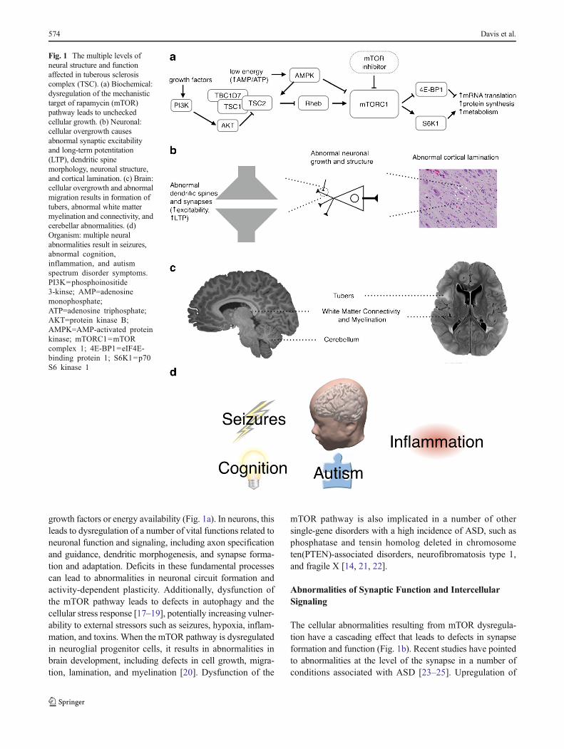
growth factors or energy availability (Fig. 1a). In neurons, thisleads to dysregulation of a number of vital functions related toneuronal function and signaling, including axon specificationand guidance, dendritic morphogenesis, and synapse forma-tion and adaptation. Deficits in these fundamental processescan lead to abnormalities in neuronal circuit formation andactivity-dependent plasticity. Additionally, dysfunction ofthe mTOR pathway leads to defects in autophagy and thecellular stress response [17–19], potentially increasing vulner-ability to external stressors such as seizures, hypoxia, inflam-mation, and toxins. When the mTOR pathway is dysregulatedin neuroglial progenitor cells, it results in abnormalities inbrain development, including defects in cell growth, migra-tion, lamination, and myelination [20]. Dysfunction of the
mTOR pathway is also implicated in a number of othersingle-gene disorders with a high incidence of ASD, such asphosphatase and tensin homolog deleted in chromosometen(PTEN)-associated disorders, neurofibromatosis type 1,and fragile X [14, 21, 22].
Abnormalities of Synaptic Function and IntercellularSignaling
The cellular abnormalities resulting from mTOR dysregula-tion have a cascading effect that leads to defects in synapseformation and function (Fig. 1b). Recent studies have pointedto abnormalities at the level of the synapse in a number ofconditions associated with ASD [23–25]. Upregulation of
Fig. 1 The multiple levels ofneural structure and functionaffected in tuberous sclerosiscomplex (TSC). (a) Biochemical:dysregulation of the mechanistictarget of rapamycin (mTOR)pathway leads to uncheckedcellular growth. (b) Neuronal:cellular overgrowth causesabnormal synaptic excitabilityand long-term potentitation(LTP), dendritic spinemorphology, neuronal structure,and cortical lamination. (c) Brain:cellular overgrowth and abnormalmigration results in formation oftubers, abnormal white mattermyelination and connectivity, andcerebellar abnormalities. (d)Organism: multiple neuralabnormalities result in seizures,abnormal cognition,inflammation, and autismspectrum disorder symptoms.PI3K=phosphoinositide3-kinse; AMP=adenosinemonophosphate;ATP=adenosine triphosphate;AKT=protein kinase B;AMPK=AMP-activated proteinkinase; mTORC1=mTORcomplex 1; 4E-BP1=eIF4E-binding protein 1; S6K1=p70S6 kinase 1
574 Davis et al.

the mTOR pathway leads to abnormal dendritic protein syn-thesis with reduced or dysmorphic dendritic spines and alter-ations in postsynaptic glutamate receptor-mediated long-termdepression [26–28]. These synaptic abnormalities have beenpostulated to contribute to deficits in learning, memory, andadaptation seen in ASD [15, 23, 29]. Changes at the synapseor electrical properties of the cells may then lead to abnormalneuronal excitability, which has been shown to be decreasedin cerebellar Purkinje cells and increased in hippocampal py-ramidal neurons of TSC mutant mice [28, 30]. In addition,decreased gamma-aminobutyric acid (GABA)-ergic inhibi-tion and increased glutamatergic excitation is implicated inthe increased seizure susceptibility seen in TSC and likely alsocontributes to cognitive dysfunction [31–33].
Abnormalities of Brain Development and NeuralConnectivity
The mTOR pathway is active during embryonic brain devel-opment and affects the initial growth and development ofneurons and glia. In addition to causing abnormalities andovergrowth of individual cells, mTOR dysregulation leads toabnormalities in neuronal migration and formation of corticallamination (Fig. 1b). These migration abnormalities appear tobe responsible for the various structural brain malformationscharacteristic of TSC, including tubers, white matterheterotopias, radial migration lines, and subependymal nod-ules (Fig. 1c) [15]. Studies of postmortem brains of patientswith TSC have also shown microscopic structural abnormal-ities in grossly normal-appearing regions, potentially indicat-ing more widespread abnormalities in brain structure thanwould be suggested by neuroimaging [34]. The variety ofphenotypes seen in TSC may be a result of variations in loca-tion of tubers and other macrostructural abnormalities, withtubers particularly in temporal and cerebellar locations beingcorrelated with ASD symptoms in TSC [35–37]. In addition toabnormalities in glial and neuronal migration, the formation ofmyelin also appears to be affected in TSC [34, 38], thus con-tributing to the white matter abnormalities found usingdiffusion-weighted imaging of patients with TSC. These pa-tients had decreased anisotropy in regions of radiographicallynormal-appearing white matter, indicating abnormal whitematter microstructure. This decrease was more severe in pa-tients with TSC with an autism phenotype throughout thewhite matter and specifically in language-related pathways[39, 40]. The abnormal neuronal formation, migration, andwiring in TSC may result in the deficits in functional neuralconnectivity that have been postulated to be at the root of ASD[41–43]. Local overconnectionmay result from clusters of cellsthat do not properly migrate and laminate and form excessiveaxonal and dendritic connections. Long-range underconnectionmay result from disorders of myelination, axonal pathfindingdefects and abnormal larger-scale neuronal migration.
Epilepsy, Development, and Environment Effects on ASDin TSC
A number of systemic factors and comorbid conditions affectthe ASD phenotype and severity in TSC, including epilepsy,cognitive impairment, and inflammation (Fig. 1d). Epilepsy isthe most prevalent neurologic comorbidity of TSC, present inabout 85 % of patients, with the majority of patients develop-ing seizures within the first year, and often within the first fewmonths, of life. Infantile spasms are particularly common,although focal seizures also often occur before, after, or inconjunction with spasms [2, 3]. Studies have shown correla-tions with earlier onset of seizures and worse cognitive out-comes in rodents and humans [44, 45]. A study of early de-velopmental trajectories in children with TSC and ASD founda trend towards more severe epilepsy in patients with TSCwith ASD, as well as a decreased nonverbal intelligence quo-tient (IQ) in patients with longer seizure durations [10]. Earlystudies suggest treatment of epileptic abnormalities in TSCprior to the onset of seizures results in improved outcomesin intellectual ability and epilepsy control [46, 47]. This sug-gests that more severe or poorly controlled epilepsy may con-tribute to worse cognitive outcomes in TSC and ASD, andcorrelations to that effect have been shown, particularly withinfantile spasms [48]. However, the correlative versus causalrelationship between epilepsy and autism remains an area inneed of further exploration [49, 50].
Patients with TSC have a high prevalence of cognitiveimpairment and intellectual disability, but there is a wide rangeof severity across patients. IQ scores in TSC appear to beroughly bimodally distributed, with one group of patients withsevere intellectual disability and another group with intelli-gence in the normal range but with a downshifted mean [3].The biological basis for this bimodal distribution remains un-clear. Investigations of early developmental trajectories ofchildren with TSC have shown correlations between cognitiveimpairment and autism, such that children with the most se-vere autism also have the most severe intellectual impairment.Even children with TSC but without an ASD diagnosis hadimpairment in their play behavior [6]. In addition, childrenwith TSC and an ASD diagnosis had a decline in their non-verbal IQ, verbal IQ, and developmental quotient in the sec-ond to third year of life, even when controlling for the durationof seizures, while their non-ASD counterparts had gains inthese domains [10]. The relationship between cognition andautism in TSC may be a result of a common underlyingneurodevelopmental dysfunction, increased difficulty withlearning due impaired social interactions, or some combina-tion thereof.
The relationship between inflammation and autism is anactive area of research, as well as one of controversy. One areaof particular relevance to autism in TSC is the relationshipbetween mTOR pathway dysregulation and altered immune
Tuberous Sclerosis and Autism 575

system activity in patients with ASD [51]. Other studies havefocused on the interaction between prenatal inflammatory trig-gers and TSC genetics, including the finding that mice withTsc2 haploinsufficiency and intrauterine immune activationwere more likely to show deficits in social behavior. Thiswas paired with an observation that, when compared withchildren with TSC without ASD or children with idiopathicASD, more children with both ASD and TSC had the thirdtrimester of their development occur during a time of peakseasonal influenza activity [8]. These findings suggest thatthe interaction of increased immune activation during late in-trauterine development with the TSC genotype may contrib-ute to the development of ASD.
Promising Treatments
mTOR Inhibitors
The first mTOR inhibitor, rapamycin (sirolimus), was discov-ered in 1975 as an antifungal agent and was subsequentlyfound to have antiproliferative and immunosuppressive prop-erties, leading to its initial uses in preventing solid organ trans-plant rejection and restenosis of coronary arteries followingangioplasty. Following the elucidation of the mTOR pathwayand the role of TSC1 and TSC2, and with the development ofderivative medications with improved pharmacokinetics andside effect profiles, over the last decade mTOR inhibitors havebegun to be studied and used in treatment of the various man-ifestations of TSC, as well as in a number of malignancies (see[52] and [53] for reviews). Sirolimus and its derivatives(Brapalogs^) such as everolimus work by binding FKBP12and then interacting with the FKBP12–rapamycin-binding do-main of mTORC1, inhibiting the serine–threonine kinase ac-tivity of mTORC1 and preventing mRNA translation. Thiseffectively restores the mTORC1 inhibition that would nor-mally be provided by the TSC1/2 complex that is dysfunction-al in patients with TSC. A second generation of mTOR kinasedomain inhibiting medications with activity against bothmTORC1 and mTORC2 are now under development and inearly clinical trials [53]. With the clinical availability of thesemedications, studies of their use in TSC have shown a numberof promising results in animal and human studies.
Therapeutic Effects of mTOR Inhibitor Treatmentin Animal Models of TSC
A number of animal models of TSC have been developed,with both spontaneous and conditional knockout of eitherTSC1 or TSC2 in neural stem cells, neurons, or glial cells(Table 1). These animals develop most of the pathologic fea-tures of TSC, including abnormal neuronal migration and lam-ination, increased cell size, and hypomyelination, as well as a
number of neurocognitive features, including seizures, impair-ment in learning and memory, and deficits in social behavior.
Trials using mTOR inhibitors in animal models of TSChave shown that blocking the action of mTOR can reverse anumber of manifestations of TSC, both at the cellular leveland in the behavior and survival of the animal. Zeng et al. [69]demonstrated in mice with conditional knockout of Tsc1 inglial cells that early treatment with rapamycin prevented thedevelopment of epilepsy, and later treatment decreased sei-zures and prolonged survival. In another mouse model withneuron-specific conditional knockout of Tsc1 in neuronalcells, treatment with rapamycin or everolimus normalizedlevels of mTOR pathway constituents with corresponding im-provement in neurofilament abnormalities, reduction of en-larged cells, and restoration of myelination [66]. Treated ani-mals showed significant improvement in seizures and overallsurvival, although these improvements were attenuated whentreatment was discontinued. These improvements were seendespite no evidence in reversal of neuronal migration abnor-malities and only minor improvement in dendritic spine den-sity and length, indicating not all mTOR-related structural andfunctional abnormalities may be equally important for behav-ioral abnormalities or seizures, or that correction of some butnot all abnormalities may be sufficient to improve outcome.
Ehninger et al. [67] showed that heterozygous Tsc2 micehad abnormalities in hippocampal long-term potentiation,with induction of late long-term potentiation at a lower thresh-old. This led to deficits in spatial learning and contextual dis-crimination, even in the absence of frank neuropathology andseizures, and these deficits were abrogated with only 5 days ofrapamycin treatment. Various approaches have been used toinvestigate the underlying mechanisms responsible for thishippocampal hyperexcitability [32], with at least 2 contribut-ing pathways implicated to date. Tavazoie et al. [28] reportedthat loss of Tsc2 is associated with increased α-amino-3-hy-droxy-5-methyl-4-isoxazolepropionic acid(AMPA)-mediatedexcitatory currents, whereas Chevere-Torres et al. [26] foundthat metabotropic glutamate-induced long-term depression isimpaired with functional disruption of Tsc2 (in the DeltaRGmouse model of TSC) that is extracellular regulated kinasedependent and rapamycin sensitive [27]. This hyperexcitabil-ity is thought to contribute to the epilepsy and learning prob-lems seen in TSC and demonstrates another mechanism inwhich mTOR inhibitors may prove beneficial for the treat-ment of ASD.
Several studies have specifically examined the underlyingmechanisms of autism-like behaviors in animals with a TSCmutation. In contrast to hippocampal neurons, where loss ofTsc1 or Tsc2 results in hyperexcitability, loss of Tsc1 in cere-bellar Purkinje cells in mice results in decreased neuronalexcitability [30]. These latter mice demonstrate an ASD-likephenotype, including abnormal social behavior and learning,repetitive behaviors, and vocalization. These changes were
576 Davis et al.

Tab
le1
Examples
ofpreclin
icalmouse
modelsof
tuberous
sclerosiscomplex
Model
Celltype
Phenotypes
Drugtreatm
ent
Reference
CNPcreCre;T
sc1fl/fl
Olig
odendrocytes
Reduced
myelin
ation
Rapam
ycin
[54]
Gbx2C
reER;T
sc1fl/fl
Thalamus
relayneurons(m
osaic)
Seizures,R
epetitive
grooming
N/A
[55]
Cam
Kllalpha
Cre;T
sc1fl/fl
Forebrainexcitatory
neurons
Kainateseizures
N/A
[32]
hGFA
PCre;T
sc1fl/–
hGFA
PCre;T
sc1fl/–;Tsc2
f/–
Radialg
lialp
rogenitorcells
Seizures,reduced
myelin
ation,lethality
N/A
[56]
L7C
re;T
sc1fl/fl
CerebellarPurkinjecells
Impaired
sociability,R
epetitive
grooming
Rapam
ycin
[30]
L7C
re;T
sc2fl/fl
CerebellarPurkinjecells
Impaired
sociability,R
epetitive
grooming
Rapam
ycin
[57]
hGFA
P2C
re;T
sc1fl/–
hGFA
P2C
re;T
sc2fl/–
Radialg
lialp
rogenitorcells
Seizures,reduced
myelin
ation,lethality
Rapam
ycin
[58]
Dlx5/6C
re;T
sc1fl/fl
GABAergicinterneurons
Reduced
seizurethreshold,lethality
N/A
[59]
EmxC
re;T
sc1fl/fl
Forebrainneuralprogenito
rcells
Seizures,reduced
myelin
ation,lethality
Rapam
ycin
[60]
SynC
re;T
sc2c
-del3
Neurons
(hypom
orphicallele)
Impaired
sociability
andlearning
N/A
[61]
EmxC
re;T
sc1fl/fl
Forebrainneuralprogenito
rcells
Abnormalcorticallamination,
expanded
SVZregion
Rapam
ycin
[62]
Nestin
-CreERT2;T
sc1fl/fl
Neuronalp
rogenitorcells
(mosaic)
ExpandedSVZregion
N/A
[63]
Nestin
-rtTA;T
etOp-Cre;T
sc1fl/fl
Neuronalp
rogenitorcells
(mosaic)
Seizures,H
yperactiv
ityRapam
ycin
[64]
hGFA
PCre;T
sc2fl/fl
Astrocytes
Seizures,L
ethality
Rapam
ycin
[65]
SynC
re;T
sc1fl/fl
Neurons
Tremor,seizures,reducedmyelin
ation,lethality
Rapam
ycin
RAD001
[38,66]
Tsc2
+/–
Constitu
tiveheterozygous
Deficits
inhippocam
pal-dependentlearning
Rapam
ycin
[67]
hGFA
PCre;T
sc2fl/–
Radialg
lialp
rogenitorcells
Abnormalcorticallamination,reducedmyelin
ation
N/A
[68]
hGFA
PCre;T
sc1fl/fl
Astrocytes
Seizures,lethality
Rapam
ycin,vigabatrin
[65,69–71]
GABA=gamma-am
inobutyricacid;S
VZ=subventricular
zone;N
/A=notapplicable
Tuberous Sclerosis and Autism 577

prevented by treatment with rapamycin. Similar results wereobtained with Tsc2 knockout in the Purkinje neurons [57].Talos et al. [72] reported that a rodent model of neonatal sei-zures results in mTORC1 overactivity, and seizures during aperiod of peak synaptogenesis contributed to deficits in a so-cial novelty assay. Treatment with rapamycin in animals withneonatal hypoxic seizures was protective against the develop-ment of epilepsy and decreased the social novelty deficit.Another mouse model with conditional knockout of Tsc1 inforebrain neurons (under CaMKIIα-cre) further links mTORhyperactivation with seizures and autism-like behaviors [73].Decreased social behavior (in the 3-chamber social approach)and increased repetitive behaviors (marble burying) were ob-served in these mutant mice. As the recurrent seizure spread tothe brainstem, the authors hypothesized that serotonergic neu-rons were specifically implicated in mediating the autistic-likebehaviors. Using a conditional knockout of Tsc1 in serotoner-gic neurons, they found that autism-like behaviors persisted,even though the mice did not develop spontaneous seizures.Treatment with rapamycin decreased mTOR hyperactivityand normalized measures of social preference and marbleburying. Taken together, these studies show a complex rela-tionship between early brain development, mTOR pathwayoveractivity in various brain regions, seizures, and the devel-opment of ASD-like behaviors, and also show that treatmentwith an mTOR inhibitor is a potentially promising method tonormalize underlying mechanisms leading to the behavioralmanifestations of ASD.
Preliminary Results of Neuropsychological Effectsof mTOR Inhibitor Trials in Humans
The first human clinical trials using mTOR inhibitors in thetreatment of TSC began in 2002, with the initial focus on reduc-ing tumor burden and growth in the brain (SEGA), kidney(angiomyolipoma), and lung (lymphangioleiomyomatosis).These targets were selected based on significant disease-related morbidity and mortality, lack of effective noninvasivetreatment options, and relative ease in objectively assessingtreatment response. Rapamycin was shown to be effective andwell tolerated for reducing SEGA tumor volume [74], renalangiomyolipoma volume [75–77], and pulmonary lung func-tion [78], as long as treatment was maintained. However, ineach case disease progression resumed upon discontinuationof treatment, similar to what had been observed in preclinicalmodels. As a result, more recent studies have evaluated thesustained efficacy and long-term safety of continuous treatment.Krueger et al. [79, 80] treated 28 patients with SEGAs witheverolimus for a median of 3 years without loss of SEGA tumorvolume reduction and no newly encountered clinical toxicities.A placebo-controlled SEGA treatment study with everolimusinvolving 117 patients, with a median follow-up of 2 years,yielded similar results [81, 82]. Bissler et al. [83] reported
safety and eff icacy of everol imus treatment forangiomyolipoma but median treatment duration was muchshorter (slightly over 6 months).
Throughout these early clinical trials, unpublished anecdot-al reports (Franz, NIH Curing Epilepsy 2007), case reports[84], and secondary end points [77, 79] suggested thatrapalogs might have benefit for central nervous system(CNS)-related manifestations in TSC beyond reduction of tu-mors and tubers. For example, Krueger et al. [79] reportedafter 6 months of treatment with everolimus, seizure frequen-cy was reduced in 56 % of participants. By 24 months, pa-tients with daily seizures were reduced from 27 % to 13 %,while patients with no seizures increased from 39 % to 65 %during the same interval [80]. These improvements correlatedwith improvement in white matter integrity as measured withdiffusion tensor imaging and were independent of SEGAtreatment response [85]. Analysis of seizure control as a sec-ondary end point in the placebo-controlled, blinded Phase IIItrial with everolimus to treat SEGA was unable to confirmthese earlier results, largely owing to significant differencesin baseline seizure frequency between the control and treat-ment groups, and the fact that in both groups the majority ofparticipants were seizure-free at time of treatment initiation[81]. Since then, smaller, mostly retrospective, studies havecontinued to report treatment benefit for epilepsy [86–89]. Todate, the only clinical trial specifically to use mTOR inhibitorsto treat epilepsy prospectively involved open-label treatmentwith everolimus for 4 months [90]. Eighteen of 20 participantsreported a reduction in seizure frequency, with 12/20 (60 %)experiencing an improvement of 50 % or more. Furthermore,treatment response appeared to improve over time, with betterresponse at 3–4 months compared with response at 1–2 months. This delayed or prolonged response time for opti-mal outcome supports involvement of mechanisms discussedin the previous sections that require weeks to months ratherthan hours to days to show effect, such as synapse remodeling,network connectivity, and neuronal plasticity. However, directevidence that this is the case and the relative contributionseach has on seizure susceptibility and treatment response re-mains to be determined. Longer-term treatment and pla-cebo-controlled, double-blind clinical trials are already inprogress to further investigate everolimus impact on seizuresand epilepsy in TSC.
Evaluating the impact of mTOR inhibitors in patients withTSC with intellectual disability and other aspects of TAND,including ASD, has been more difficult. In the initial 28-patient SEGA study, a robust neurocognitive assessment bat-tery was included but the majority of participants were devel-opmentally and/or cognitively impaired such that relativelyfew were able to complete the assessments, and no conclu-sions could be drawn [79]. A similar attempt by Davies et al.[77] in adults treated with rapamycin for angiomyolipomafared a little better, although the final size of the analysis
578 Davis et al.
cohort remained relatively small (n=8) and yielded mixedresults. They reported improvements in recall memory (7/8)and executive function (5/8) but worsening in recognitionmemory (5/8) in adults with TSC following treatment withrapamycin for 4–12 months. In another study, Chung et al.[91] reported improvement in 3 patients with TSC with co-morbid intermittent explosive disorder or adjustment disordernot otherwise specified who underwent formal psychiatricevaluations before and after initiating treatment with everoli-mus (n=2) or rapamycin (n=1). In the more recent open-labelepilepsy trial by Krueger et al. [90], an indirect, more broadapproach was utilized, sacrificing direct observational mea-sures for indirect parental report using validated assessmenttools with the goal of providing more universal assessment ofTAND-related comorbidities and domains. Using theNisonger Child Behavior Rating Form, adapted from theChild Behavioral Rating Form to allow assessment of bothcognitively impaired and normal IQ children, they reportedsmall but significant improvement in adaptive social behav-iors, conduct problems, and insecurity/anxiety compared withbaseline following treatment for 4 months. Quality of Life forChildren with Epilepsy assessments done in parallel over thesame time period identified similar improvements in multipledomains, including attention and concentration, behavior, so-cial interactions and activity, and overall quality of life.However, whether these changes were secondary to or inde-pendent of improvement in seizure control could not bedetermined.
Intense interest exists to confirm and expand these prelim-inary studies in order to determine if a longer treatment periodresults in sustained or further improvements, to verify resultsin a larger cohort using a placebo-controlled, double-blindedstudy design, and to incorporate direct observational assess-ment approaches to better determine the specific areas ofneurocognitive and ASD-related subdomains affected bymTOR inhibitor treatment and further separate which effectsare seizure control-dependent and which are seizure control-independent. Ongoing trials include Everolimus (RAD001)Therapy for Epilepsy in Patients with TSC (clinicaltrials.gov:NCT01070316); Trial of RAD001 and Neurocognition inTSC (clinicaltrials.gov: NCT01289912); Efficacy ofRAD001/Everolimus in Autism and NeuroPsychologicalDeficits in Children With Tuberous Sclerosis Complex(RAPIT) (clinicaltrials.gov: NCT01730209); A Placebo-controlled Study of Efficacy & Safety of 2 Trough-ranges ofEverolimus as Adjunctive Therapy in Patients With TSC &Re f r a c t o r y P a r t i a l - o n s e t S e i z u r e s (EX IST- 3 )(clinicaltrials.gov: NCT01713946); A Study of Everolimusin the Treatment of Neurocognitive Problems in TuberousSclerosis (TRON) (clinicaltrials.gov: NCT01954693);Rapalogues for Autism Phenotype in TSC: A FeasibilityStudy (RAPT) (clinicaltrials.gov: NCT01929642); andLong-term Follow-up for Growth and Development of
Pediatric Patients From CRAD001M2301 (EXIST-LT)(clinicaltrials.gov: NCT02338609).
Early Vigabatrin Treatment Trials
Vigabatrin is a rationally designed drug that aims to increasethe levels of GABA, the main inhibitory neurotransmitter inthe CNS. The addition of a vinyl group to GABA (vi-GABA-trin) creates a substrate that irreversibly inhibits GABA-trans-aminase, the GABA-degrading enzyme, thus increasingGABA availability in the synaptic cleft. In humans, it has aspecific and rapid mechanism of action and its antiepilepticproperties are thought to be a direct result of increased inhib-itory neurotransmission, although additional mechanisms areconsidered [92].
Side effects during initiation are those common to drugsaffecting the CNS, and include irritability, drowsiness, andhypotonia, which typically resolve over time. One importantside effect is irreversible peripheral visual field constriction,with a risk that increases cumulatively with higher doses andlonger exposure [93]. In the USA, the manufacturer has imple-mented a vigorous monitoring program for this side effect. Theclinically evident response of cessation of spasms within daysto weeks provides an opportunity to try vigabatrin at a low risk.
Other than as add-on for intractable complex partial sei-zures, vigabatrin is approved in the US as monotherapy forinfantile spasms (IS). An early small controlled trial demon-strated efficacy compared with placebo but no patients withTSC were included [94]. In studies of high- versus low-dosevigabatrin and of vigabatrin versus steroid therapy for IS, pa-tients with TSC had consistently higher response rates tovigabatrin [95, 96]. Indeed, an early review of the literaturesuggested a response rate of 95 % to vigabatrin in IS due toTSC [97], an estimate that has not changed in subsequent lit-erature [93]. Within patients with TSC, in a head-to-head com-parison with adrenocorticotropic hormone, vigabatrin showedclear superiority [98]. Based on these and other studies andreviews, the International TSC Consensus Conference recom-mends vigabatrin as first-line treatment for IS in TSC [4].
Why IS in TSC in particular respond so dramatically tovigabatrin is not understood. Other drugs affectingGABAergic transmission (e.g., barbiturates and benzodiaze-pines) do not achieve comparable efficacy. Reported addition-al effects of vigabatrin such as decreased glial GABA uptake,enhanced GABA release, and reduction of glutamate have notbeen reproduced [92]. In a TSCmouse model, vigabatrin sup-pressed the mTOR pathway, which could be an additionalexplanation for the particular efficacy in TSC [70].However, the rapid efficacy against IS is more likely basedon GABAergic changes.
Better long-term neurocognitive outcome has been report-ed with early, aggressive, and successful treatment of IS[99–101], and later cessation of IS is associated with poorer
Tuberous Sclerosis and Autism 579
outcomes [102]. For partial seizures, however, efficacy is low-er and the beneficial effects on outcome may not be presentwhen compared with treatment of IS [100, 101].
When given even earlier, in a small open-label trial ofvigabatrin initiation prior to onset of IS, good long-term out-comes were also seen [46]. If, indeed, mTOR pathway activityis modified by vigabatrin, it could contribute to these longer-term benefits. This raises the possibility that vigabatrin is notmerely antiepileptic, but it may also be disease modifying.
How to identify who would most benefit, with justifiablerisk, from early or even pre-emptive intervention withvigabatrin, is a subject of active study. For example, epilepticspasms beyond infancy respond less well to vigabatrin, eventhough these seizures are similar to IS in their electroclinicalpresentation. Thus, there appears to be a therapeutic windowin obtaining the longer-term neurodevelopmental benefitsfrom early medical intervention. In the USA, a multicenterprospective observational study is currently underway to iden-tify early developmental, neurophysiology, and neuroimagingmarkers for increased risk of epilepsy and ASD in TSC(clinicaltrials.gov: NCT01767779; NCT01780441). In a nextphase, those markers will be used to stratify patients to eitherstandard of care versus early aggressive, pre-emptive treat-ment with vigabatrin. In Europe, a similar effort is ongoing(clinicaltrials.gov: NCT02098759).
Future Directions of Treatment for ASD in TSC
We are on the cusp of a new era in targeted, disease-modifyingtreatment for TSC and other disorders associated with ASD.The initial results from a Phase II study looking specifically ateffects on neurocognition in TSC after treatment with evero-limus (clinicaltrials.gov: NCT01289912) are currently in theprocess of being analyzed. Open questions include the type ofeffects mTOR inhibitors will have on ASD symptoms inTSC and how the effects will vary based on patientcharacteristics and disease manifestations. Another cru-cial question is what end points are quantifiable anddynamic in response to treatment within the durationof the trial. Finally, it is not yet clear whether there isa critical window during which treatments will be most effec-tive. Once these questions are studied, we will be in a betterposition to determine which patients will most benefit fromwhich treatment.
Robust and early biomarkers of neurodevelopmental out-come in TSC are critical to best target treatments to patientswho will most benefit from them, to avoid unnecessarily ex-posing patients to potentially harmful and irreversible treat-ment side effects, and to measure treatment effects. For ASD,behavioral and developmental signs may be recognized earlyin high-risk populations [10], and retrospective work has iden-tified electroencephalography network properties and
diffusion tensor imaging metrics as possible biomarkers [39,103]. Diffusion imaging abnormalities may reflect changes inthe underlying neurobiology [43], and these measures mayeven respond to intervention in parallel with clinical changesseen [85]. Further research on these and other biomarkers isneeded to better characterize their utility for clinical use andfuture prospective studies.
It is a rare but fortunate occurrencewhen our understandingof a disorder matches our ability to treat based on that under-standing. We are approaching that point now for ASD in TSC.Our hope is that the recent advances described here in under-standing and treating the mechanisms contributing toASD in TSC will benefit our patients with TSC andprovide a path towards better understanding and treatmentfor all people with ASD.
Acknowledgments P.E.D. is supported by a training grant from theNational Institutes of Health (NIH) National Institute of NeurologicalDisorders and Stroke (NINDS; 3R25NS070682-04S1). J.M.P., D.A.K.and M.S. are supported by NIH (U01 NS082320, P20 NS080199,U54NS092090). The Developmental Synaptopathies Consortium(U54NS092090) is a part of the National Center for AdvancingTranslational Sciences (NCATS) Rare Diseases Clinical ResearchNetwork (RDCRN). RDCRN is an initiative of the Office of RareDiseases Research (ORDR), NCATS, funded through collaboration be-tween NCATS, National Institute of Mental Health, NINDS and NationalInstitute of Child Health and Human Development. J.M.P. is also sup-ported by Harvard Catalyst | The Harvard Clinical and TranslationalScience Center (National Center for Research Resources and theNational Center for Advancing Translational Sciences, NIH AwardUL1 TR001102). D.A.K. is also supported by Tuberous SclerosisAlliance, Clack Foundation, Novartis, and Upsher-Smith. Research inthe laboratory of M.S. is also supported by the NIH P30 HD018655,the Department of Defense, Tuberous Sclerosis Alliance, AutismSpeaks, Nancy Lurie Marks Family Foundation, Simons Foundation,Boston Children’s Hospital Translational Research Program, andNovartis, Shire and Roche. Full conflict of interest disclosures are avail-able in the electronic supplementary material for this article. Owing tolimited space, we have not quoted all the literature in this field, and weapologize to those whose articles are not referenced. Finally, we are in-debted to the children and families who participated in the studiesreviewed in this article.
Required Author Forms Disclosure forms provided by the authors areavailable with the online version of this article.
References
1. Osborne JP, Fryer A, Webb D. Epidemiology of tuberous sclero-sis. Ann N YAcad Sci 1991;615:125-127.
2. Curatolo P, Bombardieri R, Jóźwiak S. Tuberous sclerosis. Lancet2008;372:657-668.
3. Curatolo P, Maria BL. Tuberous sclerosis. Handb ClinNeurol 2013;111:323-331.
4. Krueger DA, Northrup H, International Tuberous SclerosisComplex Consensus Group. Tuberous sclerosis complex surveil-lance and management: recommendations of the 2012 InternationalTuberous Sclerosis Complex Consensus Conference. PediatrNeurol 2013;49:255-265.
580 Davis et al.
5. de Vries PJ, Whittemore VH, Leclezio L, et al., Tuberous sclerosisassociated neuropsychiatric disorders (TAND) and the TANDChecklist. Pediatr Neurol 2015;52:25-35.
6. Jeste SS, Sahin M, Bolton P, Ploubidis GB, Humphrey A.Characterization of autism in young children with tuberous scle-rosis complex. J Child Neurol 2008;23:520-525.
7. Leclezio L, de Vries PJ. Advances in the treatment of tuberoussclerosis complex. Curr Opin Psychiatry 2015;28:113-120.
8. Ehninger D, Sano Y, de Vries PJ, et al., Gestational immune acti-vation and Tsc2 haploinsufficiency cooperate to disrupt fetal sur-vival and may perturb social behavior in adult mice. MolPsychiatry 2012;17:62-70.
9. Datta AN, HahnCD, SahinM. Clinical presentation and diagnosisof tuberous sclerosis complex in infancy. J Child Neurol 2008;23:268-273.
10. Spurling Jeste S, Wu JY, Senturk D, et al. Early developmentaltrajectories associated with ASD in infants with tuberous sclerosiscomplex. Neurology 2014;83:160-168.
11. Bruining H, Eijkemans MJ, Kas MJ, et al. Behavioral signaturesrelated to genetic disorders in autism. Mol Autism 2014;5:11.
12. Laplante M, Sabatini DM. mTOR signaling in growth control anddisease. Cell 2012;149:274-293.
13. Lipton JO, Sahin M. The neurology of mTOR. Neuron 2014;84:275-291.
14. Ehninger D. From genes to cognition in tuberous sclerosis: impli-cations for mTOR inhibitor-based treatment approaches.Neuropharmacology 2013;68: 97-105.
15. Feliciano DM, Lin TV, Hartman NW, et al., A circuitry and bio-chemical basis for tuberous sclerosis symptoms: from epilepsy toneurocognitive deficits. Int J Dev Neurosci 2013;31:667-678.
16. Chen J, Alberts I, Li X. Dysregulation of the IGF-I/PI3K/AKT/mTOR signaling pathway in autism spectrum disorders. Int J DevNeurosci 2014;35:35-41.
17. Tang G, Gudsnuk K, Kuo SH, et al. Loss of mTOR-dependentmacroautophagy causes autistic-like synaptic pruning deficits.Neuron 2014;83:1131-1143.
18. Di Nardo A, Wertz MH, Kwiatkowski E, et al. Neuronal Tsc1/2complex controls autophagy through AMPK-dependent regula-tion of ULK1. Hum Mol Genet 2014;23:3865-3874.
19. Di Nardo A, Kramvis I, Cho N, et al. Tuberous sclerosis complexactivity is required to control neuronal stress responses in anmTOR-dependent manner. J Neurosci 2009;29:5926-5937.
20. LimK-C, Crino PB. Focal malformations of cortical development:new vistas for molecular pathogenesis. Neuroscience 2013;252:262-276.
21. Hoeffer CA, Klann E. mTOR signaling: At the crossroads of plas-ticity, memory and disease. Trends Neurosci 2010;33:67-75.
22. de Vries PJ. Targeted treatments for cognitive andneurodevelopmental disorders in tuberous sclerosis complex.Neurotherapeutics 2010;7:275-282.
23. Kelleher RJ, Bear MF. The autistic neuron: troubled translation?Cell 2008;13:401-406.
24. Auerbach BD, Osterweil EK, Bear MF. Mutations causingsyndromic autism define an axis of synaptic pathophysiology.Nature 2011;480:63-68.
25. Won H, Mah W, Kim E. Autism spectrum disorder causes, mech-anisms, and treatments: focus on neuronal synapses. Front MolNeurosci 2013;6:19.
26. Chevere-Torres I, Kaphzan H, Bhattacharya A, et al. Metabotropicglutamate receptor-dependent long-term depression is impaireddue to elevated ERK signaling in the DeltaRG mouse model oftuberous sclerosis complex. Neurobiol Dis 2012;45:1101-1110.
27. Hou, L, Klann E. Activation of the phosphoinositide 3-kinase-Akt-mammalian target of rapamycin signaling pathway is requiredfor metabotropic glutamate receptor-dependent long-term depres-sion. J Neurosci 2004;24:6352-6361.
28. Tavazoie SF, Alvarez VA, Ridenour DA, Kwiatkowski DJ,Sabatini BL. Regulation of neuronal morphology and functionby the tumor suppressors Tsc1 and Tsc2. Nat Neurosci 2005;8:1727-1734.
29. Santini E, Klann E. Reciprocal signaling between translationalcontrol pathways and synaptic proteins in autism spectrum disor-ders. Sci Signal 2014;7:re10.
30. Tsai PT, Hull C, Chu Y, et al. Autistic-like behaviour and cerebel-lar dysfunction in Purkinje cell Tsc1 mutant mice. Nature2012;488:647-651.
31. Zeng L-H, Ouyang Y, Gazit V, et al. Abnormal glutamate homeo-stasis and impaired synaptic plasticity and learning in a mousemodel of tuberous sclerosis complex. Neurobiol Dis 2007;28:184-196.
32. Bateup HS, Johnson CA, Denefrio CL, et al. Excitatory/inhibitory synaptic imbalance leads to hippocampal hyperexcit-ability in mouse models of tuberous sclerosis. Neuron 2013;78:510-522.
33. Curatolo P. Mechanistic target of rapamycin (mTOR) in tuberoussclerosis complex-associated epilepsy. Pediatr Neurol 2014;52:281-289.
34. Crino PB. Evolving neurobiology of tuberous sclerosis complex.Acta Neuropathol 2013;125:317-332.
35. Bolton P, Park RJ, Higgins J, Griffiths PD, Pickles A. Neuro-epileptic determinants of autism spectrum disorders in tuberoussclerosis complex. Brain 2002;125:1247-1255.
36. Eluvathingal TJ, Behen ME, Chugani HT, et al. Cerebellar lesionsin tuberous sclerosis complex: neurobehavioral and neuroimagingcorrelates. J Child Neurol 2006;21:846-851.
37. Weber AM, Egelhoff JC, McKellop JM, Franz DN. Autism andthe cerebellum: evidence from tuberous sclerosis. J Autism DevDisord 2000;30:511-517.
38. Meikle L, Talos DM, Onda H, et al. A mouse model of tuberoussclerosis: neuronal loss of Tsc1 causes dysplastic and ectopic neu-rons, reduced myelination, seizure activity, and limited survival. JNeurosci 2007;27:5546-5558.
39. Peters JM, Sahin M, Vogel-Farley VK, et al. Loss of white mattermicrostructural integrity is associated with adverse neurologicaloutcome in tuberous sclerosis complex. Acad Radiol 2012;19:17-25.
40. Lewis WW, Sahin M, Scherrer B, et al. Impaired language path-ways in tuberous sclerosis complex patients with autism spectrumdisorders. Cereb Cortex 2013;23:1526-1532.
41. Geschwind DH, Levitt P. Autism spectrum disorders: develop-mental disconnection syndromes. Curr Opin Neurobiol 2007;17:103-111.
42. Wass S. Distortions and disconnections: disrupted brain connec-tivity in autism. Brain Cogn 2011;75:18-28.
43. Peters JM, Taquet M, Prohl AK, et al. Diffusion tensor imagingand related techniques in tuberous sclerosis complex: review andfuture directions. Future Neurol 2013;8:583-597.
44. Waltereit R, Japs B, SchneiderM, deVries PJ, Bartsch D. Epilepsyand Tsc2 haploinsufficiency lead to autistic-like social deficit be-haviors in rats. Behav Genet 2011;41:364-372.
45. Jansen FE, Vincken KL, Algra A, et al. Cognitive impairment intuberous sclerosis complex is a multifactorial condition.Neurology 2008;70:916-923.
46. Jóźwiak S, Kotulska K, Domańska-Pakieła D, et al. Antiepileptictreatment before the onset of seizures reduces epilepsy severityand risk of mental retardation in infants with tuberous sclerosiscomplex. Eur J Paediatr Neurol 2011;15:424-431.
47. Domanska-Pakiela D, Kaczorowska M, Jurkiewicz E, et al. EEGabnormalities preceding the epilepsy onset in tuberous sclerosiscomplex patients—a prospective study of 5 patients. Eur JPaediatr Neurol 2014;18:458-468.
Tuberous Sclerosis and Autism 581

48. van Eeghen AM, Pulsifer MB, Merker VL, et al. Understandingrelationships between autism, intelligence, and epilepsy: a cross-disorder approach. Dev Med Child Neurol 2013;55:146-153.
49. Berg AT, Plioplys S. Epilepsy and autism: is there a special rela-tionship? Epilepsy Behav 2012;23:193-198.
50. El Achkar CM, Spence SJ. Clinical characteristics of children andyoung adults with co-occurring autism spectrum disorder and ep-ilepsy. Epilepsy Behav 2015. doi:10.1016/j.yebeh.2014.12.022
51. Careaga M, Van de Water J, Ashwood P. Immune dysfunction inautism: a pathway to treatment. Neurotherapeutics 2010;7:283-292.
52. Gentzler RD, Altman JK, Platanias LC. An overview of themTOR pathway as a target in cancer therapy. Expert Opin TherTargets 2012;16:481-489.
53. Santulli G, Totary-Jain H. Tailoring mTOR-based therapy: molec-ular evidence and clinical challenges. Pharmacogenomics2013;14:1517-1526.
54. Lebrun-Julien F, Bachmann L, Norrmen C, et al. BalancedmTORC1 activity in oligodendrocytes is required for accurateCNS myelination. J Neurosci 2014;34:8432-8448.
55. Normand EA, Crandall SR, Thorn CA, et al. Temporal andmosaicTsc1 deletion in the developing thalamus disrupts thalamocorticalcircuitry, neural function, and behavior. Neuron 2013;78:895-909.
56. Mietzsch U, McKenna J, 3rd, Reith RM, Way SW, Gambello MJ.Comparative analysis of Tsc1 and Tsc2 single and double radialglial cell mutants. J Comp Neurol 2013;521:3817-3831.
57. Reith RM, McKenna J, Wu H, et al. Loss of Tsc2 in Purkinje cellsis associated with autistic-like behavior in a mouse model of tu-berous sclerosis complex. Neurobiol Dis 2013;51:93-103.
58. Magri L, Cominelli M, Cambiaghi M, et al. Timing of mTORactivation affects tuberous sclerosis complex neuropathology inmouse models. Dis Model Mech 2013;6:1185-1197.
59. Fu C, Cawthon B, Clinkscales W, et al. GABAergic interneurondevelopment and function is modulated by the Tsc1 gene. CerebCortex 2012;22:2111-2119.
60. Carson RP, Van Nielen DL, Winzenburger PA, Ess KC. Neuronaland glia abnormalities in Tsc1-deficient forebrain and partial res-cue by rapamycin. Neurobiol Dis 2012;45:369-380.
61. Yuan E, Tsai PT, Greene-Colozzi E, et al. Graded loss of tuberin inan allelic series of brain models of TSC correlates with survival,and biochemical, histological and behavioral features. Hum MolGenet 2012;21:4286-4300.
62. Magri L, Cambiaghi M, Cominelli M, et al. Sustained activationof mTOR pathway in embryonic neural stem cells leads to devel-opment of tuberous sclerosis complex-associated lesions. CellStem Cell 2011;9:447-462.
63. Zhou J, Shrikhande G, Xu J, et al. Tsc1 mutant neural stem/progenitor cells exhibit migration deficits and give rise tosubependymal lesions in the lateral ventricle. Genes Dev2011;25:1595-1600.
64. Goto J, Talos DM, Klein P, et al. Regulable neural progenitor-specific Tsc1 loss yields giant cells with organellar dysfunctionin a model of tuberous sclerosis complex. Proc Natl Acad Sci U SA 2011;108:E1070-E1079.
65. Zeng LH, Rensing NR, Zhang B, et al. Tsc2 gene inactivationcauses a more severe epilepsy phenotype than Tsc1 inactivationin a mouse model of tuberous sclerosis complex. HumMol Genet2011;20:445-454.
66. Meikle L, Pollizzi K, Egnor A, et al. Response of a neuronalmodel of tuberous sclerosis to mammalian target of rapamycin(mTOR) inhibitors: effects on mTORC1 and Akt signaling leadto improved survival and function. J Neurosci 2008;28:5422-5432.
67. Ehninger D, Han S, Shilyansky C, et al. Reversal of learningdeficits in a Tsc2+/- mouse model of tuberous sclerosis. NatMed 2008;14:843-848.
68. Way SW,McKenna J, 3rd,Mietzsch U, et al. Loss of Tsc2 in radialglia models the brain pathology of tuberous sclerosis complex inthe mouse. Hum Mol Genet 2009;18:1252-1265.
69. Zeng LH, Xu L, Gutmann DH, Wong M. Rapamycin preventsepilepsy in a mouse model of tuberous sclerosis complex. AnnNeurol 2008;63:444-453.
70. Zhang B, McDaniel SS, Rensing NR, Wong M. Vigabatrin in-hibits seizures and mTOR pathway activation in a mouse modelof tuberous sclerosis complex. PLoS ONE 2013;8:e57445.
71. Uhlmann EJ, Wong M, Baldwin RL, et al. Astrocyte-specificTSC1 conditional knockout mice exhibit abnormal neuronal orga-nization and seizures. Ann Neurol 2002;52:285-296.
72. Talos DM, Sun H, Zhou X, et al. The interaction between early lifeepilepsy and autistic-like behavioral consequences: a role for themammalian target of rapamycin (mTOR) pathway. PLoS ONE2012;7:e35885.
73. McMahon JJ, Yu W, Yang J, et al. Seizure-dependent mTORactivation in 5-HT neurons promotes autism-like behaviors inmice. Neurobiol Dis 2015;73:296-306.
74. Franz DN, Leonard J, Tudor C, et al. Rapamycin causes regressionof astrocytomas in tuberous sclerosis complex. Ann Neurol2006;59:490-498.
75. Bissler JJ, McCormack FX, Young LR, et al. Sirolimus forangiomyol ipoma in tuberous sc leros is complex orlymphangioleiomyomatosis. N Engl J Med 2008;358:140-151.
76. Dabora SL, Franz DN, Ashwal S, et al. Multicenter phase 2 trial ofsirolimus for tuberous sclerosis: kidney angiomyolipomas andother tumors regress and VEGF- D levels decrease. PLoS ONE2011;6:e23379.
77. Davies DM, de Vries PJ, Johnson SR, et al. Sirolimus therapy forangiomyolipoma in tuberous sclerosis and sporadiclymphangioleiomyomatosis: a phase 2 trial. Clin Cancer Res2011;17:4071-4081.
78. McCormack FX, Inoue Y, Moss J, et al. Efficacy and safety ofsirolimus in lymphangioleiomyomatosis. N Engl J Med 2011;364:1595-1606.
79. Krueger DA, Care MM, Holland K et al. Everolimus forsubependymal giant-cell astrocytomas in tuberous sclerosis. NEngl J Med 2010;363:1801-1811.
80. Krueger DA, Care MM, Agricola K, et al. Everolimus long-termsafety and efficacy in subependymal giant cell astrocytoma.Neurology 2013;80:574-580.
81. Franz, D.N., E. Belousova, S. Sparagana, et al. Efficacy and safetyof everolimus for subependymal giant cell astrocytomas associat-ed with tuberous sclerosis complex (EXIST-1): a multicentre,randomised, placebo-controlled phase 3 trial. Lancet 2013;381:125-132.
82. Franz DN, Belousova E, Sparagana S, et al. Everolimus forsubependymal giant cell astrocytoma in patients with tuberoussclerosis complex: 2-year open-label extension of the randomisedEXIST-1 study. Lancet Oncol 2014;15:1513-1520.
83. Bissler JJ, Kingswood JC, Radzikowska E, et al. Everolimus forangiomyolipoma associated with tuberous sclerosis complex orsporadic lymphangioleiomyomatosis (EXIST-2): a multicentre,randomised, double-blind, placebo-controlled trial. Lancet2013;381:817-824.
84. Muncy J, Butler IJ, Koenig MK. Rapamycin reduces seizure fre-quency in tuberous sclerosis complex. J Child Neurol 2009;24:477.
85. Tillema J-M, Leach JL, Krueger DA, Franz DN. Everolimus alterswhite matter diffusion in tuberous sclerosis complex. Neurology2012;78:526-531.
86. Kotulska K, Chmielewski D, Borkowska J, et al. Long-term effectof everolimus on epilepsy and growth in children under 3 years ofage treated for subependymal giant cell astrocytoma associated
582 Davis et al.
with tuberous sclerosis complex. Eur J Paediatr Neurol 2013;17:479-485.
87. Wiegand G, May TW, Ostertag P, et al. Everolimus in tuberoussclerosis patients with intractable epilepsy: a treatment option? EurJ Paediatr Neurol 2013;17:631-638.
88. Canpolat M, Per H, Gumus H, et al. Rapamycin has a beneficialeffect on controlling epilepsy in children with tuberous sclerosiscomplex: results of 7 children from a cohort of 86. Childs NervSyst 2014;30:227-240.
89. Cardamone M, Flanagan D, Mowat D, et al. Mammalian target ofrapamycin inhibitors for intractable epilepsy and subependymalgiant cell astrocytomas in tuberous sclerosis complex. J Pediatr2014;164:1195-1200.
90. Krueger DA, Wilfong AA, Holland Bouley K, et al. Everolimustreatment of refractory epilepsy in tuberous sclerosis complex.Ann Neurol 2013;74:679-687.
91. Chung TK, Lynch ER, Fiser CJ, et al. Psychiatric comorbidity andtreatment response in patients with tuberous sclerosis complex.Ann Clin Psychiatry 2011;23:263-269.
92. Ben-Menachem E. Mechanism of action of vigabatrin: correctingmisperceptions. Acta Neurol Scand Suppl 2011:5-15.
93. Willmore LJ, AbelsonMB, Ben-Menachem E, Pellock JM, ShieldsWD. Vigabatrin: 2008 update. Epilepsia 2009;50:163-173.
94. Appleton RE, Peters AC, Mumford JP, Shaw DE. Randomised,placebo-controlled study of vigabatrin as first-line treatment ofinfantile spasms. Epilepsia 1999;40:1627-1633.
95. Elterman RD, Shields WD, Mansfield KA, Nakagawa J; USInfantile Spasms Vigabatrin Study Group, Randomized trial of
vigabatrin in patients with infantile spasms. Neurology 2001;57:1416-1421.
96. Vigevano F, Cilio MR. Vigabatrin versus ACTH as first-line treat-ment for infantile spasms: a randomized, prospective study.Epilepsia 1997;38:1270-1274.
97. Hancock E, Osborne JP. Vigabatrin in the treatment of infantilespasms in tuberous sclerosis: literature review. J Child Neurol1999;14:71-74.
98. Chiron C, Dumas C, Jambaque I, Mumford J, Dulac O.Randomized trial comparing vigabatrin and hydrocortisone in in-fantile spasms due to tuberous sclerosis. Epilepsy Res 1997;26:389-395.
99. Bombardieri R, Pinci M, Moavero R, Cerminara C, Curatolo P.Early control of seizures improves long-term outcome in childrenwith tuberous sclerosis complex. Eur J Paediatr Neurol 2010;14:146-149.
100. Jambaque I, Chiron C, Dumas C, Mumford J, Dulac O. Mentaland behavioural outcome of infantile epilepsy treated byvigabatrin in tuberous sclerosis patients. Epilepsy Res 2000;38:151-160.
101. Yum MS, Lee EH, Ko TS. Vigabatrin and mental retardation intuberous sclerosis: infantile spasms versus focal seizures. J ChildNeurol 2013;28:308-313.
102. Muzykewicz DA, Costello DJ,. Halpern EF, Thiele EA. Infantilespasms in tuberous sclerosis complex: prognostic utility of EEG.Epilepsia 2009;50:290-296.
103. Peters JM, Taquet M, Vega C, et al. Brain functional networks insyndromic and non-syndromic autism: a graph theoretical study ofEEG connectivity. BMC Med 2013;11:54.
Tuberous Sclerosis and Autism 583