Embed Size (px)
Citation preview

Trypanosoma cruzi (T. cruzi) Whole Cell Lysate Antigen ORTHO® T. cruzi ELISA Test System
REF
192 Test Kit: 6902594 480 Test Kit: 6901968 2400 Test Kit: 6901969
IVD ORTHO
Component Code: 631209801; January 2009
2009-01 Page 1 of 40

Trypanosoma cruzi (T. cruzi) Whole Cell Lysate Antigen ORTHO® T. cruzi ELISA Test System
192 Test Kit: 6902594 480 Test Kit: 6901968 2400 Test Kit: 6901969 INTENDED USE ORTHO T. cruzi ELISA Test System is an enzyme-linked immunosorbent assay for the qualitative detection of antibodies to Trypanosoma cruzi (T. cruzi) in human serum, plasma, and cadaveric specimens.
This product is intended for use as a donor screening test to detect antibodies to T. cruzi in plasma and serum samples from individual human donors, including volunteer donors of Whole Blood, blood components, source plasma, and other living donors. It is also intended for use to screen organ donors when specimens are obtained while the donor’s heart is still beating, and in testing blood specimens to screen cadaveric (non-heart-beating) donors. This test is not intended for use on samples of cord blood.
The ORTHO T. cruzi ELISA Test System is intended for use in a fully manual mode, in semi-automated mode using the Ortho Summit™ Sample Handling System (Summit) or in automated mode with the Ortho Summit™ System (OSS).
This assay is not intended for use as an aid in diagnosis.
FOR IN VITRO DIAGNOSTIC USE
SUMMARY AND EXPLANATION Trypanosoma cruzi is a flagellated, protozoan parasite, which is endemic to regions of Latin America. It is the causative agent of Chagas’ Disease. Infection is chronic, asymptomatic, untreatable, and potentially fatal. Methods of transmission are vectorial (Reduviid bug), congenital, organ transplant, and blood transfusion. Organ transplant and blood transfusion cases in the USA have been demonstrated.1,2,3,4,5
The ORTHO T. cruzi ELISA Test System is an enzyme-linked immunosorbent assay (ELISA). ELISA technology utilizes the principle that antigens or antibodies bound to the solid phase can be detected by complementary antibodies or antigens labeled with an enzyme capable of acting on a chromogenic substrate. When substrate is applied, the presence of antigens or antibodies can be detected by development of a colored end product. 6
This screening assay was developed to detect human antibodies to T. cruzi for blood screening. The assay utilizes microwells coated with a whole-cell lysate containing T. cruzi antigens as the solid phase. Any specimen that reacts in an initial test (is initially reactive) with the ORTHO T. cruzi ELISA Test System must be retested in duplicate.
2009-01 Page 2 of 40

2009-01 Page 3 of 40
PRINCIPLE OF THE PROCEDURE The assay procedure is a three-stage test carried out in a microwell coated with lysate (antigens) prepared from T. cruzi. In the first stage, test specimen, Negative Control, and Positive Calibrator are diluted directly in the test well containing Specimen Diluent, and incubated for a specified length of time. If antibodies to T. cruzi are present, antigen-antibody complexes will form on the microwell surface. If antibodies to T. cruzi are absent, complexes will not form. Unbound antibodies in the sample will be removed during the subsequent wash step.
In the second stage, murine monoclonal antibody conjugated with Horseradish Peroxidase (Conjugate) is added to the test well. The Conjugate binds specifically to the antibody portion of the antigen-antibody complex. If complexes are not present, the unbound Conjugate is removed by the subsequent wash step.
In the third stage, an enzyme detection system composed of o-phenylenediamine (OPD) and hydrogen peroxide is added to the test well. If bound Conjugate is present, the OPD will be oxidized, resulting in a colored end product. Sulfuric acid is then added to stop the reaction. The color intensity depends on the amount of bound Conjugate and, therefore, is a function of the concentration of antibodies to T. cruzi present in the specimen. The intensity of color in the substrate solution is then determined with a microwell reader (spectrophotometer) designed to measure light absorbance in a microwell.

2009-01 Page 4 of 40
REAGENTS
Label Abbreviations
192 Test Kit Product Code
6902594
480 Test Kit Product Code
6901968
2400 Test Kit Product Code
6901969 Component Description
T. cruzi 2 plates 5 plates 25 plates T. cruzi Lysate-Coated Microwell Plates (96 wells each)
CON 1 bottle (125 mL)
1 bottle (125 mL)
5 bottles (125 mL each)
Conjugate Reagent: Antibody to Human IgG (Murine Monoclonal) – anti-human IgG heavy chain (murine monoclonal) conjugated to horseradish peroxidase with bovine protein stabilizers Preservative: 1% ProClin™ 300
SD 1 bottle (190 mL)
1 bottle (190 mL)
4 bottles (190 mL each)
Specimen Diluent – phosphate-buffered saline with bovine protein stabilizers Preservative: 1% ProClin™ 300
PCal 1 vial (3 mL)
1 vial (3 mL)
5 vials (3 mL each)
Positive Calibrator (Human) Source: Human plasma containing antibodies to T. cruzi antigens and non-reactive for HBsAg and antibodies to human immunodeficiency virus type 1 (HIV-1) and type 2 (HIV-2), and hepatitis C virus (HCV). Preservative: 1% ProClin™ 300
NC 1 vial (2 mL)
1 vial (2 mL)
5 vials (2 mL each)
Negative Control (Human) Source: Human plasma nonreactive for HBsAg and antibodies to human immunodeficiency virus type 1 (HIV-1) and type 2 (HIV-2), T. cruzi, and hepatitis C virus (HCV). Preservative: 1% ProClin™ 300
OPD 1 vial (30 tablets)
1 vial (30 tablets)
5 vials (30 tablets each)
OPD Tablets – contains o-phenylenediamine • 2HCl
SB 1 bottle (190 mL)
1 bottle (190 mL)
4 bottles (190 mL each)
Substrate Buffer-G – citrate-phosphate buffer with 0.02% hydrogen peroxide Preservative: 0.1% 2-chloroacetamide
21 21 84 Plate Sealers, disposable
CAUTION: HANDLE AS IF CAPABLE OF TRANSMITTING INFECTIOUS AGENTS. STORAGE REQUIREMENT Store unopened and opened components at 2 to 8ºC

2009-01 Page 5 of 40
PRECAUTIONS 1. CAUTION: Some components of this kit contain human blood derivatives. No
known test method can offer complete assurance that products derived from human blood will not transmit infectious agents. Therefore, all blood derivatives should be considered potentially infectious. It is recommended that these reagents and human specimens be handled using established good laboratory working practices. 7,8,9,10,11,12
2. Wear disposable gloves while handling kit reagents and specimens. Thoroughly wash hands afterward.
3. All specimens should be handled as potentially infectious agents.
4. Handle and dispose of all specimens and materials used to perform the test as if they contain infectious agents. Disposal of all specimens and materials should be in accordance with applicable guidelines or regulations.13
5. 4N Sulfuric Acid (H2SO4) is a strong acid. Wipe up spills immediately. Flush the area of the spill with water. If the acid contacts the skin or eyes, flush with copious amounts of water and seek medical attention.
6. Handle OPD tablets with plastic or Teflon® -coated forceps only. Metal forceps may react with the tablets and interfere with the test results. The vial cap may be used for counting and adding tablets.
7. Avoid contact of OPD with eyes, skin, or clothing, as OPD may cause irritation or an allergic skin reaction. If OPD should come into contact with the skin, wash thoroughly with water. OPD is toxic for inhalation, ingestion, and skin contact. In case of malaise, call a physician. Following are the Risk and Safety Requirements.14
T,N,R: 20/21-25-36-40-43-50/53-68 – Harmful by inhalation and in contact with skin. Toxic if swallowed. Irritating to eyes. Limited evidence of a carcinogenic effect. May cause sensitization by skin contact. Very toxic to aquatic organisms, may cause long-term adverse effects in the aquatic environment. Possible risks of irreversible effects.
S: 26-36/37-45-60-61 – In case of contact with eyes, rinse immediately with plenty of water and seek medical advice. Wear suitable protective clothing and gloves. In case of accident or if you feel unwell, seek medical advice immediately (show the label where possible).
This material and its container must be disposed of as hazardous waste. Avoid release to the environment. Refer to special instructions/safety data sheets.
8. OPD tablets are light- and moisture-sensitive. Keep vial tightly closed when not in use. Bring vial to room temperature (15 to 30°C) before opening. The desiccant pouch must be retained in the vial at all times. Do not use tablets that are yellow, broken, or clumped.
9. Distilled or deionized water must be used for Wash Buffer preparation. Clinical laboratory reagent water Type I or Type II is acceptable.15 Store the water in nonmetallic containers.

2009-01 Page 6 of 40
10. Do not mix lot numbers of coated microwell plates, Conjugate Reagent, Negative Control, or Positive Calibrator from kits with different lot numbers. Any lot number of Substrate Buffer-G, OPD tablets, 4N Sulfuric Acid (H2SO4), and 20X Wash Buffer Concentrate may be used provided they are not used beyond the labeled expiration date.
11. All reagents and components must be at room temperature prior to use and kit components returned to 2 to 8°C after use.
12. The microwell strips are sealed in protective pouches with a humidity indicator desiccant. The desiccant, normally blue/purple in color, will turn pink if moisture is present in the pouch. If the desiccant is pink, the microwell strips should not be used.
13. Unused microwell strips are suitable for use for 30 days after opening the foil pouch when stored at 2 to 8°C with desiccant in the foil pouch. Do not use reagents beyond their labeled expiration date.
14. Cross-contamination between reagents will invalidate the test results. Permanently labeled, dedicated reservoirs for the appropriate reagents are recommended.
15. Ensure that kit control, calibrator, and specimens are added to the microwell. Failure to add specimen may produce an erroneous nonreactive result. Addition of specimens, control, and calibrator to the microwells should be verified visually and by a photometric Sample Omission Monitoring (SOM) reading at 610 nm.
NOTE: The color-coded control and calibrator used in this assay will change the color of the Specimen Diluent, once added. This color will be different than that of the wells containing specimen samples; this is normal.
16. Grossly hemolyzed specimens may not present a visible color change when added to microwells containing Specimen Diluent. Hemolyzed specimens may require visual verification that the pipetting device has delivered the specimen.
17. When using a single-channel micropipette for manual sample addition, use a new pipette tip for each specimen to be assayed. When using a multi-channel micropipette, new tips are to be used for each reagent to be added.
18. Strict adherence to the specified wash procedure is crucial to ensure optimum assay performance.
19. Do not allow the microwells to become dry once the assay has begun.
20. Do not touch the bottom exterior surface of the microwells. Fingerprints or scratches may interfere with reading the microwell. If necessary, wipe the bottom of the microwell strips carefully with a soft, lint-free absorbent tissue to remove any moisture, dust, or debris before reading.
21. Ensure that the microwell strips are level in the microwell strip holder during the test procedure.
22. Negative Control or Positive Calibrator values which are not within the expected range (refer to Quality Control Procedures section) may indicate a technique problem or product deterioration.

2009-01 Page 7 of 40
23. Do not allow sodium hypochlorite fumes from chlorine bleach or other sources to contact the microwell strips during the assay because the color reaction may be inhibited.
24. All pipetting equipment should be used with care and calibrated regularly, following the equipment manufacturer’s instructions.
25. The microwell reader should contain a reference filter with a setting at 620 or 630 nm. If an instrument without a reference filter is used, areas in the bottom of the microwells that are opaque, scratched, or irregular may cause erroneous readings.
26. ProClin™ 300 is included as a preservative in the Conjugate Reagent, Specimen Diluent, Positive Calibrator, and Negative Control. Following are the Risk and Safety Requirements: 16
R: 43 – May cause sensitization by skin contact.
S: 24-37 – Avoid contact with skin. Wear suitable gloves.
27. 2-chloroacetamide is included as a preservative in the 20X Wash Buffer Concentrate and Substrate Buffer-G. Following are the Risk and Safety Requirements: 16
R: 43 – May cause sensitization by skin contact.
S: 24-37 – Avoid contact with skin. Wear suitable gloves. 28 Delays in plate processing may affect absorbance values.
29 Room temperature is defined as 15° to 30°C.
30. Serum-separator tubes (SST) should be used with caution when using automated pipetting instrumentation. Consult the Instrument User’s Manuals for precautions.
31 Refer to “Precautions” in other Ortho-Clinical Diagnostics instruments User’s Manuals:
a. Ortho Summit™ System User’s Guide
b. Ortho Summit™ Sample Handling System User’s Guide
c. Ortho Summit™ Processor User’s Guide
d. AutoReader IV User’s Guide
e. Model 120 Incubator Operator’s Manual
f. ORTHO Training and Reference Manual
32.Visual inspections of the reagents should be performed prior to use to check for color change, cloudiness, and precipitates.
PREPARATION OF REAGENTS 1. Preparation of Wash Buffer (1X): Mix 1 part of 20X Wash Buffer Concentrate with
19 parts of distilled or deionized water (1 to 20 dilution). Wash Buffer (1X) is stable for 30 days when stored at room temperature. For longer storage (up to 60 days), store at 2 to 8°C. Record the date the Wash Buffer (1X) is prepared and the expiration date on the container. Discard the Wash Buffer (1X) if visibly contaminated.
NOTE: Any lot number of 20X Wash Buffer Concentrate may be used to prepare this reagent provided it is not used beyond its labeled expiration date.

2009-01 Page 8 of 40
2. Preparation of Substrate Solution: Clean glass or plastic vessels must be used. Prior to the end of the second incubation, transfer a sufficient amount of Substrate Buffer-G to a container and protect the contents from light. Completely dissolve the appropriate number of OPD tablets in Substrate Buffer-G prior to use.
Each microwell plate requires at least 20 mL of Substrate Solution. More Substrate Solution may be needed depending on the reagent dispenser used. See the instrument manufacturer’s instructions for additional reagent requirements. Below are guidelines for general use.
Number of Wells
Number of Plates
Number of OPD Tablets
Substrate Buffer-G (mL)
24 0.25 1 6
48 0.5 2 12
72 0.75 3 18
96 1 4 24
192 2 7 42
288 3 10 60
The Substrate Solution is stable for 60 minutes after the addition of OPD tablets when held at room temperature in the dark and should be colorless to very pale yellow when used. Record the time when the OPD tablets are added to the Substrate Buffer-G and when it will expire. If it is noticeably yellow in color, discard and prepare more Substrate Solution as required. Do not use more than a single preparation of Substrate Solution per plate.
SPECIMEN COLLECTION, STORAGE, AND HANDLING NOTE: Handle all specimens as if they are capable of transmitting infectious agents. Living Donor Specimens
A. Blood specimens collected in glass, plastic, or serum-separator tubes may be used.
B. Plasma specimens collected in EDTA (glass and plastic tubes), lithium heparin, CPD, CP2D, CPDA-1, ACD, or 4% citrate anticoagulants may be used. Plasma collected with an improper ratio of specimen to anticoagulant should not be used.
C. Whole blood may be stored up to 25°C for 24 hours from time of draw, and serum and EDTA plasma specimens may be stored up to 10 days at 2-8°C prior to centrifugation. Do not freeze whole blood.
D. Specimens may be stored for up to 10 days at 2-8°C following centrifugation, or up to 4 weeks at –20°C undergoing 5 freeze/thaw cycles, or up to 6 months undergoing 1 freeze/thaw cycle. Store specimens in appropriately qualified freezers. Mix specimen thoroughly after thawing and before testing.

Temperature (°C)
25 2 – 25°C 8 Whole 2 – 8°C 2 Blood Serum and Plasma
0 1 2 3 4 5 6 7 8 9 10 4 6 days weeks months
Collection Time (days)
E. Studies have demonstrated that specimens may be shipped at ambient temperature (up to 37°C) for up to seven days or refrigerated (2 to 8°C) for up to seven days. Upon arrival, specimens should be stored at 2 to 8°C. For shipments requiring extensive transit times (greater than seven days), specimens should be kept frozen (-20°C or below).
F. If specimens are to be shipped, they must be packaged in compliance with International Air Transport Association (IATA) and other applicable guidelines and regulations.17
G. No special preparation of the donor is required prior to specimen collection. Blood should be collected by approved medical techniques. Proper sample handling techniques should be employed to avoid microbial contamination.
H. Clear, non-hemolyzed samples are preferred. Precipitates in specimens should be removed by centrifugation.
I. No effect on reactivity was observed when 30 T. cruzi reactive and 30 nonreactive specimens were treated with up to 800 mg/dL of hemoglobin and 30 mg/dL of bilirubin.
J. No effect on reactivity was observed for lipids when 30 T. cruzi reactive and 30 nonreactive specimens were treated with up to 3000 mg/dL of triglyceride.
K. No effect on reactivity was observed in 41 T. cruzi reactive and 41 nonreactive specimens containing ≥ 9.0 g/dL total protein.
L. No interference from human anti-mouse antibodies (HAMA) was observed in a 15 member commercially available HAMA panel. No interference from heterophilic antibodies was observed in a 15 member commercially available panel.
M. Do not use heat-treated specimens. N. Specimens such as pleural fluids, saliva, urine, and nonhuman specimens have not been
evaluated with this assay and should not be used. Cadaveric Blood Specimens O. Cadaveric specimens may be collected into serum, serum-separator tubes, or EDTA blood
collection devices.
P. Cadaveric specimens may be stored for up to 10 days at 2-8°C and up to 4 weeks at –20°C undergoing 5 freeze/thaw cycles. Store specimens in appropriately qualified freezers. Specimens may be frozen and thawed up to 5 times. Mix specimen thoroughly after thawing and before testing.
Q. Studies have demonstrated that specimens may be shipped at ambient temperature (up to 37°C) for up to seven days or refrigerated (2 to 8°C) for up to seven days. Upon arrival, specimens should be stored at 2 to 8°C. For shipments requiring extensive transit times (greater than seven days), specimens should be kept frozen (-20°C or below).
2009-01 Page 9 of 40

2009-01 Page 10 of 40
R. If specimens are to be shipped, they must be packaged in compliance with International Air Transport Association (IATA) and other applicable guidelines and regulations.17
S. Proper sample handling techniques should be employed to avoid microbial contamination.
T. Clear, non-hemolyzed samples are preferred. Precipitates in specimens should be removed by centrifugation.
U. No effect on reactivity was observed when the level of hemolysis in the cadaveric specimens ranged from 0 mg/dL to 800 mg/dL of hemoglobin.
Specimen Pooling Testing of these specimens is not recommended. No data are available to interpret tests performed on pooled blood or processed plasma and products made from such pools.
PROCEDURE Operational Modes
Manual testing is performed with handheld pipette sample handling, AutoReader IV, AutoWash 96, Model 120 Incubator or equivalent microwell incubator capable of maintaining 37°C, and Ortho® Assay Software (OAS).
Automated testing is performed with the Ortho Summit System (OSS), defined as the Ortho Summit Sample Handling System (Summit), Ortho Summit™ Processor (OSP), and Ortho Assay Software (OAS).
Semi-automated testing is performed with the Ortho Summit Sample Handling System (Summit), AutoReader IV, AutoWash 96, Model 120 Incubator or equivalent microwell incubator capable of maintaining 37°C, and Ortho Assay Software (OAS).
Under circumstances of limited sample volume or limited number of samples, handheld pipette sample handling may be combined with the Ortho Summit Processor (OSP) and Ortho Assay Software (OAS).
An Ortho Assay Protocol Disk (OAPD) for ORTHO T. cruzi ELISA Test System is also used in the testing of the samples by all processing methods.
The protocol to run this test on the automated Ortho Summit System (OSS) is contained on the ORTHO T. cruzi ELISA Test System Ortho Assay Protocol Disk (OAPD) for the Ortho Assay Software (OAS). Follow the instructions in the OSS User’s Guide.

2009-01 Page 11 of 40
Materials Provided 192 Test Kit (Product Code 6902594)
480 Test Kit (Product Code 6901968)
2400 Test Kit (Product Code 6901969)
Materials Required But Not Provided • Ortho Assay Protocol Disk (OAPD) for ORTHO T. cruzi ELISA Test System
(Product Code 6902488) • ORTHO T. cruzi ELISA Test System Plate Bar Code Labels (Product Code 6902323, 1000
pkg and 6902324, 4500 pkg) to run the assay on OSS • ORTHO T. cruzi ELISA Test System Control Bar Code Label Sets (Product Code 6902325,
150 Sets of Control Labels) to run the assay on OSS • Ortho Summit System User’s Guide (Product Code 936578) and other appropriate OSS user
documentation listed in the guide to run the assay on OSS • Ortho Summit Processor, adjustable multichannel micropipettes, or equivalent reagent
dispenser capable of delivering 50 µL and 200 µL with at least ± 5% accuracy • Ortho Summit Sample Handling System, a micropipette, or equivalent pipetter-dilutor
capable of delivering 20 µL and 200 μL with at least ± 5% accuracy • 50 µL to 300 µL disposable pipette tips or equivalent • 20 µL disposable pipette tips or equivalent • Appropriately sized serological pipette or graduated cylinder • Multichannel micropipette reservoirs or equivalent containers • Ortho Summit Processor, AutoWash 96, or a multichannel microwell aspirator-washer device
capable of at least 5 cycles of wash by dispensing and aspirating at least 700 µL of fluid per well and leaving a full well of fluid to soak at least 20 seconds. (Consult the device operator’s manual for additional technical information.)
• Ortho Summit Processor or AutoReader IV or a dual wavelength microwell reader capable of reading at 490 or 492 nm with a reference filter of 620 or 630 nm. A 610 nm filter is required for performing Sample Omission Monitoring (SOM) reads. If an instrument without a reference filter is used, areas in the bottom of the microwells that are opaque, scratched, or irregular may cause erroneous readings. Linearity of the microwell reader must range from at least 0 to 2.5 absorbance units. Consult the instrument manufacturer's specifications.
• Ortho Summit Processor or equivalent 37°C ± 1°C microwell incubator (dry) • 20X Wash Buffer Concentrate (Product Code 933730) - phosphate buffer with sodium
chloride and detergent. Preservative: 2% 2-chloroacetamide. • 4N Sulfuric Acid (H2SO4) - available in the United States from Ortho-Clinical Diagnostics,
Inc. (Product Code 933040) or equivalent. NOTE: To determine the suitability of another source of acid, prepare Substrate Solution as described under PREPARATION OF REAGENTS. Add 200 μL of Substrate Solution to three microwells, and then add 50 μL of 4N Sulfuric Acid (H2SO4) to be tested to each microwell. Read the microwells at a wavelength of 490 or 492 nm with a reference filter of

2009-01 Page 12 of 40
620 or 630 nm at “0” time and “60 minutes.” All absorbance values at each time interval must be less than or equal to 0.050.
• Distilled or deionized water; clinical laboratory reagent water Type I or Type II is acceptable.15 (See the PRECAUTIONS section.)
• 5.25% sodium hypochlorite (chlorine bleach) • Plastic or Teflon®-tipped forceps • Uncoated microwell strips
Test Procedure 1. Approximately 30 minutes prior to the beginning of the procedure, bring kit
components to room temperature (15 to 30°C). Invert liquid reagents gently several times, but avoid foaming. Check the incubator temperature; maintain at 37°C ± 1°C.
2. Determine the total number of wells needed for the assay. In addition to specimens, one substrate blank, two Negative Controls, and three Positive Calibrators must be included on each plate or partial plate. Unused wells should be stored at 2 to 8ºC in the supplied foil pouch with desiccant, tightly sealed and used within 30 days of opening the foil pouch. Record the date the pouch is opened and the expiration date of the unused wells in the space provided on the pouch.
Performing the test on less than a full plate is permitted as long as the following conditions are met.
Microwell strips from different plates can be mixed to assemble full or partial plates as long as they are from the same lot, are within the open pouch expiration date, and are from plates that have previously demonstrated proper response to kit controls.
When assembling a plate which contains strips from a newly opened, previously untested plate, one of these strips should be placed at the beginning of the plate and receive the full complement of kit controls.
CAUTION: Handle microwell strips with care. Do not touch the bottom exterior surface of the wells.
3. Assemble the microwell strips in the microwell strip holder, if necessary. Microwell strips must be level in the microwell strip holder. For incomplete plates, add uncoated microwell strips that are readily distinguishable from the test kit microwell strips.

2009-01 Page 13 of 40
4. Prepare a record (plate map) identifying the placement of the control, calibrator, and specimens in the microwells.
Arrange the assay control wells so that well 1A is the substrate blank. From 1A, arrange all controls in row (horizontal) or column (vertical) configuration. The configuration is dependent upon software.
Well 1A Substrate Blank Negative Control Negative Control Positive Calibrator Positive Calibrator Positive Calibrator
5. Verify that any manual dispensing equipment is set to deliver the specified volumes as stated in the procedure, following the equipment manufacturer's instructions.
Follow the equipment manufacturer’s guidelines for specimen integrity when using automatic dispensing equipment. Add control, calibrator, and specimens to the microwells as follows:
Sample Addition a. Add 200 µL of Specimen Diluent to all wells, including 1A using the Ortho
Summit Sample Handling System, a micropipette, or an equivalent pipetter-dilutor capable of delivering 200 μL with at least ± 5% accuracy.
b. Add 20 µL of the calibrator, control, or specimens to the appropriate wells using the Ortho Summit Sample Handling System, a micropipette, or an equivalent pipetter-dilutor capable of delivering 20 µL with at least ± 5% accuracy. To ensure the complete addition of calibrator, control, or specimen, mix the sample and Specimen Diluent in the well by flushing the pipette tip several times.
Visually inspect the microwells upon addition of specimens, control, and calibrator to the wells containing specimen diluent. A color change from green to blue-green indicates that the specimen, calibrator, or control has been added to the microwell.
The maximum allowable time from the completion of pipetting to the start of first incubation is 40 minutes.
6. Sample Omission Monitoring (SOM) is performed photometrically as follows:
a. If necessary when processing manually, carefully wipe moisture from the bottom of the microwell strips with a soft, lint-free absorbent tissue before reading.
b. If necessary, level the strips in the microwell holder. Bubbles in the reader’s optical path (center of the well) may cause erroneous SOM results.
c. Read the microwell strip plate at a wavelength of 610 nm. For manual calculations, SOM values are determined by dividing the optical density at 610 nm for each microwell by the optical density at 610 nm for the 1A well.
d. Each Positive Calibrator, Negative Control, or specimen should be interpreted using the Interpretation of SOM Results table.

2009-01 Page 14 of 40
Interpretation of SOM Results
SOM Result of Quality Control Samples
SOM Result of the Test
Specimen Microplate Processing
Status Specimen Status
Test Specimen ≥1.400
Continue processing of microplate. Follow Quality
Control Procedures to determine plate validity.
Follow INTERPRETATION OF RESULTS section
2 or more of the Positive Calibrators ≥1.400
AND
both T. cruzi Negative Controls ≥1.400
Test Specimen <1.400
Continue processing of microplate. Follow Quality
Control Procedures to determine plate validity.
Follow INTERPRETATION OF RESULTS section
If specimen is nonreactive, retest specimen in a single
well. Visually verify specimen addition.
OR
If specimen is reactive, specimen must be repeated in duplicate. Visually verify
specimen addition.
OR
SOM Retest
If specimen is nonreactive and is a retest due to a
previous SOM failure, follow INTERPRETATION OF
RESULTS section
2 or more of the Positive Calibrators <1.400
AND/OR
either T. cruzi Negative Control <1.400
N/A Discontinue processing of microplate. Assay is invalid
and must be repeated.
Invalid
7. For manual processing of microwell plates, cover the microwell strip holder with a plate sealer. When using an automated microplate processor for incubation, follow the instrument manufacturer's recommendations with regard to microwell plate sealing. (Plate sealers are not required when processing plates with the Ortho Summit Processor.) Incubate at 37°C ± 1°C for 60 minutes ± 5 minutes.

2009-01 Page 15 of 40
8. Level the strips in the microwell strip holder, if necessary. With the AutoWash 96 or a multichannel aspirator-washer device, aspirate and wash all wells five times with Wash Buffer (1X).
CAUTION: Strict adherence to the specified wash procedure is crucial to ensure optimum assay performance. Follow the steps specified in order to ensure thorough washing.
a. Aspirate the sample solutions from the microwells. Continuously dispense and aspirate with approximately 700 µL (600-800 µL) of Wash Buffer into the microwell, leaving the microwell filled with 380 µL of Wash Buffer to soak for approximately 20 seconds (10-30 seconds). Do not allow the wells to overflow.
b. Complete the aspirate/dispense sequence four additional times (5 times total).
c. Completely aspirate wells. If processing manually, invert the plate and firmly tap on an absorbent paper towel to remove excess Wash Buffer, if necessary.
9. Add 200 µL of Conjugate to all wells except 1A using an adjustable multichannel micropipette or equivalent reagent dispenser capable of delivering 200 µL with at least ± 5% accuracy. Conjugate must be added to the microwells within 10 minutes of the last wash cycle.
10. Conjugate Omission Monitoring (COM) is performed photometrically as follows:
a. If necessary when processing manually, carefully wipe moisture from the bottom of the microwell strips with a soft, lint-free absorbent tissue before reading.
b. If necessary, level the strips in the microwell holder.
c. Read the microwell strip plate at a wavelength of 490 or 492 nm. Do not blank the reader on well 1A.
d. COM Optical Density (OD) values are not blank-adjusted.

2009-01 Page 16 of 40
Interpretation of COM Results COM Result of Quality Control
Samples
COM Result of the Test Specimen
Microplate Processing
Status Specimen Status
Test Specimen ≥0.700
Continue processing of
microplate. Follow Quality Control Procedures to
determine plate validity.
Follow INTERPRETATION OF RESULTS section
2 or more of the Positive Calibrators
≥0.700
AND
both T. cruzi Negative Controls ≥0.700
Test Specimen <0.700
Continue processing of
microplate. Follow Quality Control Procedures to
determine plate validity.
Follow INTERPRETATION OF RESULTS section
If specimen is nonreactive, retest specimen in a single well.
OR
If specimen is reactive, specimen must be repeated in duplicate.
2 or more of the Positive Calibrators
<0.700
AND/OR
either T. cruzi Negative Control <0.700
N/A Discontinue processing of
microplate. Assay is invalid and must
be repeated.
Invalid
11. For manual processing of microwell plates, cover the microwell strip holder with a new, unused plate sealer. When using an automated microplate processor for incubation, follow the instrument manufacturer's recommendations with regard to microwell plate sealing. (Plate sealers are not required when processing plates with the Ortho Summit Processor.) Incubate at 37°C ± 1°C for 30 minutes ± 1 minute.
12. Prepare sufficient Substrate Solution prior to use in Step 14 to allow time for the OPD tablets to dissolve completely. See the PREPARATION OF REAGENTS section. Do not use more than a single preparation of Substrate Solution on a plate.
13. After the second incubation, wash the wells as described in Step 8.
14. Add 200 µL of Substrate Solution to all wells, including 1A using an adjustable multichannel micropipette or equivalent reagent dispenser capable of delivering 200 µL with at least ± 5% accuracy. Substrate must be added to the microwells within 10 minutes of the last wash cycle.
15. Incubate at room temperature (15 to 30°C) in the dark for 30 minutes ± 1 minute.

2009-01 Page 17 of 40
16. Add 50 µL of 4N Sulfuric Acid (H2SO4) to all wells, including 1A using an adjustable multichannel micropipette or equivalent reagent dispenser capable of delivering 50 µL with at least ± 5% accuracy.
17. To ensure proper mixing, the 4N Sulfuric Acid (H2SO4) should be added forcibly in a steady stream. If necessary, gently tap the plate to mix the contents. Care should be taken to avoid splashing the contents of the microwells.
18. If necessary, level the strips in the microwell strip holder. Read the microwell strip plate at a wavelength of 490 or 492 nm with a reference wavelength at 620 or 630 nm. Blank the reader on well 1A according to the instrument manufacturer's instructions.
19. For manual calculation, calculate the blank-adjusted absorbance values for the final OD read by subtracting the absorbance value of well 1A from all calibrator, control, and specimen well absorbance values.
NOTE: Microwell strip plates must be read within 60 minutes following the addition of 4N Sulfuric Acid (H2SO4). Plates must be stored in the dark until read.
Quality Control Procedures18,19
1. Substrate Blank Acceptance Criteria
The absorbance value of the substrate blank well (well 1A) must be >-0.010 and < 0.050 OD. The plate is invalid if the substrate blank well is invalid.
2. Positive Calibrator Acceptance Criteria
• Positive Calibrator absorbance values must be ≥ 0.300 and ≤ 1.800 OD. If one of the three Positive Calibrator values is outside the specified OD limits, the well is invalid. If two or more Positive Calibrator wells are invalid, the plate is invalid.
• Positive Calibrator values (OD) will be applied to the Positive Calibrator Outlier Test (as described below).
• Positive Calibrator Mean Requirements
The Positive Calibrator mean shall be calculated from all valid Positive Calibrator wells.
• Positive Calibrator Outlier Test
Calculate the acceptable range for each Positive Calibrator OD value as follows:
0.85 x PCal Mean = Lowest acceptable OD for each PCal OD
1.15 x PCal Mean = Highest acceptable OD for each PCal OD
A. If all the Positive Calibrators are valid after the limit tests specified for SOM, COM, and final read are performed and one of the three Positive Calibrator values is outside the Outlier Test limits, then that Positive Calibrator value shall be invalid. If the larger of the 2 remaining Positive Calibrators is not within 15% of the smaller then all of the Positive Calibrators are invalid.
2009-01 Page 18 of 40
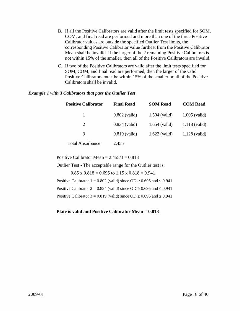
B. If all the Positive Calibrators are valid after the limit tests specified for SOM, COM, and final read are performed and more than one of the three Positive Calibrator values are outside the specified Outlier Test limits, the corresponding Positive Calibrator value furthest from the Positive Calibrator Mean shall be invalid. If the larger of the 2 remaining Positive Calibrators is not within 15% of the smaller, then all of the Positive Calibrators are invalid.
C. If two of the Positive Calibrators are valid after the limit tests specified for SOM, COM, and final read are performed, then the larger of the valid Positive Calibrators must be within 15% of the smaller or all of the Positive Calibrators shall be invalid.
Example 1 with 3 Calibrators that pass the Outlier Test
Positive Calibrator Final Read SOM Read COM Read
1 0.802 (valid) 1.504 (valid) 1.005 (valid)
2 0.834 (valid) 1.654 (valid) 1.118 (valid)
3 0.819 (valid) 1.622 (valid) 1.128 (valid)
Total Absorbance 2.455
Positive Calibrator Mean = 2.455/3 = 0.818
Outlier Test - The acceptable range for the Outlier test is:
0.85 x 0.818 = 0.695 to 1.15 x 0.818 = 0.941
Positive Calibrator 1 = 0.802 (valid) since OD ≥ 0.695 and ≤ 0.941
Positive Calibrator 2 = 0.834 (valid) since OD ≥ 0.695 and ≤ 0.941
Positive Calibrator 3 = 0.819 (valid) since OD ≥ 0.695 and ≤ 0.941
Plate is valid and Positive Calibrator Mean = 0.818
2009-01 Page 19 of 40
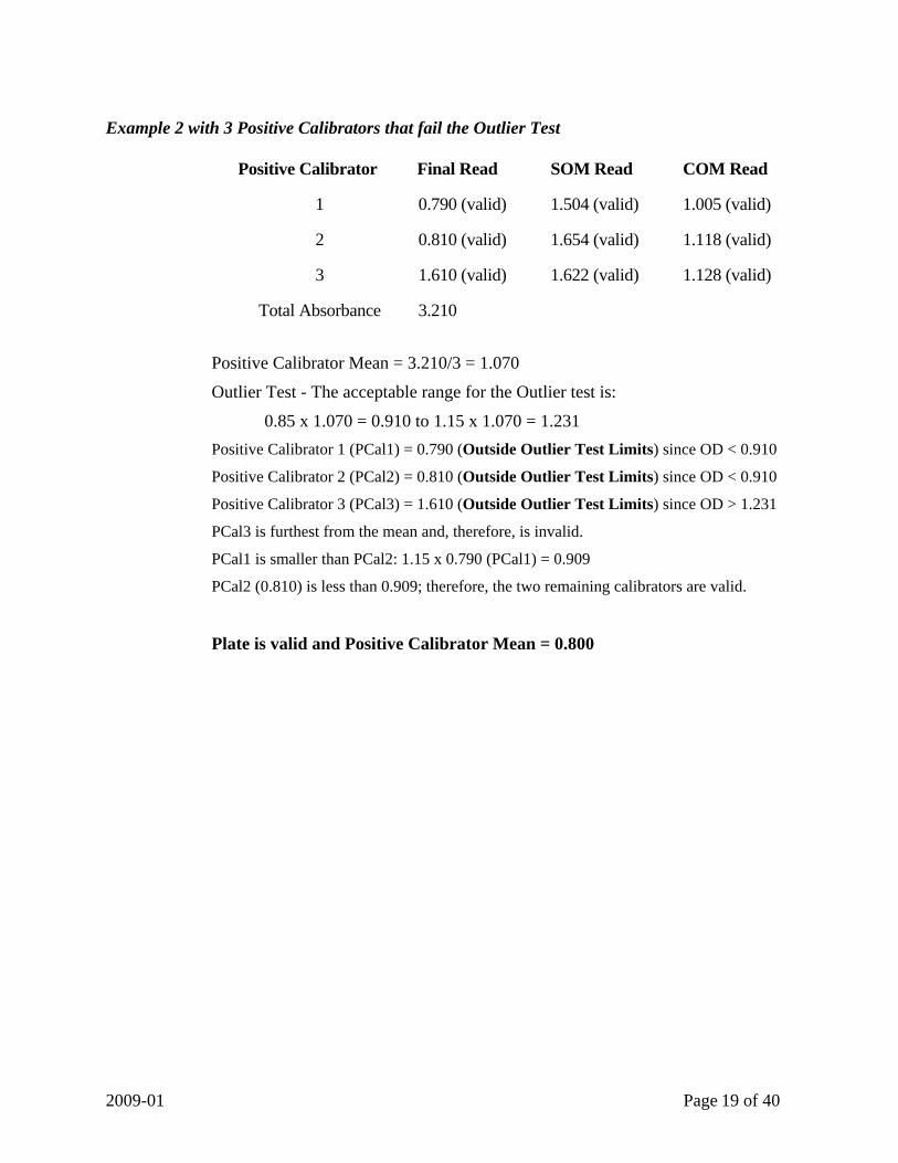
Example 2 with 3 Positive Calibrators that fail the Outlier Test
Positive Calibrator Final Read SOM Read COM Read
1 0.790 (valid) 1.504 (valid) 1.005 (valid)
2 0.810 (valid) 1.654 (valid) 1.118 (valid)
3 1.610 (valid) 1.622 (valid) 1.128 (valid)
Total Absorbance 3.210
Positive Calibrator Mean = 3.210/3 = 1.070
Outlier Test - The acceptable range for the Outlier test is:
0.85 x 1.070 = 0.910 to 1.15 x 1.070 = 1.231 Positive Calibrator 1 (PCal1) = 0.790 (Outside Outlier Test Limits) since OD < 0.910
Positive Calibrator 2 (PCal2) = 0.810 (Outside Outlier Test Limits) since OD < 0.910
Positive Calibrator 3 (PCal3) = 1.610 (Outside Outlier Test Limits) since OD > 1.231
PCal3 is furthest from the mean and, therefore, is invalid.
PCal1 is smaller than PCal2: 1.15 x 0.790 (PCal1) = 0.909
PCal2 (0.810) is less than 0.909; therefore, the two remaining calibrators are valid.
Plate is valid and Positive Calibrator Mean = 0.800

2009-01 Page 20 of 40
Example 3 with 2 Positive Calibrators that fail the Outlier Test
Positive Calibrator Final Read SOM Read COM Read
1 0.700 (valid) 1.504 (valid) 1.005 (valid)
2 1.000 (valid) 1.654 (valid) 1.118 (valid)
3 1.400 (valid) 1.622 (valid) 1.128 (valid)
Total Absorbance
3.100
Positive Calibrator Mean = 3.100/3 = 1.033
Outlier Test - The acceptable range for the Outlier test is:
0.85 x 1.033 = 0.878 to 1.15 x 1.033 = 1.188 Positive Calibrator 1 (PCal1) = 0.700 (Outside Outlier Test Limits) since OD < 0.878
Positive Calibrator 2 (PCal2) = 1.000 (valid) since OD ≥ 0.878 and ≤ 1.188
Positive Calibrator 3 (PCal3) = 1.400 (Outside Outlier Test Limits) since OD > 1.188
PCal3 is furthest from mean and, therefore, is invalid.
PCal1 is smaller than PCal2: 1.15 x 0.700 (PCal1) = 0.805
PCal2 (1.000) is greater than 0.805 and, therefore, is invalid.
All calibrators were invalid; therefore, the plate is invalid.

2009-01 Page 21 of 40
3. Calculation of the Cutoff Value
a. Determine the mean of the valid Positive Calibrator values.
b. Calculate the cutoff value:
Cutoff value = the mean OD of the Positive Calibrator multiplied by 0.425 (cutoff constant)
Example: PCal mean of 0.800 x 0.425 = cutoff of 0.340
4. Calculation of Signal to Cutoff (S/C)
a. Calculate Signal to Cutoff (S/C) values for Negative Controls and individual specimens by dividing each absorbance values (OD) by the cutoff value.
Example: Absorbance of 0.500/0.340 cutoff = S/C of 1.471
b. Report the S/C to 3 decimal places.
5. Negative Control Acceptance Criteria
Negative Control signal to cutoff must be ≥ -0.012 and ≤ 0.300. If either of the two values is outside this limit, the plate is invalid and all the samples on the plate must be repeated.

2009-01 Page 22 of 40
INTERPRETATION OF RESULTS NOTE: Before interpreting the test results, interpret the SOM and COM results. Refer to the Interpretation of SOM Results Table in Step 6 in the Test Procedure section and Interpretation of COM Results Table in Step 10 in the Test Procedure section.
1. Specimens with absorbance values less than -0.020 OD should be retested in a single
microwell. The specimen should be considered nonreactive if the retest absorbance value is less than the cutoff value, even if the retest absorbance value remains less than -0.020 OD.
2. Specimens with absorbance values greater than or equal to -0.020 OD and less than the cutoff value are considered nonreactive. Further testing is not required.
3. Specimens with absorbance values greater than or equal to the cutoff value are considered initially reactive and should be retested in duplicate before final interpretation.
4. After retesting an initially reactive specimen, the specimen is considered repeatedly reactive for antibodies to T. cruzi if either or both duplicate determinations are reactive, i.e., greater than or equal to the cutoff value.
5. After retesting an initially reactive specimen, the specimen is considered nonreactive for antibodies to T. cruzi if both duplicate determinations are nonreactive, i.e., less than the cutoff value.
LIMITATIONS OF THE PROCEDURE The Test Procedure and Interpretations of Results for the ORTHO T. cruzi ELISA Test System must be followed closely when testing for the presence of antibodies to T. cruzi in human serum or plasma. A laboratory that uses the ORTHO T. cruzi ELISA Test System should have a program that will train personnel on the proper use and handling of the product.
Because the ORTHO T. cruzi ELISA Test System was designed to screen individual units of blood or plasma, most data regarding its interpretation were derived from testing individual specimens. Insufficient data are available to interpret tests performed on other body fluids including pooled blood, or processed plasma and products made from such pools; testing of these specimens is not recommended.
Failure to add specimen or reagent may result in an erroneous result.
Specimens with abnormally low protein levels may cause a false SOM failure even in the presence of sample addition. The operator should visually verify sample addition during repeat testing for a SOM failure result.
The Positive Calibrator in the test kit is not to be used to quantitate assay sensitivity.
The ORTHO T. cruzi ELISA Test System detects antibodies to T. cruzi in blood and thus is useful in screening blood and plasma donated for transfusion and further manufacture in establishing prior infection with T. cruzi. It is recommended that repeatedly reactive specimens be investigated by additional testing for antibodies to T. cruzi before a specimen

2009-01 Page 23 of 40
is considered positive, indicating T. cruzi infection. Additional testing for Leishmania, Malaria, Syphilis, and Paracoccidioides brasiliensis (P. brasiliensis) should be considered.
A nonreactive test result does not exclude the possibility of exposure to T. cruzi. Levels of antibodies to T. cruzi may be below the detectable limit of the assay or undetectable during an early stage following exposure to T. cruzi.
PERFORMANCE CHARACTERISTICS In addition to the following studies, data from analytical testing and clinical trials demonstrated equivalent results for all modes of operation of the ORTHO T. cruzi ELISA Test System.
Clinical Specificity The specificity of the ORTHO T. cruzi ELISA Test System is based on a population of presumably healthy volunteer blood donors from four geographically distinct sites in the United States.
A total of 40,665 human serum and plasma samples were tested by the automated processing method. Among the 40,665 volunteer blood donor samples tested, 40,661 (99.990%) were nonreactive, 4 (0.010%) were initially reactive, and 3 (0.007%) were repeatedly reactive. The three repeatedly reactive samples were negative by T. cruzi Radioimmune Precipitation Assay (RIPA), which was used as a confirmatory test. Rates of reactivity for the four sites are shown in Tables 1 and 2. The observed specificity of the ORTHO T. cruzi ELISA Test System in the volunteer blood donor population in this study was 99.993% (40,662/40,665) with a 95% exact confidence interval of 99.978% to 99.999%.
Table 1. Frequency of the ORTHO T. cruzi ELISA Test System Reactivity in Volunteer Blood Donors: Ortho Summit System [Ortho Summit Sample Handling System (Summit), Ortho Summit Processor (OSP) and Ortho Assay Software (OAS)]
Test Site
Number of Samples
Sample Matrix Nonreactive (%) Repeatedly
Reactive (%)
Confirmed Positive with
RIPA
1 4523 Serum
4522 (99.978)
1 (0.022) 0
2 9219 Serum
9218 (99.989)
1 (0.011) 0
3 12118 Plasma 12117 (99.992) 1 (0.008) 0 4 14805 Plasma 14805 (100.000) 0 (0.000) NA
Total N = 40665
40662 (99.993)
3 (0.007) 0
The ORTHO T. cruzi ELISA Test System was used to test 2,121 additional donor samples by both automated and semi-automated processing methods at three sites. Semi-automated processing consists of the Ortho Summit Sample Handling System (Summit) with the AutoWash 96, Model 120 Incubator, AutoReader IV, and Ortho Assay Software (OAS). Automated processing consists of the Ortho Summit System (OSS) defined as the Summit, Ortho Summit Processor (OSP), and OAS. There was 100% agreement

2009-01 Page 24 of 40
between the T. cruzi ELISA results of automated and semi-automated processing methods.
Table 2. Frequency of the ORTHO T. cruzi ELISA Test System Reactivity in Volunteer Blood Donors by Processing Method
Ortho Summit System [Summit, OSP and OAS]
Semi-Automated Processing (Summit, AutoWash 96,
AutoReader IV, and OAS) Test Site Number of Samples Sample Matrix
Nonreactive (%) Nonreactive (%) 1 713 Serum 713 (100.00) 713 (100.00) 2 738 Serum 738 (100.00) 738 (100.00) 3 670 Plasma 670 (100.00) 670 (100.00)
Total N = 2121 2121 (100.00) 2121 (100.00)
An additional study was conducted using volunteer blood donor samples from three geographic locations in the United States, including one site where previous cases of T. cruzi have been reported.20 A total of 148,989 human serum and plasma samples were tested by the automated processing method. Among the 148,989 volunteer blood donor samples tested, 148,935 (99.964%) were nonreactive, 54 (0.036%) were initially reactive, and 50 (0.034%) were repeatedly reactive, twenty-nine of which were confirmed positive and 21 negative by the T. cruzi Radioimmune Precipitation Assay (RIPA) used as a confirmatory test. Rates of reactivity for the three sites are shown in Table 3. The observed specificity of the ORTHO T. cruzi ELISA Test System in random, presumably healthy, linked, volunteer blood donors in these specific geographic locations was 99.986% (148,939/148,960) with a 95% exact confidence interval of 99.977% to 99.992%.
Table 3. Frequency of the ORTHO T. cruzi ELISA Test System Reactivity in Volunteer Blood Donors from a High Prevalence Areaa
Site Number of Samples Nonreactive (%) Repeatedly
Reactive (%)
Confirmed Positive with
RIPA
A 95,674
95,635 (99.959)
39 (0.041) 23
B 29,306
29,298 (99.973)
8 (0.027) 4
C 24,009
24,006 (99.988)
3 (0.012) 2
Total N=148,989
148,939 (99.966)
50 (0.034) 29
aTesting was performed with the Ortho Summit System

2009-01 Page 25 of 40
Clinical Sensitivity The sensitivity of the ORTHO T. cruzi ELISA Test System in a positive population was evaluated by testing a total of 106 samples from subjects included as parasite positive by historical identification of T. cruzi parasites by one of the following methods: blood smear (i.e., Giemsa), hemoculture, or xenodiagnosis. The samples were obtained from the endemic countries of Bolivia, Chile, Colombia, and Nicaragua. Testing was performed at one site by the automated and semi-automated processing methods. All specimens initially reactive with the ORTHO T. cruzi ELISA Test System were retested in duplicate. Table 4 shows the overall results of the testing of the 106 positive samples by the automated processing method. Equivalent results were obtained with the semi-automated processing method.
Table 4. Frequency of ORTHO T. cruzi ELISA Test System Reactivity in Positive Samplesa
Number of Samples Repeatedly Reactive (%) Nonreactive (%) 106 106 (100.0) 0 (0.0)
aTesting was performed on the automated and semi-automated systems with the same outcomes The overall sensitivity of the ORTHO T. cruzi ELISA Test System in this study was observed to be 100.0% (106/106) for parasite positive samples with a 95% exact confidence interval of 96.6% to 100.0%.
Sensitivity and Specificity in a High Risk Population A total of 574 samples from study subjects from countries endemic for T. cruzi infection were tested with the ORTHO T. cruzi ELISA Test System and a T. cruzi IFA to determine sensitivity and specificity in a population at risk. The samples were obtained from the endemic countries of Bolivia, Colombia, Guatemala, Mexico, and Nicaragua. Testing was performed at two sites by the semi-automated processing method. Table 5 compares the ORTHO T. cruzi ELISA Test System results with the most probable T. cruzi antibody status for the High Risk population.
Table 5. ORTHO T. cruzi ELISA Test System Results and Most Probable T. cruzi Antibody Status for High Risk Samples
Most Probable T. cruzi Antibody Status Observed Resultsa
Positive Negative Indeterminate TOTAL Repeatedly Reactive 92b 5 b 0 97
Nonreactive 1 b 476c 0 477 TOTAL 93 481 0 574
aTesting was performed by the semi-automated processing method bBased on RIPA results c Based on negative T. cruzi IFA results
The observed sensitivity of the ORTHO T. cruzi ELISA Test System in the High Risk population in this study was 98.9% (92/93) with a 95% exact confidence interval of 94.2% to 100.0%.

2009-01 Page 26 of 40
The observed specificity of the ORTHO T. cruzi ELISA Test System in the High Risk population in this study was 99.0% (476/481) with a 95% exact confidence interval of 97.6% to 99.7%.
Additional Positive Performance Data In addition to the samples from parasite positive individuals, another group of samples that were serological presumed positive were tested. A total of 810 samples were included in this T. cruzi serological positive population. The samples were obtained from the endemic countries of Bolivia, Brazil, Chile, Guatemala, Mexico, and Nicaragua. Serological presumed positive samples were included based upon two positive serological tests for T. cruzi antibodies (i.e., ELISA, IFA, RIPA, hemagglutination, or complement fixation). Testing was performed at two sites by the semi-automated processing method. All specimens initially reactive with the ORTHO T. cruzi ELISA Test System were retested in duplicate. Six hundred sixty-four (664) samples gave repeatedly reactive results with the ORTHO T. cruzi ELISA Test System. Two of the 664 repeatedly reactive samples had S/C results <1.500 and both were tested with RIPA. Both samples were RIPA negative. The agreement between the ORTHO T. cruzi ELISA Test System and most probable T. cruzi antibody status was 100% (662/662) for samples with a T. cruzi antibody status of positive. All 146 samples that were ORTHO T. cruzi ELISA nonreactive were negative by RIPA.
Table 6 shows the ORTHO T. cruzi ELISA Test System results for the serological presumed positive population compared to the most probable T. cruzi antibody status.
Table 6. ORTHO T. cruzi ELISA Test System Results and Most Probable T. cruzi Antibody Status for Serological Presumed Positive Samples
Most Probable T. cruzi Antibody Status Observed Resultsa
Positive Negative Indeterminate TOTAL
Repeatedly Reactive 662 2b 0 664 Nonreactive 0 146b 0 146
TOTAL 662 148 0 810 aTesting was performed by the semi-automated processing method, except for 20 samples with limited volume that were pipetted manually bMost probable T. cruzi antibody status was determined by RIPA for samples that were nonreactive or had S/C results <1.500 in the T. cruzi ELISA

2009-01 Page 27 of 40
Analytical Sensitivity (Dilutional Panel Precision Study) Analytical sensitivity was determined by testing a 20-member dilutional panel and comparing results across multiple sites and multiple kit lots. Three replicates of each panel member were tested on a single occasion per day on three different days by one technologist at three sites, for a total of 540 observations. The dilutional panel was prepared from five unique T. cruzi antibody positive plasmas/serums, each diluted to provide 4 samples (dilutional levels) with signal to cutoff (S/C) values targeted in descending order around the cutoff of 1.000. Analytical sensitivity testing was performed by the automated processing method. The reactive panel members were reactive across all sites with all kit lots and the nonreactive panel members were nonreactive across all sites with all kit lots. The mean S/C, standard deviation (SD), and coefficient of variation (CV%) results are shown in Table 7 for each dilutional level.
Table 7. Dilutional Panel Member Precision by Dilutional Levela
Between Site* Between Lot† Total‡ Dilutional
Level
Mean ORTHO T. cruzi ELISA
S/C SD CV (%) SD CV (%) SD CV (%)
Number of Observations
DL1 5.404 0.000 0.0 0.145 2.7 0.526 9.7 135 DL2 2.616 0.000 0.0 0.000 0.0 0.241 9.2 135 DL3 1.935 0.064 3.3 0.000 0.0 0.274 14.2 135 DL4 0.293 0.029 N/A+ 0.000 N/A+ 0.108 N/A+ 135
aTesting was performed by the automated processing method *Between Sites: Variability of the assay performance from site to site †Between Lot: Variability of the assay performance from lot to lot ‡Total: Variability of the assay incorporating factors of site and lot + % CVs are not meaningful when S/C is very small

2009-01 Page 28 of 40
Analytical Specificity – Potentially Cross Reacting Samples The specificity of the ORTHO T. cruzi ELISA Test System was evaluated using 616 samples from individuals with infections or clinical conditions that might potentially exhibit cross reactivity when tested with the assay. This testing was performed by the semi-automated processing method. Samples from the following conditions or disease states were included in the testing: Leishmania; Malaria; Schistosomiasis; Syphilis; Influenza Vaccine; Paraproteins, Autoantibodies and Alloantibodies; Virally Infected and other Disease States. Table 8 shows the numbers and types of samples tested.
Table 8. Reactivity of the ORTHO T. cruzi ELISA Test System with Samples from Subjects with Potentially Cross Reacting Conditions or Disease Statesa
Potentially Cross Reacting Condition or Disease State
Number of
Samples Nonreactive (%) Repeatedly
Reactive (%) Positive with
RIPA (%)
Leishmania 100 21 (21.0) 79 (79.0) 21 (21.0)* Malaria 96 94 (97.9) 2 (2.1) 0 (0)
Schistosomiasis 30 30 (100.0) 0 (0) 0 (0) Syphilis 30 29 (96.7) 1 (3.3) 0 (0)
Influenza Vaccine A 70 70 (100.0) 0 (0) 0 (0) Paraproteins, Autoantibodies,
and Alloantibodies B 120 120 (100.0) 0 (0) 0 (0)
Virally Infected and Other Disease States C 170 168 (98.8) 2 (1.2) 2 (1.2)**
Total 616 532 (86.4) 84 (13.6) 23 (3.7)* aTesting was performed by the semi-automated processing method *Leishmania specimens cannot reliably be confirmed as T. cruzi antibody positive by RIPA. Leishmania samples
were collected in India where T. cruzi is not endemic and these samples are presumed to be T. cruzi antibody negative
**These two RIPA positive samples were P. brasiliensis specimens that were obtained from Argentina, where T. cruzi infection is endemic
A. Unlinked Paired Pre- and Post-Vaccination Samples from 35 Persons Receiving the Influenza Vaccine
B. Unlinked Samples from Individuals with Paraproteins, Autoantibodies, and Alloantibodies: Lupus Erythematosus (N=30, ANA titer > 1:640), Rheumatoid Arthritis (N= 30, RF > 30 IU or titer > 1:320), Polyclonal Gammopathies (N=15), Monoclonal Gammopathies (N=15), Multiple Leukocyte Alloantibodies (N=15), Multiple Red Cell Alloantibodies (N=15)
C. Unlinked Samples from Individuals with Antibodies: Cytomegalovirus (N=20), Epstein-Barr Virus (N=20), Herpes Simplex Virus Type 1 (N=20), Rubella (N=20), Hepatitis C (N=20), Hepatitis B (N=20), Human Immunodeficiency Virus (N=20), Human T-Cell Lymphotropic Virus (N=20), Toxoplasma gondii (N=5), Paracoccidioides brasiliensis (N=5)
Among the 100 subjects with Leishmania infection, 19 (19.0%) were nonreactive, 81 (81.0%) were initially reactive, and 79 (79.0%) were repeatedly reactive. Although 21 (21.0%) of the samples were positive by RIPA, the samples were obtained in India where T. cruzi is not endemic and, therefore, the most probable T. cruzi antibody status of the 100 Leishmania samples is negative. The ORTHO T. cruzi ELISA Test System may yield falsely reactive results among test subjects with Leishmania infection.

2009-01 Page 29 of 40
Of the 516 non-Leishmania samples, 510 (98.8%) were nonreactive, six (1.2%) were initially reactive, and five (1.0%) were repeatedly reactive. Three of the five repeatedly reactive samples (one syphilis and two malaria, P. falciparum) were RIPA negative. Two of the five repeatedly reactive samples were obtained from among the five test subjects with P. brasiliensis infection. These two samples were RIPA positive and were obtained from a T. cruzi endemic area. Whether these represent false positive for T. cruzi infection due to cross reactivity in both ELISA and RIPA or co-infection with P. brasiliensis and T. cruzi is not known.
Reproducibility The intra-assay (within plate) and inter-assay (between plate) reproducibility of the ORTHO T. cruzi ELISA Test System was evaluated using an eight-member reproducibility panel. The reproducibility panel consisted of three moderate to strongly reactive samples, three reactive samples near the assay cutoff (approximately 1.5 – 2.0 S/C), and two nonreactive samples. The panel was tested at three external sites using three different kit lots and both the automated and semi-automated processing methods. Ten replicates each of the eight-member panel were assayed on a single occasion per day on nine different days by two technologists for a total of 4319 observations (one observation for R7 was a statistical outlier on both processing methods) per processing method. Mean signal to cutoff (S/C), standard deviation (SD), and coefficient of variation (CV%) results are presented in Table 9 and Table 10 for the two processing methods.
Table 9. Reproducibility Panel Testing: Ortho Summit Sample Handling System (Summit), AutoWash 96, AutoReader IV, and Ortho Assay Software (OAS)
Inter-assay* Intra-assay† Total‡ Panel Member
Number Tested
Mean ORTHO T. cruzi ELISA
S/C SD CV(%) SD CV(%) SD CV(%)
R1 540 5.954 0.258 4.3 0.324 5.4 0.492 8.3 R2 540 6.424 0.306 4.8 0.324 5.0 0.501 7.8 R3 540 6.647 0.338 5.1 0.345 5.2 0.554 8.3 R4 540 1.946 0.089 4.6 0.143 7.3 0.189 9.7 R5 540 1.909 0.097 5.1 0.128 6.7 0.180 9.4 R6 540 2.173 0.113 5.2 0.134 6.2 0.207 9.5 R7 539 0.084 0.011 N/A+ 0.025 N/A+ 0.031 N/A+ R8 540 0.101 0.013 N/A+ 0.029 N/A+ 0.035 N/A+
*Between Plate (Between Run (Lot x Site x Technologist)): Variability of the assay performance from plate to plate †Within Plate (Between Replicate): Variability of the assay performance from replicate to replicate ‡Total: Inter-assay and Intra-assay variability + % CVs are not meaningful when S/C approaches zero

2009-01 Page 30 of 40
Table 10. Reproducibility Panel Testing: Ortho Summit System (OSS) [Summit, Ortho Summit Processor (OSP), and OAS]
Inter-assay* Intra-assay† Total‡ Panel Member
Number Tested
Mean ORTHO T. cruzi ELISA
S/C SD CV(%) SD CV(%) SD CV(%)
R1 540 5.198 0.128 2.5 0.307 5.9 0.371 7.1 R2 540 5.524 0.139 2.5 0.348 6.3 0.396 7.2 R3 540 5.730 0.166 2.9 0.331 5.8 0.411 7.2 R4 540 1.820 0.056 3.1 0.145 8.0 0.169 9.3 R5 540 1.777 0.065 3.7 0.121 6.8 0.142 8.0 R6 540 2.026 0.076 3.8 0.119 5.9 0.156 7.7 R7 539 0.054 0.008 N/A+ 0.010 N/A+ 0.014 N/A+ R8 540 0.062 0.007 N/A+ 0.011 N/A+ 0.014 N/A+
*Between Plate (Between Run (Lot x Site x Technologist)): Variability of the assay performance from plate to plate †Within Plate (Between Replicate): Variability of the assay performance from replicate to replicate ‡Total: Inter-assay and Intra-assay variability + % CVs are not meaningful when S/C approaches zero

2009-01 Page 31 of 40
PERFORMANCE CHARACTERISTICS OF CADAVERIC SPECIMEN TESTING Reproducibility Reproducibility of ORTHO T. cruzi ELISA Test System was assessed using 20 cadaveric (post-mortem) and 20 living donor sera. These specimens were spiked with anti-T. cruzi positive plasma to give reactivity near the assay cutoff (approximately 2.0 S/C). Each of the specimens was tested once on six different days on each of three lots of ORTHO T. cruzi ELISA Test System at one site. Reproducibility testing was performed by both manual and automated processing methods. For each processing method, cadaveric and living donor specimens were 100% reactive across kit lots and the %CVs were comparable for both specimen groups. Kit Lot 1
Number of Donors Replicates % Positive Mean S/C CV(%)
Cadaveric 20 120 100 1.906 15.2 Manual
Living Donor 20 120 100 1.583 12.4
Cadaveric 20 120 100 1.933 15.9 Automated
Living Donor 20 120 100 1.684 11.8
Kit Lot 2
Number of Donors Replicates % Positive Mean S/C CV(%)
Cadaveric 20 120 100 1.900 12.4 Manual
Living Donor 20 120 100 1.613 12.4
Cadaveric 20 120 100 1.912 11.3 Automated
Living Donor 20 120 100 1.693 10.6
Kit Lot 3
Number of Donors Replicates % Positive Mean S/C CV(%)
Cadaveric 20 120 100 2.121 16.8 Manual
Living Donor 20 120 100 1.766 14.3
Cadaveric 20 120 100 2.111 15.7 Automated
Living Donor 20 120 100 1.828 11.3

2009-01 Page 32 of 40
Specificity Specificity was evaluated using 50 cadaveric specimens collected up to 23.7 hours after death and 50 living donor specimens. Testing was performed across three lots of ORTHO T. cruzi ELISA Test System by both manual and automated processing methods. For the manual method, the mean signal to cutoff (S/C) ratio was 0.268 for the cadaveric specimens, and the mean S/C ratio was 0.122 for the living donor specimens. For the automated method, the mean signal to cutoff (S/C) ratio was 0.196 for the cadaveric specimens, and the mean S/C ratio was 0.093 for the living donor specimens. While the cadaveric mean results (0.268 and 0.196) are statistically different from the living donor specimens (0.122 and 0.093) for both processing methods, they are well below the assay cutoff of 1.000 signal to cutoff and no false positives were observed. The results are presented in Table 11. Table 11 Reactivity with the ORTHO T. cruzi ELISA Test System
Population Number of Specimens Nonreactive Initially Reactive Manual Processing
Cadaveric 50 50 (100.0%) 0 (0.0%) Living Donor 50 50 (100.0%) 0 (0.0%)
Automated Processing
Cadaveric 50 50 (100.0%) 0 (0.0%) Living Donor 50 50 (100.0%) 0 (0.0%)
The ORTHO T. cruzi ELISA Test System has an estimated specificity in cadaveric specimens of 100.0% (50/50) with a 95% exact confidence interval of 92.9% to 100.0%.

2009-01 Page 33 of 40
Sensitivity Sensitivity was evaluated using 50 cadaveric specimens collected up to 23.7 hours after death and 50 living donor specimens. All specimens were screened for anti-T. cruzi and were found to be nonreactive. All specimens were spiked with anti-T. cruzi positive plasma to give reactivity near the assay cutoff. Testing was performed approximately 47 hours after spiking using three lots of ORTHO T. cruzi ELISA Test System by both manual and automated processing methods. Since the specimens were spiked to be reactive, duplicate repeat testing was not performed for initially reactive specimens. For the manual method, the mean signal to cutoff (S/C) ratio was 1.836 for the cadaveric specimens, and the mean S/C ratio was 1.570 for the living donor specimens. The calculated difference between the cadaveric specimens and the living donor specimens tested by the manual method was 0.266 S/C, which was determined by the F-test to be statistically significant (p<0.0001). However, all results for the cadaveric and living donor specimens were reactive with the ORTHO T. cruzi ELISA Test System resulting in 100.0% reactivity. For the automated method, the mean signal to cutoff (S/C) ratio was 1.861 for the cadaveric specimens, and the mean S/C ratio was 1.597 for the living donor specimens. The calculated difference between the cadaveric specimens and the living donor specimens tested by the automated method was 0.264 S/C, which was determined by the F-test to be statistically significant (p<0.0001). However, all results for the cadaveric and living donor specimens were reactive with the ORTHO T. cruzi ELISA Test System resulting in 100.0% reactivity. The results are presented in Table 12. Table 12 Reactivity with the ORTHO T. cruzi ELISA Test System
Population Number of Specimens Nonreactive Initially Reactive Manual Processing
Cadaveric 50 0 (0.0%) 50 (100.0%) Living Donor 50 0 (0.0%) 50 (100.0%)
Automated Processing
Cadaveric 50 0 (0.0%) 50 (100.0%) Living Donor 50 0 (0.0%) 50 (100.0%)
The ORTHO T. cruzi ELISA Test System has an estimated sensitivity in spiked cadaveric specimens of 100.0% (50/50) with a 95% exact confidence interval of 92.9% to 100.0%.

2009-01 Page 34 of 40
Summary of Revisions for January 2009 version
Front Cover: Added “192 Test Kit:” with the product code 6902594. Intended Use: Added the use of cadaveric (non-heart-beating) specimens. Reagents: Added the 192 Test Kit product code 6902594 with the component configuration to the table Precautions: Updated statements:
Number 7 - Added the “T” in front of “N,R: 20/21-25-36-40-43-50/53-68”, removed the number “28” from the Safety Statement S: 26-36/37-45-60-61, and deleted the sentence “After contact with skin, wash immediately with plenty of water.” Number 26 – Updated R & S statements for ProClin 300™ Number 27- Added Precaution and R & S Statements for 2-chloroacetamide Number 30- Added caution statement: Serum-separator tubes (SST) should be used with caution when using automated pipetting instrumentation. Consult the Instrument User’s Manuals for precautions. Re-numbered precautions as appropriate.
Specimen Changed section title to Specimen Collection, Storage, and Handling Collection Updated specimen collection to include serum-separator tubes and added the statement and Prep: “Cadaveric specimens may be collected into serum, serum-separator tubes or EDTA blood collection devices.” Included cadaveric specimens in the storage requirements section. Added claims
for total protein, human anti-mouse antibodies (HAMA) and heterophilic antibodies. Reformated section into three (3) subsections, Living Donor Specimens, Cadaveric Blood Specimens, and Specimen Pooling. Bulleted information format introduced. Revised statement on Specimen Pooling to read “Testing of these specimens is not recommended. No data are available to interpret tests performed on pooled blood or processed plasma and products made from such pools.”
Performance Added the reproducibility, specificity and sensitivity performance claims for cadaveric Characteristics: specimens

2009-01 Page 35 of 40

2009-01 Page 36 of 40
BIBLIOGRAPHY 1. Leiby DA, Lenes BA, Tibbals MA, et al.1999. Prospective evaluation of a patient with
Trypanosoma cruzi infection transmitted by transfusion. N. Engl. J. Med.341:1237-39.
2. Cimo PL, Luper WE, Scouros MA. 1993. Transfusion-associated Chagas’ disease in Texas: report of a case. Texas Med. J.89:48-50.
3. Grant IH, Gold JW, Wittner M, et al. 1989.Transfusion-associated acute Chagas disease acquired in the United States. Ann. Intern. Med. 111:849-51.
4. Nickerson P, Orr P, Schroeder M-L,et al. 1989. Transfusion-associated Trypanosoma cruzi infection in a nonendemic area. Ann. Intern. Med. 111:851-53.
5. Lane DJ, Sher G, Ward B, et al. 2000. Investigation of the second case of transfusion transmitted Chagas disease in Canada. Blood 96:60a (pt. 1).
6. Engvall E, Perlmann P. Enzyme-linked immunosorbent assay (ELISA): quantitative assay of IgG. Immunochemistry 1971; 8:871-74.
7. Centers for Disease Control. Update: Universal Precautions for Prevention of Transmission of Human Immunodeficiency Virus, Hepatitis B Virus, and Other Bloodborne Pathogens in Health-Care Settings. MMWR 1988; 37(24);377-88.
8. Clinical Laboratory Standards Institute. Protection of Laboratory Workers from Occupationally Acquired Infections; Approved Guideline – Third Edition. CLSI document M29-A3 (ISBN 1-56238-567-4). Clinical Laboratory Standards Institute, 940 West Valley Road, Suite 1400, Wayne, Pennsylvania 19087–1898 USA, 2005.
9. Occupational Exposure to Hazardous Chemicals in Laboratories, 29 CFR 1910.1450.
10. Occupational Exposure to Bloodborne Pathogens, 29 CFR 1910.1030.
11. World Health Organization, Laboratory Biosafety Manual. 3rd ed, 2004.
12. Technical Manual. 15th ed. Bethesda, MD: American Association of Blood Banks, 2005. Facilities and Safety.
13. Block SS. Disinfection, sterilization and preservation, 4th ed, Lea & Febiger, 1991.
14. Council Directive of 31 May 1999 on the approximation of the laws, regulations and administrative provisions of the Member States relating to the classification, packaging, and labeling of dangerous preparations (1999/45/EC).
15. Clinical Laboratory Standards Institute. Preparation and Testing of Reagent Water in the Clinical Laboratory; Proposed Guideline - Fourth Edition. CLSI document C3-P4 [ISBN 1-56238-570-4]. Clinical Laboratory Standards Institute, 940 West Valley Road, Suite 1400, Wayne, Pennsylvania 19087–1898 USA, 2005.
16. Commission of Directive 2001/60/EC of 7 August 2001 adapting to technical progress Directive 1999/45/EC of the European Parliament and of the Council concerning the approximation of the laws, regulations, and administrative provisions of the Member States relating to the classification, packaging, and labeling of dangerous preparations.
17. International Air Transportation Association (IATA): Dangerous Goods Regulations.

2009-01 Page 37 of 40
18. NCCLS. Specifications for Immunological Testing for Infectious Diseases; Approved Guideline – Second Edition. NCCLS document I/LA18-A2 (ISBN 1-56238-445-7). NCCLS, 940 West Valley Road, Suite 1400, Wayne, Pennsylvania 19087-1898 USA, 2001.
19. NCCLS. Statistical Quality Control for Quantitative Measurements: Principles and Definitions; Approved Guideline Second Edition. NCCLS document C24-A2 [ISBN 1-56238-371-X]. NCCLS, 940 West Valley Road, Suite 1400, Wayne, Pennsylvania 19087-1898 USA, 1999.
20. Leiby, DA, Herron, RM, Read, EJ, et al. 2002. Trypanosoma cruzi in Los Angeles and Miami blood donors: impact of evolving donor demographics and seroprevalence and implications for transfusion transmission. Transfusion 42:549-55.

2009-01 Page 38 of 40 2009-01 Page 38 of 40
2009-01 Page 39 of 40

2009-01 Page 40 of 40 2009-01 Page 40 of 40