Embed Size (px)
Citation preview

Trunk muscle activity while lifting objects ofunexpected weight
著者 Watanabe Masahiro, Kaneoka Koji, Okubo Yu,Shiina Itsuo, Tatsumura Masaki, MiyakawaShumpei
journal orpublication title
Physiotherapy
volume 99number 1page range 78-83year 2013-03権利 (C) 2011 Chartered Society of Physiotherapy.
Published by Elsevier Ltd.NOTICE: this is the author’s version of awork that was accepted for publication inPhysiotherapy. Changes resulting from thepublishing process, such as peer review,editing, corrections, structural formatting,and other quality control mechanisms may notbe reflected in this document. Changes mayhave been made to this work since it wassubmitted for publication. A definitiveversion was subsequently published inPUBLICATION, 99(1) (2013)DOI:10.1016/j.physio.2011.09.005
URL http://hdl.handle.net/2241/118590doi: 10.1016/j.physio.2011.09.005

Abstract
Objective: To determine trunk muscle activities during lifting of an object heavier than
expected, which may contribute to the development of low back pain.
Design: Electromyographic evaluation of trunk muscle activities
Setting: University Spine Laboratory
Participants: Eleven healthy men (mean age ± SD, 24.0 ± 2.2 years)
Interventions: Trunk muscle activities were measured when the participants lifted an
object with their right arm in immediate response to a light stimulus. Surface and wire
electrodes were used to measure the activities of the rectus abdominis, external oblique,
and erector spinae muscles and those of the transversus abdominis and lumbar
multifidus muscles, respectively. The lifting tests were performed in three different
settings: lifting an expected 1 .0 kg object; an unexpected 4.0 kg object (erroneously
recognized as 1 kg); and an expected 4.0 kg object.
Main Outcome Measures: Comparison was made among the muscle activities, each
being induced when the participants listed different weights of objects, by calculating
the root mean square (RMS) at rest and % maximum voluntary contraction (MVC).

Results: When the participants were aware of the weight of the object to be lifted, the
activities of the external oblique, transversus abdominis, erector spinae, and lumbar
multifidus muscles were elevated immediately after lifting. While, the elevation of these
activities was delayed (P < .05) after lifting.
Conclusions: Our findings of this study suggest that when participants lift an object
much heavier than expected, their trunk muscles may not be able to function
appropriately.
Keywords: Electromyography; Trunk muscles; Estimate; Feedforward; Low back pain;
Rehabilitation;

1
Trunk Muscle Activity while Lifting of Objects with Unexpected Weights
Introduction
Low back pain (LBP) is one of the most frequent physical complaints worldwide. To
address this problem, the guidelines for clinical evaluation of LBP have been
established in various countries in the world (1). In these guidelines, no evaluation and
treatment methods have been specified from the standpoint of physical therapy and no
definitive views on the function of the trunk muscles, which is one of factors playing an
important role in lifting, have been set forth.
However, to control the trunk stably, the muscle activities of the trunk muscles,
especially the deep-seated muscles are essential (2,3) and it has been verified that
feedforward control of the trunk muscles occur prior to any motion(4).A study has
reported that the subject make a change in responses of the trunk muscles depending on
the weight of the object to be lifted for the stable control of the trunk muscles (5).
On the other hand, various factors such as the delayed contraction of the transverses
2
abdominis muscles, which are the deep-seated muscles of the trunk muscles and the
attenuated muscle activities of the back muscles (6), including psychological factors
such as fear-avoidance cycle (7), have the potential effects on chronic LBP.
The causes of etiology include lifting objects heavier than expected and taking
unintentional behavior (8, 9). Although the mechanism of unstable control of the trunk
muscles may be assumed from various aspects, the reaction of the body trunk muscles
has not been clarified. A previous study has reported that the muscle activities of the
trunk muscles, especially of the back muscles occur when an object is lifted with one
hand (10) while almost no studies have reported on the reactions of the trunk muscles
when the subject lifted an object heavier than expected.
Against this background, to reveal the effects of anticipation of the weight of an
object to be lifted on the trunk muscles, this study analyzed the muscle activities of the
trunk muscles occurring when an object heavier than expected was lifted using surface
electrodes and wire electrodes, and conducted a comparative study.
Participants and Methods
3
1. Participants
Eleven adult men without LBP at the start of the study, who provided us with their
informed consent, were enrolled in this study (age, 24.0 ± 2.2 years; height, 172.1 ± 6.6
cm; weight, 67.2 ± 7.9 kg; all right-handed; right arm length, 71.8 ± 5.1 cm). The
exclusion criteria included a history of lumbar spine disorder, neurological disorder,
and/or spine surgery.
This study was conducted in the presence of an orthopedic surgeon and was approved
by the Ethics Committee of the Waseda University Faculty of Sport Sciences (Approval
No. 08-027).
2. Tests
The test was started on the participant, who had sat up straight on a stool (in an elect
sitting posture) with the bottoms of his feet in contact with the floor surface (and the
knee joints and hip joints 90-degree flexed). The upper right limb grasped an object

4
on a table with the elbows straight and the upper left limb naturally dropped downward
along the body side. Each participant was instructed to lift an object on the table up to
the eye level with his right arm in response to a light stimulus (lifting test).
The five steps of the test procedure were sequentially performed (Table 1.)
Two kinds of materials of the same size but different weights, 1.0 kg of sand and 4.0
kg of lead, were used for the objects to be lifted. These materials were put in the same
containers to make it impossible to distinguish between them based on their external
appearances.
3. Electromyography
The activities of 10 types of muscles were measured including the right and left
rectus abdominis, external oblique, transverses abdominis, lumbar multifidus, and
erector spinae.
The EMG signals of the muscles of bilateral transverses abdominis and lumbar
multifidus were recorded using fine-wire bipolar electrodes fabricated from two strands

5
of urethane-coated stainless-steel wire (diameter, 0.05 mm; Unique Medical Co, Ltd,
Tokyo, Japan).
The fine wire was threaded into hypodermic needles (23 gauge × 60 mm) with 2 mm
of urethane cut off and the tips bent back to form 1- and 2-mm hooks. Wire electrodes
were sterilized in an autoclave (HighClave HVE-50; Hirayama Manufacturing Corp,
Saitama, Japan) at 121°C for 20 minutes. The electrodes were inserted into the muscles
of bilateral transverses abdominis (approximately midway between the rib cage and the
iliac crest) (11) and lumbar multifidus (approximately 2 cm lateral to the L5 spinous
process) (12) under the guidance of ultrasound imaging. Once the electrodes reached the
targeted muscle, it was stimulated by an electrical stimulation and muscle contraction
was visually confirmed by ultrasound imaging.
Before the surface electrodes were attached, the skin was rubbed with a skin abrasive
and alcohol to reduce the skin impedance to the level below 2 kΩ. Pairs of disposable
Ag/AgCl surface electrodes (Vitrode F-150S; Nihon Kohden Corporation, Tokyo,
Japan) were bilaterally attached, parallel to the muscle fibers, with a center-to-center
distance of 2 cm, to the following muscles: the rectus abdominis (3 cm lateral to the

6
umbilicus) (13-15), the external oblique (midway between the costal margin of the ribs)
and the iliac crest (approximately 45° to the horizontal)(15,16), and the erector spinae (3
cm lateral to the L3 spinous process)(14,17). A reference electrode was placed over the
sternum.
4. Tests on Maximum Voluntary Contraction
For normalization of the EMG data, a test on maximum voluntary contraction (MVC)
was performed on the individual muscles of interest while the EMG signal amplitude
was recorded. The test positions were consistent with those demonstrated in manual
muscle testing books commonly used by physical therapists, but in some cases
additional manual resistance was applied. Manual resistance was applied gradually, with
the maximum level held for 3 seconds. Correct electrode placement was further
confirmed by observing the EMG signal amplitude during the manual muscle tests.
For the rectus abdominis muscles, MVC was measured in a partial sit-up posture with
the knees flexed, hands behind the head, and the trunk flexed, while resistance was
applied onto the shoulder in the trunk extension direction. For the external oblique
7
muscles on the right side, the participants were in a supine position with their knees
flexed and hands behind the head, while trunk was being flexed and rotated to the left.
Resistance was applied onto the shoulders in the trunk extension and right rotation
directions. For the external oblique muscles on the left side, the trunk was, instead,
flexed and rotated to the right, with the resistance applied onto the shoulders in the
trunk extension and left rotation directions. The MVC levels for the muscles of lumbar
multifidus and erector spinae were measured with prone trunk extension while
resistance was being applied onto the upper thoracic area in the trunk flexion direction.
MVC for the transverses abdominis muscles was recorded when a maximal expiratory
maneuver occurred with the abdominal hollowing in a sitting position (18, 19). Similar
verbal encouragements were given to eleven participants for each of the MVC tests to
ensure full extent of their power throughout the 3 seconds, and after the MVC test, the
participants were asked if they thought it required full extent of their power. If not, the
MVC was repeated. The MVC tests were performed at the intervals of one minute.
EMG data were collected for the 3-second period of isometric phase. The MVC level
was calculated for the 1-second period, in which the highest signal activity was
observed.

8
5. Measurement and data analysis
The electrical signals obtained from the individual electrodes during the period from
the delivery of the light stimulus to the end of the lifting test were converted to the
digital values at a sampling frequency of 1000 Hz to import into a personal computer.
To normalize the muscle activities, MVC was measured for each of the muscles; the
root mean square (RMS), which showed the muscle activity, was calculated. During the
baseline measurement, RMS was calculated for the 50-ms period (20), during which the
participant held the object with his right arm and muscular potential was stable (at rest).
During each of the lifting tests, the time points when the object left the table and the
sensor potential decreased were defined as 0 ms and RMS analysis was performed
during the period from -200 to +200 ms. This period was divided into eight 50-ms
phases and RMS was calculated for each of these 8 phases. The RMS value calculated
in this way was divided by the RMS at the time of MVC to find % MVC. A comparison
was made between the muscle activities by calculating the RMS at rest and %MVC,
which was obtained by dividing the RMS for each of the 8 phases of the individual
9
lifting tests by the RMS at the time of MVC.
6. Statistical analysis
For each of the muscles, comparisons were made between activities at rest and those
during the individual phases, as well as between the tests for each phase. In
comparison, ANOVA was used and if a significant difference was observed in any of
test items, the Dunnett’s multiple comparison test was conducted.
Statistical analyses were performed using SPSS (15.0). In all analyses, P<.05 was
regarded as statistically significant.
Results
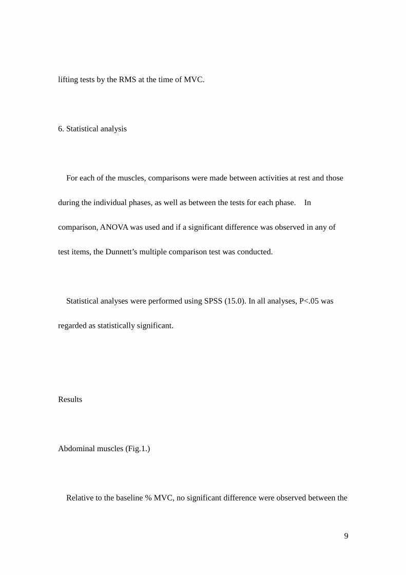
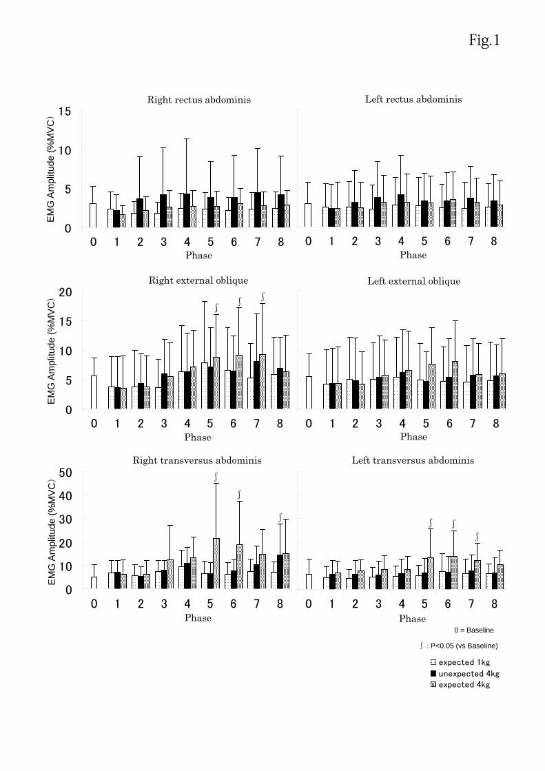
Abdominal muscles (Fig.1.)
Relative to the baseline % MVC, no significant difference were observed between the

10
muscles of rectus abdominis and external oblique. There was no significant increase in
the muscle activities of the transverses abdominis muscles did not significantly increase
during lifting of the expected 1 kg object. However, when the participant lifted the
expected 4 kg object, a significant increase in the muscle activity of right transverses
abdominis was recorded during Phases 5 and 6 (immediately after the start of the lifting
test). Increases were also observed in the muscle activity of right external oblique and
left transverses abdominis during Phases 5 through 7. When the participant lifted the
unexpected 4 kg object, a significant increase was observed only in the muscle activity
of right transverses abdominis during Phase 8.
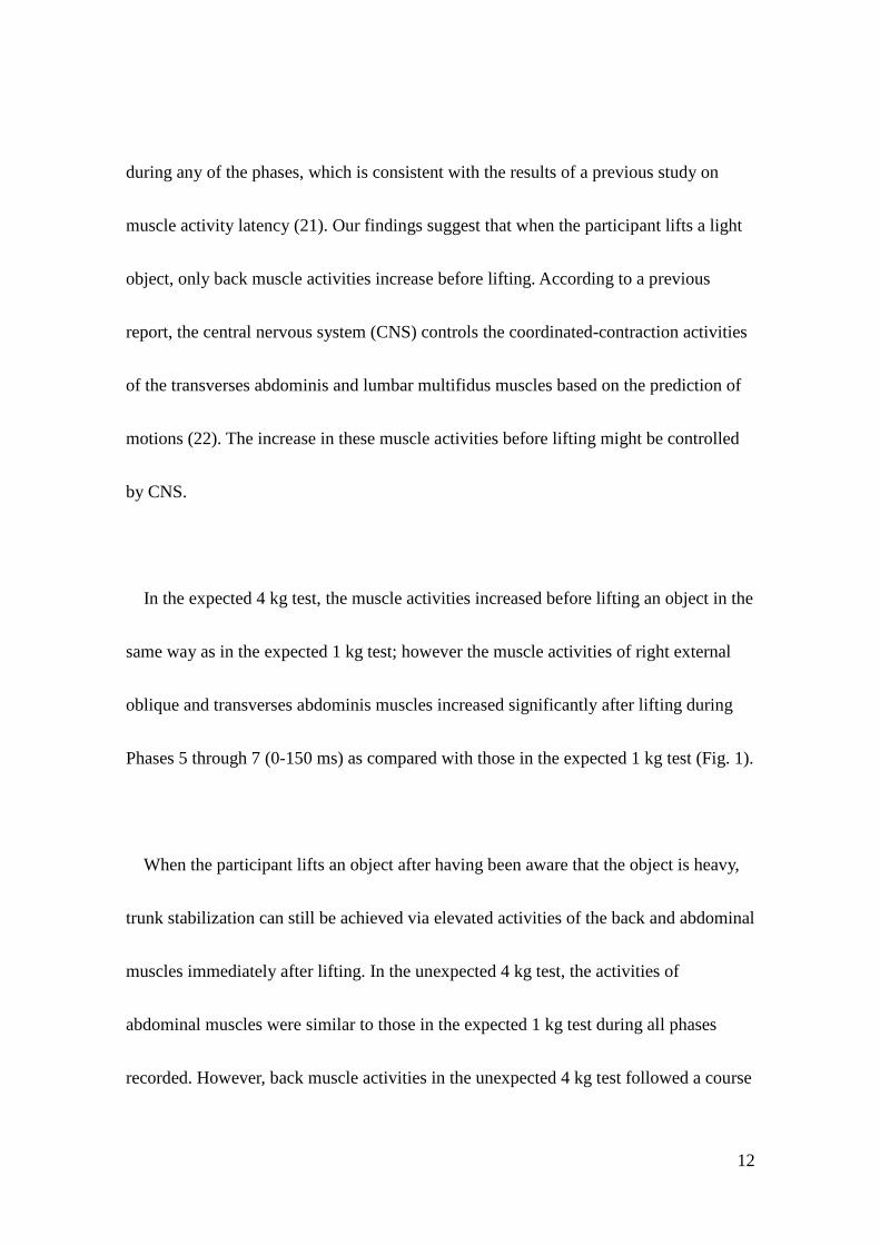
Back muscles(Fig.2.)
Relative to the baseline % MVC, both of the muscle activities of the erector spinae
and lumbar multifidus muscles significantly increased during Phases 3 and 4
(immediately before the start of the lifting test) in each of the three-different test settings.
Furthermore, these back muscle activities also increased during Phases 5 through 7
when the participant lifted the expected 4 kg object. When the participant lifted the
expected 1.0 kg or unexpected 4 kg object, no significant increase in the muscle activity

11
was recorded during Phase 5 (immediately after the start of the lifting test), but the
muscle activities of the right erector spinae and right lumbar multifidus muscles
increased during Phases 6 through 8, while those of the left erector spinae and left
lumbar multifidus muscles increased during Phases 7 and 8.
Discussion
We investigated paraspinal muscle activities during three-different lifting tests: 1) the
participants were aware of the object’s actual weight, which was 1 kg (expected 1 kg);
2) the participants believed the object’s weight was 1 kg, but it actually weighed 4 kg
(unexpected 4 kg); and 3) the participants were aware of the object’s actual 4 kg weight
(expected 4 kg).
The present study revealed that in the expected 1 kg test, the activities of back
muscles (erector spinae and lumbar multifidus) increased only during Phases 3 and 4
(-200 to 0 ms) before the start of the lifting test with no increase during the subsequent
phases. On the other hand, none of the abdominal muscles showed increased activities
12
during any of the phases, which is consistent with the results of a previous study on
muscle activity latency (21). Our findings suggest that when the participant lifts a light
object, only back muscle activities increase before lifting. According to a previous
report, the central nervous system (CNS) controls the coordinated-contraction activities
of the transverses abdominis and lumbar multifidus muscles based on the prediction of
motions (22). The increase in these muscle activities before lifting might be controlled
by CNS.
In the expected 4 kg test, the muscle activities increased before lifting an object in the
same way as in the expected 1 kg test; however the muscle activities of right external
oblique and transverses abdominis muscles increased significantly after lifting during
Phases 5 through 7 (0-150 ms) as compared with those in the expected 1 kg test (Fig. 1).
When the participant lifts an object after having been aware that the object is heavy,
trunk stabilization can still be achieved via elevated activities of the back and abdominal
muscles immediately after lifting. In the unexpected 4 kg test, the activities of
abdominal muscles were similar to those in the expected 1 kg test during all phases
recorded. However, back muscle activities in the unexpected 4 kg test followed a course
13
similar to those in the expected 1 kg test until Phase 5 (-200 to 50 ms), while the
activities of erector spinae and lumbar multifidus muscles after Phase 6 (50-100 ms)
were higher than those in the expected 1kg test. This suggests that the onsets of erector
spinae and lumbar multifidus muscle contraction were delayed in the unexpected 4kg
test as compared with those in the expected 4kg test (Fig. 2).
In general, the activities of the lumbar multifidus muscles begin with a short latency
response to stimuli and spread in a bilateral and multi-segmental manner (23-25). The
activated motor cortex may induce the postural control of the trunk muscles for stable
spine. In the present study, CNS’s failure to appropriately function in the unexpected 4
kg test might cause a delay in muscle activities of the back muscles.
It has been reported that feedforward control of the deep-seated muscles is delayed in
the patients with LBP (4) and in the patients with unstable lumbar spine, feedforward
reaction of the erector spinae muscles is delayed (26). According to these reports and
our findings, we postulate that this delay of muscle activities in the unexpected 4kg test
may be related to the development of LBP.

14
We conducted this study in a sitting posture to eliminate the influence of lower limbs
motion. Pre-training or priming was conducted on the participants in this study. Thus, it
is not completely simulate the lifting motion in the real world. These are the limitations
of this study.
Conclusion
The findings of this study suggest that when an unexpectedly heavy weight of object
is lifted, the trunk muscles may not function appropriately. We hypothesize that these
findings might be related to the LBP onset, however further investigation is needed.
Ethical approval: Waseda University Faculty of Sport Sciences (Approval No. 08-027).
Funding: This study was supported by a Grant-in-Aid for Scientific Research (B) from
the Japan Society for the Promotion of Science (grant 22300224).
Conflict of interest: None declared.

15
References
(1) Koes BW, van Tulder M, Lin CW, Macedo LG, McAuley J, Maher C. An updated overview of clinical guidelines for the management of non-specific low back pain in primary care. Eur Spine J 2010 Dec;19(12):2075-2094.
(2) Brown SH, McGill SM. The relationship between trunk muscle activation and trunk stiffness: examining a non-constant stiffness gain. Comput Methods Biomech Biomed Engin 2010 Dec;13(6):829-835.
(3) Kim K, Kim YH. Role of trunk muscles in generating follower load in the lumbar spine of neutral standing posture. J Biomech Eng 2008 Aug;130(4):041005.
(4) Hodges PW, Richardson CA. Contraction of the abdominal muscles associated with movement of the lower limb. Phys Ther 1997 Feb;77(2):132-42; discussion 142-4.
(5) Oliveira Ade S, Goncalves M. Lumbar muscles recruitment during resistance exercise for upper limbs. J Electromyogr Kinesiol 2009 Oct;19(5):737-745.
16
(6) Taechasubamorn P, Nopkesorn T, Pannarunothai S. Comparison of physical fitness between rice farmers with and without chronic low back pain: a cross-sectional study. J Med Assoc Thai 2010 Dec;93(12):1415-1421.
(7) Hansen Z, Daykin A, Lamb SE. A cognitive-behavioural programme for the management of low back pain in primary care: a description and justification of the intervention used in the Back Skills Training Trial (BeST; ISRCTN 54717854). Physiotherapy 2010 Jun;96(2):87-94.
(8) Heiss DG, Shields RK, Yack HJ. Balance loss when lifting a heavier-than-expected load: effects of lifting technique. Arch Phys Med Rehabil 2002 Jan;83(1):48-59.
(9) van der Burg JC, Kingma I, van Dieen JH. Effects of unexpected lateral mass placement on trunk loading in lifting. Spine (Phila Pa 1976) 2003 Apr 15;28(8):764-770.
(10) Butler HL, Hubley-Kozey CL, Kozey JW. Activation amplitude patterns do not change for back muscles but are altered for abdominal muscles between dominant and non-dominant hands during one-handed lifts. Eur J Appl Physiol 2009 May;106(1):95-104.
(11) McCook DT, Vicenzino B, Hodges PW. Activity of deep abdominal muscles increases during submaximal flexion and extension efforts but antagonist co-contraction remains unchanged. J Electromyogr Kinesiol 2009 Oct;19(5):754-762.
(12) Stokes IA, Henry SM, Single RM. Surface EMG electrodes do not accurately record from lumbar multifidus muscles. Clin Biomech (Bristol, Avon) 2003 Jan;18(1):9-13.
(13) Juker D, McGill S, Kropf P, Steffen T. Quantitative intramuscular myoelectric activity of lumbar portions of psoas and the abdominal wall during a wide variety of tasks. Med Sci Sports Exerc 1998 Feb;30(2):301-310.
(14) Souza GM, Baker LL, Powers CM. Electromyographic activity of selected trunk muscles during dynamic spine stabilization exercises. Arch Phys Med Rehabil 2001 Nov;82(11):1551-1557.
(15) Stevens VK, Coorevits PL, Bouche KG, Mahieu NN, Vanderstraeten GG, Danneels LA. The influence of specific training on trunk muscle recruitment patterns in healthy subjects during stabilization exercises. Man Ther 2007 Aug;12(3):271-279.
17
(16) Ng JK, Kippers V, Richardson CA. Muscle fibre orientation of abdominal muscles and suggested surface EMG electrode positions. Electromyogr Clin Neurophysiol 1998 Jan-Feb;38(1):51-58.
(17) Preuss RA, Grenier SG, McGill SM. Postural control of the lumbar spine in unstable sitting. Arch Phys Med Rehabil 2005 Dec;86(12):2309-2315.
(18) Ferreira PH, Ferreira ML, Hodges PW. Changes in recruitment of the abdominal muscles in people with low back pain: ultrasound measurement of muscle activity. Spine (Phila Pa 1976) 2004 Nov 15;29(22):2560-2566.
(19) Marshall PW, Murphy BA. Core stability exercises on and off a Swiss ball. Arch Phys Med Rehabil 2005 Feb;86(2):242-249.
(20) Moseley GL, Hodges PW, Gandevia SC. External perturbation of the trunk in standing humans differentially activates components of the medial back muscles. J Physiol 2003 Mar 1;547(Pt 2):581-587.
(21) Masahiro Watanabe, Koji kaneoka, Koichiro Oka, Shumpei Miyakawa. Trunk muscle contraction during lifting a mass with or without estimating its weight. Journal of Spine Research 2010;1(7):1283-1289.
(22) Hodges PW, Gandevia SC, Richardson CA. Contractions of specific abdominal muscles in postural tasks are affected by respiratory maneuvers. J Appl Physiol 1997 Sep;83(3):753-760.
(23) Reeves NP, Narendra KS, Cholewicki J. Spine stability: the six blind men and the elephant. Clin Biomech (Bristol, Avon) 2007 Mar;22(3):266-274.
(24) Indahl A, Kaigle A, Reikeras O, Holm S. Sacroiliac joint involvement in activation of the porcine spinal and gluteal musculature. J Spinal Disord 1999 Aug;12(4):325-330.
(25) Solomonow M, Zhou BH, Baratta RV, Lu Y, Harris M. Biomechanics of increased exposure to lumbar injury caused by cyclic loading: Part 1. Loss of reflexive muscular stabilization. Spine (Phila Pa 1976) 1999 Dec 1;24(23):2426-2434.

18
(26) Silfies SP, Mehta R, Smith SS, Karduna AR. Differences in feedforward trunk muscle activity in subgroups of patients with mechanical low back pain. Arch Phys Med Rehabil 2009 Jul;90(7):1159-1169.
∫:P<0.05 (vs Baseline)
expected 1kg
unexpected 4kgexpected 4kg
0
5
10
15
0 1 2 3 4 5 6 7 8 0 1 2 3 4 5 6 7 8Phase
Right rectus abdominis Left rectus abdominis
Phase
0 = Baseline
0
10
20
30
40
50
0 1 2 3 4 5 6 7 8
∫
∫
∫
Phase Phase 0 1 2 3 4 5 6 7 8
∫
∫
∫
Right transversus abdominis Left transversus abdominis
0
5
10
15
20
0 1 2 3 4 5 6 7 8
∫
∫
∫
Right external oblique
Phase Phase 0 1 2 3 4 5 6 7 8
Left external oblique
EM
G A
mpl
itude
(%M
VC)
E
MG
Am
plitu
de (%
MV
C)
E
MG
Am
plitu
de (%
MV
C)
Fig.1

0 1 2 3 4 5 6 7 80
10
20
30
40
50
60
0 1 2 3 4 5 6 7 8
Right erector spinae Left erector spinae
Phase Phase
∫:P<0.05 ∬:P<0.01(vs Baseline)
*:P<0.05 **:P<0.01
∫
∫
∫
∫
∫
∫
∬
∬
∬
∬
∬
∬
∬
∫
∫
∫
∬
∬
∬
*
**
** **
*
**
** **
** *
**
**
** *
*
0
10
20
30
40
50
60
70
0 1 2 3 4 5 6 7 8 0 1 2 3 4 5 6 7 8
∬
∬
∬
∬
∬
∬
∬
∬
∫
∫
∫
∫
**
** ** ** **
∫
∬
∬
∬
∬
∬
∫
∫
∫
∫
**
** *
*
** *
*
Right lumbar multifidus Left lumbar multifidus
Phase Phase
0 = Baseline
expected 1kg
unexpected 4kgexpected 4kg
EM
G A
mpl
itude
(%M
VC)
EM
G A
mpl
itude
(%M
VC)
Fig.2

1
Figure legend
Fig. 1. Abdominal muscles
In the expected 4.0 kg test, a significant elevation was observed in the muscle activities
of the right external oblique and left and right rectus abdominis muscles relative to the
baseline (at rest) immediately after lifting. In the unexpected 4.0 kg test, the muscle
activities at rest were at the same level as that obtained from the expected 1.0 kg test
with no significant difference but a significant elevation in the muscle activities of the
right rectus abdominis muscle in Phase 8.
Fig. 2. Back muscles
In all the tests, a significant elevation was observed in the muscle activities relative to
the baseline (at rest) immediately before lifting (Phases 3 and 4). In the expected 4.0
kg test, an elevation was observed in all the muscle activities immediately after lifting
(Phase 5). In the unexpected 4.0 kg test, the muscle activities were at the same level as
that obtained from the expected 1.0 kg test up to Phase 5 with no significant difference
but a significant elevation in Phase 6 and its subsequent phases.

Table 1. The five test steps (1). Recognizing a 1.0 kg
object The participant lifted an object of 1.0 kg as expected 10 times to achieve familiarization with the expected 1.0 kg weight. The repetitive trials allowed the participants to learn the most appropriate way to lift the object.
(2). Lifting the expected 1.0 kg object and measuring the muscle activities
An object identical to that used in the step (1) in external appearance and weight was placed on the table and the muscle activities were measured while the participant lifted it (one session). A sensor was placed between the table and the object to immediately detect the removal of the object from the table, generating an electromyographic signal.
(3). Lifting the unexpected 4 kg object and measuring the muscle activities
An object identical to that used in the step (1) in external appearance but different in weight (4.0 kg) was placed on the table and the muscle activities were measured while the participant lifted it. The participant had not be aware of a difference in weight between this object and that used in the step (1).
(4). Recognizing the 4.0 kg object
The participant lifted an 4.0 kg object placed on the table 10 times to achieve familiarization with the 4 kg weight as expected.
(5). Lifting the expected 4 kg object and measuring the muscle activities
An object identical to that used in the step (4) was placed on the table and the muscle activities were measured while the participant lifted it.


![Muscle Mechanics - Chris Evanschrisevans3d.com/files/reference/muscle_mechanics_sellers.pdf · lifting weights slowly. Figure 2 Muscle twitch & tetanus [Keynes & Aidley 2001]](https://img.dokumen.tips/doc/110x75/600d9fe42f01bc5b0d04c9bb/muscle-mechanics-chris-lifting-weights-slowly-figure-2-muscle-twitch-tetanus.jpg)


![Motion Capture BIOMECHANICS. T – trunk, El – elbow, SH – shoulder, Fl – flexion EX extention MUSCLE GROUP X [Nm]WojtekLuciaLi-TingSamuelAndrew FL T M](https://img.dokumen.tips/doc/110x75/56649cbf5503460f949854eb/motion-capture-biomechanics-t-trunk-el-elbow-sh-shoulder-fl.jpg)