Embed Size (px)
Citation preview

TRUNK ABNORMALITIES IN ADOLESCENCE
A school health care based epidemiological cohort study
CIP-DATA KONINKLIJKE B!BLIOTHEEK, DEN HAAG
Hazebroek-Kampschreur, A.A.J.M.
Trunk abnormalities in adolescence: a school health care based epidemiological cohort study/ A.A.J.M. Hazebroek-Kampschreur. - [SJ.:s.n.J.-llL Thesis Rotterdam. - with ref. ISBN 90-9006278-5 NUGJ742 Subject headings: scoliosis; adolescents
Correspondence: Koerillen 2 2904 VL Capelle aan den lJssel The Netherlands
Cover: Geert P. Hazebroek.
No part of this thesis may be reproduced, stored in a retrieval system, or transmitted in any form or by any means, without the permission of the author.
TRUNK ABNORMALITIES IN ADOLESCENCE
A school health care based epidemiological cohort study
Afwijkingen van de wervelkolom bij adolescenten
Een epidemiologisch cohort onderzoek vanuit de jeugdgezondheidszorg
Proefschrift
ter verkrijging van de graad van doctor
aan de Erasmus Universiteit Rotterdam
op gezag van de rector magnificus
Prof. Dr P.W.C. Akkermans M.Lit.
en volgens besluit van het College van Dekanen.
De openbare verdediging zal plaatsvinden op
woensdag 22 september 1993 om 13.45 uur.
door
Alice Augusta Johanna Maria Hazehroek-Kampschreur
geboren te 's Gravenhage

PROMOTIE COMMISSIE
Promotores:
Overige !eden:
Prof. Dr B. van Linge
Prof. Dr A Hofman
Prof. Dr E. van der Does
Prof. Dr T.W.J. Schulpen

He is a better physician that keeps diseases off us, than he that cures them being on us.
Thomas Adams, 1621-1653
Ask questions, for they are the keys that unlock the storehouse of knowledge.
Blaise Pascal, 1623-1662
Voor Frans
Eric Martijn Rutger Geert
Voor allen die ik liefheb

Acknowledgements
This thesis presents the joint work of several departments. The author gratefully acknowledge the collaboration with the department of Orthopedic Surgery, University Hospital Rotterdam Dijkzigt and Sophia Children's Hospital (B. van Linge, A. Diepstraten, A. Lengkeek, P. Tangkau), the department of Orthopedic Surgery, Hospital Eudokia Rotterdam (B. Verbiest), the department of Epidemiology & Biostatistics, Erasmus University Medical School (A. Hofman), the department of Informatics (C.J. Wulffraat, B. van de Meeberg, M. Terlouw, A. Wessels), the department of Epidemiology (H.F.L. Garretsen, J.A.M. van Oers, A.Ph. van Dijk), the department of Youth Health Care, Municipal Health Service Rotterdam (N.W. Dekema-Kiaasse, J. Schipper, S.W. Ordonez-van de Reijden, J. Belder, B. van de Kamp, J. van der Valk-de Jonge, R. Badal, R. Boer, J. Breedveld, M. van Burgh, W. Pluymers). Also acknowledged is the collaboration with the school physicians and school nurses of the department of Youth Health Care, Municipal Health Service Rotterdam (G. Asma, J. Bernsen, C. BlaauwWitteveen, R. den Boer-Reuben, L. Bosselaar, M. ten Bruggcncate-Bruyncn, X. de Bruyn-van Leeuwen, W. Cheng, A. Daniels-ten Bakke! Huinink, G. van dcr EndeKremer, V. Erjavec, E. Franken, R. Heemskerk, H.Hoogeveen-Schroot, G. Ivens, G. Iwema, M. Jacobs, F. Keiser, M. Klompenhouwer, J. Mcs-Warmolts, R. Noordermeer-Nauta, P. Nugteren-de Jager, G. van Omme, P. Osman-Handoko, G. Paul-Alderlieste, !. Pieterse-Hooft, B. Pouw-Tan, R. Reys, Y. Selier, Y. Sevenbergen, R. Slier-Schreur, A. Spaans, B. Smit-Boekelman, M. Steilen, A. Taverner-Hansen, L. Tetrode, P. Thiebaut, A. Vossenaar, S. Wiarda), and the family physicians and orthopedic surgeons in Rotterdam.
Financial support for the publication of this thesis by the Stichting Orthopedic Rotterdam and the Rotterdam Medical Research Foundation is gratefully acknowledged.

Contents
1. Introduction 11
2. Scoliosis and kyphosis: a review of the literature 15
3. School screening: a review of the literature 31
4. Prevalence of trunk abnormalities 39
5. Incidence of trunk abnormalities 57
6. Determinants of trunk abnormalities 67
7. Is scoliosis screening by nurses advisable? 79
8. Follow-up in children referred for trunk abnormalities 91
9. General discussion 105
Summary 115
Samenvatting 119
Epiloog 123
Curriculum vitae 127

Publications and manuscripts based on studies in this thesis
Chapter 4 Hazebroek-Kampschreur AAJM, Hofman A, Dijk APh van, Linge B van. Prevalence of trunk abnormalities in eleven-year-old schoolchildren in Rotterdam, The Netherlands. J Ped Orthop 1992;12:480-4.
Chapter 5 Hazebroek-Kampschreur AAJM, Hofman A, Dijk APh van, Linge B van. Two-year cumulative incidence of trunk abnormalities in a schoolpopulation m Rotterdam, The Netherlands. Submitted.
Chapter 6 Hazebroek-Kampschreur AAJM, Hofman A, Dijk APh van, Linge B van. Determinants of trunk abnormalities. Submitted.
Chapter 7 Hazebroek-Kampschreur AAJM, Hofman A, Dijk APh van, Linge B van. Is scoliosescreening door schoolverpleegkundigen zinvol? T Soc Gezondheidsz 1993;71:288-91. (in Dutch).
Chapter 8 Hazebroek-Kampschreur AAJM, Tangkau PL, Linge B van. Follow-up in children referred for trunk abnormalities. Submitted.

Chapter 1
Introduction

Chapter 1
Introduction
In 1981, the foundation of the Dutch Association of Scoliosis Patients and their Patents instigated members of the Lower House of Parliament in The Netherlands to ask questions about the role of the school physician in the early detection of scoliosis, and about the relation between early detection and onset of adequate treatment.' At the time, orthopedic surgeons in the USA and Sweden reported a 60% decrease of severe scoliosis since the introduction of a scoliosis school screening program in which children aged 10 to 15 years are annually screened for scoliosis by school nurses.2 In The Netherlands, examination for spinal deformities is pact of the periodical medical examinations by school physicians. The intervals between the scheduled periodical medical examinations may be too long, because progression of structural scoliosis is associated with growth of the spine during puberty. Therefore, annual screening for scoliosis during (pre)adolescence was recommended.'
In Rotterdam, the periodical medical examination is held four times during the school career of each child, i.e. at age 4, 7 and 11 years, and during the second year of secondary school (13-14 yeats). However, prevalence and incidence data of trunk abnormalities ace not routinely collected. A positive forward bending test is an indication for referral to the family physician for further assessment of scoliosis. Direct referral to the orthopedic surgeon by the school physician is not possible under the Dutch health care and health insurance system. Although the school physician has an important role in the early detection of spinal deformities, it is no guarantee for early, adequate treatment. A difference of opinion between family physician and school physician concerning the necessity for referral to an orthopedic surgeon may cause a delay. The children and their patents may also cause a delay in not following the advice of the physicians. Intervals between the scheduled periodical medical examinations may be too long for early detection. This is why consultation between orthopedic surgeons of the University Hospital Rotterdam and Eudokia Hospital, and representatives of the Department of Youth Health Care of the Municipal Health Service Rotterdam was initiated.
The rationale for the studies presented in this thesis was the whish to answer the question whether an extra scoliosis screening should be added to the two periodical medical examinations during adolescence. It was decided to use data collected in a prospective follow-up study conducted since 1984 to answer the following subquestions: 1. What is the prevalence of trunk abnormalities, including scoliosis and
Scheuermann's disease? 2. What is the incidence of trunk abnormalities? 3. What ace the determinants of trunk abnormalities?
11
Chapter 1
4. Are the current frequency of periodical medical examinations during adolescence, and the applied method of examining posture and back, adequate for early detection of adolescent idiopathic scoliosis?
5. Is it feasible to have scoliosis screening performed by school nurses? 6. What happens in the period between referral for scoliosis by the school
physician and the onset of observation or treatment by the orthopedic surgeon?
Chapter 2 contains a review of the literature on trunk abnormalities, focussing on scoliosis and kyphosis. Chapter 3 contains a review of the literature on scoliosis school screening programs. Chapter 4 describes the results of the baseline examination at age 11 years in school year 1984-1985 (prevalence). Chapter 5 describes the results of the reexamination at age 13 years in school year 1986-1987 (two-year cumulative incidence). In Chapter 6, determinants of trunk abnormalities are discussed. Chapter 7 describes the results of the additional screening carried out by school nurses in school year 1985-1986. Chapter 8 describes the follow-up of children referred for trunk abnormalities. This study was completed in 1990. In Chapter 9 the findings of the presented studies are discussed.
References l. Tweede Kamer der Staten-Generaal. Zitting 1981-1982. Aanbangsel van de Handelingen (309). 1981. 2 Torell G. Nordwall A. Nachcmson A The changing pattern of scoliosis treatment due to effective
screening. J Bone Joint Surg [Am] 1981;63:337-41. 3. Steenaert BAJM. De Haas-Michgelsen M. Perre-Bulder AJM. Ponsiocn AMAJ. Schoolsa'eening op
scoliose. T Jeugdgezondheidsz 1981;13:82-8.
12

Chapter 2
Scoliosis and kyphosis:
a review of the literature


Chapter 2
Scoliosis and kyphosis: a review of the literature
Introduction
The normal spine is composed of vertebrae, intervertebral disks, and their related ligaments and muscles. The stability of the vertebral column is provided by its intrinsic structures, and by extrinsic support of the rib cage and the muscles of the trunk. The normal movement of the spine is flexion, extension, lateral flexion and rotation in the cervical region, rotation in the thoracic region, and flexion-extension and lateral flexion in the lumbar region. The normal spine contour, when viewed in the frontal plane, is straight, and when viewed in the lateral plane has physiological cervical and lumbar lordosis, and thoracic kyphosis.
There are three basic types of spinal deformity: scoliosis, kyphosis and lordosis. Each may occur singly or in combination. Scoliosis is the commonest type of deformity, it is an unphysiological curving laterally from the midline. Pure lordosis is extremely rare. In 1950, Ponseti and Friedman presented a classification of idiopathic scoliosis based on the site and number of primary curves.1 Table 2.1 shows the classification of spinal deformity based on the associated conditions as compiled by the terminology committee of the Scoliosis Research Society.>
Table 2.1. Classification of spinal deformities
Primary, progressive or structural deformities 1 Idiopathic deformities
- idiopathic scoliosis early-onset
- idiopathic kyphosis
2 Congenital deformities
late-onset Type I ctassic Scheuermann's disease Type ll 'apprentice's spine'
3 Neuromuscular deformities: poliomyelitis, cerebral palsy 4 Deformities in association with neurofibromatosis 5 Mesenchymal deformities 6 Traumatic deformities 7 Deformities due to infection 8 Deformities due to tumors 9 Miscellaneous conditions 10 Spondylolisthesis
II Secondary, non-progressive or non-structural deformities 1 Postural scoliosis, kyphosis, lordosis 2 Pelvic tilt scoliosis, due to leg-lenght inequality or/and pelvic asymmetry 3 Irritative lesions associated with the spine 4 Hysterical scoliosis
15

Cho.pter 2
Poliomyelitis and rachitis used to be the major causes of structural scoliosis and kyphosis. In some less developed countries, paralytic scoliosis as a result of poliomyelitis still is the commonest type.' In Western countries, 80% of the structural scoliosis is idiopathic, and 10-15% is congenital.
Orthotic and surgical treatment became more effective since the 1950s.' Since the foundation of the Scoliosis Research Society in 1966, the knowledge about the epidemiology, etiology, natural history, diagnosis and treatment of scoliosis has increased. Data from scoliosis school screening programs and long-term follow-up studies of idiopathic scoliosis have added new insight to the natural history of the condition.5-
13
Scoliosis
Scoliosis is derived from the Greek word meaning curvature. When used in medical literature, it signifies a lateral curvature of the spine. A normal spine has physiological curvatures when viewed from the side, but there is no lateral deviation when viewed anteriorly or posteriorly. These lateral curvatures must be defmed as non-structural or functional scoliosis and structural scoliosis. Nonstructural or functional scoliosis is a lateral curvature which is totally correctable by bending forward. Clinically and radiologically it does not show rotation, and it is without structural changes. It is frequently due to leg-length inequality, or to irritative phenomena associated with the spine. Structural scoliosis is a lateral curvature identified clinically when the child on bending forward shows fixed posterior vertebral rotation including rib rotation on the convexity of the primary curve. Radiography reveals rotation, and structural changes of wedging and obliquity in the vertebrae; the size of the curve is measured using the Cobb method. Rib rotation posteriorly without scoliosis on radiography can occasionally occur. Bunnell11 stated that spinal curvatures less than 10 degrees may be diagnosed as postural scoliosis, and curves of 10 degrees and more with rotation and wedging as structural.
Idiopathic scoliosis is a structural lateral curvature of the spine with vertebral rotation and wedging, the underlying cause of which is unknown. It is diagnosed "per exclusionem". Idiopathic scoliosis occurs in the thoracic and lumbar vertebrae, its onset may be seen between birth and the end of skeletal growth. James' classified three types of idiopathic scoliosis according to the age of onset (Table 2.2).
Table 22. Idiopathic scoliosis according to age of onset
infantile scoliosis
juvenile scoliosis
adolescent scoliosis
16
between birth and 4 years of age
between 4 years and 10 years of age (onset of puberty)
from 10 years of age to the end of skeletal growth

Scoliosis and kyphosis
Many hypotheses about the etiology of idiopathic scoliosis have been proposed, such as abnormal vertebral growth, spinal muscle imbalance, postural alterations, endocrine and metabolic changes, and genetic factors.
One· interesting theory is the concept of biplanar asymmetry.'4•15 Scoliosis is a
three-dimensional deformity; thoracic idiopathic scoliosis is usually a combination of loss of physiologic kyphosis, convex vertebral body rotation, and lateral bending." Somerville17 and Roaf18 have stated that hypokyphosis (lordosis) and rotation are the primary lesions of idiopathic scoliosis, with coronal plane deformity occurring only secondarily. This lordosis was thought to arise from failure of growth of the posterior elements of a segment of the spine. According to Dickson14
"15
, spinal asymmetry in both coronal and median planes is essential for the development of a progressive curve. Biomechanically, it can be shown that only when median plane asymmetry exists the spine does have any tendency to rotate. Spinal curvatures in the median plane change during growth and in normal children thoracic kyphosis reduces in size from age 8 to 14 years. When it is at its minimum, girls are going through adolescent peak growth velocity, and this may explain greater progression potential in girls. In boys, adolescent peak growth velocity is approximately two years later when their thoracic kyphosis becomes maximal, which may explain why boys are particularly prone to developing Scheuermann's disease. In idiopathic scoliosis there is an increased anterior vertebral height at the apex of the curve with posterior end-plate irregularity. This is analogous but opposite to the anterior wedging observed in Scheuermann's disease. However, the underlying disturbances of growth could not be demonstrated experimentally, and the exact pathogenetic mechanics of idiopathic scoliosis (and idiopathic kyphosis) remain controversial.
Frequency
The prevalence of scoliosis in the adult population is mainly based on reports of mass-population roentgenographic screenings for tuberculosis, and ranges from 1% to 2%. These reports include no data about lumbar curves19;w
Data obtained through scoliosis school screening programs showed large differences in prevalence, depending on the method used for diagnosis and the minimum deformity required to determine a positive screening test. Prevalence figures range from 2% to 16%. Table 2.3 gives an overview of prevalence studies of scoliosis."'7•
21-53 The minimum curvature required to make a diagnosis
of adolescent idiopathic scoliosis is 10" as measured by the Cobb method. At the age of 16 years, approximately the end of growth in girls, the prevalence of idiopathic scoliosis of more than 10" is approximately 2% to 3%. In larger curves, the prevalence decreases. Approximately 100 per 1,000 adolescents have minor degrees of curvature, only 2 per 1,000 warrant treatment because of curve progression.11,54,ss Because of misuse of epidemiological terms with regard to incidence versus prevalence, school screening surveys on incidence of scoliosis21=-" are actually prevalence studies. Incidence data are lacking.
17

Table 2.3, Scoliosis pre~·alence rtudlu
Name Place Numlx'r Age or grade Screening method Number lo second Numbu rererred or all Scoliosis >10~ screened scrtener children prel·alenre
SegiJ11 1974 fobannesburg, S. Africa 1,016 Nrkans N.S. N.S. N.S, N,S, 1.6% 919 Caucasians
Drooks7 1975 California~ USA 3,492 gr. 7 & 8 N.S. N.S. 13.6% (>5°) N.S.
Span"" 1976 Jerusalem, Israel IO,OJO 10- 16 N.S. 13.0% X-ray 3.0% 1.5%
Dr.:nnanu 1977 Colorado, USA 58,314 10- 13 nurses 5.2% 1.9% o.2% (>15.)
LonslelnM 1971 Minnesota, USA 571,722 gr.5-10 nurses 8.3% 4.0% 1.1%
Flynnv; 1971 Florida, USA 38,710 gr. 7- 9 physical cduc.ation 10.6% 1.8% N.S. teachers (PET)
DunnZ6 1978 Virginia, USA 5,000 gr. 6- 8 nurses and PET 13.1% 3.3% 1.2%
HowdJl1 1978 Edmonton, Canada 609 (girls only) 11 - 17 nurses and 45.0% 14.0% 6.4% physiotherapists
Rogala» 1978 Montreal, Canada '26,947 12- 14 nurses or pb)·sldan 10.0% 6.5% 2.2%
Smyrnls29 1979 Athens, Greece 3,494 11 - 12 doctors 10.0% 6.4% 4.6% girls 1.1% boys
Dlckson30 1900 Oxford, Great-Bri!aln 1,764 13 - 14 physiotherapist 8.3% 6.9% 2.5%
Gohlbug11 1900 Dublin, Irdand 604 10- 14 doctors 21.9% 6.4%
Ascanl12 1900 Rome, l!aly 16,104 6- 13 N.S. 7.2% N.S. 4.6% 2,302 6 21.6% 5.9% 2,102 II 19.5%
Owenn 1900 Uverpool, Great-Britain 15,();)(1 age 5, age 9, age 11 nurses and S<:hool 3/2% 1.0% N.S. medical offker
O'Brien" 1900 Om·estry, Great-Britain 903 11 - 14 health \'isitors N.S. 3.3% 2.0%
TayJorlS 1900 Perth, West-Australia !,200 11 - 14 N.S. N.S. 5.6 - 13.9% 1.6-6.4%
GoreJ6 1981 Wisconsin, USA 8,393 gr.5-10 \'Oiun!eers (1" stage), 34.5% 7.5% 2.0% physlo!herapists and nurses (2M stage)
Lonsldn11 1982 Minneso!a, USA 250,000 10- 14 PET W stage). N.S. 3.4% 1.2% nurses (2M stage)
\\'lllnc~ 1982 Malm6, Swtden 17,1!il 7- 16 nurses and S<:hoo!doctors N.S. 4.3% Rirls 3.2% Rlrls

LaUIUM'' l'JIS"l venmarK 11U.H l_gUIS Vlll)"J lV- 1/ <'1..,\NIUV\.-\VU ~
mo!r6 topography
GoldbergH 1983 Dublin, Ireland 21,000 10 - 14 PET, schoolnurses, 10.0% 1.4% 0.2% schooldoctors
Randalr'l 1983 Alabama, USA 561 5-8 N.S. 19.8% 11.2% 4.0%
Wynne 01 1984 Vancouver, Canada 8,010 12- 14 nurses, physiotherapists N.S. 2.1% 0,5%
Adler44 1984 Callfomla, USA 237 (girls only) gr. 5-8 fOJWard bending t 21.4% N.S. N.S. moir6 lopography
U High Pin41 1985 Hunan, People's R~publ!c 8,615 6- 15 doctors; 9.2% 8.0% 2.4% of China X-ray in 2~ slage
Flynn« 1985 Florida, USA 496,965 gr. 6-9 PET, nurses N.S. 5-8% N.S.
TilOmpson" 1985 Dublin, Ireland 182 (girls only) 12 - 16 forward bending + 20.9% 14,3% (>5") 6.0% molrt topography
aum« 1986 Adelaide, South Austral!a 3,660 14 - 16 nurses N.S. 3,9% 21%
Sa\·lnl~ 9 1986 Bologna, Italy 12,832 11 - 14 doctors 10.1% 7.1% 1.4% (>15")
Drembergso 1986 S.veden 7,531 gr. 1, 4, 8 school doctors N.S. N.S 2.7% 10,644 gr. 1 - 8 scboolnurses N.S. N.S. 2.3%
Mltlnl1 1987 Patalla, India 25,376 5 - 18 dinkal eJJ:aminatlon + N.S. N.S. 0.13% idiopathic: sro!lometer 1 in 5
Frands11 1988 Utah, USA 3,210 (girls only) 17 - 21 12.3% N.S. 5.4% (>20")
Ohlrukrls:t 1988 Oliba, Japan 1,245,798 gr. 5 & 6 1) clinical examination N.S. N.S. O.Q7- 1.77 (>15") + moin! topography
gr.l&II 2) low dose radiography 3) X-ray
Nlsslnen11 1989 H~lslnkl, Finland 1,060 9- 13 cl!n!cal eum/nalion + N.S. N.S 4.1% molr6 topography
N.S. = not stated PET= physical education teacher

Chapter 2
Risk factors
The etiology of adolescent idiopathic scoliosis is unknown, but there is a relationship between gender, growth, maturation, and the occurrence of scoliosis.'1s6 A familial occurrence of idiopathic scoliosis has been reported suggesting a genetic basis.58
Prognosis
The main area of concern in the skeletally immature patient is the probability of curve progression. Information about progression is derived from studies of follow-up of untreated patients. Progression is defined as an increase of 5° for curves of 20° or more, and as an increase of 10° or more for curves less than 20°.10= 6 Lonstein and Carlson10 reported progression in 23% of 727 scoliotic patients with curves between 5° and 29°. In scoliotic children detected in a school screening program, spontaneous improvement occurred in 3%-22%, and curve progression occurred in 7%-15%Y'
Certain factors have been found to be related to curve progression: gender, age and maturity, and magnitude and pattern of the curve9 -nss The risk of curve progression in girls is tenfold the risk in boys. Age is another risk indicator. The younger the patient at time of diagnosis, the greater the risk of progression. A critical period for rapid progression is during rapid growth at puberty. Physiologic age as determined by skeletal maturity (Risser sign or bone age) and onset of menarche in girls are better indicators for the risk of progression than the chronologie age.56 Curve magnitude and curve pattern are also related to curve progression; the larger the magnitude at time of detection the higher the risk. Thoracic and double-major curves have the highest risk of progression, thoracolumbar curves have an intermediate level of risk, and lumbar curves have the lowest risk. Lonstein and Carlson10 used the factors age, curve magnitude, and maturity as measured by the Risser sign to develop a progression factor formula for curves between 20° and 29° in individual cases. Risks of curve progression decrease with increasing skeletal maturity. After skeletal maturity, curves less than 30° do not progress. Slight progression of larger curves may occur.9
In adults, scoliosis is a disabling disease and affects the quality of life. Severe scoliosis causes back pain, cardiorespiratory disturbances, psychological and socioeconomic problems, and may lead to death. Long-term follow-up studies of non-treated scoliotic patients, forty to fifty years after diagnosis, showed that the average mortality of scoliotic patients was twice that in the general population.5·'S' Cardiac and pulmonary diseases were the causes of death in 60% of the patients who died. Backache in adult life was a common complaint. Many patients were unable to work. Women, in particular, were less likely to marry.'·6S' Bengtsson et al.60 found in 26 adult women with severe scoliosis that their superficial psychosocial adjustment was good. However, the results of the personality-psychological examination indicated that their adjustment was less
20

Scoliosis and kyphosis
well. Their lives had become marked by their deformity. They were characterised by hypersensitivity and insecurity; the psychological adjustment deteriorated with increasing degree of deformity.
In early adolescence, deformities of the trunk may cause psychological problems too. Scoliosis and its treatment often collide with the most sensitive years of social development. Clayson" found that for adolescent girls, scoliosis had its greatest impact on their sense of worth to others, and for boys, scoliosis presented the greatest threat to their sense of self-worth. Kahanovitz and Weiser"' compared in 72 adolescent girls, aged 12 to 16 years, the psychological aspects of the various treatments for scoliosis: observation for mild curves, brace for moderate cnrves, and surgery for severe cnrves. In addition, the attitudes of the patients' mothers toward scoliosis were assessed. It was found that type of treatment was not related to the girl's adjustment to the condition. However, mothers' and children's attitudes toward scoliosis were strongly related to the children's adjustment to their condition.
Treatment
When confronted with children referred with trunk abnormalities, the decision facing the clinician is discharge of those children whose scoliosis will never be a health problem, observation of those with small curves at risk of progression, or treatment of those with significant curves by bracing or surgery.
Observation is important in the management of any patient. The primary goal is to determine whether the patient will need active treatment. At the initial evaluation, cnrves between 10° and 25° to 30° in juvenile and skeletally immature adolescent children should be observed. The risk of progression is 20% in a skeletally immature child with a 20° curve, and 60% in a similar patient with a 30° curve. Skeletally mature children with curves under 30° do not need observation. Contra-indications for observation are progressive cnrves, and cnrves greater than 30° in skeletally immature patients on their first visit. Restriction of physical activity is not necessary!·"
A progressive curvature in a skeletally immature patient is an indication for treatment by bracing, if the magnitude of the curve is between 25° and 40°. Prerequisites for a successful brace treatment are the cooperativeness and willingness of the patient and the family to continue a treatment program until the end of skeletal growth. The original brace system is the Milwaukee brace which is a cervical-thoracic-lumbar-sacral orthosis. Of the underarm braces (thoraco-lumbo-sacral orthoses), the Boston brace is the most widely used.
Indications for surgical treatment are severe curvatures of 40° and more in skeletally immature patients, and of 50° and more in skeletally mature patients, to prevent respiratory insufficiency. An unacceptable cosmetic appearance, failure of brace treatment, and a progressive thoracic curvature associated with increased thoracic lordosis are other indications for surgery.
21
Chapter 2
Kyphosis
Thoracic kyphosis is a physiologic posterior convex angulation of the spine in the sagittal plane. Normally, this kyphosis measures between 20" and 40" by the Cobb method.64 Kyphosis of more than 40", and kyphosis less than 20" in the thoracic spine are considered abnormal. White et a!. 65 suggest that any significant amount of posterior curvature in the adult cervical and lumbar spine should be considered abnormal.
Juvenile kyphosis remains one of the most frequently neglected trunk deformities during childhood and adolescence. The minimal roundback deformity of the spine is often regarded as a problem of poor posture. Postural kyphosis is especially common in adolescent girls because breast development sometimes makes them extremely self-conscious. They assume a round-shouldered slouch in order to hide their breasts, especially if they are tall for their age. The roundback deformity may prove to be a manifestation of structural deformities of the spine.
Holger Scheuermann, in 1921, published a report describing a category of juvenile patients, almost exclusively male, with increased thoracic kyphosis. Radiography revealed vertebral wedging and changes similar to osteochondritis deformans juvenilis coxae. Scheuermann labelled the kyphosis dorsalis juvenilis as ostechondritis deformans juvenilis dorsi, now known as Scheuermann's disease." Scheuermann's juvenile kyphosis is defined as a fixed round back deformity in the growing spine associated with wedging of at least three adjacent vertebrae at the apex of the curve of 5" or more with specific x-ray changes. The major radiologic criteria for the diagnosis of the classic form of Scheuermann's disease are: (1) irregular vertebral end plates, (2) narrowing of the intervertebral disk space, (3) one or more vertebrae wedged 5" or more, ( 4) an increase in normal kyphosis beyond 40".67 The etiology of Scheuermann's disease is unknown; mechanical factors, heavy physical work, genetic or environmental abnormalities, muscle weakness and inflammation have been implicated in the etiology. Digiovanni et al.68 found a distinct anterior elongation of the vertebral body in skeletons with Scheuermann's kyphosis. It appeared to be the result of an alteration of normal growth in the immature spine.
The typical patient is between 13 and 17 years old and complains of poor posture, fatigue, stiffening and/or pain near the kyphos. The kyphosis is thoracic in about 75% of the patients (Type I Scheuermann's disease), and thoracolumbar in the other 25% (Type H Scheuermann's disease). According to Leatherman and Dickson2
, Type I Scheuermann's disease appears to be precisely the opposite deformity to idiopathic scoliosis, in being another example of median plane spinal asymmetry with anterior vertebral wedging and end-plate irregularity. In idiopathic scoliosis, there is median plane spinal asymmetry with posterior vertebral wedging. Lumbar lordosis is increased. Mild, generally non-progressive scoliosis is assumed to be present in 30% to 40% of patients with Scheuermann's disease. Deacon et al.69 demonstrated in 50 cases of thoracic Scheuermann's disease that a lateral curvature of the spine was present in 85%. It is still debated whether Type I Scheuermann's disease produces back pain.
22
Scoliosis and kyphosis
Bradford et al.70, and Stoddard and Osbom71 consider that it does. In Type ll
Scheuermann's disease, pain is a common complaint, often associated with increased physical activity. It occurs almost solely in boys. There is an increased prevalence of spondylolysis and spondylolisthesis. In adults with low back pain, low Scheuermann's disease is often overlooked by both clinicians and radiologists.72
An atypical form of Scheuermann's disease may present itself in two fashions: vertebral body changes without wedging or increased kyphosis, or increased kyphosis without vertebral body changes. In the second case, we see a clinical appearance of classic Scheuermann's disease (a stmctural kyphosis) in a teenager without radiographic changes of endplate irregularity or vertebral wedging.73
A kyphoscoliosis is a combination of a tme kyphosis and a lateral curve. This is very rare, and nearly always due to a congenital anomaly74
, except in Scheuermann's disease where the scoliosis is usually mild and nonprogressive. Scoliosis can clinically simulate a kyphoscoliosis, because the vertebral rotation carries the nb backwards with it on the convex side to produce a hump. The term kyphoscoliosis is a misnomer; it would be better to describe this condition as kyphosing scoliosis.75
Frequency
Prevalences ranging from 0.4% to 8.3% have been reported, depending on whether the diagnosis is based on clinical (8.3%) or radiographic criteria (0.4%).76 From school screening programs, prevalences ranging from 0.1% to 1% have been reported.58
•77 The male to female ratios reported range from 1:1 to
1:2!7•76 The age of onset is difficult to establish because radiographic changes
typical of Scheuermann's disease are generally not seen before the age of 11 years. Kyphosis tends to appear at a later age than scoliosis, and it progresses later as well. Incidence has not been studied.
Risk factors
The etiology of Scheuermann's disease is unknown. Mechanical factors have been implicated in the development of kyphosis66 A familial occurrence has been described.'6 Patients with Scheuermann's disease were taller than average, and the degree of skeletal maturity was advanced beyond the chronological age78
Prognosis
Prognosis and clinical course of Scheuermann's disease have not been systematically investigated, as has been done in adolescent idiopathic scoliosis. Complications of Scheuermann's disease are cosmetic deformity, back pain, and neurological complications. Cosmetically unacceptable appearance occurs when the kyphosis is above 65° or 70°, because of increased compensatory lumbar and
23

Chapter 2
cervical lordosis. Back pain in the untreated adult is a common complaint, especially in Type II Scheuermann's disease76
•79
, but probably no more frequent than in the normal population." Neurological complications occur, but are exceedingly rare.2
Treatment
Treatment depends on the severity of the problem and the radiographic changes in the vertebrae. In pre-adolescent children, exercises alone are usually adequate for managing postural roundback or postural increased lordosis. In Scheuermann's disease, (Milwaukee) brace treatment is the most effective therapy. In adult life, surgical treatment is rarely indicated for severe Scheuermann's disease. Other types of kyphosis, such as the congenital kyphosis, are treated operatively73
$1- 83
References 1. James JIP. Idiopathic scoliosis. J Bone Joint Surg [Dr] 1954;36:36-49. 2. Leatherman KD, Dickson RA. The management of spiool defonnitics. London: Wright,
1988:10-23. 3. Minal RL. Aggerwal R. Sarwal AK. School screening for scoliosis in India. The evaluation of a
scoliometcr. Int Orthop 1987;11:335-8. 4. Bunnell \VP. Treatment of idiopathic scoliosis. Orthop Clin N Amer 1979;10:813-27. 5. Nachemson A A long term follow-up study of non-treated scoliosis. Acta Orthop Scand 1968;
39,466-76. 6. Nilsonne U, Lundgren K-D. Long-term prognosis in idiopathic scoliosis. Acta Orthop Scand
1968;39,456-65. 7. Brooks HL, Azen SP, Gcrbcrg E, Brooks R, Chan, L Scoliosis: a prospective epidemiological
study. J Bone Joint Surg [Am] 1975;57:968-72. 8. Leaver JM, Alvik A, Warren MD. Prescriptive screening for adolescent idiopathic scoliosis: a
review of the evidence. Int J Epidemiol 1982;11:101-11. 9. Weinstein SL,. Ponseti N. Curve progression in idiopathic scoliosis. J Bone Joint Surg [Am]
1983;65,447-55. 10. Lonstein JE,. Carlson JM. The prediction of curve progression in untreated idiopathic scoliosis
during growth. J Bone Joint Surg (Am] 1984;66:1061-71. 11. Bunnell \VP. The natural history of idiopathic scoliosis. Cin Orthop 1988;229:20-5. 12. Rinsky I.A,. Gamble JG. Adolescent idiopathic scoliosis. West J Mcd 1988;148:182-91. 13. Kehl DK, Monissy RT. Brace treatment in adolescent idiopathic scoliosis. Oin Orthop 1988;
229:34-43. 14. Dickson RA., Lawton JO, Archer IA, Butt WP. The pathogenesis of idiopathic scoliosis. J Bone
Joint Surg [Br]1984;66,8-15. 15. Dickson RA, Lawton JO, Archer IA, Butt \VP, Bcrkin CR, Bliss P, Somerville EW, Jobbins B,
Dawson D. Bi-planar spinal asymmetry: the pathogenesis of idiopathic scoliosis. J Bone Joint Surg [Br]1984;66,143-144.
16. Ogilvie JW. Biomechanics. In: Bradford DS, Lonstein JE, Moe JH, Ogilvie JW, Winter RB, eds. Moe's Textbook of scoliosis and other spinal deformities. Philadelphia: \VB Saunders, 1987:7-23.
17. Somerville EW. Rotational lordosis: the development of the single curve. J Bone Joint Surg [Br] 1952;34:421-7.
18. Roaf R. The basic anatomy of scoliosis. J Bone Joint Surg [Br] 1966;48:786-92. 19. Shands AR, Eisbcrg HB. The incidence of scoliosis in the state of DelawOlie. J Bone Joint Surg
[Br] 1955;37,1243-9.
24
Scoliosis and kyphosis
20. Bellyei A. Czeizel A. Barta 0, Magda T, Molnar. Prevalence of adolescent idiopathic scoliosis in Hungary. Acta Orthop Scand 1977;48:177-80.
21. Segil CM. The incidence of idiopathic scoliosis in the Bantu and white population groups in Johannesburg. J Bone Joint Surg [Br}1974;56:393.
22. Span Y, Robin G, Makin M. Incidence of scoliosis in school children in Jerusalem. J Bone Joint Surg [Br] 1976;58:379.
23. Drennan JC. Campbell JB, Ridge H. Denver: a metropolitan public school scoliosis survey. Pediatrics 1977;60:193-6.
24. Lonstein JE. Screening for spinal deformities in Minnesota schools. Clin Orthop 1977;126: 33-42.
25. Flynn JC, Riddick MF, Keller TL. Screening for scoliosis in Florida schools. J Florida M A 1977;64:159-61.
26. Dunn BH,. Hakala MW, McGee :ME. Scoliosis screening. Pediatrics 1978;61:794-6. 27. Howell JM, Craig PM, Dawe BG. Problems in scoliosis screening. Can J Public Health 1978;
69:293-6. 28. Rogala EJ, Drummond DS, Gurr J. Scoliosis: incidence and natural history. J Bone Joint Surg
[Am] 1978;60:173-6. 29. Smymis PN, Valavanis J, Alexopoulos A. Siderakis G, Giannestras NJ. School screening for
scoliosis in Athens. J Bone Joint Surg [Br] 1979;61:215-7. 30. Dickson RA, Stamper P, Sharp A. Harker P. School screening for scoliosis: cohort study of
clinical course. Br Med J 1980;281:265-7. 31. Goldberg C. Thompson F. Dowling F, Regan BF, Blake NS. Pilot study for a scoliosis screening
project in South Dublin. Ir Med J 1980;73:265-8. 32. Ascani E, Giglio GC, Salsano V. Scoliosis screening in Rome. In: Zorab PA, Siegler D, eds.
Scoliosis 1979. London: Academic Press, 1980:39-44. 33. Owen R. Taylor JF, McKendrick 0, Dangerfield P. Current incidence of scoliosis in school
children i•1 the city of Liverpool. In: Zorah PA,. Siegler D, eds. Scoliosis 1979. London: Academic Press, 1980:31-4.
34. O'Brien JP. The incidence of scoliosis in Oswestry. In: Zorah PA, Siegler D. eels. Scoliosis 1979. London: Academic Press,. 1980:19-29.
35. Taylor JR.,. Slinger BS. Scoliosis screening and growth in Western Australian students. Med J Aust 1980;1:475-8.
36. Gore DR, Passehl R. Sepic S, Dalton A Scoliosis screening: results of a community project. Pediatrics 1981;67:196-200.
37. Lonstcin JE,. Bjorklund S. Wanninger MH, Nelson RP. Voluntary school screening for scoliosis in Minnesota. J Bone Joint Surg [Am] 1982;64:481-8.
38. Willner S, Uden A A prospective prevalence study of scoliosis in Southcm Sweden. Acta Orlhop Scand 1982;53:233-7.
39. Miller D, Lever CS. Scoliosis screening: an approach used in the school. J Sch Health 1982; 98-101.
40. l.aulund T. Sojbjerg JO, Horlyck E. Moire topography in school screening for structural scoliosis. Acta Orthop Scand 1982;53:765-8.
41. Goldberg C. Fogarty EE~ Blake NS, Dowling F, Regan BF. School scoliosis screening: a review of 21,000 children. Ir Med J 1983;76:247-9.
42. Randall FM, Denton 1E. Scoliosis screening: a school survey. Alabama J Med Sci 1983;20: 395-6.
43. Wynne EJ. Scoliosis: To screen or not to screen. Can J Public Health 1984;75:277-80. 44. Adler NS, Csongradi J, Bleck EE. School screening for scoliosis. One experience in California
using clinical examination and moin~ topography. West J Med 1984;141:631-3. 45. Li High Pin, Liu Yong Mo, Li Lin, Li Kan Hua, Hei Pei Hui, Den Shi Hui. Bao Dong Chang,
Yuin Yuin Chang. Early diagnosis of scoliosis based on school-screening. J Bone Joint Surg [Am] 1985;67:1202-5.
46. Flynn JC. Riddick :MF'~ Price cr. Keller TL. Present status of scoliosis screening in Florida schools. J Florida M A 1985;72:847-51.
47. Thompson F, Walsh M, Colville J. Moire topography: a method of screening for adolescent idiopathic scoliosis. Ir Med J 1985;78:162-5.
25

Chapter 2
48. Chan A. Moller J, Vimpani G. Patterson D. Southwood R.,. Sutherland, A The case for scoliosis screening in Australian adolescents. Med J Aust 1986;145:379-83.
49. Savini R.,. Cervellati S, Ponzo I.., Ciani A, Palmisani M, Rizqallah Y, Gargiulo G, Nardi S, Di Silvestre M. L'incidenza della scoliosi idiopatica nel Comune di Bologna. Chir Org Mov 1986; 81:119-25.
50. Bremberg S. Nilsson-Berggren B. School screening for adolescent idiopathic scoliosis. J Pediatr Orthop 1986;6:564-7.
51. Francis RS. Scoliosis screening of 3,000 college-aged women. The Utah study - phase 2. Phys Thor 1988;68:1513-6.
52. Ohtsuka Y, Yamagata M, Arai S, Kitahara H, Minami S. School screening for scoliosis by the Ch.iba university medical school screening program. Results of 1.24 million students over an 8-year period. Spine 1988;13:1251-7.
53. Nissinen M, Heliovaara M,. Tallroth K. Poussa M. Trunk asymmetry and scoliosis. Anthropometric measurements in prepuberal school children. Acta Paediatr Scand 1989;78:747-53.
54. Kane WJ. Scoliosis prevalence: a call for a statement of terms. Qin Orthop 1977;126:43-6. 55. Weinstein SL Adolescent idiopathic scoliosis: prevalence and natural history. Instr Course Lect
1989;38:115-28. 56. Duval-BeaupCre G. Pathogenic relationship between scoliosis and growth. In: Zorah PA,. ed.
Scoliosis and Growth. Edinburgh: Churchill Livingstone, 1971:58-64. 57. Wynne-Davies R. Familial (idiopathic) scoliosis: a family survey. J Bone Joint Surg [Br] 1968;
50:24-30. SS. Drummond DS, Rogala E, Gurr J. Spinal dcfonnity: natural history and the role of school
screening. Orthop Clio N Am 1979;10:751-9. 59. Weinstein SL. Zavala DC. Ponseti IV. Idiopathic scoliosis. Long-tcnn follow-up and prognosis
in untreated patients. J Bone Joint Surg [Am] 1981;63:702-12. 60. Bcngtsson G, Fallstrom K. Jansson B, Nachcmson A A psychological and psychiatric
investigation of the adjustment of female scoliosis patients. Acta Psychiat Scand 1974;50:50-9. 61. Clayson D. Psychological problems of scoliosis in early adolescence. In: Zorab PA,. Siegler D,
eds. Scoliosis 1979. London: Academic Press,. 1980:227-231. 62. Kahanovitz N, Weiser S. The psychological impact of idiopathic scoliosis on the adolescent
female. A preliminary multi-center study. Spine 1989;14:483-5. 63. Tolo vr. Treatment, follow-up. or discharge. Spine 1988;13:1189-90. 64. Bradford DS. Kyphosis. Qin Orthop 1977;128:2-4. 65. White AA,. Panjabi MM. Thomas CL The clinical biomechanics of kyphotic dcfonnitics. Clin
Orthop 1977;128:8-17. 66. Scheuermann HW. Kyphosis dorsalis juvenilis. Clin Orthop 1977;128:5-7. 67. Bradford DS. Juvenile k-yphosis. Clin Orthop 1977;128:45-55. 68. Digiovanni BF. Scoles PV, Latimer BM. Anterior extension of the thoracic vertebral bodies in
Scheuermann's kyphosis. An anatomic study. Spine 1989;14:712-6. 69. Deacon P, Berkin CR. Dickson RA Combined idiopathic kyphosis and scoliosis. An analysis of
the lateral spinal curvatures associated with Scheuermann's disease. J Bone Joint S [Br] 1985; 67:189-92.
70. Bradford DS. Moe JH,. Montalvo FJ, Winter, RB. Scheuermann's kyphosis and roundback deformity. J Bone Joint Surg [Am} 1974;56:740-58.
71. Stoddard A. Osborn JF. Scheuermann's disease or spinal osteochondrosis. Its frequency and relationship with spondylosis. J Bone Joint Surg [Br] 1979;61:56-8.
72. Lings S. Mikkelsen L Scheuermann's disease with low localization. A problem of underdiagnosis. Scand J Rehab Mcd 1982;14:77-9.
73. Bradford DS. Juvenile Kyphosis. In: Bradford DS, Lonstein JE. Moe JH, Ogilvie JW, Winter RB, eds. Moe's Textbook of scoliosis and other spinal deformities. Philadelphia: WB Saunders, 1987:347-68.
74. James JIP. Scoliosis. Edinburgh: Livingstone, 1967:115-36. 75. Wmter RB. Classification and terminology. In: Bradford DS, Lonstein JE, Moe JH, Ogilvie JW,
Wmter RB. eds. Moe's Textbook of scoliosis and other spinal deformities. Philadelphia: WB Saunders,. 1987:41-5.
76. Sorensen KH. Scheuermann's juvenile kyphosis. Munksgaard, Copenhagen 1964.
26

Scoliosis and kyphosis
77. Ascani E. Salsano V, Giglio G. The incidence and early detection of spinal deformities. ltal 1 Onhop Traumatol 1977;3:111-7.
78. Ascani E. Ippolito E. Montanaro A. Scheuermann's kyphosis: histological, histochemical and ultrastructUral studies. Orthop Trans 1982;7:28-32.
79. lO.ing TF, Bensinger RN. Scheuermann's disease: natural history, current concepts and management. In: Dickson RA, Bradford DS, eds. Management of spinal deformities. London: Butterworth 1984:252-74.
80. Haanen HCM. Rugklachten. In: Grobbee DE. Hofman ~ eels. Epidemiologic van ziektcn in Nederland Utrechc Bunge,. 1989:213-8.
81. Keirn HA. The adolescent spine. New York: Grone & Stratton, 1976. 82. Moe JH. Scheuennann's disease - surgical treatment. Die Wirbelsaulc in Forschung und Pra.-cis
1978;72:125-9. 83. Ooij A van. Operatieve behandeling van vcrschillendc vonnen van kyfosc van de wervclkolom.
Maastricht: Thesis 1985.
27


Chapter 3
School screening:
a review of the literature


Chapter 3
Scoliosis school screening programs: a review of the literature
Introduction
Screening is an activity directed at secondary prevention. It attempts to identify the disease when symptoms are present, but at a much earlier stage of the disease than when the symptoms would normally become obvious. Screening aims at separating those who do have the disease from those who do not have the disease. Screening is not a substitute for healthcare. It is actually an effort to bring those who are thought to have the disease into further channels for diagnosis and treatment. The goal of scoliosis school screening is to improve early detection, diagnosis and treatment of this condition. A second goal of the school screening programs is to gain information on prevalence, incidence, etiology, and natural history of idiopathic scoliosis.
In the absence of school screening programs, scoliosis in an early stage is mostly detected by chance because of its insidious and painless early natural history. Scoliosis screening was introduced in Delaware, USA, in the late 1950s by Dr. A. Shands. In reviewing 50,000 minifilms taken for a chest disease survey, he found that 1.9% of the population had a scoliosis of 10° or more.1 In the 1960s, a school nurse started a local scoliosis screening program in Aitken, Minnesota, USA, because her own daughter had required treatment for the deformity. In 1973, the State Department of Health in Minnesota introduced a statewide screening program. The American Academy of Orthopedic Surgeons officially recommended that children should be screened during the years that they are most at risk. Since then, school screening programs are being run in most states of the USA, and also in many other countries.2
...
Screening program
Most scoliosis school screening programs in the USA are established as a three-tier system with the nurse-coordinator as the most important individual on the team. She coordinates the school nurses or other first-tier screeners, helps in the examination of difficult cases, and is involved with the documentation, follow-up, and education on scoliosis screening. The first-tier examination is done by trained volunteers, physical education teachers, or school nurses. All suspect cases, based on trunk asymmetry on forward bending, are rescreened later; from 10% to 45% of the children have a suspected deformity7
-11 The
second-tier examination is done by either the nurse-coordinator or the physician-consultant. At this examination other screening techniques are used, such as an inclinometer, formulator body-contour tracer, moire topography or low dose radiography. About 5% to 10% of screenees are referred for the
31
Chapter 3
tertiary examination.2,3.7-10
•12
-21 In the third-tier screen, a thorough examination of
the locomotor system is done, including leg-length determination. A standing posterioanterior radiography is taken and any scoliotic curve is measured by the Cobb method. In children suspected of having kyphosis, standing lateral radiography is done as welL After this stage, approximately 4% of all children screened are referred for further orthopedic evaluation and treatment Two percent will have an idiopathic curve of 10 degrees or more, and only 0.1% to 0.3% will require brace or operative treatment 22
One of the problems in scoliosis screening is the detection of a large number of small curvatures subsequently exposed to unnecessary radiation and treatment. Besides, it is not possible to identify in an early phase those adolescents with a high risk of significant curve progression.
Additional screening methods
A rib hump on the forward bending test is taken as an alerting sign for the existence of a spinal deformity such as scoliosis. As the forward bending test detects also minor deformities and false-positive cases, the need for more reliable and valid screening techniques arose. Five techniques have been developed: moire topography, the inclinometer or scoliometer, the formulator body-contour tracer, thermography, and computerized ultrasonic digitization. The first two techniques are used in the second stage of school screening programs.
Moire topography is a method of projecting contour lines on the body using an interference fringe technique with a light source passing through a grid. Photographs of a moire pattern on the human back will permit to assess body shape and symmetry of the back. ArmstrongZ' and Adair' consider the moire technique a more sensitive screening method than the forward bending test, although the percentages of false positive results were approximately the same for both methods. Willner' compared the range of the asymmetry of the moire pattern with clinical findings and x-ray findings in patients with structural scoliosis. Asymmetry of at least one fringe interval was regarded as a positive result. All the observed asymmetries less than one fringe interval had a lateral deviation of the spine of less than 10• by the Cobb method. Laulund26 did not find a correlation between the degree of scoliosis and moire asymmetry. An explanation is that an X-ray examination of the spine shows lateral deviation whereas moire topography expresses the rotation of the vertebrae. Most authors describe moire topography as easy to apply, rapid to perform, noninvasive, and inexpensive. Other advantages of the moire photography are less radiation exposure of minor spinal deformities, a decrease in the number of cases referred, and the possibility of three-dimensional documentation of the status of the back which allows a better comparison between two observations.23.24.27.2S Other authors suggest that moire topography as a screening device should be reserved for use in the second tier of screening, since the forward bending test is an effective and cheap method for the first tier of a school screening program. When used in longitudinal observation of scoliosis, moire topography can diminish the
32
School screening
frequency of radiographic examinations.29-32
The second method to improve the screening results of the forward bending test is measurement of rib hump height and of angle of trunk rotation with an inclinometer or scoliometer. Axial rotation of the vertebrae is one of the constant features of structural scoliosis. The spinous processes rotate toward the concavity of the curve. Rotation of thoracic vertebrae causes rotation and deformity of the attached rib cage, with elevation on the side of the convexity and depression on the side of the concavity. A commonly used method employs a spirit level and a ruler to measure the height of the rib hump at the apex of the scoliosis." The index of rotation is defined as the relation between the height of the rib hump and the distance of measuring points. Bunnell'" designed an inclinometer (scoliometer) in order to measure the angle of trunk rotation. This inclinometer (scoliometer of Bunnell) consists of a single-radius, u-shaped tube that is filled with fluid to dampen the motion of a ball. The ball seeks the point that is lowest in the tube and from which the angle of rotation can be read directly. When the child is bending forward, the scoliometer is placed on the back at the apex of the deformity. The minimum significant deformity justifying referral for orthopedic evaluation is a five-degree angle of trunk rotation at any level of the spine. Children with a lesser degree of deformity should be rescreened in six to twelve months. In The Netherlands, Pruijs et al.35 designed a similar device. They concluded that neither moire topography nor angle of trunk rotation allow a sharp distinction between normal and pathological cases. Instead, it was preferable to define a borderline in terms of a danger zone of rib hump height of 5-10 mm, of rotation of 3-7°, and of moire topography of 1-3 lines.
Another screening method, not so widely used, is the formulator body contour tracer. To record the outline of the deformity, Thulbourne and Gillespie" , and Burwell et aLJ.7 used measuring devices consisting of a series of movable strips which could be locked in position by a lever on the frame. The central strip was marked and the frame carried a spirit level. The instrument was placed across the back in a forward-bent patient, centred on the spinous process of the apical vertebra, and perpendicular to the spine. Each movable strip of the horizontal instrument had to be in contact with the skin. The strips were locked in place. The resulting contour of the back was transferred to graph paper as a chart.
Thermography is a sensitive means of measuring differences in temperature of the back. It consists of a scanning camera sensitive to infrared radiation, and a display unit. The thermogram consists of dark and light tones indicating areas at different temperatures. In scoliotic children, the thermograms showed thermal asymmetry about the midline.33 As far as we know, this method has not been used in large-scale surveys.
In order to reduce the need for multiple radiographs in follow-up examinations, Letts et a!." developed the computerized ultrasonic digitization method of identifying and documenting spinal curvatures. A probe is run along the spinous processes emitting an ultrasonic sound, which is picked up by four sound receivers. The signal is fed in a micro-computer which calculates the magnitude of the curve. This method appeared to have most accuracy in curves over 30°, and therefore it is not a method to be used for screening.
33
Chapter 3
Scoliosis screening in The Netherlands
Examination for trunk deformities, such as scoliosis and kyphosis, is a routine procedure in the periodical medical examination of schoolchildren. During adolescence, children in grade 7 of elementary school (approximately 11 years of age) and in second year of secondary school (approximately 14 years of age) are eligible for the periodical medical examinations. In 1981, annual screening for early detection of scoliosis was recommended by orthopedic surgeons."' In answer to this recommendation, Dikkeboer41 devised a screening program to be performed by physical education teachers in secondary schools. Cooperation with school physicians was essential because approximately 20% of all children were expected to be referred for reexamination.
At the same time, scoliosis surveys were started in various parts of The Netherlands to collect prevalence data. Komips et al.42 found among 11 to 12-year-old children a positive forward bending test in 12.9%, and among 13 to 15-year-old in 22.7%. After main§ topography, 2.4% and 6.9%, respectively, were referred to the family physician and/or orthopedic surgeon. Pessers et al.43
screened 10,251 schoolchildren of grades 7 and 8 of elementary school, and of the first two grades of secondary school; measurement of height of rib hump was included. A rib hump or lumbar prominence was found in 11.6%; referral for orthopedic evaluation occurred in 1.8%. ln the central part of The Netherlands, Pruys et al.44 did a scoliosis survey among 28,970 schoolchildren, aged 10, 12 and 14 years. Physical examination in the first phase was performed by school physicians, 5.6% of all children had a positive forward bending test. In the second phase, measurement of height of rib hump, measurement of rotation, and moire topography were performed.35 Of all children examined, 3.3% were found to be positive after the second phase.
References 1. Shands AR. Eisbcrg Hl3. The incidence of scoliosis in the state of Delaware. J Bone Joint Surg
[Br]1955;37:1243-9. 2. Owen R, Taylor JF, McKendrick 0, Dangerfield P. Current incidence of scoliosis in school
children in the city of Liverpool. In: Zorab PA, Siegler D, eds. Scoliosis 1979. London: Academic Press, 1980:31-4.
3. Willner S, Uden A A prospective prevalence study of scoliosis in Southern Sweden. Acta Orthop Scand 1982;53:233-7.
4. Li High Pin, Liu Yong Mo. Li Lin, Li Kan Hua,. Hei Pei Hui. Den Shi Hui, Bao Dong Chang. Yuin Yuin Chang. Early diagnosis of scoliosis based on school-screening. J Bone Joint Surg [Am] 1985;67:1202-5.
5. Kane WJ The North American decision in historical perspective. Spine 1988;13:1191. 6. Lonstein JE. Natural history and school screening for scoliosis. Orthop Qin North Am 1988;
19:227-37. 7. Drennan JC, Campbell JB, Ridge H. Denver: a metropolitan public school scoliosis survey.
Pediatrics 1977;60:193-6. 8. Lonstein JE. Screening for spinal deformities in Minnesota schools. Clin Orthop 1977;126:
33-42. 9. Flynn JC, Riddick MF, Keller TL. Screening for scoliosis in Florida schools. J Rorida M A
1977;64:159-61. 10. Dunn BH, Hakala MW, McGee ME. Scoliosis screening. Pediatrics 1978;61:794-6.
34

School screening
11. Gore DR. Passehl R Sepic S, Dalton A. Scoliosis screening: results of a community project. Pediatrics 1981;67:196-200.
12. Span Y, Robin G, Makin M. Incidence of scoliosis in school children in Jerusalem. J Bone Joint Surg [Br] 1976;58:379.
13. Howell JM, Craig PM, Dawe BG. Problems in scoliosis screening. Can J Public Health 1978; 69:293-6.
14. Rogala EJ, Drummond DS, Gurr J. Scoliosis: incidence and natural history. J Bone Joint Surg [Am] 1978;60:173-6.
15. Smymis PN, Valavanis J, Alexopoulos A. Siderakis G, Gianncstras NJ. School screening for scoliosis in Athens. J Bone Joint Surg [Br]1979;61:215-7.
16. Dickson RA,. Stamper P, Sharp A. Harker P. School screening for scoliosis: cohort study of clinical course. Br Med J 1980;281:265-7.
17. O'Brien JP. The incidence of scoliosis in Oswestry. In: Zorah P A, Siegler D, eds. Scoliosis 1979. London: Academic Press, 1980:19-29.
18. Taylor JR., Slinger BS. Scoliosis screening and growth in Western Australian students. Med J Aust 1980;1:475-8.
19. Gore DR, Passehl R. Sepic S, Dalton A. Scoliosis screening: results of a community project. Pediatrics 1981;67:196-200.
20. Lonstein JE,. Bjorklund S, Wanninger MH, Nelson RP. Voluntary school screening for scoliosis in Minnesota. J Bone Joint Surg [Am] 1982;64:481-8.
21. Miller D, Lever CS. Scoliosis screening: an approach used in the school. J Sch Health 1982; 98-101.
22. Weinstein SL Adolescent idiopathic scoliosis: prevalence and natural history. Instr Course Lect 1989;38:115-28.
23. Armstrong GWD, Adair IV, Van Wijk MR. The evaluation of moire topography as a method for scoliosis screening progranuncs. J Bone Joint Surg {Br] 1977;59:505.
24. Adair IV, Van Wijk MC. Armstrong GWD. Moire topography in scoliosis screening. Qin Orthop 1977;129:165-71.
25. Willner S. Moire topography for the diagnosis and documentation of scoliosis. Acta Orthop Se<md 1979;50:295-302.
26. Laulund T. Sojbjerg JO, Horlyck E. Moire topography in school screening for structural scoliosis. Acta Orthop Scand 1982;53:765-8.
27. Bremberg S, Nilsson-Berggren B. School screening for adolescent idiopathic scoliosis. J Pediatr Orthop 1986;6:564-7.
28. Willner S. A comparative study of the efficiency of different types of school screening for scoliosis. Acta Orthop Scand 1982;53:769-74.
29. Thompson F, Walsh M, Colville J. Moire topography: a method of screening for adolescent idiopathic scoliosis. Ir Med J 1985;78:162-5.
30. Daruwalla JS. Balasubramaniam P. Moire topography in scoliosis. Its accuracy in detecting the site and sU:e of the curve. J Bone JointS [Br] 1985;67:211-3.
31. Sahlstrand T. The clinical value of moire topography in the management of scoliosis. Spine 1986;11:409-17.
32. Nissinen M, HeliOvaara, Ylikoski M. Poussa M. Trunk asymmetry and screening for scoliosis: a longitudinal study of pubertal schoolchildren. Acta paediatr 1993;82:77-82.
33. GOtze H-G. Der Rotationsindex bei idiopathischen Thorakalskoliosen. Z Orthop 1973;111: 737-43.
34. Bunnell \VP. An objective criterion for scoliosis screening. J Bone Joint Surg [Am} 1984;66:-1381-7.
35. Pruijs JEH, Keessen W, Mecr R van der, Wieringen JC van, Hageman MAPE. School screening for scoliosis: methodologic considerations. Part 1: external measurements. Spine 1992;17:431-6.
36. Thulbourne T, Gillespie R. The rib hump in idiopathic scoliosis. J Done Joint Surg [Br] 1976; 58:64-71.
37. Burwell RG, James NJ, Johnson F, Webb JK, Wilson YG. Standardised trunk asymmetry scores. J Bone Joint Surg [Br] 1983;65:452-63.
38. Cooke ED, Carter LM, Pilcher MF. Identifying scoliosis in the adolescent with thermography. Clin Orthop 1980;148:172-6.
35
Chapter 3
39. Letts M. Quanbury ~ Gouw G. Kolsun W, Letts E. Computerized ultrasonic digitization in the measurements of spinal curvature. Spine 1988;13:1106-10.
40. Steenaert BAJM, De Haas-Michgelsen M, Perre-Bulder AJM. Ponsioen AMAJ. Schoolscreening op scoliose. T Jeugdgezondheidszorg 1981;13:82-6.
41. Dikkeboer B. Het opsporen van houdings- en bewegingsafwijkingen. Lich Opv 1983;1:26-31. 42. Komips FHM. Slot GH. van den Broeck IA. De scoliometer van Bunnell; cen onderzoek naar de
betrouwbaarheid en de handzaambeid van deze meetrp.ethode bij de vroegtijdige onderkenning van idiopathische scoliose bij schoolkinderen. Nijmegen: GGD. 1985.
43. Pessers FGM. Floraek: ElM, Zielhuis GA. Is screening op scoliosc aan tc bevelen? T Soc Gezondheidsz 1986;64:451-5.
44. Pruys JEH. vd Meer R. Keessen w. van Wieringen JC. Prevalentie en incidentic van idiopathische scoliose in de provincie Utrecht en in Hilversum en klinische follow-up van scoliosepati.entcn. Methodologisch onderzoek:. Utrecht: GGD. 1986.
36

Chapter 4
Prevalence of trunk abnormalities


Chapter 4
Prevalence of trunk abnormalities
Abstract
The prevalence of trunk abnormalities was studied in 4,915 children aged 11 years (2,528 boys, 2,387 girls); 33% of the children were of non-Dutch origin. The following measurements were recorded: height, weight, signs of puberty, and menarche. Trunk abnormality was assessed in the erect child (asymmetry of shoulders, waistline, imbalance of the trunk, scoliosis, lordosis, kyphosis, swayback and flexibility), and by the forward bending test (rib hump or lumbar prominence, persistence of scoliosis, kyphosis, deviant lateral aspect): 85.9% of boys and 81.3% of girls were symmetric, and abnormal forward bending test was noted in 7.1% of boys and 10.6% of girls. In non-Dutch girls, trunk abnormalities were more prevalent.
Introduction
Idiopathic scoliosis is a structural deformity of unknown etiology. Screening for scoliosis has been established as a valuable method for early detection, which makes prevention of severe deformity possible. Prevalence data regarding adolescent idiopathic scoliosis were obtained from early studies based on chest radiographs taken for tuberculosis screening and later from studies based on school screening for scoliosis. Data regarding trunk asymmetries in a normal population during the growth period are not so abundant. Schoolchildren in Rotterdam, The Netherlands, are examined before and after onset of puberty by school physicians. Children with abnormal fmdings who need further clinical assessment are referred to the family physician and/or medical specialist, but we did not have prevalence and incidence data regarding trunk deformities in our population. Neither did we know whether the (for scoliosis and kyphosis) referred children received timely and adequate treatment. We wished to obtain prevalence and incidence data regarding trunk asymmetries, including scoliosis and kyphosis, and their determinants. We now present data concerning tbe prevalence of trunk abnormalities in schoolchildren aged 11 years in Rotterdam, The Netherlands.
Methods
Subjects All schoolchildren in Rotterdam, born in 1973 were eligible for the regular
medical examination by the school physician between September 1984 and April 1985 (n=5,167); 51.5% were boys. The average age of the children was 11 years
39

Chapter 4
and 5 months. Of the eligible children, 92.4% were in elementary school and 7.6% were in special schools for nonmentally retarded and nonphysically handicapped children with learning disabilities. They attended 227 schools. Rotterdam has a multiracial population; the distribution of the children according to ethnic (origin) was 65.7% Dutch; 8.6% of the children were from Turkey, 3.2% were from Morocco, 8.8% were from Surinam and the Caribbean, and 12.7% were from other countries (primarily Mediterranean). Of the 5,167 eligible subjects, 4,915 (95.7%) participated in the baseline examination. Subjects were qualified as not participating if they did not respond twice to the invitation for the optional regular medical examination. There was no difference in the ratio of males to females between participants and nonparticipants, but there was a difference between the different ethnic groups. Of the children of Turkish and of Moroccan origin, 10.8% did not participate.
Measurements Although the school physicians were experienced in the physical examination
of the trunk, including forward bending test, as being part of the regular examination, they received oral, audiovisual, and written training, and demonstration of patients before the study started to guarantee standardization of measurements. The following measurements were the object of study: height in centimeters, weight in 0.1 kilograms. Signs of puberty were examined: breast development and menarche in girls, testis development in boys, pubic hair development and onset of the rapid growth spurt in both sexes. Grading was performed according to the method of Tanner! First, pelvic tilt was determined and leg length inequality was corrected by placing one or more 0.5-cm boards under the short leg until horizontal symmetry of iliac crests and posterior iliac spines was obtained. The correction was noted in centimeters. After correction, the child's posture was observed. The standing child was viewed from front and back for symmetries in the shoulders, scapulae and waistline. The balance of the thorax over the pelvis was assessed with a plumbline. Flexibility of the spine was examined in flexion, extension, and side bending. The child was viewed from the side for areas of hyper- or hypokyphosis and hyper- or hypolordosis. Trunk asymmetries and abnormal curvatures in the median plane in standing position were recorded as either absent or present, according to the proposed limits for structural trunk asymmetries of Vercauteren et al.Z The forward bending test was performed, with the child standing with feet together and knees straight, bending at the waist, with the arms dangling and held with fingers and palms opposed. The back was viewed head on for symmetry; both sides were compared. The back was also viewed from the side to assess kyphosis and rib humps. The following four components of the forward bending test were listed as either absent or present: (a) rib humps and lumbar prominences as signs of vertebral rotation (a rib hump is not always associated with a scoliosis in upright position), (b) persistence of the standing scoliosis on forward bending to discriminate between postural and structural scoliosis (it will usually be associated with a rib hump or lumbar prominence), (c) correctability of the kyphosis to discriminate between postural and structural kyphosis, and (d) deviant lateral aspect.
40
Prevalence
Flexibility of the hyperkyphosis was also tested by the prone hyperextension evaluation. Flexibility of lordosis was demonstrated in the forward bending test, but also with the child bending in a knee-chest position. We considered a forward bending test abnormal if at least one of the four components listed above was present.
Data analysis We calculated the prevalence for each sign of puberty and each trunk deformity
separately. We also calculated the proportion of children with no trunk abnormalities on standing examination and forward bending test. We calculated the prevalence of a positive forward bending test and the number of abnormalities on the forward bending test.
Results
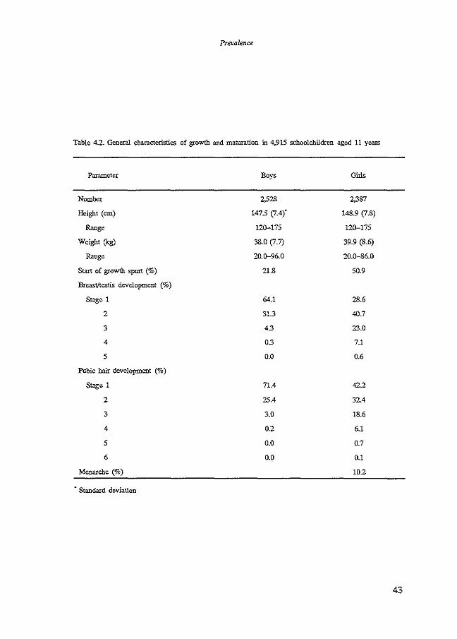
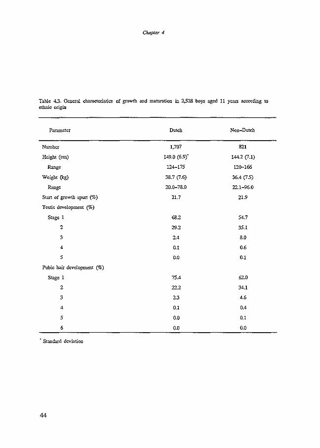
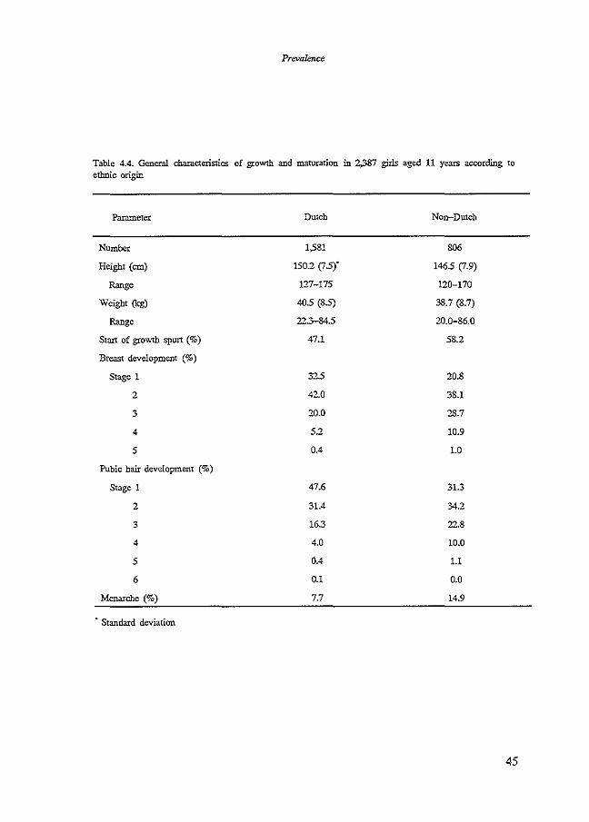
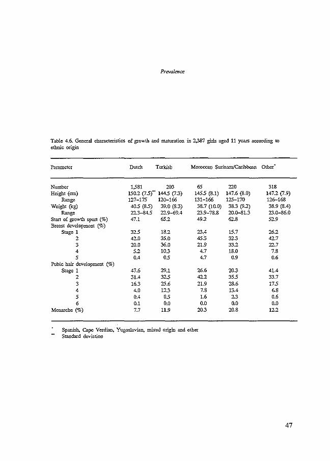
Baseline characteristics of the participant and nonparticipant groups are shown in Table 4.1. Table 4.2 shows the means (:!:SD) and ranges of height and weight, the onset of growth spurt and stages of puberal development for boys and girls. Tables 4.3 and 4.4 show these data for boys and girls separately. More detailed data according to ethnic origin are given in Tables 4.5 and 4.6. At age 11 years, girls were slightly taller and heavier; onset of growth spurt had occurred more among girls (50.9 vs. 21.8%), and pubertal development was faster. Ten percent of all girls had reached menarche. A subgroup analysis according to ethnic origin showed that although the mean height and weight of Dutch children were greater than those of children of other origin, development of puberty occurred later, except for onset of growth spurt in boys.
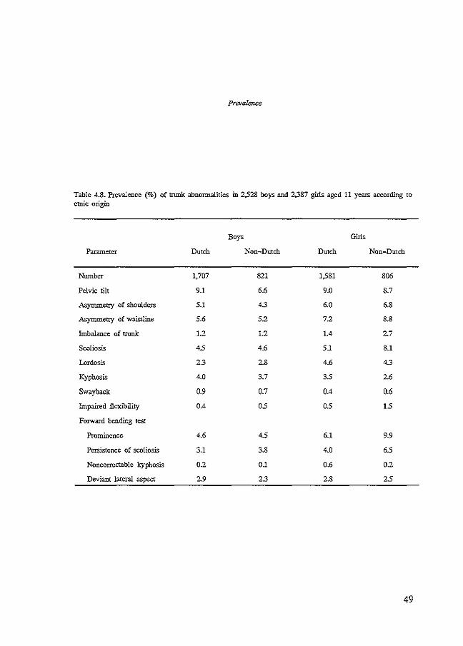
Results of the clinical findings are shown in Tables 4.7 and 4.8. Detailed data according to ethnic origin are given in Tables 4.9 and 4.10. Pelvic tilt owing to leg length inequality was noted in 8.3% of boys and 8.9% of girls (36% of these required a correction of ;,1 em). The prevalences of most trunk abnormalities were higher in girls than in boys, and the highest in non-Dutch girls. Scoliosis in upright position was noted in 115 (4.5%) of boys and in 146 (6.1 %) of girls, 5.1% of Dutch and 8.1% of non-Dutch origin. In 84 (3.3%) of boys and 115 (4.8%) of girls, the scoliosis did not disappear in the forward bending test. Kyphosis was noted in 98 (3.9%) of boys and 76 (3.2%) of girls in standing examination; most of the kyphoses were correctable. A rib hump or lumbar prominence was noted in 116 (4.6%) of boys and in 177 (7.4%) of girls, 6.1% of Dutch and 9.9% of non-Dutch origin.
The majority of boys (2,170, 85.9%) and girls (1,943, 81.3%) were symmetric on all 12 parameters of the standing examination and forward bending test. An abnormal forward bending test was noted in 177 (7.1%) of boys and in 254 (10.6%) of girls, 9.5% of Dutch and 12.9% of non-Dutch origin (fable 4.11).
41

Chapter 4
Table 4.1. Baseline characteristics of 5,167 schoolchildren in the participant and nonparticipant groups
Characteristics Participants Nonparticipants All
Number 4,915 252 5,167
Sex(%)
Male 51.4 51.8 515
Female 48.6 48.2 485
Ethnic origin (%)
Dutch 66.9 42.3 65.7
Turkish 8.1 19.4 8.6
Moroccan 3.0 7.3 3.2
Surinam/Caribbean 8.9 7.3 8.8
01her 12.8 11.7 12.7
Missing 0.3 12.1 09
· Spanish. Cape Verdian, Yugoslavian. mixed origin and other
42
Prevalence
Table 4.2. General characteristics of growth and maturation in 4,915 schoolchildren aged 11 years
Parameter Boys Girls
Number 2,528 2,387
Height (ern) 1475 (7.4)" 148.9 (7.8)
Range 120-175 120-175
Weight (kg) 38.0 (7.1) 39.9 (8.6)
Range 20.D-96.0 20.0-86.0
Start of growth spurt (%) 21.8 50.9
Breast/testis development (%)
Stage 1 64.1 28.6
2 31.3 40.7
3 43 23.0
4 0.3 7.1
5 0.0 0.6
Pubic hair development (%)
Stage 1 71.4 42.2
2 25.4 32.4
3 3.0 18.6
4 02 6.1
5 0.0 0.7
6 0.0 0.1
Menarche (%) 10.2
• Standard deviation
43
Chapter 4
Table 43. General characteristics of growth and maturation in 2.528 boys aged 11 years according to ethnic origin
Parameter Dutch Non-Dutch
Number 1,707 821
Height (em) 149.0 (6.9)" 144.2 (7.1)
Range 124-175 120-166
Weight (kg) 38.7 (7.6) 36.4 (7.5)
Range 20.Q-78.0 22.1-96.0
Start of growth spun (%) 21.7 21.9
Testis development (%)
Stage 1 68.2 54.7
2 29.2 35.1
3 2.4 8.0
4 0.1 0.6
5 0.0 0.1
Pubic hair development (%)
Stage 1 75.4 62.0
2 22.2 34.1
3 2.3 4.6
4 0.1 0.4
5 0.0 0.1
6 0.0 0.0
• Standard deviation
44
Prevalence
Table 4.4. General characteristics of growth and maturation in 2,387 girls aged 11 years according to ethnic origin
Parameter Dutch Non-Dutch
Number 1,581 806
Height (em) 150.2 (J.5)" 146.5 (J.9)
Range 127-175 120-170
Weight (kg) 40.5 (8.5) 38.7 (8.7)
Range 22.3-34.5 20.0-86.0
Start of gtowth spurt (%) 47.1 58.2
Breast development (%)
Stage 1 32.5 20.8
2 42.0 38.1
3 20.0 28.7
4 5.2 10.9
5 0.4 1.0
Pubic hair development (%)
Stage 1 47.6 31.3
2 31.4 34.2
3 163 22.8
4 4.0 10.0
5 0.4 1.1
6 0.1 0.0
Menarche (%) 7.7 14.9
• Standard deviation
45

Chapter 4
Table 45. General characteristics of growth and maturation in 2,.528 boys aged 11 years according to ethnic origin
Parameter Dutch Turkish Moroccan Surinam/Caribbean
Number 1,707 194 83 Height (em) 149.0 (6.9)"" 141.8 (6.8) 142.6 (5.8)
Range 124-175 120-166 128-161 Weight (kg) 38.7 (7.6) 36.2 (7.3) 35.0 (5.8)
Range 20.0-78.0 23.0-70.7 23.9-52.0 Start of growth spurt (%) 21.7 22.7 21.7 Testis development (%)
Stage 1 68.2 48.1 44.6 2 29.2 37.6 48.2 3 2.4 12.7 7.2 4 0.1 1.1 0.0 5 0.0 05 0.0
Pubic hair development (%) Stage 1 75.4 50.8 48.2
2 22.2 413 47.0 3 23 6.9 4.8 4 0.1 05 0.0 5 0.0 05 0.0 6 0.0 0.0 0.0
Spanish, Cape Vcrdian, Yugoslavian,. mixed origin and other Standard deviation
46
220 144.9 (6.8) 128-163
35.2 (7.8) 225-96.0 25.9
49.8 42.3 65 1.4 0.0
60.9 33.0
5.1 0.9 0.0 0.0
Other·
324 145.6 (7.4) 126-166 37.8 (7.6) 22.1-65.5 18.6
66.3 27.0
6.7 0.0 0.0
74.6 22.2
3.2 0.0 0.0 0.0
Prevalence
Table 4.6. General characteristics of growth and maturation in 2.387 girls aged 11 years according to ethnic origin
Parameter Dutch Turkish Moroccan Surinam/Caribbean
Number 1,581 203 65 Height (em) 150.2 (IS)"" 144S (1.3) 145S (8.1)
Rmge 127-175 12Q-!66 131-166 Weight (kg) 40S (85) 39.0 (8.3) 38.7 (10.0)
Rmge 22.3-84.5 22.9-69.4 23.9-78.8 Start of growth spurt (%) 47.1 65.2 49.2 Breast development (%)
Stage 1 325 18.2 23.4 2 42.0 35.0 45.3 3 20.0 36.0 21.9 4 5.2 10.3 4.7 5 0.4 OS 4.7
Pubic hair development (%) Stage 1 47.6 29.1 26.6
2 31.4 325 42.2 3 16.3 25.6 21.9 4 4.0 12.3 7.8 5 0.4 OS 1.6 6 0.1 0.0 0.0
Menarche (%) 7.7 11.9 20.3
Spanish. Cape Verdian, Yugoslavian. mixed origin and other Standard deviation
220 147.6 (8.0) 125-170 38.3 (9.2) 20.0-81.3 62.8
15.7 32.3 33.2 18.0
0.9
20.3 35S 28.6 13.4 2.3 0.0
20.8
Other .
318 147.2 (1.9) 126-168 38.9 (8.4) 23.0-86.0 52.9
26.2 42.7 22.7 7.8 0.6
41.4 33.7 17S 6.8 0.6 0.0
12.2
47

Chapter 4
Table 4.7. Prevalence (%) of trunk abnormalities in 4,915 schoolchildren aged 11 years
Parameter ll<>ys Girls All
Number 2,528 2,387 4,915
Pelvic tilt 8.3 8.9 8.6
Asymmetry of shoulders 4.8 6.3 55
Asymmetry of waistline 5.5 7.8 6.6
Imbalance of trunk 1.2 1.8 1.5
Scoliosis 4.5 6.1 5.2
Lordosis 2.4 4.5 3.5
Kyphosis 3.9 3.2 3.5
Swayback 0.9 05 0.7
Impaired flexibility 0.4 0.8 0.6
Forward bending test
Prominence 4.6 7.4 6.0
Persistence of scoliosis 3.3 4.8 4.0
Noncorrectable kyphosis 0.2 0.5 0.3
Deviant lateral aspect 2.7 2.7 2.7
48
Prevalence
Table 4.8. Prevalence (%) of trunk abnormalities in 2,.528 boys and 2.387 girls aged 11 years according to ctnic origin
Boys Girls
Parameter Dutch Non-Dutch Dutch Non-Dutch
Number 1,707 821 1,581 806
Pelvic tilt 9.1 6.6 9.0 8.7
Asynunetry of shoulders 5.1 4.3 6.0 6.8
Asymmetry of waistline 5.6 5.2 72 8.8
Imbalance of trunk 1.2 1.2 1.4 2.7
Scoliosis 45 4.6 5.1 8.1
Lordosis 2.3 2.8 4.6 4.3
Kyphosis 4.0 3.7 35 2.6
Swayback 0.9 0.7 0.4 0.6
Impaired flexibility 0.4 05 05 15
Forward bending test
Prominence 4.6 45 6.1 9.9
Persistence of scoliosis 3.1 3.8 4.0 65
Noncorrectable kyphosis 0.2 0.1 0.6 0.2
Deviant lateral aspect 2.9 2.3 2.8 25
49

Cf.opter 4
Table 4.9. Prevalence (%) of trunk abnormalities in 2.528 boys aged 11 years according to ethnic origin
Parameter Dutch Turkish Moroccan Surinam/Caribbean Other·
Number 1,707 194 83 220 324 Pelvic tilt 9.1 5.2 2.4 7.7 7.3 i\symmetry of shoulders 5.1 4.1 9.6 3.6 3.5 i\symmetry of waistline 5.6 5.2 12.0 4.1 4.4 Imbalance of trunk 1.2 1.6 1.2 0.5 1.6 Scoliosis 4.5 3.1 4.8 4.5 5.7 Lordosis 2.3 2.6 2.4 5.0 1.6 Kyphosis 4.0 3.6 6.0 4.1 2.8 Swayback 0.9 0.5 1.2 1.4 0.3 Impaired :flexibility 0.4 0.5 0.0 09 0.3 Forward bending test
Prominence 4.6 2.6 3.6 6.8 4.4 Persistence of scoliosis 3.1 2.6 2.4 4.5 4.4 Noncorrectable kyphosis 0.2 0.0 0.0 0.0 0.3 Deviant lateral aspect 2.9 3.1 1.2 3.2 1.6
• Spanish, Cape Verdian, Yugoslavian, mixed origin and other
Table 4.10. Prevalence (%) of trunk abnormalities in 2.387 girls aged 11 years according to ethnic origin
Parameter Dutch Turkis.\ Moroccan Surinam/Can"bbean Other
Number 1,581 203 65 220 318 Pelvic tilt 9.0 11.8 6.2 8.3 7.7 i\symmetry of shoulders 6.0 7.4 7.7 5.0 7.1 Asymmetry of waistline 7.2 8.3 9.2 5.5 11.6 Imbalance of trunk 1.4 2.5 1.5 1.8 3.9 Scoliosis 5.1 6.9 6.2 8.3 9.4 Lordosis 4.6 2.0 3.1 5.0 5.2 Kyphosis 3.5 3.4 1.5 2.8 1.6 Swayback 0.4 1.0 o.o 0.0 1.0 Impaired flexibility 0.5 2.0 1.5 2.3 0.6 Forward bending test
Prominence 6.1 10.3 6.2 11.5 9.7 Persistence of scoliosis 4.0 5.4 1.5 8.3 7.1 Noncorrectable kyphosis 0.6 0.0 0.0 0.5 0.3 Deviant lateral aspect 2.8 2.5 1.5 3.2 2.3
·Spanish, Cape Verdian, Yugoslavian, mixed origin and mhcr
50
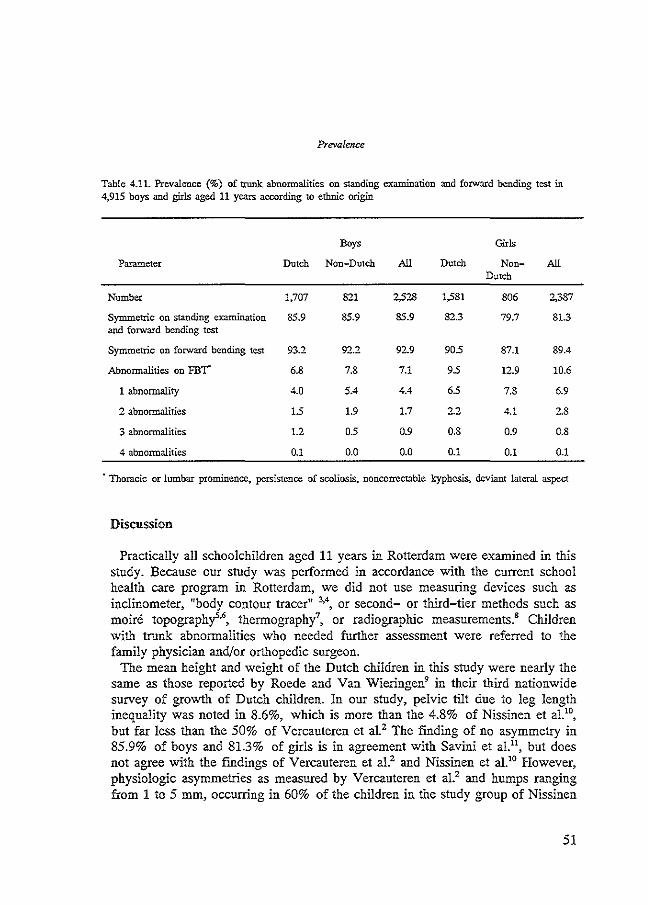
Prevalence
Table 4.11. Prevalence (%) of trunk abnormalities on standing examination and forward bending test in 4,915 boys and girls aged 11 years according to ethnic origin
Boys GIT!s
Parameter Dutch Non-Dutch All Dutch Non- All Dutch
Number 1,707 821 2,528 1,581 806 2;387
Symmetric on standing examination 85.9 85.9 85.9 82.3 79.7 81.3 and forward bending test
Symmetric on forward bending test 93.2 92.2 92.9 905 87.1 89.4
Abnormalities on FBT" 6.8 7.8 7.1 95 12.9 10.6
1 abnormality 4.0 5.4 4.4 65 7.8 6.9
2 abnormalities 15 1.9 1.7 2.2 4.1 2.8
3 abnormalities 1.2 0.5 0.9 0.8 0.9 0.8
4 abnormalities 0.1 0.0 0.0 0.1 0.1 0.1
· Thoracic or lumbar prominence, persistence of scoliosis, noncorrcctable kyphosis, deviant lateral aspect
Discussion
Practically all schoolchildren aged 11 years in Rotterdam were examined in this stndy. Because our study was performed in accordance with the current school health care program in Rotterdam, we did not use measuring devices such as inclinometer, 11body contour tracer,, 3
•4, or second- or third-tier methods such as
moire topography'·', thermography7, or radiographic measurements.• Children
with trunk abnormalities who needed further assessment were referred to the family physician and/or orthopedic surgeon.
The mean height and weight of the Dutch children in this stndy were nearly the same as those reported by Roede and Van Wieringen9 in their third nationwide survey of growth of Dutch children. Jn our study, pelvic tilt due to leg length inequality was noted in 8.6%, which is more than the 4.8% of Nissinen et al.10
,
but far less than the 50% of Vercauteren et al2 The fmding of no asymmetry in 85.9% of boys and 81.3% of girls is in agreement with Savini et a!!', but does not agree with the findings of Vercauteren et al.2 and Nissinen et al.10 However, physiologic asymmetries as measured by Vercauteren et al.Z and humps ranging from 1 to 5 mm, occurring in 60% of the children in the stndy group of Nissinen
51

Chapter 4
et al.10, were considered normal in our study and were not listed as asymmetries. Studies of school screening for scoliosis show a wide range of prevalence; from 0.3% to 13.6%.'2-
15 These differences are due to differences in age groups, definitions of scoliosis, and different detection methods. We noted one or more abnormalities on the forward bending test in 7.0% of boys and in 10.6% of girls. Most of those abnormalities were rib humps or lumbar prominences, and persistence of the standing scoliosis. Both are considered alerting signs for a structural scoliosis. The prevalence of Scheuermann's disease varies from 0.4 to 8.3% of the general population, depending on whether the diagnosis is based on radiographic or clinical criteria!' Ascani et al.17 reported a prevalence of kyphotic curves of 3.1% among children, aged 6-14 years (nonstructural curves included). Drummond et al. 18 reported 1.2 cases in 1,000 screened children. 1n our group of children aged 11 years, we noted a prevalence of noncorrectable kyphosis of 0.3%. A variation in prevalence of scoliosis between ethnic groups has also been reported in other studies!'-21 We noted a higher prevalence of trunk abnormalities among non-Dutch girls than among Dutch girls.
In their review on adolescent idiopathic scoliosis, Leaver et al.13 reported that 3 to 22% of all children aged 6-16 years were abnormal on first- or second-stage examination. The children were referred for diagnosis, including, for many, a roentgenogram of the spine. Three percent were referred to a treatment clinic. Two percent of all children screened had curvature :dO degrees. Brace or operative treatment was required in only 0.1-0.2% of all children screened. In our study, the population consisted of one age group (children aged 11 years). The examination was not performed as a scoliosis school screening program, but as an integral part of the regular medical examination. At least one abnormal finding on standing examination and forward bending test was noted in 14.1% of boys and in 18.7% of girls. Although 7.0% (n;177) of boys and 10.6% (n;254) of girls had an abnormal forward bending test, none had had radiographic examination before referral. Referral for diagnostic examination was made only in 1.7% (n;42) and 3.1% (n;74), respectively. This agrees with the referral rate in U.S. screening programs. The 53% and 7.5% not referred were reexamined for progression within 12 months by school physicians. All children of the study population will be examined during the regular medical examination in 2 years. Incidence of trunk abnormalities will be studied.
References 1. Tanner JM. Growth at adolescence. 2nd ed. Oxford: Blackwell Scientific Publications, 1962:32-39. 2. Vercauteren M, Van Bcncdcn M, Vcrplaetse R, Croene Ph, Uyttendaele D, Verdonk R. Trunk
asymmetries in a Belgian school population. Spine 1982;7:555-62. 3. Bunvcll RG, James NJ, Johnson F, Webb JK. Wilson YG. Standardised trunk asymmetry scores. J
Bone Joint Surg [Br] 1983;65:452-63. 4. Bunnell WP. An objective criterion for scoliosis screening. J Bone Joint Surg [Am] 1984;66:1381-
7. 5. Adair IV, Van Wijk MC, Armstrong GWD. Moire topography in scoliosis screening. Clin Orthop
1977;129:165-71. 6. Willner S. MoirC topography for the diagnosis and documentation of scoliosis. Acta Orthop Scand
1979;50:295-302.
52
Prevalence
7. Cooke ED, Carter LM. Pilcher ?vfF. Identifying scoliosis in the adolescent with thermography. Qin Orthop 1980;148:172-6.
8. Dickson RA. Stamper P. Sharp A, Harker P. School screening for scoliosis: cohort study of clinical course. Br Med J 1980;281:265-7.
9. Roede MJ, van Wieringen JC. Growth diagrams 1980, Netherlands third nation-wide survey. Tijdschr Soc Gezondheidszorg 1985;63:(suppl):1-34.
10. Nissinen M, HeliOvaara M. Tallroth K. Poussa M. Trunk asymmetry and scoliosis. Anthropometric measurement in prepuberal school children. Acta Paediatr Scand 1989;78:747-53.
11. Savini R,. Cervcllati s. Ponzo L. Cioni A, Palmisani M, Rizqallah Y. Gargiulo G. Nard s. Di Silvestre M. L'incidenza della scoliosi idiopatica nel Comune di Bologna. Chir Org Mov 1986;71: 119-25.
12. Brooks HL. Azen SP, Gerberg E, Brooks R, Chan L Scoliosis: a prospective epidemiological study. J Bone Joint Surg [Am]1975;57:968-72.
13. Leaver JM, Alvik A, Warren MD. Prescriptive screening for adolescent idiopathic scoliosis: a review of the evidence. Int J Epidemiol 1982;11:101-11.
14. Lonstein .JE, Bjorklund S, Wanninger MH, Nelson RP. Voluntary school screening for scoliosis in Mirmcsota. J Bone Joint Surg [Am] 1982;64:481-8.
15. Rogala EJ, Drummond DS, Gurr J. Scoliosis: incidence and natural history, a prospective epidemiological study. J Bone Joint Surg [Am.Jl978;60:173-6.
16. Sorensen KH. Scheuennann's juvenile kyphosis. Copenhagen: Munksgaard, 1964. 17. Ascani E, Salsano V, Giglio G. The incidence and early detection of spinal deformities. Ita! J
Orthop Traumatol 1977;3:111-7. 18. Drummond DS, Rogala EJ, Gurr J. Spinal deformity: natural history and the role of school
screening. Orthop Clin N Am 1979;10:751-9. 19. Daruwalla JS, Balasubramaniam P, Chay SO, Raj an U, Lee HP. Idiopathic scoliosis. Prevalence and
ethnic distribution in Singapore school children. J Bone Joint Surg [Br} 1985;67:182-9. 20. Segil CM. The incidence of idiopathic scoliosis in the Bantu and 'White population groups in
Johannesburg. J Bone Joint Surg [Br) 1974;56:393. 21. Smymis PN, Valavanis J, Alexopoulos A, Siderakis G, Gianncstras NJ. School screening for
scoliosis in Athens. J Bone Joint Surg [Br}1979;61:215-7.
53


Chapter 5
Incidence of trunk abnonnalities


Chapter 5
Incidence of trunk abnormalities
Abstract
We conducted a study of the two-year cumulative incidence of trunk abnormalities in a cohort of 2,819 eleven-year-old children (1,507 boys, 1,312 girls). The following data were recorded: height, weight, signs of puberty, menarche. Trunk abnormality was assessed in the erect child (asymmetry of shoulders and waistline, imbalance of the trunk, scoliosis, lordosis, kyphosis, swayback and flexibility) and by the forward bending test (FBT) (rib hump or lumbar prominence, persisting scoliosis, kyphosis, deviant lateral aspect). A normal FBT both at baseline and at follow-up was found among 84.1% of the boys and among 79.4% of the girls. The two-year cumulative incidence of an abnormal FBT was 10.1% in boys and 13.0% in girls.
Introduction
Trunk abnormalities in children, such as scoliosis and kyphosis, have been widely studied. School screening programs for scoliosis have given insight in its prevalence and incidence, and in its natural history. However, in many studies prevalence and incidence data are mixed. Prevalence refers to the number of individuals with the disease existing at any time as a proportion of the number exposed to that risk, while cumulative incidence refers to the number of individuals developing the disease in a specified period of time as a proportion of the number exposed to that risk. The cumulative incidence provides a good estimate of the risk to develop the disease during a specified period of time. Also methods of diagnosing trunk abnormalities (scoliosis) are different in the various studies. Some are based on full clinical examination by physicians, others only on the forward bending test (one-minute test) by school nurses or trained laymen.
There is no consensus about the most appropriate age for scoliosis screening. In many countries 10 to 16-year-old children are screened annually. In Rotterdam, The Netherlands, there is no specific screening program for scoliosis, but all school children are invited for a full medical examination by school physicians at the age of eleven and in the second grade of secondary education (age 13-14).
We conducted a prospective follow-up study of trunk abnormalities in a cohort of eleven-year-old children in order to obtain prevalence and two-year cumulative incidence data. The results of the prevalence study in eleven-yearold children have already been described.' We now report the two-year cumulative incidence of trunk abnormalities in the same cohort.
57

Chapter 5
Methods
Subjects All school children in Rotterdam, born in 1973, who had participated in the
prevalence study (1984/1985) were eligible for follow-up. The study was incorporated in the regular medical examination of the second grade of secondary school, between September 1986 and July 1987. Of the approximately 6,000 second grade students, 4,663 children were born in 1973. Of 3,071 children, data of baseline examination were available; 62.5% of the cohort of eleven-year-old children (Figure 5.1). The mean age of the children was 13 years and seven months; 52.8 percent were male. A number of the eligible children was examined later than August 1987, they are considered as lost-to-follow-up in the present study. Other reasons for lost-to-follow-up were moving home or going to a school outside Rotterdam, or not responding to the invitation for the optional examination twice. There was no difference in baseline characteristics between the follow-up and lost-to-follow-up groups (Table 5.1). Anthropometrical data of examinations at baseline and at follow-up are given in Table 5.2. Table 5.3 shows prevalence of clinical findings at baseline and two years later. In more than half of the 252 children having an abnormal forward bending test (FBT) at baseline examination, the FBT was normal at follow-up examination.
The current analysis of the two-year cumulative incidence was based on 2,819 children free of trunk abnormalities at baseline examination.
BASELINE EXAMINATION
FOLLOW-UP EXAMINATION
1,844
OUTFLOW
3,071
3,071 1,592
COHORT OF STUDY INFLUX
Figure 5.1. Number of participating subjects of cohort 1973 in baseline and in follow-up examination
Measurements Although the school physicians were experienced in the physical examination
of the trunk, including the forward bending test, as being part of the regular examination, they received oral, audiovisual and written training and demonstration of patients prior to the study to guarantee standardisation of measurements. All family physicians and orthopedic surgeons in Rotterdam were informed. The following measurements were object of the study: height in centimeters, weight in 0.1 kilogrammes. Physical maturity was ascertained by
58

Incidence
Table 5.1 Baseline characteristics of 3,071 cohort subjects and 1,844 subjects lost-to-follow-up
Boys Girls
Parameter Cohort Lost-to-follow-up Cohort Lost-to-follow-up
Number 1,621 907 1,450 937
Height (em) 147.6 (7.2)' 147.2 (7.6) 149.3 (7.8) 148.4 (7.8)
Range 128-175 120-171 125-172 120-175
Weight (kg) 38.0 (7.7) 37.9 (7.7) 39.9 (85) 39.8 (8.8)
Range 22.1-96.0 20.0-78.0 20.0-84.5 223-86.0
Start of growth spurt (%) 20.8 23.7 50.4 51.5
Breast/testis development (%)
Stage 1 66.2 60.2 28.8 28.2
2 29.8 34.1 39.3 43.0
3 3.8 52 23.4 22.3
4 0.2 0.3 7.9 5.9
5 0.0 0.0 0.6 0.7
Pubic hair development (%)
Stage 1 73.4 67.6 42.1 42.3
2 24.1 27.7 31.8 33.5
3 2.4 4.3 18.7 18.3
4 0.2 02 6.5 5.4
5 0.0 0.1 0.8 0.5
6 0.0 0.0 0.1 0.0
Menarche (%) 10.0 10.5
· Standard deviation
59
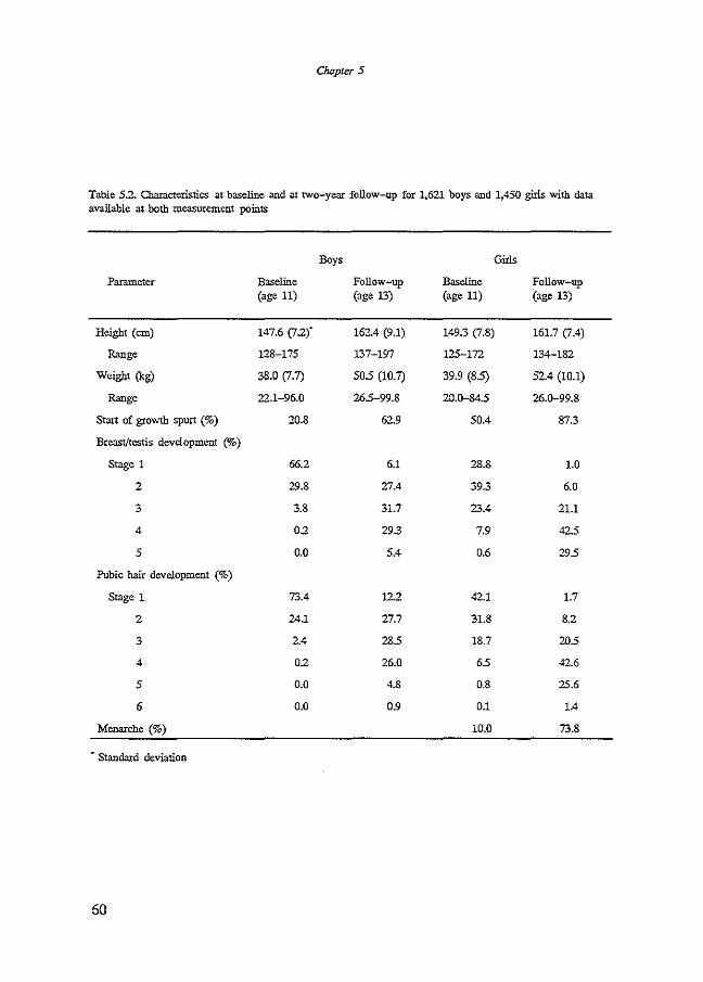
Chapter 5
Table 5.2. Characteristics at baseline and at two-year follow-up for 1.621 boys and 1,450 girls with data available at both measurement points
Boys Girls
Parameter Baseline Follow-up Baseline Follow-up (age 11) (age 13) (age 11) (age 13)
Height (em) 147.6 (J.2)" 162.4 (9.1) 149.3 (7.8) 161.7 \1.4)
Range 128-175 137-197 125-172 134-182
Weight (kg) 38.0 (7.7) 50S (10.7) 39.9 (8.5) 52.4 (10.1)
Range 22.1-96.0 265-99.8 20.D-84S 26.0-99.8
Start of growth spurt (%) 20.8 62.9 50.4 873
Breast/testis development (%)
Stage 1 66.2 6.1 28.8 1.0
2 29.8 27.4 39.3 6.0
3 3.8 31.7 23.4 21.1
4 0.2 29.3 7.9 425
5 0.0 5.4 0.6 295
Pubic hair development (%)
Stage 1 73.4 12.2 42.1 1.7
2 24.1 27.7 31.8 8.2
3 2.4 285 18.7 205
4 0.2 26.0 6S 42.6
5 0.0 4.8 0.8 25.6
6 0.0 09 0.1 1.4
Menarche (%) 10.0 73.8
• Standard deviation
60

Incidence
Table 5.3. Prevalence (%) of trunk abnormalities in 1,621 boys and 1,450 girls at baseline (age 11) and at two-year follow-up (age 13)
Boys Girls
Parameter Baseline Follow-up Baseline Follow-up (age 11) (age 13) (age 11) (age 13)
Pelvic tilt 9.1 13.1 8.7 11.6
Asymmetry of shoulders 4.1 75 6.1 7.8
Asymmetry of waistline 5.2 115 7.6 11.3
Imbalance of trunk 1.1 35 1.7 3.1
Scoliosis 4.4 6.4 5.4 8.8
Lordosis 2.3 25 4.4 3.7
Kyphosis 3.9 6.6 2.9 6.4
Swayback 0.7 1.0 0.6 0.9
Impaired flexibility 0.3 15 0.8 1.4
Forward bending test
Prominence 4.3 8.6 6.1 12.3
Persistence of scoliosis 3.3 3.0 4.3 5.2
Noncorrectable kyphosis 0.2 1.1 0.3 1.1
Deviant lateral aspect 25 2.3 2.8 2.6
61

Chapter 5
assessing testis development in boys, breast development and menarche in girls, pubic hair development and onset of the rapid growth spurt in both sexes. Grading was performed according to Tanner.2 First, pelvic tilt was looked for and leg length inequality was corrected by placing one or more boards of 0.5 em under the short leg until horizontal symmetry of iliac crests and posterior iliac spines was obtained. The correction was noted in centimeters. After correction, the standing child was observed from the front and the back for any obvious deformities such as asymmetry of the shoulders, scapulae and waistline. The balance of the thorax over the pelvis was assessed with the plumbline. Flexibility of the spine was examined in flexion, extension and side-bending. The standing child was viewed from the side for areas of hyper- or hypolordosis and hyperor hypokyphosis. Trunk asymmetries and abnormal curvatures in the median plane in upright position were recorded as either absent or present, according to the proposed limits for structural trunk asymmetries by Vercauteren et al.3 The forward bending test (FBT) was performed, with the child standing with knees straight and feet together, bending at the waist with arms hanging and palms together. The back was viewed from the child's head, both sides were compared for symmetry from the upper thoracic area to the lumbosacral area. The spine was also viewed from the side in the forward-bending position to evaluate the contour of the back for kyphotic angulation. Flexibility of hyperkyphosis was also tested by prone hyperextension. Flexibility of lordosis was shown in the FBT and also with the child bending in a knee-chest position. The following four components of the forward bending test (FBT) were listed as either absent or present: (a) Rib humps and lumbar prominences as signs of vertebral rotation. A rib hump is not always associated with a scoliosis in upright position; (b) Persistence of the standing scoliosis, in order to discriminate between postural and structural scoliosis. It will usually be associated with a rib hump or lumbar prominence; (c) Correctability of the kyphosis, in order to discriminate between postural and structural kyphosis; (d) Deviant lateral aspect. We considered a FBT as abnormal if at least one of the four components was present.
Data analysis The two-year cumulative incidence was calculated for each trunk abnormality
separately, as well as for a positive FBT.
Results
Table 5.4 shows the two-year cumulative incidence of trunk abnormalities, and of abnormal FBT. Scoliosis in upright position was found in 84 (5.6%) of the boys and 80 (6.1%) of the girls; and did not disappear on forward bending in 36 (2.4%) and 46 (3.5%), respectively. A rib hump or lumbar prominence was found in 107 (7.1 %) of the boys and 127 (9.7%) of the girls. An abnormal FBT was found in 152 (10.1 %) and 170 (13.0%), respectively.
62

Incidence
Table 5.4. Two-year cumulative incidence (%) of trunk abnormalities and of abnormal forward bending test (FB!) in 1,387 boys and 1,197 girls aged 13 yeaxs
Parameter Boys Girls All
Pelvic tilt 9.2 8.0 8.7
Asymmetry of shoulders 6.8 6.0 65
Asymmetry of waistline 10.4 9.1 9.8
Imbalance of trunk 2.7 2.1 2.4
Scoliosis 5.6 6.1 5.8
Lordosis 1.9 2.8 2.4
Kyphosis 5.4 5.8 5.6
Swayback 0.7 0.8 1.0
Impaired flexibility 1.2 0.8 1.0
Forward bending test
Prominence 7.1 9.7 8.3
Persistence of scoliosis 2.4 35 2.9
Noncorrectable kyphosis 0.7 0.9 0.8
Deviant lateral aspect 1.4 15 1.5
Abnormal on forward bending test 10.1 13.0 11.4
Discussion
In our prospective follow-up study, embedded in the current school health care program, 62.5 percent of the ioitial cohort of eleven-year-old children were examined for follow-up. In regard to baseline characteristics, there was no differential lost-to-follow-up. Compared to the 1980 biometrical survey\ the mean height and weight of boys were nearly the same. However, girls were heavier and slightly shorter. Children with distinct trunk abnormalities who needed further assessment were referred to the family physician and/or orthopedic surgeon. Children with lesser trunk abnormalities were not referred. They were reexamined by the school physician in 6 to 12 months.
The principal finding of this part of our study is that the cumulative incidence, and thereby the absolute risk for trunk abnormalities is 11%. Most studies of adolescent idiopathic scoliosis are based on annual screening programs in 10 to 15-year-old children; and data concern mostly prevalence, not cumulative incidence. Our survey was conducted in one birth cohort. All children were examined twice. First, in a prepuberal or early puberal phase at age 11; the second examination in a puberal phase at age 13. Prevalences at age 11 and at age 13 in our study were similar to prevalences of 10% to 20% of the first tier
63

Chapter 5
of scoliosis screening programs, and prevalence increased with age.5-7 However,
we found that the prevalence at the examination at age 13 mainly consisted of the two-year cumulative incidence. Chan et al.8 found in retrospect that 21 of 49 scoliosis patients had been symmetric at an earlier screening.
As in most studies, we used an abnormal FBT as an alerting sign for a structural deformity. Many children with an abnormal FBT will have mild and nonprogressive or resolving scoliosis, but some children will have curves that progress. It is impossible to separate progressive from nonprogressive scoliosis on the basis of one clinical examination. Regression of scoliosis has been described by various authorss~"0 In their groups of scoliosis patients, spontaneous improvement or regression had occurred in 3% to 22%, and in approximately half of the patients the magnitude of the curvatures had remained unchanged. In our study group, one-half to two-thirds of the children with an abnormal FBT at age 11 had improved to normal at age 13. Only few of these children had been referred for further assessment; most children referred after the first examination did have an abnormal FBT at both measuring moments.
What do our findings mean for school health programs? It appears necessary to perform at least two examinations for trunk abnormalities during adolescence. Referral on one sole examination, i.e. one abnormal FBT, should only be done in case of distinct findings; in case of less distinct fmdings reexamination in six months is necessary.
References 1. Hazebroek-Kampscbreur AAJM. Hofman A. Dijk APh van. Linge B van. Prevalence of trunk
abnormalities in eleven-year-old schoolchildren in Rotterdam. The Netherlands. J Pediatr Orthop 1992;12:480-4.
2. Tanner JM. Growth at adolescence. Oxford: Blackwell Scientific Publications, 1962:32-9. 3. Vercauteren M. Van Beneden M,. Verplaetse R, Croene Ph. Uyttendaele D, Verdonk R. Trunk
asymmetries in a Belgian school population. Spine 1982;7:555-62. 4. Roede MJ. Wieringen JC van. Growth diagrams 1980. Netherlands third nation-wide survey. T Soc
Gezondheidszorg 1985;63:(suppl):1-34. 5. Brooks HL. Azen SP, Gcrberg E. Brooks R, Chan L. Scoliosis: a prospective epidemiological
stody. J Bone Joint Surg [Am]1975;57:968-72. 6. Morais T, Bernier M. Turcotte F. Age- and sex-specific prevalence of scoliosis and the value of
school screening programs. Am J Public Health 1985;75:1377-80. 7. Pessers FGM, Florack: EIM, Zielhuis GA. Is screening op scoliose aan te bevelen? T Soc Gezond
heidsz 1986; 64,451-5. 8. Chan .A,. Moller J, Vunpa."li G, Patterson D, Southwood R, Sutherland A The case for scoliosis
screening in Australian adolescents. Med J Aust 1986;145:379-83. 9. Li High Pin. Liu Yong Mo, Li Lin, Li Kan Hua,. Hei Pei Hui, Den Shi Hui, Bao Dong Chang,
Yuin Yuin Chang. Early diagnosis of scoliosis based on school-screening. J Bone Joint Surg [Am} 1985;67: 1202-5.
10. Rogala EJ, Drummond DS, Gurr J. Scoliosis: incidence: and natural history. J Bone Joint Surg [Am] 1978; 60:173-6.
64

Chapter 6
Determinants of trunk abnormalities


Chapter 6
Determinants of trunk abnormalities
Abstract
The relationship between gender, height, weight, onset of adolescent growth spurt, pubertal phase, menarche and incidence of trunk abnormalities was studied in 2,819 children aged eleven years who were symmetric at baseline examination. For all children, tbe two-year cumulative incidence of trunk abnormalities was 11%. The relative risk for boys was 0.78 (95% confidence interval 0.63-0.96). Height was tbe strongest predictor for the development of trunk abnormalities in botb sexes. The relative risks increased after adjustment for weight, onset of growth spurt, pubertal phase, and menarche. For girls with onset of adolescent growth spurt, tbe relative risk was 1.45 (95% confidence interval 1.08-1.95) compared to girls who had not yet started with adolescent growth spurt. For girls who had reached menarche, the relative risk was 0.48 (95% confidence interval 0.24-0.96) compared to tbose who had not reached menarche. Our findings suggest tbat taller eleven-year-old girls with onset of adolescent growth spurt have an increased risk of future trunk abnormalities, but that having reached menarche has a protective effect.
Introduction
Trunk abnormalities, particularly scoliosis and k-yphosis, are frequently found in adolescents. The unknown etiology of idiopathic scoliosis, and the relationship between growth, maturation, and progression of idiopathic scoliosis have prompted a number of anthropometric studies_,_, 1n most of these studies children with an already developed scoliosis were compared to a nonscoliotic control group. 1n Rotterdam, The Netherlands, all school children are invited for a full medical examination by school physicians at tbe age of eleven years and in the second grade of secondary school (age 13-14 years). Assessment of height, weight, pubertal development, and examination for trunk abnormalities are routine procedures in tbese examinations. Between 1984 and 1987, we conducted a two-year prospective follow-up study in a cohort of eleven-year-old children in order to obtain prevalence and two-year cumulative incidence data regarding trunk asymmetries, including scoliosis and kyphosis. The results of the prevalence study have already been described."
The purpose of tbe present study was to identify predictive factors for trunk abnormalities as defined by a positive forward bending test.
67

Chapter 6
Methods
Subjects The study was embedded in the current school health care program for
adolescents, i.e. a medical examination in a cohort of eleven-year-old children and in second graders. Of the 5,167 eligible subjects, born in 1973 and attending a Rotterdam school, 4,915 participated in the baseline examination at age 11 years (schoolyear 1984/1985). During the regular medical examination of the second year of secondary school (schoolyear 1986/87), data of 3,071 children were collected.
Not included in this study are data of 1,506 children examined later than August 1987, and data of 338 children who attended school outside Rotterdam. There was no difference in baseline characteristics between the follow-up group and the groups who were not reexamined at time of follow-up.
For this study, data of 3,071 children were available. An abnormal forward bending test (FBT) at baseline examination was noted in 252 (8.2%) of the children; these were excluded from the data analysis. The population at risk for future trunk abnormalities consisted of 1,507 boys and 1,312 girls with a normal FBT at baseline. Data of these 2,819 children were used for analysis.
Measurement of determinants The children were examined by school physicians during the (optional) regular
school medical examination, which included assessment of the following determinants: height, weight, onset of growth spurt, pubertal development and menarche.
Height was measured in the child standing straight, barefooted, with heels, buttocks, midthoracic portion of the trunk, and the back of the head touching the wall. The heels were kept together, the feet forming an angle of 45°. Metal yardsticks, either attached to the wall or transportable, were used. The horizontal indicator of the yardstick was brought down to the child's head which was held in the Frankfurt plane, i.e. the line from the lateral comer of the eye to the upper edge of the attachment of the ear is horizontal. Height was noted in centimeters. Weight was measured using a step-scale and recorded in hectogrammes. Sigus of puberty were examined: pubic hair development differentiated into six stages (PHI - PH6) in both boys and girls, testis development differentiated into five stages (Gl - G5) in boys, and breast development differentiated into five stages (Ml - M5) in girls. Grading was performed according to the method of Tanner', stage 6 of pubic hair was added according to the criteria of The Netherlands Biometric Survey.' For analysis, we constructed the following five categories of pubertal phase: I Prepubertal phase: both pubertal sigus were graded stage 1 II Early pubertal phase: one pubertal sign was graded stage 1 and the other
pubertal sign was graded stage 2-4 m Pubertal phase: both pubertal sigus were graded stage 2-4 IV Early adult phase: one pubertal sigu was graded stage 2-4 and the other
pubertal sign was graded stage 5 or 6
68

Determinants
V Adult phase: both pubertal signs were graded stage 5 or 6 Menarche refers to the age at the first menstrual flow. We used the 'status quo'
method by asking each girl in colloquial speech whether she had reached menarche. Onset of individual adolescent growth spurt was assessed by comparing the attained height with previous measurements noted in the growth diagram, and if necessary by means of information provided by the parents. Onset of growth spurt was noted as "no", "yesn or considered "indistinct~~.
Measurement and definition of outcome Pelvic tilt was determined and leg length inequality was corrected by placing
one or more 0.5 em boards under the short leg until horizontal symmetry of iliac crests and posterior iliac spines was obtained. After correction, the child's posture was observed. The standing child was viewed for symmetries in the shoulders, scapulae and waistline. The balance of trunk over the pelvis was assessed with a plumbline. Flexibility of the spine was examined in flexion, extension, and side bending. The child was viewed from tlte side for areas of hyper- or hypokyphosis and hyper- or hypolordosis. Trunk asymmetries and abnormal curvatures in the median plane in standing position were recorded as either absent or present, according to the proposed limits of Vercauteren et a!." The forward bending test was performed, with the child standing with feet together and knees straight, bending at the waist, with the arms dependent and held with fingers and palms opposed. The back was viewed head on for symmetry; both sides were compared. The back was also viewed from the side to assess kyphosis and rib humps. We noted four components in the forward bending test: (a) rib humps and lumbar prominences as signs of vertebral rotation, (b) persistence of the standing scoliosis on forward bending to discriminate between postural and structural scoliosis, (c) correctability of the kyphosis to discriminate between postural and structural kyphosis, and (d) deviant lateral aspect. We considered a forward bending test abnormal if at least one of the four components listed above was present
New (incident) cases of trunk abnormalities are defmed as subjects with a normal forward bending test on baseline examination having an abnormal forward bending test as listed above at age 13 years.
Data analysis The two-year cumulative incidence was calculated for each variable, i.e.
gender, height, weight, onset of adolescent growth spurt, pubertal phase, and menarche.
Univariate analysis of categorical data was performed with calculation of the relative risk (risk ratio) and its 95 percent confidence interval. Stratum-specific relative risks with 95 percent confidence interval were calculated of all variables for strata of gender, height, change in height, growth spurt, pubertal phase, and menarche. Numerically continuous variables, such as height, change in height, weight, and change in weight were divided in four strata (quartiles) using the lowest quartile as reference group.
69
Chapter 6
Logistic regression analysis was applied to estimate the adjusted relative risk (and 95 percent confidence interval) of trunk abnormalities associated with the independent variables at baseline examination: gender, month of birth, height, change in height, weight, change in weight, onset of adolescent growth spurt, pubertal phase, and menarche. Indicator variables were accomplished as follows: height, weight, change in height, change in weight, and month of birth were entered as numerically continuous variables. Using the five categories of pubertal phase, a new binary variable was created. The combined pubertal categories III, IV and V were used as refence category for the combined pubertal categories I and H. Onset of adolescent growth spurt was used as reference category for no onset of adolescent growth spurt. Menarche was used as reference category for no menarche.
In a final analysis the predictive value of various variables for the occurrence of trunk abnormalities was assessed by entering gender, height, weight, pubertal phase, onset of adolescent growth spurt, and menarche in a discriminant function with trunk abnormalities as outcome variable.
Results
Table 6.1 shows the two-year cumulative incidence of trunk abnormalities. For boys, the relative risk of trunk abnormalities was 0.78 (95% confidence interval [CI] 0.63-0.96) compared to girls. Therefore, we controlled for gender in all further analyses.
In the assessment of single factors (univariate analysis), the outcome was significantly associated with height in boys and girls, and with menarche and onset of growth spurt in girls (Tables 6.2 - 6.5). After adjustment for weight, onset of growth spurt, pubertal phase, and menarche, the relative risks relating height at baseline examination to trunk abnormality at age 13 years increased (Tables 6.2, 6.3). Month of birth, weight, and pubertal phase were not associated with the outcome.
At adolescence, height is related to the particular stage of pubertal development at that moment. After stratification for height (above and below median), the relative risk for girls with onset of growth spurt was still higher than the risk for girls in whom onset of growth spurt had not yet occurred, but this was not significant (above median: RR 1.06, 95% Cl 0.72 to 1.57; below median: RR 1.57, 95% Cl 0.98 to 2.51). As all incident girls who had reached menarche had a height above median, stratification for height showed no changes in relative risk. For girls with early maturation (onset of growth and menarche), and late maturation (no onset of growth spurt, no menarche), the relative risks were 0.35 (95% CI 0.16-0.78) and 0.69 (95% CI 0.51-0.92) compared to girls with normal maturation (onset of adolescent growth spurt, but not yet menarche).
Tables 6.6 and 6.7 show the results of the logistic regression analysis. In boys, height had a less pronounced effect than in the univariate analysis. In girls, the greatest effect was found for height and onset of growth spurt, with an inverse effect of menarche.
70

Determinants
Table 6.1. Population at risk for future trunk abnormalities
Boys
Girls
Total
Total at risk
1,507
1,312
2,819
2y CI: two-year cumulative incidence
Table 6.2. Height in boys
Total at risk New cases
Quartile 1 426 34
Quartile 2 345 27
Quartile 3 372 40
Quartile 4 364 51
New cases
152
170
322
2ya
0.080
0,078
0.108
0.140
RR 95% ci
1
0.98 0.61-159
135 0.87-2.08
1.75 1.16-2.64
2y CI
0.101
0.130
0.114
RR"
1
1.30
1.84
2.17
2y CI: two year cumulative incidence; RR: relative risk; 95% ci: 95% confidence interval · adjusted for weight, onset of growth spurt. pubertal phase
Table 6.3. Height in girls
Total at risk New cases 2ya RR 95% ci RR"
Quartile 1 358 35 0.098 1 1
Quartile 2 306 30 0.098 1.01 0.63-1.60 1.23
Quartile 3 374 59 0.158 1.62 1.09-2.40 2.41
Quartile 4 274 46 0.168 1.72 1.14-259 2.94
2y CI: two year cumulative incidence; RR: relative risk; 95% ci: 95% confidence interval • adjusted for weight, onset of growth spurt. pubertal phas~ menarche
95% ci.
0.81-2.08
1.02-3.30
1.11-4.22
95% cf
0.77-1.95
1.33-4.36
152-5.66

Table 6.4. Onset of growth spurt in girls
Yes
No
Total at Jiskl
656
622
Chapter 6
New cases2
100
65
1 Onset of growth spurt is indistinct in 34 girls 2 Onset of growth spurt is indistinct in 5 new cases
2y Cl
0.152
0.105
RR
1.45
1
2y CI: two year cumulative incidence; RR: relative risk; 95% ci: 95% confidence interval
Table 65. Menarche
Total at risk!
Yes
No
121
1,174
New ca=f
8
161
1 Data of menarche are missing in 17 girls 2 Data of menarche are missing in one new case
2y Cl
0.066
0.137
RR
0.48
1
2y CI: two year cumulative incidence; RR: relative risk; 95% ci: 95% confidence interval
Table 6.6. Multiple logistic regression analysis for boys
Variable
Month of birth
Onset of growth spurt
Pubertal phase
Height (em)
Change m height (em)
Weight (hg)
Change m weight (hg)
Constant
1 logistic regression coefficient 2 standard error of B
B'
-0.036
-0.011
0.131
0.398
0.299
0.141
-0.135
1.285
SE'
0.027
0.229
0.216
0.224
0.212
0225
0.217
0.404
3 odds ratio: relative increase in risk per unit change in the determinant 4 95% ci: 95% confidence interval
72
Exp(B)'
0.98
0.99
1.11
1.49
1.35
1.15
0.87
95% ci
1.08-1.95
95% ci
024-0.96
95% ci4
0.97-1.09
0.63-1.55
0.75-1.74
0.96-2.31
0.89-2.04
0.74-1.34
057-1.34
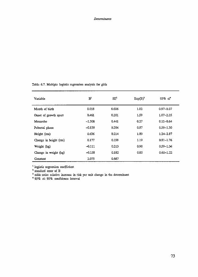
Determinants
Table 6.7. Multiple logistic regression analysis for girls
Variable B' SE' Exp(B)' 95% cr'
Month of birth 0.018 0.026 1.02 0.97-1.07
Onset of growth spurt 0.461 0.201 1.59 1.07-2.35
Menarche -1.308 0.441 0.27 0.11-0.64
Pubertal phase -0.139 0.204 0.87 0.59-1.30
Height (em) 0.636 0.214 1.89 1.24-2.87
Change in height (em) 0.177 0.199 1.19 0.81-1.76
Weight (hg) -0.111 0.213 0.90 0.59-1.36
Change in weight (hg) -0.158 0.182 0.85 0.60-1.22
Constant 2.075 0.667
1 logistic regression coefficient 2 standard error of B 3 odds ratio: relative increase in risk per unit change in the determinant 4 95% ci: 95% confidence interval
73

Chapter 6
In discriminant analysis, the predictive value of the discrintinant function was small; 56% in boys and 60% in girls.
Discussion
The main finding of our study is that two-year cumulative incidence of trunk abnormalities was associated with gender, and with height at baseline in both sexes, and with menarche and onset of growth spurt at baseline in girls. Our study differed from most studies of growth and scoliosis in a few aspects. First, we conducted our study prospectively in a large group of unselected (pre-) adolescent children embedded in the current school health care program. Most other studies concerned only girls with diagnosed adolescent idiopathic scoliosis (AIS) who were compared to historical or contemporary controls. Second, we studied prevalence and incidence of trunk abnormalities not limited to diagnosed idiopatlllc scoliosis. Therefore, small scoliotic curves not in need of orthopedic observation or treatment were also included, as well as abnormal kyphosis. Third, we had excluded all children with trunk abnormalities at age 11 years from the study of determinants of future trunk abnormalities.
Studies in girls with AIS have shown that girls with AIS were on the average taller than controls.'-u Retrospectively, girls with AIS appeared to be taller before time of diagnosis', and before onset of pubertal growth spurt.13 The difference in height disappeared towards the end of growth.' There is a difference of opinion whether growth is faster or more intense in scoliotic girls than in their non-scoliotic peers14
•15
, or that their growth pattern is similar in time and in quantity.'·u Dickson and Sevitf reported that although children with progressive scoliosis were significantly taller than children with non-progressive scoliosis, they were growing at a similar rate. In their opinion, increased height should be regarded not as an etiological factor but as an indicator of bad prognosis. Our study showed a difference in height two years prior to the detection of trunk abnormalities. Children with a height above median at baseline examination had a significant higher risk for future trunk abnormalities than children with a height below median at baseline examination.
The variation in the age of onset, duration, maximum and overall gain of adolescent growth spurt, and of sexual maturation is wide. Menarche is a rather late event during the period of adolescent growth spurt, occurring about two years after stage 2 of breast and pubic hair development, and about one year before cessation of rapid growth. Although most studies showed no difference of mean age of menarche between girls with AIS and controls3.s.16, a positive relationship between the age at menarche and the age at the time of diagnosis has been reported.' Progression of scoliosis is also most rapidly in the period between the appearance of the first signs of puberty and menarche.'7 Drummond and Rogala10 reported a delay of onset of menarche in scoliotic girls. Smyrnis et al.l8 reported a slightly higher prevalence of scoliosis among girls who had either early or delayed menarche. In our study, we had excluded all children with prevalent trunk abnormalities at age 11 years. Two-year cumulative incidence of
74

Determinants
trunk abnormalities was lower among girls who had reached menarche at time of baseline examination. The relative risk for future trunk abnormalities was signilicantly greater in girls who had started with the adolescent growth spurt but had not yet reached menarche. Girls who had not started with the adolescent growth spurt at age 11 years had a significantly lower risk for trunk abnormalities at age 13 years. It might be possible that these girls still develop trunk abnormalities at an older age. However, the incidence of progressive idiopathic scoliosis decreases with increasing chronological age.19.20 Our findings suggest that the risk of trunk abnormalities is highest in the period between onset of adolescent growth spurt and menarche.
Our study of determinants of future trunk abnormalities was conducted in one cohort-of-birth of eleven-year-old schoolchildren. The results would not be valid if bias should have occurred. There are three categories of bias: selection bias, information bias and confounding bias. It is unlikely that selection bias has occurred. Practically all schoolchildren aged 11 years were examined in our study. Response to follow-up was high: data of the examination at age 13 years were obtained in 63% of the population at baseline examination. No selection had occurred between follow-up group and lost-to-follow-up group with respect to anthropometric baseline characteristics. Information bias, such as observer bias and response bias, occurs when data collection differs between index and control groups. It is unlikely that information bias has occurred in our study. All children were examined during the regular school medical examination with standardized methods of measurements. Moreover, there was an interval of two years between measurement of the independent variables (height, weight, pubertal signs, menarche, and onset of growth spurt) and of the outcome variable (abnormal forward bending test). As for response bias, we pointed out that the reponse to follow-up was high without selection between follow-up and lost-tofollow-up groups. A last point in the evaluation is control of confounding. Our study population consisted of children of the same year of birth. Therefore, the maximal difference in chronological age was 12 months.
In summary, the findings of this follow-up study among eleven-year-old children suggest that height, onset of adolescent growth spurt, and menarche are not only associated with diagnosed AIS, but also with incidence of (smaller) trunk abnormalities. Taller girls who had started adolescent growth spurt were found to have an increased risk of future trunk abnormalities; having reached menarche seemed to have a protective effect.
References 1. Duval-BeaupCre G. La croissance des scoliotiqucs. Acta Orthop Belg 1972;38:365-76. 2. Dickson RA, Sevin EA. Growth and idiopathic scoliosis: a longitudinal cohort study. J Bone
Joint Surg [Br] 1982;64:385. 3. Normelli H. Sevastik: J. Ljung G. Aaro s. JOnsson-SOderstrOm AM. Anthropometric data
relating to nonnal and scoliotic Scandinavian girls. Spine 1985;10:123-6. 4. Hazebroek-Kampscbreur AAJM. Hofman A, van Dijk APh. van Unge B. Prevalence of trunk
abnormalities in eleven-year-old schoolchildren in Rotterdam. The Netherlands. J Ped Orthop 1992;12:48Q-4.
5. Tanner JM. Growth at adolescence. 2nd ed. Oxford: Blackwell Scientific,. 1962:32-9.
75

Chapter 6
6. Roede MJ, van Wieringen JC Growth diagrams 1980. Netherlands third nation-wide survey. Tijdschr Soc Gezondheidszorg 1985;63(suppl):l-34.
7. Vercauteren M,. Van Bcneden M. Verplactse R,. Croene Ph, Uynendaele D, Verdonk R Trunk asymmetries in a Belgian school population. Spine 1982;7:555-62.
8. Willner S. A study of height, weight and menarche in girls with idiopathic structural scoliosis. Acta Orthop Scand 1975;46:71-83.
9. Nordwall ~ Willner S. A study of skeletal age and height in girls with idiopathic scoliosis. Clin Orthop 1975;110:6-10.
10. Drummond DS, Rogala EJ. Growth and maturation of adolescents with idiopathic scoliosis. Spine 1980;S,S07-ll.
11. Archer IA. Dickson RA. Stature and idiopathic scoliosis. A prospective study. J Bone Joint S [Br] 1985;6?,185-8.
12. Leong JCY, Low WD, Mok CK. Kung LS, Yau ACMC. Linear growth in Southern Chinese female patients with adolescent idiopathic scoliosis. Spine 1982;7:471-5.
13. Hagglund G, Karlberg J, Willner S. Growth in girls with adolescent idiopathic scoliosis. Spine 1992;17:108-1.
14. Loncar-Dusek M,. Pecina M. Prcberg Z. A longitudinal study of growth velocity and development of secondary gender characteristics versus onset of idiopathic scoliosis. Clio Orthop 1991; 270:278-282.
15. Buric M, Momcilovic B. Growth pattern and skeletal age in school girls with idiopathic scoliosis. Qin Orthop 1982;170:238-42.
16. Low WD, Mok CK,. Leong JCY, Yau ACMC, Lisowski FP. The development of southern Chinese girls with adolescent idiopathic scoliosis. Spine 1978;3:152-6.
17. Duval-Beaupere G. Rib hump and supine angle as prognostic factors for mild scoliosis. Spine 1992;17:103-7.
18. Smymis PN. Valavanis J, Alexopoulos A. Siderakis G, Giannestras NJ. School screening for scoliosis in Athens. J Bone Joint Surg [Br] 1979;61:215-7.
19. Lonstein JE,. Carlson JM. The prediction of curve progression in untreated idiopathic scoliosis during growth. J Bone Joint Surg [Am] 1984;66:1061-71.
20. Bunnell WP. The natural history of idiopathic scoliosis before skeletal maturity. Spine 1986;11: 773-6.
76

Chapter 7
Is scoliosis screening by nurses advisable?

Chapter 7
Is scoliosis screening by nurses advisable?
Abstract
The aim of this study was to establish whether an additional screening for trunk abnormalities in young adolescents ought to be added to the regular school health care program. To this aim 775 twelve-year-old first graders were screened by nurses for eight variables of standing examination (asymmetry of shoulders, waistline, imbalance of the trunk, scoliosis, lordosis, kyphosis, swayback and flexibility) and four variables of the forward bending test (rib hump or lumbar prominence, persistence of scoliosis, kyphosis, deviant lateral aspect). Children with at least one abnormal fmding on the forward bending test (FBT) (21%) were rescreened by school physicians. In half of these cases, 11% of the screened population, the abnormalities were confirmed. Almost 2% of the screened population was referred to the family physician for further examination. The consistency of an abnormal FBT in three consecutive years was low. Addition of an extra screening to the regular program is not worthwhile. In the absence of a regular school health care program, however, screening for scoliosis will contribute to early detection.
Introduction
Scoliosis is associated with a lateral curvature of the spine. The risk of progressive development is highest during periods of rapid growth. Screening is a method by which scoliosis and other trunk abnormalities can be timely detected. In several countries 10 to 16-year-old children are annually screened either by physical education teachers or by school nurses. In Rotterdam, The Netherlands, examination for trunk abnormalities is a routine procedure in the full medical examination performed by school physicians in children at the age of 4 years and six months, 7 years, 11 years, and 13 or 14 years (when in second grade of secondary school). Children with abnormalities requiring further examination or treatment are referred to the family physician.
In the early 1980s introduction of annual screening for scoliosis in children aged from 10 to 15 years was recommended in The Netherlands after the subject had been discussed in the Lower House of Parliament. In order to be able to answer the question whether it was necessary to add such a screening program to the regular school health care program we needed information about prevalence and incidence of trunk abnormalities, and information about the follow-up of children referred for such abnormalities to the family physician. Moreover, we wanted to know whether annual screening performed by school nurses would contribute to timely detection of trunk abnormalities.
79

Chapter 7
To the latter aim we set up a three-year prospective follow-up study in a cohort of eleven-year-old children. The prevalence and incidence study was embedded in the regular school health care program, and has already been reported.1 This paper reports the findings of the extra screening performed by school nurses in the second year of the study.
Methods
Subjects Between December 1985 and March 1986, a randomly selected number of 831
children born in 1973 were invited for the screening; 775 (93.3%) of them took part. Reasons for not attending the screening were absence because of illness, or no consent from the parents because of earlier referral ( 4 children). The mean age of the children was 12 years and seven months. They were all in the first grade of secondary school, 53% were boys, and 69% was of Dutch origin.
All 775 students had the previous year participated in a prevalence study of trunk abnormalities among 4,915 eleven-year-old children.' They also participated in the two-year cumulative incidence study among the same population. Five boys and ten girls had already been referred to the orthopedic clinic in the first year of the study.
Measurements The examinations were performed by five school nurses, the reexaminations by
five school physicians. Since none of the nurses had screening experience, they received training in advance. The children, their families and school staff were informed in writing.
First, the undressed, standing child was observed for possible leg length inequality. If found, it was corrected by placing one or more boards of 0.5 em under the short leg until horizontal symmetry of iliac crests and posterior iliac spines was obtained. The standing child was then observed from the front and behind for shoulder, scapular, or waistline asymmetry, and unequal arm-to-flank spaces. A plumbline was used to check whether the spine was set upright. Flexibility of the spine was examined in flexion, extension, and side-bending. The standing child was viewed from the side for areas of hyper- or hypolordosis and hyper- or hypokyphosis. Trunk asymmetries and abnormal curvatures in the median plane in upright positon were recorded as either absent or present, according to the proposed limits for structural trunk asymmetries by Vercauteren et al.2 Next, the forward bending test was performed, with the child standing with knees straight and feet together, bending at the waist with the arms dangling forward and palms together. The back was viewed from the child's head, both sides were compared for symmetry from the upper thoracic area to the lumbosacral area. The spine was also viewed from the side in the forward-bending position to evaluate the contour of the back for kyphotic angulation. Four components of the forward-bending test (FBJ) were listed: (a) Absence or presence of rib humps and lumbar prominences as signs of vertebral rotation; (b)
80

Screening by schoolmuses
Persistence or disappearance of the standing scoliosis, in order to discriminate between postural and structural scoliosis; (c) Correctable or incorrectable kyphosis, in order to discriminate between postural and structural kyphosis; (d) Absence or presence of deviant lateral aspect. An FBT was considered abnormal if one or more of the components was present. In that case the child was referred to the school physician. The school physician decided whether or not the child should be referred to the family physician for further examination, taking into account the child's physical maturity.
Data analysis We calculated the prevalence of each of the 12 parameters of the standing
examination and the FBT. We also calculated the proportion of children without any trunk abnormality, the proportion with an abnormal FBT, and the number of abnormalities on the FBT. Of the children referred for reexamination, we calculated the agreement between the nurses' and the school physicians' findings. To compare the findings of the screening at age 12 to the findings of the physical examinations at age 11 and age 13, we composed three groups: (1) Group Normal, children with no trunk abnormalities on standing examination nor on FBT; (2) Group FBT, children with one to four abnormalities on FBT; (3) Group Poor Posture, children with abnormalities on standing examination, but no abnormalities on FBT. The observations in the three years were made independently of each other.
Results
Table 7.1 shows the prevalence of the twelve parameters of the standing examination and FBT separately and combined, and the percentage of children referred for reexamination. There was hardly any difference between boys and girls. On standing examination, scoliosis was found in 20% of the children. On the FBT, 21% had one or more abnormalities; rib humps and lumbar prominence, and persistence of scoliosis found at standing examination were most frequent. Because the nurses also referred some children who had only abnormal results on standing examination, the percentage of referred children was higher than the percentage of children with an abnormal FBT (24% versus 21%).
Referred to the school physician were 95 boys and 88 girls, but six boys and six girls refused the reexamination, so that 171 children were reexamined. Table 7.2 shows for each parameter the agreement between nurses' and physicians' findings. In total 2,052 data were recorded by nurses and physicians (171 children x 12 parameters). The nurses registered 671 abnormal findings, 263 of which the physicians confirmed (39%, range 11 %-62%). Of the 1,381 findings judged normal by the nurses, 120 were judged as abnormal by the physicians (9%, range 3%-28%).
Table 7.3 shows how the nurses and the physicians classified the children on the FBT. In 87 (51%) of the 171 children the physicians confirmed the positive
81

Chapter 7
Table 7.1. Prevalence (%) of trunk abnormalities on standing examination and forvvard bending test in 775 twelve-year-old children. proportion without any trunk abnormality, proportion with abnormal forward bending test, and percentage referred for reexamination
Parameter Boys Girls (n=408) (n=367)
Standing examjnatjon % %
Pelvic tilt 17,2 15,0
Asymmetry of shoulders 11,2 10,6
Asymmetry of waistline 12,0 8,4
f:mbalance of trunk 12,7 11,2
Scoliosis 20,8 19,6
Lordosis 2,9 7,1
Kyphosis 8,8 5,7
Swayback 4,9 4,4
Impaired flexibility 2,2 1,1
Forward bendjng test lFBT)
Prominence 17,9 16,3
Persistence of scoliosis 11,5 10,4
Noncorrectable kyphosis 3,4 1,9
Deviant lateral aspect 3,9 3,5 --------Symmetric on standing examination and on FBT 74,0 76,3
Symmetric on FBT 79,2 79;3
Abnormal on FBT" 20,8 20,7
1 abnormality 6,6 10,9
2 abnormalities 12,7 8,2
3 abnormalities 1,0 1,6
4 abnormalities 0,5 0,0
Referral to school physician 3,3 24,0
• Thoracic or lumbar prominence, persistence of scoliosis,. noncorrectable kyphosis, deviant lateral aspect
82
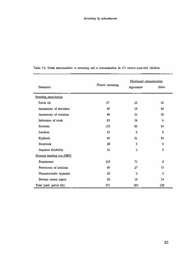
Screening by schoolnurses
Table 7.2. Trunk abnormalities at screening and at reexam.ination in 171 twelve-year-old children
Parameter
Standing examination
Pelvic tilt
Asymmetry of shoulders
Asymmetry of waistline
Imbalance of trunk
Scoliosis
Lordosis
Kyphosis
Swayback
Impaired flexibility
Forward bending test CFBD
Prominence
Persistence of scoliosis
Noncorrectable kyphosis
Deviant lateral aspect
Total (excl. pelvic tilt)
Nurses' screening
57
60
66
82
135
13
41
28
10
115
80
15
26
671
Physicians' reexamination
Agreement
35 20
18 16
24 20
16 6
63 14
6 6
21 14
3 0
2 6
71 8
27 15
2 5
10 14
263 120
83

Chapter 7
Table 7 .3. Number of trunk abnormalities on FBT at screening and at reexamination of 171 children
Reexamjnatjon
Number of abnormalities on FBT
0 1 2 3 4 Total
Screening o· 19 8 2 3 2 34
1 22 23 5 2 0 52
Number of abnormalities 2 25 25 20 0 3 73 on FBT
3 3 2 3 2 0 10
4 0 2 0 0 0 2
Total 69 60 30 7 5 171
normal FBT but abnormal standing examination
Table 7.4. Qassification of trunk abnormalities in 775 children. at school medical examination at age 11 year and age 13 year~ and at nurses' screening at age 12 year
Nurses' screening
SsfuoQl medical Total Normal FBT" Poor posture examination
Normal 11 + 13 year 511 408 80% 57 11% 46 9%
FBT 11 year 47 25 53% 18 38% 4 9%
FBT 13 year 82 26 32% 42 51% 14 17%
FBT 11 + 13 year 24 5 21% 15 63% 4 17%
Poor posture 111 61 55% 28 25% 22 19%
Total 775 525 68% 160 21% 90 12%
• FBT: Group Abnormal Forward Bending Test
84

Screening by schoolnurses
FBT. Fifty-two children had one abnormality on the FBT, 28 had two, 4 had three, and 3 had all four. These 87 children constituted 11% of the original population. At reexamination it appeared that in 50 (29%) children the FBT was not abnormal. In 15 (9%) children with normal FBT at screening, who had abnormalities on standing examination, the FBT was found to be abnormal as well.
Table 7.4 shows the results of the nurses' screening in relat;on to the results of the physical examination one year earlier and one year later. In 408 (53%) of the 775 children the FBT was considered normal on all three occasions: in 231 (57%) boys and 177 (48%) girls. In 238 (31%) children the FBT was considered abnormal at least on one occasion. An abnormal FBT on all three occasions was found in 15 (2%) children, five (1.2%) boys and ten (2.7%) girls.
In the first year the school physicians had referred 15 children of the screened population to the family physician for further assessment. In the year of screening two of them were referred to the family physician again. Fourteen other children were now referred to the family phyician for the first time. Only one of those had had an abnormal FBT in the previous year. The year after screening all 16 children had an abnormal FBT. The percentage of new referrals at age 12 amounted to 1.9% of the screened population.
Discussion
In our study, one in four children was referred for reexamination by the school physician. The large number of children screened as positive is one of the problems in scoliosis screening. In most countries the screening programs are on a three-tier basis. The first screening is done by a trained layman or school nurse. The second screening, to which is added a moire topography is done by a specially trained nurse. The third tier consists of X-ray and examination by an orthopedic surgeon. If necessary the child is then referred to a scoliosis center. Depending on the experience of the first screener, referral rates to the second screener range from 15% to 45%. None of the nurses in our study had previous experience in screening for trunk abnormalities. The percentage of referrals found in our study (24%) is in accordance with that in other studies. Referral rate for recheck will decrease with the growing experience of the first screener.:>-6
Mild truncal asymmetries are common in the normal population. Burwell et al.' found that about 15% of adolescents showed evidence of asymmetry of the trunk on quick visual examination of the back, including the FBT. When the surface of the back was measured with a "formulator body contour tracer" the rate of asymmetry rose to 25%. The FBT does not only produce a large number of false positives, it also identifies a fair number of true positives with small to serious abnormalities. Many slight scoliotic curvatures will never require treatroent. It cannot be predicted, however, which (slight) curvatures will progress. The second and third screenings will diminish the number of false-positives, and only those children will be referred that need orthopedic examination or treatment. School physicians strive at discerning the ones with "no-significant problem" from those
85

Chapter 7
"at risk" and those 11needing treatment". However, there is no way of foretelling which curves will progress.
In our study, 11.2% of the screened population was found positive on reexamination and only 2% of the screened population was referred for orthopedic assessment. Other studies also showed that 10% is abnormal on reexamination, and that between 2% and 5% of all screenees were referred for orthopedic assessment.J..s-n
The findings of the reexamination by school physicians were compared with the nurses' findings at screening. The school physicians confirmed from 11% to 62% of the single parameters; an abnormal FBT was confirmed in 51%. The best agreement was found on scoliosis and kyphosis in the standing child and on rib hump/lumbar prominence in the bending child. The nurses failed to detect 9% of single parameters and 9% of abnormalities on FBT. Other studies showed an even higher percentage of false-negatives, approximately 25% for scoliotic curves between 10 and 20 degreesY2 We were only able to determine the number of false-negatives in the group referred for reexamination. The specificity of the FBT is high13
; one in four children was singled out for reexamination. We therefore presume that there is little chance that scoliotic curvatures needing treatment have been overlooked in the other children.
The value of an additional screening program depends on how much it contributes to the regular school health care program, for instance early detection and treatment at an earlier stage. Comparing the results of the additional screening with the results of the two regular medical examinations we found that the consistency of an abnormal FBT was low. We think that this is caused by three factors: (a) the high specificity of the FBT (also very slight deviations are detected), (b) the child's stage of puberty development, and (c) the natural course of trunk abnormalities. Spontaneous correction may also occur in scoliosis with curves larger than 15 degrees.'' Not until after repeated measurements is it possible to determine whether scoliosis shows progression or regression. In our study population only a small group had an abnormal FBT on all three occasions. All newly referred children had been classified in Group Normal on the examination the previous year, and they were classified in Group FBT the following year. Although their trunk abnormality had now been detected one year earlier, it still remains doubtful if addition of a screening program to the regular examination by the school physician at age 11 and age 13 will be cost-effective. In those countries that have no health care program during adolescence, for instance the United States, an annual screening program for trunk abnormalities will contribute to early identification of children at risk
References 1. Hazebroek-Kampscbreur AA.JM. Hofman A. Dijk APh van. Linge B van. Prevalence of trunk
abnormalities in eleven-year-old schoolchildren in Rotterdam,. The Netherlands. J Pcd Orthop 1992;12:48o-4.
2. Vercauteren M. Van Bencdcn M. Verplaetse R,. Croene Ph, Uyttendacle D. Verdonk R Trunk asymmetries in a Belgian school population. Spine 1982;7:555-62.
86

Screening by schoolnurses
3. Annstrong GWD, Livermore NB, Suzuki N, Armstrong JG. Nonstandard vertebral rotation in scoliosis screening patients. Spine 1982;7:50-4.
4. Dunn BH, Hakala MW, McGee r..ffi. Scoliosis screening. Pediatrics 1978;61:794-6. 5. Howell JM, Craig PM, Dawe BG. Problems in scoliosis screening. Can J Public Health 1978;
69:293-6. 6. Gore DR. Passehl R. Sepic S, Dalton A. Scoliosis screening: results of a community project.
Pediatrics 1981;67:196-200. 7. Burwell RG, James NJ, Johnson F, Webb JK. Wilson YG. Standardised tnmk asymmetry scores.
J Bone Joint Surg [Br] 1983;65:452-63. 8. Lonstein JE. Screening for spinal deformities in Minnesota schools. Clin Orthop 1977;126:
33-42. 9. Flynn JC, Riddick MF, Keller TL. Screening for scoliosis in Florida schools. J Florida M A
1977;64:159-61. 10. Rogala EJ, Drummond DS, Gurr J. Scoliosis: incidence and natural history. J Bone Joint Surg
[Am] 1978;60:173-6. 11. Goldberg C. Fogarty EE, Blake NS, Dowling F, Regan BF. School scoliosis screening: a review
of 21.000 children. lr Mcd J 1983;76:247-9. 12. Viviani GR. Budgell L, Dok C. Tugwell P. Assessment of accuracy of the scoliosis school
screening examination. Am J Public Health 1984;74:497-8. 13. Wynne EJ. Scoliosis: to screen or not to screen. Can J Public Health 1984;75:277-80.
87


Chapter 8
Follow-up in children
referred for trunk abnormalities


Chapter 8
Follow-up in children referred for trunk abnormalities
Abstract
We conducted a follow-up study in 210 children referred for scoliosis and/or kyphosis. All had participated in a prospective follow-up study on prevalence and two-year cumulative incidence of trunk abnormalities among 6,507 children born in 1973. In 97%, follow-up data were obtained within 3 months, and at 3 to 6 years after referral. Orthopedic assessment occurred in 139 children, of whom 81 appeared to have a structural deformity. Ten children were braced. In most children, the initial curve did not exceed 20 degrees. Compliance with orthopedic observation was low. Observation of mild curves by school physicians may increase compliance.
Introduction
During the past decades screening programs for idiopathic scoliosis have been adopted in many countries. These programs aim at early detection of spinal deformities, for the earlier a curvature is recognized, the easier treatment and prevention of severe disabilities. In Rotterdam, The Netherlands, school physicians examine the trunk of all children during general health examinations at age 4 years, 7 years, 11 years, and in the second year of secondary education (age 13-14 years). Children with abnormal findings who need further clinical assessment are referred to the faroily physician and/or medical specialist. Although the Department of Youth Health Care of the Municipal Health Service Rotterdam did not have prevalence and incidence data regarding trunk deformities in the population, data regarding the number of children referred to the family physician were available. But we did not know whether children referred for scoliosis and kyphosis received timely and adequate treatment. Delay in treatment occurs as either patients' delay (non-compliance regarding referral or 'failed appointments'), or doctors' delay caused by difference of opinion between family physician and school physician regarding the necessity for orthopedic assessment.
Earlier, we set up a two-year prospective follow-up study in a cohort of children aged 11 years (born in 1973) in order to obtain prevalence and two-year cumnlative incidence data regarding trunk asymmetries, including scoliosis and kyphosis, and their determinants.' The present study deals with follow-up of children referred for scoliosis and/or kyphosis.
91

Chapter 8
Methods
Subjects Between September 1984 and August 1987, school physicians performed 9,771
exanrinations in 6,507 children (51.5% were boys: 3,354 boys, 3,153 girls). (Table 8.1). All were born in 1973. Referral for trunk abnormalities occurred 223 times concerning 210 children (38.1% were boys: 80 boys, 130 girls), this means in 2.3% of screening occasions and in 3.2% of the screened population.
Table 8.1. Populatio~ number of examinations~ number of referrals
Period
Sept 1984 - Aug 1985
Sept 1985 - Aug 1986
Sept 1986 - Aug 1987
Total
Examinations in 2y C.I. group
3,071
193
3,071
6,335
2y C.I.: two-year cumulative incidence
Method
Subjects with one examination
1,844
1,844
1,592
1,592
The follow-up study was done in two periods:
All
193
4,663
9,771
Referral
122
21
80
223
Period I: Follow-up within three months following referral by means of reply letters of family physician and/or orthopedic surgeon. The following items were noted: (1) compliance with referral, (2) family physician's policy (radiographic exanlination, physiotherapy, recheck, referral for orthopedic assessment), (3) results of initial orthopedic assessment (roentgenogram, degree and nature of deformity, discharge, observation or treatment decision). Period II: Between September 1989 and September 1990, 3 to 6 years after referral, follow-up was done retrospectively by means of (a) review of school health records, (b) questionnaire to parents and children regarding follow-through with recommendation of school physician, family physician and/or orthopedic surgeon. Parents' opinion to the referral was asked for, and all children were invited for physical examination of the trunk, (c) review of patient records of orthopedic outpatient clinic (degree and nature of deformity at initial and last examination, initial and last roentgenogram, treatment decision, duration of observation or treatment, number of visits, date of last visit, and compliance).
After consent was obtained physical examination of the trunk was perforroed by a physiotherapist (medical student) between January and September 1990. The examination was perforroed according to the same protocol (with addition of a scoliometer) as used between September 1984 and August 1987. The action which in retrospect should have been taken was recorded.
92

Follaw-up of referrals
Measurements Pelvic tilt was determined, and leg length inequality was corrected by placing one
or more 0.5 em boards under the short leg until horizontal symmetry of iliac crests and posterior iliac spines was obtained. After correction, the child's posture was observed. The standing child was viewed from the front and the back for symmetries in shoulders, scapulae and waistline. Balance of the thorax over the pelvis was assessed with a plumbline. Flexibility of the spine was examined in flexion, extension and side bending. The child was viewed from the side for areas of hyperor hypokyphosis and hyper- or hypolordosis. Trunk asymmetries and abnormal curvatures in the median plane in standing position were recorded as either absent or present, according to the proposed limits for structural trunk asymmetries by Vercauteren et ai.Z The forward bending test was performed, with the child standing with feet together and knees straight, bending at the waist, with the arms dependent and held with fingers and palms opposed. The back was viewed head on for symmetry; both sides were compared. The back was also viewed from the side to view kyphosis and rib humps. The following four components of the forward bending test were listed as either absent or present: (a) rib humps and lumbar prominences as signs of vertebral rotation (a ri b hump is not always associated with a scoliosis in upright position), (b) persistence of the standing scoliosis on forward bending to discriminate between postural and structural scoliosis (it will usually be associated with a rib hump or lumbar prominence), (c) correctability of the kyphosis to discriminate between postural and structural kyphosis, and (d) deviant lateral aspect of the trunk.
Flexibility of the hyperkyphosis was also tested by the prone hyperextension evaluation. Flexibility of lordosis was demonstrated in the forward bending test, but also with the child bending in a knee-chest position. We considered a forward bending test as abnormal if at least one of the four above mentioned components was present.
Measurement of the angle of trunk rotation was accomplished with a scoliometer developed by Pruijs et a!.', and comparable to the scoliometer of Bunnell.4 The angle of trunk rotation is the angle between the horizontal plane and a plane across the posterior part of the trunk at the point(s) of maximum deformity. The scoliometer was placed on the back at the apex of the deformity, the angle of the inclination is read from the scale and is noted in degrees. Criterion for referral to an orthopedic clinic is >5° of trunk rotation according to Bunnell4
, and > 7° according to Pruijs et al.3 Because Pruijs' criterion had to be established at the time of our study, we used the criterion according to Bunnell4 in the decision which action should have been taken.
Results
Table 8.2 shows data regarding abnormal forward bending test (FBT) and referral at baseline and two-year follow-up examination noted by school physicians. In 49 of 210 children, data of two-year follow-up examination were absent. lnformation
93

cr.u.pter 8
Screened population n = 6,507
(9,771 examinations)
Referral to family physician n = 210 (3.2%)
Family physician Failed Unknown n = 191 (2.9%) appointments
n = 13
I n=6
Referral to specialist No referral n = 138 (2.1%) n =53 (0.8%)
_l
Failed appointments Specialist Specialist n=2 n = 136 n=3
I I
Orthopedic assessment n = 139 (2.1%)
I I I
Stru~rn! NQnstructural Other !;]efQrrnities No defolJ!!i!Y: n = 81 (1.2%) n = 25 (0.4%) n = 22 (0.3%) n = 11 (0.2%)
scoliosis 66 scoliosis 14 kyphosis 8 kyphosis 11
kyph!sroL 7
Figure 8.1. Follow-up of children referred for scoliosis and/or kyphosis
94
Follow-up of referrals
regarding FBT could be obtained from school health records in 15 of these children, abnormal FBT was noted in 7 children and normal FBT in 8 children at the age of 16 years. In the other 34 children, who had moved outside Rotterdam, no information regarding FBT could be obtained.
Of the screened population, 210 (3.2%) children were referred for trunk abnormalities (scoliosis 2.5%, kyphosis 0.5%, kyphosis and scoliosis combined 0.2%). The male:female ratio in the screened population was 1:1, and in the referred population 1:1.6. Scoliosis was the main reason for referral, namely in 54 (67.5%) boys and in 107 (82.3%) girls, ratio 1:2. Referral for kyphosis and for kyphosis combined with scoliosis occurred in 18 (22.5%) and 8 (10.0%) boys, and in 17 (13.1 %) and 6 (4.6%) girls, respectively.
Follow-up data were collected in 204 children regarding 217 referrals; no information at all could be obtained in 6 (3%) children who had moved outside The Netherlands (Figure 8.1). In 43 referrals, the family physician had sent a reply-toreferral letter to the school physician (12 children of whom were sent to the orthopedic outpatient clinic). In all 139 children sent to the orthopedic clinic, the school physician received a reply-to-referral letter after first orthopedic examination. In 53 referrals, the family physician sent no reply letter, and information regarding findings and decision of family physician was then obtained from parents and family physicians by phone.
Failure to report to the family physician occurred in 17 referrals for reasons of indifference, fear, or domestic matters. Difference of opinion between family physician and school physician occurred in 14 (7%) of 200 referrals, they were dismissed after examination. Agreement between family physician and school physician was in 186 children (191 referrals). Of these, 138 children (140 referrals) were sent to an orthopedic outpatient clinic. In 53 children (60 referrals), the family physician did not recommend orthopedic assessment but he ordered a roentgenogram of the spine in 10 referrals, prescribed physiotherapy in 33 referrals, and second examination in 23 children. We do not have information regarding compliance with physiotherapy or second examination.
Orthopedic assessment took place in 139 children (2.1% of the screened population), two children had failed to report, and three other children went to the orthopedic outpatient clinic on their parents' initiative. Table 8.3 shows the diagnosis and treatment decision after fust examination, including radiographic examination in 117 of 139 children. Structural deformity was noted in 81 children (1.2% of screened population), 66 of whom had a structural scoliosis (male:female ratio was 1:3). Nonstructural deformity was noted in 25 children (male:female ratio was 1:1 for nonstructural scoliosis and 1:0.4 for nonstructural kyphosis). Other orthopedic deformities such as spondylolysis, spondylolisthesis, or vertebral malformation, were noted in 22 children, and in 11 children no orthopedic deformity was noted. Treatment decision was discharge (n=27) (with or without observation by family or school physician), observation (n=108) or brace treatment (n=4).
95
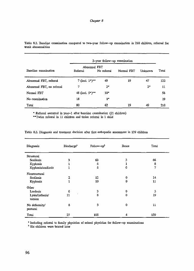
Chapter 8
Table 8.2. Baseline examination compared to two-year follow-up examination in 210 children, referred for trunk abnormalities
2-year follow-up examination
Abnormal FBT Baseline ex:anllnation Referral No referral Normal FBT Unknown
Abnormal FBT~ referral
Abnormal FBT~ no referral
Normal FBT
No examination
Total
7 (incL 1•)••
7
49
2'
10'
1'
62
• Referral occurred in year-1 after baseline examination (21 children) .. Twice referral in 11 children and thrice referral in 1 child
19 47
2'
19 49
Table 8.3. Diagnosis and treatment decision after first orthopedic assessment in 139 children
Diagnosis Discharge• Follow-up~> Brace
Structural Scoliosis 3 60 3 Kyphosis 1 6 1 Kyphosis/scoliosis 1 6 0
Nonstructural Scoliosis 2 12 0 Kyphosis I !0 0
Other Lordosis 0 3 0 L ysisllisthesisl 11 8 0 torsion
No deformity/ 8 3 0 postural
Total 27 108 4
a Including referral to family physician of school physician for follow-up examinations ~> Six children were braced later
96
Total
66 8 7
14 11
3 19
11
139
Total
122
11
58
19
210

Follow-up of referrals
Table 8.4. Radiographic examination in 117 children
Follow-up X-ray Parameter First X-ray No X-ray
Progression Unchanged Regression
Scoliosis 71 11 36 2 22 <100 35 2 18 0 10-19" 26 5 13 1 20-29" 5 2 2 1 ..:30" 5 2 3 0
Kyphosis 24 M. Scheuermann 9
Other deformities 5
No deformities 17
Table 85. Interval after first orthopedic assessment,. number of visits, duration of observation, and number of follow-up radiographic examinations in 116 children
Range Mean
Interval (months) 3- 12 5.8
Number of visits 1-23 6
Length of observation (months) 3-75 26
Radiographic examination" 2- 15 5
• In 64 chl1dren
Table 8.6. Compliance in 116 chl1dren receiving observation in orthopedic outpatient clinic
Drop out after first visit
Drop out during observation
Discharge from observation
Still observation or treatment
25
35
31
25
97

Chapter 8
Table 8.7. Response to questionnaire by referral to orthopedic clinic in 80 boys and 130 girls
No referral to Referral to orthopedic All Questionnaire orthopedic clinic clinic
No response boys 11 15 26 girls 21 13 34
No consent to examination boys 5 12 17 girls 8 32 40
Consent to examination boys 9 28 37 girls 17 39 56
Table 8.8. Treatment decision,. number of visits. length of observation and state of treatment in 210 nonrespondents and respondents with or without examination at age 17 years
Respondents Nomespondcnts No examination h-amination
No referral to orthopedic clinic 32 15 24
Orthopedic assessment 28 50 61 Discharge after first visit 7 3 13 Observation 20 39 47 Brace 1 8 1
Number of visits Range 1-7 1-24 1-14 Mean 2.2 6.3 3.2
Length of observation (months) Range 0-28 0-66 0-75 Mean 8 26 13
State of treatment Discharge from observation 4 6 21 Observation or treatment 3 16 6 Noncompliance 14 25 21
98
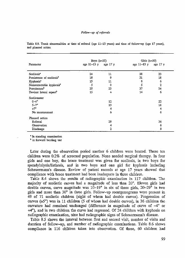
Follow-up of referrals
Table 8.9. Trunk abnormalities at time of referral (age 11-13 years) and time of follow-up (age 17 years), and planned action
Parameter
Scoliosis' Persistence of scoliosis~> Kyphosis" Noncorrectable kyphosisb Prominenceb Deviant lateral aspect'>
Scoliometer 0-4·
>7" No measurement
Planned action Referral Observation Discharge
• In standing examination ~> in forward bending test
Boys (n•35) age 11-13 y age 17 y
24 11 18 9 15 11 5 0
25 23 15 4
12 13 4 6
29 4 2
Girls (n-50) age 11-13 y age 17 y
33 23 31 18
8 6 5 2
37 34 14 6
22 18 4 6
36 6 8
Later during the observation period another 6 children were braced. These ten children were 0.2% of screened population. None needed surgical therapy. In four girls and one boy, the brace treatment was given for scoliosis, in two boys for spondylolysis/listhesis, and in two boys and one girl for kyphosis including Scheuermann's disease. Review of patient records at age 17 years showed that compliance with brace treatment had been inadequate in three children.
Table 8.4 shows the results of radiographic examination in 117 children. The majority of scoliotic curves had a magnitude of less than 20•. Eleven girls had double curves, curve magnitude was 10-19° in six of these girls, 20-29° in two girls and more than 30° in three girls. Follow-up roentgenograms were present in 49 of 71 scoliotic children (eight of whom had double curves). Progression of curves (~0) was in 11 children (5 of whom had double curves), in 36 children the curvature had remained unchanged (difference in magnitude of curve of -4 o or +4°), and in two children the curve had regressed. Of 24 children with kyphosis on radiographic examination, nine had radiographic signs of Scheuermann's disease.
Table 8.5 shows the interval between first and second visit, number of visits and duration of follow-up, and number of radiographic examinations. Table 8.6 shows compliance in 116 children taken into observation. Of these, 60 children had
99
Chapter 8
discontinued observation or treatment prematurely (25 children even after the first examination).
Of 210 mailed questionnaires, 150 (71.4%) were returned, and in 93 (44.3%) of referred children consent was given to physical examination, eight of whom eventually failed to report. No new address could be obtained in 10 children and 50 children failed to return the questionnaire even after reminder. There seemed to be no difference in response between boys and girls, but the group of nonrespondents contained less children referred to the orthopedic clinic and more failed appointments (fable 8.7). Asked three to six years after referral, parents' opinion to the referral was positive in 84.0% (126 cases), in 13 children (9.0%) the referral had worried the parents and in 11 children (7.6%) parents had thought the referral unnecessary. Although we do not know parents opinion in the group of nonrespondents, we know that compliance with referral to the family physician was in 91%. Of 150 respondents to the questionnaire, 111 had been sent to the orthopedic outpatient clinic, and 61 of whom had consented to examination at age 17 years. The group of nonrespondents and the groups with and without examination were not homogeneous regarding (initial) treatment decision, number of visits and duration of observation, and state of treatment 1n the group of respondents who refused examination, more children still had observation or treatment. This group contained probably more children with more severe deformities (Table 8.8). Table 8.9 shows the results of physical examination prior to referral (age 11 through 13 years) and of examination at follow-up (age 17 years) in 85 children. Rib hump or lumbar prominence were the most consistent findings. Five or more degrees of trunk rotation was measured in almost half of these children. The results of examination at time of referral and at follow-up were compared. In most children, the same decision should have been taken.
Discussion
The referral rate in our study, 3.2%, falls within the range of referral rates from scoliosis screening programs (2.1% to 7.5%)-'-'
We obtained follow-up data in 97% of referred children, including reported 'failed appointments' with family physician. The response to the questionnaire was also very good, considering the gap of 3 to 6 years between referral by the school physician and mailing of the questionnaire. Other authors reported less favorable administrative and medical follow-up data.5
•1o-11 Follow-up, administrative and
medical, is an essential aspect of any screening program. Screening is not diagnosis but an attempt to early detection of a disease. Screening is meaningless without referral for diagnosis and necessary treatment of children with positive or suspicious findings, unless it is done purely for epidemiologic research. Although response to the questionnaire was good, consent to examination was low and biased.
100
Follow-up of referrals
We do not apply the three-tier scoliosis screening. The trunk is examined as part of the general medical examination, in which radiographic examination is not included. Children with positive findings are directly referred to the family physician (children with less positive or suspicious findings will have a second examination in six months). The rate of 'failed appointments' with the family physician in our stndy group is comparable to that of failure to return to the final screening session in a three-tier system' or other 'failed appointments'.12
School physicians have more experience in examination of the adolescent trunk than family physicians (ratio school physician : examination was 1:300, ratio family physician : referral was 1:1). But in our health system the family physician makes the final decision to refer a child for orthopedic assessment. To stimulate referral for orthopedic assessment and to prevent delay in diagnosis and necessary treatment as much as possible, the family physician received additional information and a special referral letter. This has proved successful as is shown in the referral (for orthopedic assessment) rate of 72%. Although the family physicians and school physicians were in agreement in the majority of referrals, the family physician did not refer one in four children for further orthopedic assessment. In these children, we could only collect data immediately following referral by the school physician. No data were available regarding natnre and degree of deformity, or recheck. Judging from the questionnaire and school health records, we think that many children did not return for reexamination by the family physician. We do not think that these children included many with large curves in need of treatment But some of these children needed a second referral by the school physician before necessary treatment was started. At that time one of them had a curve of 43°.
One of the problems reported from screening programs is the high number of children undergoing radiographic examination who subsequently appear to have either negative roentgenograms or nonstructural deformities.~7-13 In our stndy group, less than 2% of the screened population was radiographically examined. Prevalence of nonstructnral deformities was less than reported in other studies.~13 Prevalence of proved scoliosis (1.1 %) was the same as reported by Lonstein et al." Prevalence of postnral kyphosis and Scheuermann's disease was in agreement with Savini et al8
,
but Ascani et al.14 reported a higher prevalence. Noncompliance with recommendation of orthopedic observation was high,
especially after first assessment. In most (86%) scoliotic patients, the initial curvatnre did not exceed 20°. It might be that a contradictory message is given to parents (on the one hand reassurance regarding the deformity, on the other hand recommendation for observation). Lack of information regarding natural history of scoliosis and necessity of a second examination (to exclude progression of the curve) might enhance drop out of observation. We recommend that after orthopedic assessment on size and natnre of the curvatnre, children with mild curves will be re-referred to the school physician for observation. The school health system, as part of the public health system, has an outreaching call-up system. Children will
101

Cho.pter 8
be called for observation once or twice yearly, and progression of mild curves will be detected in tbese dropouts. Noncompliance after several visits seemed related to increasing lengtb of interval between visits, and as patients stated, to the inversed length of time spent in waiting room aud in consulting room.
References 1. Hazebroek-Kampscbreur AAJM, Hofman A,. van Dijk APh. van Linge B. Prevalence of trunk abnor
malities in eleven-year-old schoolchildren in Rotterdam, The Netherlands. J Ped Orthop 1992;12:480-4.
2. Vercautcren M. Van Beneden M, Vcrplaetse R. Croene Ph,. Uyttendaele D, Verdonk R. Trunk asymmetries in a Belgian school population. Spine 1982;7:555-62.
3. Pruijs JEH,. Keessen W, Meer R van dcr, Wieringen JC van, Hageman MAPE. School screening for scoliosis: methodological considerations. Part I: external measurements. Spine 1992;17:431-6.
4. Bunnell \VP. An objective criterion for scoliosis screening. J Bone Joint Surg [Am.J 1984;66: 1381-7. 5. Flynn JC. Riddick MF, Price cr. Keller TL Present status of scoliosis screening in Florida schools. J
Florida Med Ass 1985;72:847-51. 6. Gore DR. Passehl R. Sepic S, Dalton A. Scoliosis screening: results of a community project. Pediatrics
1981;67,196-200. 7. Lonstein JE. Bjorklund S, Wanninger MH, Nelson RP. Voluntary school screening for scoliosis in
Minnesota. J Bone Joint Surg [Am] 1982;64:481-8. 8. Savini R. Cervellati S, Ponzo L,. Ciani A. Palmisani M. Rizqallah Y. Gargiulo G, Nardi S, Di Silvestre
M. L'incidenza dclla scoliosi idiopatica nel Comune eli Bologna. Chit Org Mov 1986;81:119-25. 9. Wynne EJ. Scoliosis: to screen or not to screen. Can J Public Health 1984;75:227-8.
10. Adler NS, Csongradi J, Bleck EE. School screening for scoliosis. One experience in California using clinical examination and moir6 topography. West J Med 1984;141:631-3.
11. Brooks HL. Azen SP, Gerberg E, Brooks R. Chan, L Scoliosis: a prospective epidemiological study. J Bone Joint Surg (Am]1975;57,968-72.
12. Steen JJ van der. Tcrugkomtrouw in de huisartspraktijk. Thesis, Leiden 1987. 13. Walker AP, Dickson RA. Schoolscreening and pelvic tilt scoliosis. Lancet 1984;2:152-153. 14. Ascani E, Salsano V, Giglio G. The incidence and early detection of spinal deformities. Ital J Orthop
Traumatol 1977;3:111-7.
102

Chapter 9
General discussion


Chapter 9
General discussion
This prospective follow-up study was set up to gain insight whether an extra screening for trunk abnormalities should be added to the current school health care program during adolescence. Annual screening of 6,000 to 7,000 students of the first grade of secondary school, including reexamination by school physicians if the screening is performed by school nurses, would mean an extra investment of manpower and money by the Department of Youth Health Care, Municipal Health Service Rotterdam, The Netherlands. In this chapter, I will first focus on the studies of prevalence, cumulative incidence and determinants of trunk abnormalities. In addition, the subquestions with respect to efficacy of the current frequency and the applied method of examination, feasibility of school nurses' screening, and follow-up of referred children will be discussed. I will then deal with recommendations, and implications for future research and policy-making.
Study findings
In many studies of scoliosis and scoliosis screening, the terminology of prevalence and incidence is mixed up.'_. Most of these studies, however, are prevalence studies. Different methods of discerning scoliotic from nonscoliotic children are used, varying from the one-minute forward bending test by trained laymen, physiotherapists or school nurses to a full clinical examination by orthopedic surgeons. A three-tier system is frequently used in scoliosis screening programs. An abnormal forward bending test on first screening is found in 10% to 45% of the children. After rescreening, 3 to 15% is still considered positive and will receive radiographic or other, Jess harmful diagnostic examinations, such as moire topography, prior to referral for orthopedic assessment Scoliosis of 10 degrees of more is found in 1 to 6.5% of the screened population, treatment of progressive scoliosis is necessary in 0.2 to 0.5% of the screened population. Abnormal kyphosis and other deformities are found in 0.1 to 2% of the screened population. The prevalence of Scheuermann's disease in the general population varies from 0.4 to 8% in the literature depending on whether the diagnosis is based on radiographic or clinical criteria. (Chapter 2 and Chapter 3). The findings of our study, as described in Chapter 5 and Chapter 6, are in agreement with other studies on scoliosis screening. At age 11 years, approximately 7% of the boys and 11% of the girls had an abnormal forward bending test. At age 13 years, the two-year cumulative incidence of an abnormal forward bending test was 10% and 13%, respectively. At age 11 years, prevalence of abnormal kyphosis on standing examination was 3.5%; the two-year cumulative incidence was 5.6%. These kyphoses could not be corrected in 0.3% and 0.8%, respectively. Of the screened population, 2.4% (boys 1.7%, girls 3.1 %) were
105

Chapter 9
referred to the family physician at age 11 years. Two years later, the referral rate was 2% (boys 1.7%, girls 2.3%).
Many authors have studied the relationship between growth and maturation on the one hand, and scoliosis on the other hand. The pubertal growth spurt is important in the natural history of scoliosis.>-' There is a wide variation in age of onset, duration, maximum and overall gain of adolescent growth spurt, and of sexual maturation. In our study of determinants, as described in Chapter 6, gender, height at baseline in both sexes, and onset of growth spurt and menarche at baseline in girls were associated with incidence of trunk abnormalities. In the discriminant analysis, however, the predictive value of the discriminant function was low. This means that no specific population at risk could be discerned. Logistic regression showed that the highest risk of future trunk abnormalities was in girls of height above median, who had already started with the adolescent growth spurt but had not yet reached menarche.
In Rotterdam, The Netherlands, the current school health care program for adolescents consists of periodical medical examinations at age 11 years and in the second grade of secondary school (age 13-14 years). The trunk is examined by observing the standing child from front, back and side, and on forward bending. An abnormal forward bending test is an alerting sign to look for clinical evidence of structural scoliosis. In the study design, we used an abnormal forward bending test as criterion for referral to the family physician. This criterion was very strict, and it turned out not to be usable in everyday practice. Of all children with an abnormal forward bending test (:10%), only one out of 3 to 4 was referred. Apparently, the others were judged not to be in need of referral yet. They were eligible for follow-up visit in 6 to 12 months. Although the referral rate appears to be low compared to the prevalence of abnormal forward bending tests, it is about thrice the usual referral rate of trunk abnormalities in our department. This is probably due to making trunk abnormalities object of registration. At the beginning of the present study, a pilot study was conducted to evaluate registration forms and procedure of referral. Five school physicians examined 1,200 eleven-year-old children during the periodical medical examination; the referral rate in the pilot group was 2.3%. In the same period, another 1,700 eleven-year-old children were examined by seven school physicians who did not participate in the pilot study. The referral rate in this group was 0.7% (unpublished). Of 4,926 eleven-year-old children examined during the periodical medical examination of schoolyear 1989/90, 5.2% needed intervention for trunk abnormalities, i.e. follow-up by the school physician in 4.5% and referral to the family physician in 0.7%.10 Several authors reported that while 2% of the screened children would be diagnosed as having adolescent idiopathic scoliosis, only 1-2 per 1,000 children screened would need treatment, either bracing or surgery!·11
-13 In addition to prevalence and
cumulative incidence data regarding trunk abnormalities, it is imperative to get information on the nature and degree of deformity in referred children. As described in Chapter 8, 3% of the screened population was referred to the family physician. Orthopedic assessment took place in two-thirds of referred children. Not all referred children were examined radiographically. A structural deformity
106

General discussion
was clinically diagnosed in 1.2% of the screened population. On radiographical examination, scoliosis of 10° or more was found in 0.6%, and of more than 20° in 0.2%. As in other studies, many referred children had small scoliotic curves, either physiological or pathological ones. It still remains impossible to identify those children whose curves are destined to progress. On radiographical examination, abnormal kyphosis was diagnosed in 0.4%, including signs of Scheuermann's disease in 0.2%. This is less than reported in other studies. However, our study population was younger than in most studies. Onset of Scheuermann's disease is rather late in adolescence, and early Scheuermann's disease can easily be confused with postural kyphosis!"-" Screening at a later age might detect more adolescents with Scheuermann's disease. In some children referred for scoliosis combined with kyphosis, the scoliotic curve proved to be less important than the kyphosis during follow-up. In summary, it appears that both the current method and frequency of trunk examination contribute effectively to early detection of trunk abnormalities. Besides, some of the problems usually encountered in school screening programs can be prevented.
In three-tier screening programs, children found to be positive on rescreening are subjected to radiographical examination. This means that many children with nonstructural deformities or with small curves receive unnecessary radiation. We did not encounter this problem on a large scale. In The Netherlands, family physicians are the core of the health care system. Under the Dutch health insurance system, school physicians neither refer directly for specialistic assessment nor order radiographical examination. In our study, whereas only very few screenees were referred to the family physician, even less were examined radiographically. Another problem encountered in the three-tier screening programs is the large number of children screened as positive. This may produce anxiety in children and their parents, and subsequently generate an unnecessary visit to the family physician or orthopedic surgeon. This phenomenon of unintended and inappropriate overreferral of children with 'physiological' asymmetries has been called 'schooliosis'.17 In The Netherlands, contrary to these screening programs performed by nurses or trained laymen, school physicians examine the backs of children during periodical health examinations, and decide whether or not to refer. We usually do not encounter the problem of overreferral on a large scale.
The results of the school nurses' screening, as described in Chapter 7, are consistent with the results of school screening programs organised according to the three-tier system. Referred on screening was 24%, positive on reexamination was 11%, and the referral rate was 2%. All screened children were eligible for the regular school medical examination one year after the screening. We assume that only in exceptional cases parents of nonreferred children have sought further medical assessment. After adequate training, nurses are well able to perform screening for trunk abnormalities. It is to be expected that the large number of referrals for reexamination will decrease with the nurses' growing experience.
The benefit of screening lies, however, not only in early detection but also in the early application of effective treatment. Administrative and medical followup is an essential aspect of any screening program and periodical medical
107

Chapter 9
examination m school health care. Children with suspected structural spinal deformities are referred to the family physician for further assessment. Radiological examination of the thoracolumbar spine is the most accurate way to evaluate type, site, magnitude and apex of the deformity, and structural changes of vertebrae, both in diagnosis and treatment. Feedback of this information is essential for school physicians in evaluating their criteria for referral. Having used a special referral form in this study, we received more reply-to-referral letters than usual. In this study, two-thirds of referred children were examined in an orthopedic outpatient clinic (Chapter 8). No orthopedic examination occurred in the others, and therefore no further evaluation of type and magnitude of the deformity was done. On the one hand, because children and parents did not comply with referral to the family physician; on the other hand, because the family physician deemed orthopedic assessment unnecessary irrespective of agreement of findings. For some children, this meant a delay in early application of effective treatment. When confronted with a child referred with trunk asymmetry, the orthopedic surgeon has to decide between treatment, follow-up, or discharge, if necessary after radiological examination. ln most cases, followup was decided on, even in children with diagnosed nonstructural deformities or with curves smaller than 10°. Noncompliance with recommendation of orthopedic follow-up proved to be high, especially after the first visit to the orthopedic department. When patients fail to turn up for a second visit, most orthopedic outpatient clinics do not take initiatives for a new appointment. Decision of discharge in some children went together with re-referral to the family physician, or to the school physician. The school health service, as part of the public health system, has an outreaching call-up system. Children needing observation for trunk abnormalities, either after routine examination by the school physician or after re-referral, will be called up once or twice yearly for observation.
Finally, the purpose of screening is early detection of a disease or deformity. In our study, the additional screening has been successful in this attempt, at any rate for a small number of children. However, considering that the method and frequency of periodical medical examinations during adolescence have proved to be effective in the early detection of trunk abnormalities, and taken into account the cost/benefit aspects of an additional screening, we conclude that an extra screening need not be added to the current school health care program during adolescence. Still, it remains necessary to examine the trunk at least twice during adolescence because of the variation in age of onset, duration, maximum and overall gain of adolescent growth spurt, and of sexual maturation.
Recommendations
The periodical medical examination provides the Department of Youth Health Care with a good instrument for epidemiological research, health care research, and, moreover, for quality management.
The two periodical medical examinations performed during adolescence effectively contribute to timely recognition of trunk abnormalities. Addition of an
108

General discussion
extra screening is not necessary. A reduction of the number of periodical examinations is undesirable, because of the variation in age of onset of adolescent growth spurt and sexual maturity. Gender and height in both boys and girls, onset of pubertal growth spurt in girls, and menarche are related to having an abnormal forward bending test. Tall girls undergoing the pubertal growth spurt, but who have not yet experienced menarche, have an increased risk of an abnormal forward bending test. Thus, physical growth and maturation ought to be taken into account in the decision making leading to referral.
About 10% of the population of (pre)adolescents appear to have an abnormal forward bending test. Identification of children with a progressive scoliosis is not (yet) possible at an early stage. Referral to the family doctor on the basis of a single forward bending lest is only advisable if distinct abnormalities are found. In case of less distinct abnormalities, re-examination (after six months) by the school physician is desirable. In the decision whether someone found to have a deformity should be referred or merely observed, status of growth and maturation has to be taken into account. Half of the children referred with minor structural or non-structural abnormalities did not participate in the follow-up examination at the orthopedic clinic. This situation might be improved by instituting a policy of active calling by the specialist, or by re-referral to the school physician for half-yearly observation. Besides, feedback to the first level of referral (school physician) is important in view of the guidance of the individual child to be provided by youth health care, and with regard to quality control, i.e. feedback to the school physician's own policy.
School physician, family physician and specialist each specifically contribute to the process of detection and treatment of trunk abnormalities in children. Good communication and insight into one another's (im)possibilities will add to optimal proceeding of this process.
Implications for future research and policy-making
At the onset of our study, annual screening for scoliosis and other trunk deformities was recommended during the period of adolescent growth (10 through 16 years). In our opinion, annual screening for trunk abnormalities is not necessary, but because of the large variation of onset and duration of adolescent growth spurt it is imperative to examine all adolescents at least twice in this period. In the second half of the 1980s, the public health budget in our country was cut down. This had consequences for the school health care programs. Many school health care services have either abolished one or both periodical medical examinations in adolescence, or have substituted these by a screening program by nurses (e.g. trunk abnormalities, blood pressure, visual and hearing impairments, height and weight) or a health questionnaire. We think that the costs and benefits of these screening programs must be compared to the costs and benefits of periodical medical examinations. Apart from the costs of the screening program itself, other costs have to be taken into consideration, such as costs of training of each new school nurse, and the costs of reexamination by the school physician of
109
Chapter 9
those outside the norm. Besides, we observe a leveling out of the difference between salaries of school physicians and school nurses. Consequently, it might be that screening for trunk abnormalities by nurses will be just as, or even more, expensive as the examination for trunk abnormalities as part of the periodical medical examination. As for the benefits, we assume that the number of adolescents benefiting from a screening program is less than that benefiting from the periodical medical examination. Screening is focused on one or two health problems only, whereas in periodical medical examinations school physicians assess status of health, growth, development, and maturation, and the factors that affect them; dependent on the findings, interventions are taken. In our opinion, substitution of periodical medical examinations by screening will mean a deterioration in preventive adolescent health care.
Scoliosis screening programs detect a large number of children with smaller curves. The natural history of scoliosis, including the progression of the curves, is not fully understood. At the time, it cannot be predicted which of the children with minor curves will progress to severe ones. Therefore, curves between 10 and 20° in the immature child must be monitored for progression. ln order to reduce overtreatment, these children can be re-referred for observation to the school physician. The initial orthopedic assessment should preferably include an objective measurement, such as the angle of trunk rotation by using a scoliometer according to Pruijs et al.18 or Bunnell.19 ln monitoring these children, school physicians can measure the angle of trunk rotation with a similar instrument. It is necessary to establish an upper limit for non-orthopedic follow-up of these mild cases. Our own experience with the scoliometer is limited. Pruijs et al.18 define the distinction between normal and pathological cases as a borderline or danger zone of 3-7° of rotation.
ln children with smaller curves, noncompliance with the advice to return for a second visit to the orthopedic outpatient clinic was high. Noncompliance may be reduced if the orthopedic surgeon refers the child back to the school physician; because of the outreaching policy of preventive health care, initiatives for an appointment for observation are taken by the school health service. In order to enhance compliance with observation or treatment, the orthopedic outpatient clinic can also take the initiative for a new appointment in those who fail to turn up. We have the impression that orthopedic surgeons do not apply the same decision-making process concerning children with scoliotic curves under 20°. A standard of management has to be developed with regard to assessment at first visit, and contents of reply-to-referral letter, as well as to follow-up, especially in children with mild scoliotic curves. Questions to be dealt with concern frequency of and intervals between check-ups, the duration of follow-up, and the choice of the physician (orthopedic surgeon, family physician or school physician) who will follow the child with mild scoliosis. ln case the child is followed by the family physician or school physician, it should be entirely clear when the child should be rechecked by the orthopedic surgeon.
110

General discussion
References 1. Brooks HL,. Azen SP, Gerberg E. Brooks R, Chan, L Scoliosis: a prospective epidemiological
study. J Bone Joint Surg [Am]1975;57,968-72. 2. Rogala EJ, Drummond DS, Gurr J. Scoliosis: incidence and natural history. J Bone Joint Surg
[Am]1978;60,1T.>-6. 3. Owen R, Taylor JF, McKendrick 0, Dangerfield P. Current incidence of scoliosis in school
children in the city of Liverpool. In: Zorab PA, Siegler D, cds. Scoliosis 1979. London: Academic Press, 1980:31-4.
4. Lonstein JE, Bjorklund S, Wanninger MH, Nelson RP. Voluntary school screening for scoliosis in Minnesota. J Bone Joint Surg [Am}l982;64:481-8.
5. Duval-BeaupCre G. Pathogenic relationship between scoliosis and growth. In: Zorab PA ed Scoliosis and growth. Edinburgh, London: Churchill Livingstone. 1971;58-64.
6. Willner S. A study of growth in girls with adolescent idiopathic structural scoliosis. Qin Orthop 1974;101,129-35.
7. Willner S. A study of height, weight and menarche in girls with idiopathic structural scoliosis. Acta Orthop Scand 1975;46o?l -83.
8. Dickson RA. Sevin EA Growth and idiopathic scoliosis: a longitudinal cohort study. J Bone Joint Surg [br]1982;64,385.
9. NlSSinen M,. HeliOvaara M, Ylikoski M,. Poussa M. Trunk asymmetry and screening for scoliosis: a longitudinal cohort study of pubertal schoolchildren. Acta Paediatr 1993;82:77-82.
10. Jansen w. Hazebroek-Kampschreur AAJM. Basisscboollccrlingen: ons een zorg?! Rotterdam: GGD, JZ-01, 1992,48.
11. Kane WJ, Moe JH. A scoliosis-prevalence survey in Minnesota. Clin Orthop 1970;69:216-8. 12. Dickson RA. Stamper P. Sharp ~ Harker P. School screening for scoliosis: cohort study of
clinical course. Br Med J 1980;281:265-7. 13. Leaver JM, Alvik ~ Warren r..ID. Prescriptive screening for adolescent idiopathic scoliosis: a
review of the evidence. Int J Epidcm.iol 1982;11:101-11. 14. Kling TF, Hensinger RN. Scheuermann's disease: natural history, current concepts and
management. In: Dickson RA,. Bradford D S. Management of spinal deformities. London: Butterwortbs, 1984:252-74.
15. Deacon P. Berkin CR. Dickson RA Combined idiopathic kyphosis and scoliosis. An analysis of the lateral spinal curvatures associated with Scheuermann's disease. J Bone Joint S [Br} 1985; 6H89-92.
16. Mau H. Scoliosis screening in West Germany and its pitfalls with Scheuermann's disease. Arch Ortbop Trauma Surg.1985;104:201-6.
17. Dvonch VM. Siegler AH. Qoppas CC. Bunch WH. The epidemiology of "schooliosis". J Pediatr Orthop 1990;10:206-7.
18. Pruijs JEH. Keessen W, Meer R van der, Wieringen JC van, Hageman MAPE. School screening for scoliosis: methodologic considerations. Part 1: external measurements. Spine 1992;17:431-6.
19. Bunnell WP. An objective criterion for scoliosis screening. J Bone Joint Surg [Am] 1984;66: 1381-7.
111


Summary


Swnmary
In Chapter 1 the rationale for this study is given: questions asked in the Lower House of Parliament on the subject of the foundation of the Association of Scoliosis Patients and their Parents, and reports in the medical literature describing a decrease in the number of severe cases of scoliosis since the introduction of annual scoliosis screening in children in the age group 10-15 years. The aim of the study was stated as: to assess whether an extra screening should be added to the two periodical medical examinations for (pre )adolescents.
Chapter 2 contains a survey of the literature on the epidemiology of trunk abnormalities, such as scoliosis and kyphosis. Chapter 3 contains a survey of the literature on scoliosis screening.
Chapter 4 describes the results of the prevalence study done in 1984/1985. This study was embedded in the health care program for eleven-year-old children; all were born in 1973 and attended schools in Rotterdam. Examination of back and posture inclnded recording of eight variables of the standing examination (symmetry of shoulders, symmetry of waistline, imbalance of the trnnk, scoliosis, lordosis, kyphosis, swayback and flexibility) and four variables of the forward bending test (FB1) (rib hump or lumbar prominence, persistence of scoliosis noted in the standing examination, correction of abnormal kyphosis, lateral aspect). Besides several background variables (gender, ethnic origin, month of birth, school), anthropometric data such as height, weight, puberty characteristics, onset of growth spurt and menarche were recorded. In 84% of the 4,915 children no abnormalities on standing examination and/or the FBT were seen. Abnormalities on the FBT were seen in 9%, and 2% of the children were referred to the family physician for further examination. For boys the corresponding figures were 86%, 7%, and 1.7%; for girls 81%, 11%, and 3%.
Chapter 5 reports the results of a two-year cumulative incidence study done in 1986/1987. The study was embedded in the school health care program in the second grade of secondary school, and included 2,819 children at risk. The twoyear cumulative incidence was 11% (boys 10%, girls 13%). A normal FBT both at baseline and at follow-up was found in 82% of the children. In more than half of the children who had an abnormal FBT at the age of 11 years, it was normal two years later. Children should only be referred for additional examination if a distinctly abnormal or a repeatedly abnormal FBT is found. Two percent of the population was referred to the family doctor.
Chapter 6 goes into the relation between gender, anthropometric factors, and pubertal development at the age of 11 years, and the incidence of trnnk abnormalities (defined as abnormal FBT) at the age of 13 years. First the different variables were processed in cross tables; cumulative incidence and relative risk, with the 95% reliability interval, were then calculated. The logistic model was then used to take into account all variables simultaneously. Boys had a lower risk of having an abnormal FBT than girls. Height was strongest related to having an abnormal FBT, both in boys and in girls. After correction for weight, onset of pubertal growth spurt, stage of pubertal development, and
115
menarche, the relative risk increased. Girls experiencing pubertal growth spurt had an increased risk of an abnormal FBT compared to girls who had not yet started growth spurt. Menarche was inversely related to the risk of an abuormal FBT. From the results of analyses in symmetric eleven-year-old children it appeared that tall girls undergoing the pubertal growth spurt, but who have not yet experienced menarche, have an increased risk of an abnormal FBT at the age of 13 years. To complete the analysis, all variables measured at the age of 11 years were put in a discriminant function with the FBT at age 13 years as outcome variable. The predictive value of the discriminant function appeared to be low.
Chapter 7 reports the results of the screening in 1985/1986. Five school nurses examined 775 children from the cohort for 12 variables of back and posture examination. Children with an abnormal FBT (21 %) were re-examined by the school physician. In half of them the abnormal FBT was confirmed: this involved well over 11% of the screened population. About 2% of the screened population was referred to the family doctor. The consistency of an abnormal FBT in three consecutive years was low (2%). But in 31% of the children the FBT had been abnormal at one of the three occasions at least; 53% of the children showed symmetry at all three consecutive examinations.
Chapter 8 reports the results of the follow-up study in 210 children, born in 1973, who had been referred for trunk abnormalities betweeu 1984 and 1987. Data about observing the referral, the policy of the family doctor, and the findings and policy at the first orthopedic examination were collected within three months after the referral; an additional follow-up study was done in 1989/1990. Between 1984 and 1987 a total of 9,771 examinations had been performed in 6,507 children born in 1973; in 3% of children referral to the family doctor occurred. The family doctor had referred 2% of the populatiou for further orthopedic examination. At orthopedic examination a structnral abnormality was diagnosed in 1.2%, and 0.2% had a scoliosis exceeding 20°. Almost all children were invited to undergo a second examination, even when the abnormality was minor or non-structuraL But it appeared that more than half of the children had not complied with it; they had not made a new appointment. If re-referral to the school physician should be institnted, these children will be called for examination every six months.
Chapter 9 concludes this thesis with a discussion answering the questions fundamental to this stndy. The study has showu that addition of an extra scoliosis screening to the two regular school medical examinations for adolescents is not necessary. The method employed in examination of back and posture and the frequency of the periodical medical examinations contributed sufficiently to early recognition of trunk abnormalities. Whether treatment is indeed started early enough, possibly depends on other factors. Scoliosis screening by school nurses was quite feasible. Before deciding to replace one of the school medical examinations during adolescence by a scoliosis screening, it is advisable to make a cost-benefit analysis.
116

Samenvatting

Sam en vatting
In hoofdstuk 1 wordt de aanleiding tot dit onderzoek beschreven: vragen gesteld in de Tweede Karner der Staten-Generaal over de oprichting de Vereniging van Scoliose Patienten en hun Ouders, en berichten in de medische literatuur over de afname van het aantal emstige gevallen van scoliose sinds de invoering van jaarlijkse scoliosescreening bij kinderen in de leeftijdsgroep 10 tot en met 15 jaar. Het doe! van het onderzoek werd omschreven als: nagaan of een extra screeningsonderzoek moet worden toegevoegd aan de twee periodieke geneesknndige onderzoeken (PGO) die tijdens de (pre-)adolescentie plaatsvinden.
Hoofdstuk 2 bevat een overzicht van de literatuur over de epidemiologie van afwijkingen van de wervelkolom, zoals scoliose en kyphose. Hoofdstuk 3 bevat een overzicht van de literatuur over scoliosescreening.
Hoofdstuk 4 beschrijft de resultaten van het prevalentie-ouderzoek in 1984/85. Het onderzoek was ingebed in het PGO bij elfjarige kinderen; allen waren geboren in 1973 en in Rotterdam schoolgaand. Bij het rug- en houdingsonderzoek werden twaalf variabelen geregistreerd, respectievelijk acht variabelen bij staand onderzoek (symmetrie van schouders, symmetrie van tailledriehoek, in het !ood staan van de wervelkolom, scoliose, lordose, kyphose, swayback, flexibiliteit) en vier variabelen van de buigtest (gibbus of Jumbale torsie, het blijven bestaan van de scoliose uit het staand onderzoek, corrigeerbaarheid van de abnormale kyphose, lateraal aspect). Naast enkele achtergrondvariabelen (geslacht, etnische afkomst, geboortemaand, school) werden anthropometrische gegevens als lengte, gewicht, puberteitskenmerken, groeispurt en menarche geregistreerd. Bij 84% van de 4915 kinderen werden noch afwijkingen op het staand onderzoek noch op de buigtest genoteerd. Bij 9% van de kinderen was de buigtest afwijkend, 2% van de kinderen werd naar de huisarts verwezen voor verder onderzoek. Voor de jongens was dit respectievelijk 86%, 7% en 1,7%, en voor de meisjes 81%, 11% en 3%.
Hoofdstuk 5 beschrijft de resultaten van het twee-jaars cumulatieve incidentie onderzoek in 1986/87. Het onderzoek was ingebed in het PGO in de tweede klas van het voortgezet onderwijs; aan het onderzoek namen 2819 kinderen nit het cohort dee!. De twee-jaars cumulatieve incidentie was 10% (9% jongens, 11% meisjes). Bij 82% van de kinderen was de buigtest normaal, zowel bij dit onderzoek als ook twee jaar eerder. Bij meer dan de helft van de kinderen bij wie op elfjarige leeftijd een afwijkende buigtest was geconstateerd, was de buigtest twee jaar later niet meer afwijkend. Verwijzing voor verder onderzoek dient aileen plaats te vinden bij een duidelijk afwijkende of herhaald afwijkende buigtest. Verwijzing naar de huisarts vond plaats voor 2% van de populatie.
Hoofdstuk 6 behandelt het verband tussen geslacht, anthropometrische factoren en puberteitsontwikkeling op elfjarige leeftijd, en de incidentie van wervelkolomafwijkingen (gedefinieerd als afwijkende buigtest) op dertienjarige leeftijd. Berst zijn de verschil!ende variabelen verwerkt in kruistabellen; cumulatieve incidentie en relatief risico, met het 95% betrouwbaarheidsinterval, werden berekend. Daarna werd het logistisch model gebruikt om met alle variabelen
119
Samenvatting
tegelijk rekening te houden. J ongens had den een Jager risico van optreden van een afwijkende buigtest dan meisjes. De lengte was het sterkst gerelateerd aan het optreden van een afwijkende buigtest, zowel bij de jongens als bij de meisjes. Na correctie voor gewicht, begin van puberteitsgroeispurt, fase van puberteitsontwikkeling en menarche nam het relatief risico toe. Meisjes in de puberteitsgroeispurt hadden een verhoogd risico van een afwijkende buigtest, vergeleken met meisjes die nog niet in de puberteitsgroeispurt waren. Menarche was omgekeerd gerelateerd aan het risico van een afwijkende buigtest. Uit de resultaten van de analyses bij symmetrische elfjarige kinderen bleek, dat lange meisjes bij wie wel de puberteitsgroeispurt begonnen is, maar bij wie de menarche nog niet is opgetreden, een verhoogd risico van optreden van een positieve bnigtest op dertienjarige leeftijd hebben. Ter afronding van de analyse werden aile variabelen, gemeten op elfjarige leeftijd, in een discriminantfunctie gebracht met als uitkomstvariabele de buigtest op dertienjarige Jeeftijd. De predictieve waarde van de discriminantfunctie bleek gering.
Hoofdstuk 7 beschrijft de resultaten van he! screeningsonderzoek in 1985/86. Vijf schoolverpleegkundigen hebben 775 kinderen uit het cohort gescreend op 12 variabelen van bet rug- en houdingsonderzoek. Kinderen met een afwijkende bnigtest (21 %) kwamen voor heronderzoek bij de schoolarts. In de belft van de gevallen werd de afwijkende buigtest bevestigd; dit betrof ruim 11% van de gescreende populatie. Verwijzing naar de buisarts vond plaats bij ongeveer 2% van de gescreende populatie. De consistentie van een afwijkende buigtest in drie opeenvolgende jaren was Jaag (2%). Wel was bij 31% van de kinderen op tenminste een van de drie onderzoeksmomenten de buigtest afwijkend geweest; 53% van de kinderen was op alle drie onderzoeksmomenten symmetrisch.
Hoofdstuk 8 beschrijft de resultaten van het follow-up onderzoek bij 210 kinderen, geboren in 1973, die tussen 1984 en 1987 wegens afwijkingen van de wervelkolom verwezen waren. Gegevens betreffende het opvolgen van de verwijzing, het beleid van de huisarts, en de bevindingen en het beleid bij bet eerste orthopedisch onderzoek werden binnen drie maanden na de verwijzing verzameld; aanvullend follow-up onderzoek werd verricbt in 1989/90. Tussen 1984 en 1987 waren 9771 onderzoeken bij 6507 kinderen, geboren in 1973, verricht; bij 3% van de kinderen vond verwijzing naar de huisarts plaats. Door de huisarts werd 2% van de populatie doorverwezen voor orthopedisch onderzoek. Bij orthopedisch onderzoek werd bij 1,2% een structurele afwijking vastgesteld, 0,2% had een scoliose groter dan 20°. Voor bijna alle kinderen werd een tweede onderzoek afgesproken, ook wanneer de afwijking gering of nietstructureel was. Meer dan de helft van de kinderen bleek hieraan geen gehoor gegeven te hebben, een nieuwe afspraak werd niet gemaakt. Indien terugverwijzing naar de schoolarts plaatsvindt, worden deze kinderen halfjaarlijks opgeroepen voor onderzoek.
Hoofdstuk 9 sluit het proefschrift af met een discussie, waarin de vraagstellingen van het onderzoek worden beantwoord. Het onderzoek beeft aangetoond, dat toevoeging van een screening op scoliose aan de twee PGO's tijdens de adolescentie niet noodzakelijk is. De gebruikte methode van rug- en houdingsonderzoek en de toegepaste frequentie van het PGO droegen in
120
Samenvatting
voldoende mate bij aan de vroegtijdige onderkenning van afwijkingen van de wervelkolom. Of er ook tijdig met adequate behande!ing begonnen wordt, is mogelijk van andere factoren afhankelijk. Screening op scoliose door schoolverpleegknndigen was goed uitvoerbaar. Macht men toch overwegen een PGO in de adolescentie te vervangen door een screening op sco!iose, dan is het gewenst eerst een kosten-baten analyse uit te voeren.
121


Epiloog
Een proefschrift is meer dan een verslag van een onderzoek. Het is een verhaal waarin velen een rol hebben gespeeld.
Allereerst Frans, met liefde en dankbaarheid denk ik aan de positieve bekrachtiging en de ruimte die je mij altijd gegeven hebt. Samen hebben wij deze belangrijke fase afgesloten, en ik kijk emaar uit om samen met jou de volgende fase in te gaan. Aan jou, Frans, draag ik dit boekje op.
Mijn beide promotores, Prof. Dr B. van Linge, em. hoogleraar Orthopedic, en Prof. Dr A. Hofman, hoogleraar Epidemiologic, ben ik vee! dank verschuldigd voor hun begeleiding, steun en niet aflatend vertrouwen in de goede afloop.
Hooggeleerde Van Linge, beste Bert, jouw aanbod om het onderzoek als promotor te begeleiden heb ik na enige aarzeling aangenomen. Nu het afgerond voor mij ligt, denk ik met vee! genoegen terug aan de stimulerende gesprekken, waarin de visie vanuit de k!inische situatie en vanuit de jeugdgezondheidszorg elkaar mochten ontmoeten.
Hooggeleerde Hofman, beste Bert, jouw kritische houding en epidemiologische deskundigheid waren onmisbaar bij de voltooiing van het proefschrift Het was een inspirerende ervaring om nu op een geheel andere wijze met jou samen te werken dan eertijds bij het Zuigelingenonderzoek.
Prof. Dr E. van der Does, hoogleraar Huisartsgeneeskunde, en Prof. Dr T.W.J. Schulpen, hoogleraar Sociale Pediatric, dank ik voor hun kritische beoordeling van het manuscript en voor hun bereidheidheid om zitting te nemen in de promotiecommissie.
Hooggeleerde Van der Does, beste Emile, ik ben je zeer erkente!ijk dat ik het manuscript met jou mocht doorspreken. De in het proefschrift onderbelichte rol van de huisarts doet geen recht aan de werkelijkheid, maar kwam voort uit de gekozen onderzoeksopzet.
Hooggeleerde Schulpen, beste Tom, ik dank je voor de stimulerende discussies die verder gingen dan het onderwerp van dit proefschrift.
Prof. Dr F. Sturmans, Prof. Dr F.C. Verhulst en Dr A.F.M. Diepstraten dank ik voor hun bereidheid om zitting te nemen in de promotiecommissie.
Barend Verbiest, ortbopedisch chirurg, dank ik voor zijn enthousiaste inbreng bij de opzet en uitvoering van de trainingsbijeenkomsten. Beste Barend, jij verzorgde niet aileen de theoretische instructie binnen de GGD, maar je schiep ook de mogelijkheid voor de instructie in de praktijk door ons de gastvrijheid van je afdeling aan te bieden. Jouw voorstel aan mij om een voordracht over het onderzoek op de jaarvergadering van de Nederlandse Orthopaeden Vereniging te houden heeft uiteindelijk tot dit proefschrift geleid.
De gehele afdeling Jeugdgezondheidszorg heeft aan het 'scolioseproject' meegewerkt, afdelingsleiding, artsen, verpleegkundigen, doktersassistenten en administratieve medewerkers.
Allereerst wil ik Dr N.W. Dekema-Klaasse bedanken. Lieve Nel, samen met jou stond ik aan het begin van het scolioseproject. Jij hebt mij steeds gestimuleerd om hierop te promoveren. Ik ben zeer verheugd dat jij paranimf bij
123

Epiloog
de promotie bent. Met jou te hebben mogen samenwerken was een unieke ervaring, het zijn ook mijn prettigste jaren binnen de GGD geweest.
Sera Ordonez, Joke Belder en Linda van de Kamp hebben in verschillende fasen van het onderzoek secretariele ondersteuning verleend. Dhr Jan Schipper zorgde ervoor dat aile benodigde formulieren op tijd klaar waren. Jannie van der Valk, Wil Pluymers, Jopie Breedveld, Rene Boer, Roepa Badal en Marga van Burgh waren altijd bereid om de adressenlijst te actualiseren. Hiervoor ben ik hen dankbaar.
Het welslagen van het scolioseproject is uiteindelijk te danken geweest aan de medewerking van de kinderen en hun ouders, de huisartsen en de orthopeden in de regia, maar vooral aan de artsen en verpleegkundigen die het onderzoek bij de kinderen uitvoerden en de gegevens registreerden. Jullie hebben altijd op positieve wijze mijn verzoeken om medewerking aan epidemiologische onderzoeken gehonoreerd. Hiermee hebben jullie bijgedragen aan het beeld van de afdeling Jeugd(gezondheids)zorg GGD Rotterdam als een afdeling waar jeugdgezondheidszorg en epidemiologisch onderzoek samengaan. lk ben aile collega's, verpleegkundigen en doktersassistenten zeer erkentelijk voor de sarnenwerking, in he! bijzonder Annemarie Tavenier-Hansen en Fifi Pouw-Tan voor hun inbreng bij het follow-up onderzoek.
Zonder Peter Tangkau was het follow-up onderzoek beperkt gebleven tot de eerste drie maanden na verwijzing. Beste Peter, niet aileen heb je op nauwgezette wijze he! onderzoek uitgevoerd, je maakte je ook in een snel tempo vertrouwd met Wordperfect, dBase en SPSS. Vee! dank voor aile inzet!
Belangrijk voor het onderzoek was ook de samenwerking met andere afdelingen van de GGD, zoals Informatievoorziening & Automatisering, Epidemiologic en de Reproduktie.
M:ujan Terlouw en Annelies Wessels voerden aile gegevens in. Bert van de Meeberg zorgde voor de koppeling van de verschillende bestanden.
Hans van Oers gaf hulp bij he! schonen van de bestanden en deed de eerste analyse.
Ad van Dijk heeft uren achter de computer doorgebracht bij het uitvoeren van de analyses. Voor zijn deskundige statistische adviezen en continue bereidheid tot discussie ben ik hem zeer erkentelijk. Ad, ik verheug mij op onze toekomstige samenwerking.
Annette Lengkeek zorgde met grate accuratesse dat aangevraagde literatuur en dia's op tijd gereed waren. Tevens wist zij altijd op korte termijn een plekje in de agenda van de 'Prof te vinden. Ik wil haar hiervoor hartelijk bedanken.
Ko Hagoort dank ik voor de zeer deskundige hulp bij het redigeren van het proefschrift en het zorgvuldig nalezen van de tekst op spel- en typefouten.
Vee! mensen hebben bijgedragen aan mijn vorming tot jeugdarts-epidemioloog, drie van hen wil ik hier speciaal noemen. Mw Christine Cramer gaf mij de gelegenheid mij te bekwamen in de jeugdgezondheidszorg, in haar afdeling werd uitgegaan van de onlosmakelijke band tussen jeugdgezondheidszorg en epidemiologisch onderzoek. Prof. Dr Hans Valkenburg bood mij de mogelijkheid om als jeugdarts in zijn afdeling zelfstandig epidemiologisch onderzoek te doen. Bij Dr Nel Dekema-Klaasse kreeg ik als adjunct hoofd van haar afdeling de kans
124

Epiloog
om deze beide aspecten ten valle te ontplooien in onderzoek, beleid en management. Christine, Hans en Nel, jullie waren zeer bijzondere afdelingshoofden, ju!lie beschikken over de zeldzame eigenschap van respect voor en vertrouwen in medewerkers. Jullie gaven mij een grate ruimte om te kunnen functioneren, tegelijkertijd waren ju!lie altijd bereid om waar nodig advies te geven, en jullie baden morele en feitelijke steun in tijden van tegenslag. lk beschouw het als een voorrecht jullie leerling te zijn geweest.
lk dank mijn ouders voor de gelegenheid die zij mij geboden hebben om mijn opleiding te volgen. Het verheugt mij zeer dat mijn moeder deze dag meebeleeft. Mamma, al die jaren was jij bereid om in te springen als de eisen van het moederschap en mijn loopbaan met elkaar op gespannen voet stonden. lk ben je daar zeer dankbaar voor.
Mijn vrienden, en ik zal jullie hier niet met name noemen, jullie ben ik dankbaar voor je vriendschap in Iichte en donkere tijden.
Vanaf de eerste dag van de opleiding Sociale Geneeskunde op het N!PG zijn Maljon Lehmann en ik 'maatjes' geweest. Lieve Maljon, jij en Nel zijn terecht mijn paranimfen.
Wanneer niet aileen je vader maar oak je moeder een proefschrift schrijft, kan dat voor de kinderen zowel een belasting als een stimulans zijn. lk hoop evenwel dat dit boekje, waarvan Geert de omslag heeft gemaakt, voor jullie 66k een inspiratiebron mag zijn. Eric, Martijn, Rutger en Geert, ik ben dankbaar en trots jullie moeder te mogen zijn. lk heb vee! door jullie en van jullie geleerd!
125


Curriculum vitae
Alice Augusta Johanna Maria Hazebroek-Kampschreur werd geboren op 6 januari 1944 te 's-Gravenhage. In 1962 behaalde zij het eindexamen Gymnasium-beta aan het St.Maartenslyceum te Voorburg. In datzelfde jaar startle zij met de stndie geneesknnde aan de Rijks Universiteit Leiden, waar zij in 1967 het doctoraal examen en in 1969 het artsexamen behaalde. Na assistentschappen kinderpsychiatrie en interne geneesknnde, begon zij in 1970 met de op!eiding Sociale Geneesknnde, tak Jeugdgezondbeidszorg, aan bet Nederlands Institnut voor Preventieve Geneesknnde te Leiden. In 1976 volgde inschrijving als sociaal-geneesknndige, tak jeugdgezondbeidszorg. Zij was werkzaam als consultatiebureau-arts voor zuigelingen en kleuters in verschillende gemeentes in Zuid-Holland, en van 1972 tot 1977 als schoolarts bij de afdeling Geneesknndig Schooltoezicht, GGD Haarlem (vml. hoofd: Mw C. Cramer). Na een docentschap Moederschapszorg en Kinderbygiene (Haagse Sociale Academie, opleiding Maatscbappelijke Gezondbeidszorg), was zij van 1979 tot medio 1982 werkzaam bij het Institnut Epidemiologie, Erasmus Universiteit Rotterdam (vml. boofd: Prof. Dr HA Valkenburg), en verricbtte onderzoek naar de relatie tussen zoutinname en bloeddruk bij zuigelingen. Vanaf 1983 is zij adjunct-hoofd van de afdeling Jeugdgezondbeidszorg, GGD Rotterdam e.o. (hoofd: Dr N.W. DekemaKiaasse; vanaf 1-3-1991 afdeling Jeugdzorg, hoofd: Dr H. Raat). Met ingang van september 1993 zal zij een andere functie binnen de GGD Rotterdam e.o. aanvaarden.
Naast bestnurlijke activiteiten op bet terrein van kerk en van het onderwijs, is zij !hans lid van de Medisch Ethiscbe Commissie van het Sophia Kinder Ziekenhuis te Rotterdam, en voorzitter van de Commissie van Deskuudigen Jeugdgezondbeidszorg van de Sociaal Geneeskundigen Registratie Commissie. Zij is sinds 1970 getrouwd met Dr Frans Hazebroek, kindercbirurg. Zij bebben vier zoons, geboren in 1971, 1972, 1975 en 1977.
127