Embed Size (px)
Citation preview

Troubleshooting The Orthokeratology Fit
Paul Levine, OD, FAAO, FIAO Vision Care Specialists, P.C.
Southborough, MA
Vision By Design Boot Camp April 24, 2014
No parts of this presentation may be duplicated without prior consent. All rights reserved. 2014

Disclosures
I have no commercial or financial interests in any of the companies, products or services mentioned in
this presentation. This presentation is given without commercial bias.

Course Objectives
• Learn how to recognize and deal with the various challenges that can present during the process of fitting an orthoK lens
• Less than optimal lens positions
• Adverse physiological response

Corneal Topography
• Corneal topography, do you really need it?
• No because you can order and fit lenses empirically with keratometry and refraction
• Yes because you have absolutely no idea what is happening at the cornea especially if your treatment needs troubleshooting
• Fluorescein patterns are helpful, but not as precise as topography for treatment management • Fluorescein does not fluoresce when less than 20 microns
thick. What looks like touch is not necessarily touch.
• Topography measures up to 9+ mm of corneal shape as opposed to 3 mm on keratometer
• OrthoK fitting happens at about the 8 mm chord of the cornea

Corneal Topography • Lens manufacturers may tell you OrthoK can be
done without topography. THEY’RE WRONG!
Is this enough data? This is the central 3 mm the keratometer gives you.

Corneal Topography
Here’s the rest of the story. Still think you don’t need a topographer?

Starting the Fitting Process GOOD TOPOGRAPHY

Starting the Fitting Process BAD TOPOGRAPHY
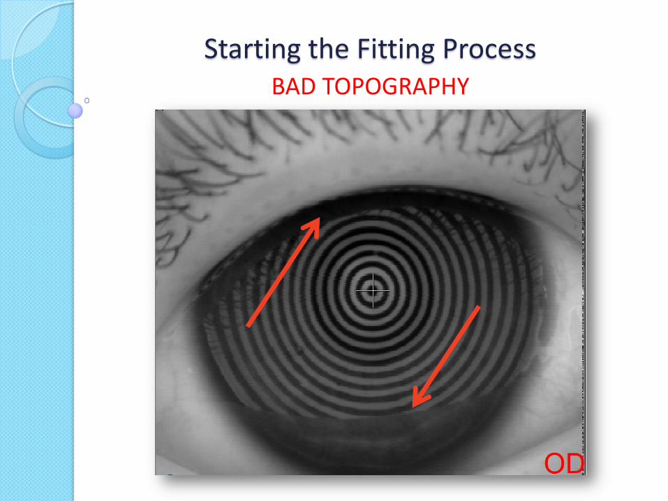
Starting the Fitting Process BAD TOPOGRAPHY

Understanding OrthoK Is it all about flattening the central cornea?

Understanding OrthoK Is it all about flattening the central cornea?
42.98D – 37.33D = 5.65D
So how did this -7.75D myope achieve 20/20 vision?
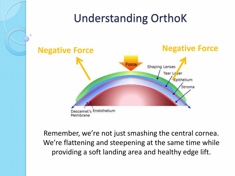
Understanding OrthoK
Negative Force Negative Force
Remember, we’re not just smashing the central cornea. We’re flattening and steepening at the same time while
providing a soft landing area and healthy edge lift.

Troubleshooting Smiley Face Topography Pattern
Typically indicates a fit that is relatively too flat. The lens decenters superiorly with the resulting topography showing a crescent-shaped area of steepening within the pupil zone and the area of apical flattening decentered upwards.

Troubleshooting Smiley Face Topography Pattern
When lens sag is insufficient (RZD too
small) will show superior
decentration and little or no peripheral
touch
When LZA is too small, there will be excessive edge lift and the lens will land inwardly toward the return zone. There will be
excessive fluorescein at the edge

Troubleshooting Fixing Smiley Face Topography Pattern
• Increase sagittal depth of lens • Alignment zone is too flat, therefore steepen
• Return Zone too shallow, need to increase • Edge lift is too great, therefore decrease
• Landing Zone too flat, therefore steepen • Tight lids can pull a lens up as well
• Could apply base down prism (on some lens designs) • Increase lens thickness to increase mass and help
lens “drop” a bit • Increase OAD to assist centration • More viscous insertion solution may be helpful • Watch out for toric periphery. If 90/270 much
steeper than 0/180, lens may pull up (or down) • Toric peripheral curves may be needed

Troubleshooting Frowney Face Topography Pattern
Indicates a fit that is relatively too steep. The lens decenters inferiorly with the resulting topography showing a crescent-shaped area of steepening within the pupil zone and the area of apical flattening decentered downwards.

Troubleshooting Frowney Face Topography Pattern
When lens sag is too great (RZD too great) will show little or no
TxZ
When LZA is too great, there will be insufficient edge lift and the lens will
land too far outwardly toward the periphery.
There will be insufficient fluorescein at the edge

Troubleshooting Fixing Frowney Face Topography Pattern
• Decrease sagittal depth of lens • Alignment zone is too steep, therefore flatten
• Return Zone too deep, need to decrease • Edge lift is insufficient, therefore increase
• Landing Zone too steep, therefore flatten • Tight lids can push a lens down as well
• Decrease lens thickness to decrease mass so lens will not drop as much
• Increase OAD to assist centration • Watch out for toric periphery. If 90/270 much
steeper than 0/180, lens may slide down (or up) • Toric peripheral curves may be needed

Troubleshooting Smiley Face vs Frowney Face
• Superiorly/inferiorly decentering lenses can occur when the sagittal depth of the lens does not match the cornea. Steep lenses can decenter up or down as can flat lenses. If both conditions could be caused by a lens being too steep or too flat, how can you tell the difference?
• Let your fluorescein patterns guide your decision.
• If you see no identifiable treatment zone, you know the lens is too steep (sagittal depth too great)
• If edges look like they are flaring, or the treatment zone appears especially dark, you know the lens is too flat (sagittal depth insufficient)
• Central SPK often indicates a lens that is too flat

Troubleshooting Central Island
Image courtesy of Randy Kojima, FAAO, FIAO

Troubleshooting Central Island
• Indicates a fit that is relatively too steep. • Refraction will show more myopia than pre-fit level.
• When a lens is too steep centrally, the cornea may steepen centrally rather than flatten as we would anticipate when performing OrthoK for myopia.
• Recheck your topography patterns • Recheck your keratometric values
• Was there an error in K reading or SimK readings? • Make sure the patient did not switch their lenses • Recheck base curve of lenses on Radiuscope if available

Troubleshooting Smiley Face with Fake Central Island
Image courtesy of Randy Kojima, FAAO, FIAO

Troubleshooting Smiley Face with Fake Central Island
• Indicates a fit that underestimates the corneal sag. • The lens sag is significantly less than the corneal sag
leading to heavy central baring and epithelial disruption. • The refraction should not show greater myopia than pre-fit
level • This is the difference between central islands and fake
central islands. Fake islands are caused by SPK. • May need to reassess fit, condition of lenses, insertion
techniques, insertion solutions used • In the event that lens sag is correct and the SPK was caused
by other conditions (ie dirty lens), may need to give lenses more time before making changes

Troubleshooting Lateral Decentration
Lateral decentration can be the most difficult fitting anomaly to correct. The reason is because there could be several causes of decentration. Critical examination of NaFl pattern should guide the decision making

Troubleshooting Lateral Decentration
Causes of lateral decentration include: • Insufficient lens diameter
• Lens should consume 92-97% of HVID. Inspect VVID and DDID as well when deciding on lens diameter
• Lens sag insufficient • Flat lenses will “rub” the apex of the cornea
and cause SPK. This will also cause the lens to move on the eye
• Lens sag excessive • If a lens is too steep it may drop and become
laterally decentered

Troubleshooting Lateral Decentration
• Eye lid forces • Asian eyes can be a challenge
• Sleeping position • Sometimes need to counsel patients on sleeping
positions • Lagophthalmos
• May need to use hyper viscosity drops or gels • Erroneous topography data
• MAKE SURE YOU HAVE GOOD MAPS!!! • Compare SimKs to Manual Ks
• Just because they want to • Sometimes they just decenter no matter what you
try. May opt for CorneoScleral design (highly advanced OrthoK)

Troubleshooting Lateral Decentration
• Deposits on lenses • Must stress/insist on compliance with
cleaning regimen • Can use Progent in office • Hand polishing machine • Lens cleaning sponge • Review/change care
system • Consider lens replacement

Troubleshooting
HUH?
Day 1 Post
Dispense

Troubleshooting
HUH?
Pre Dispense
NaFl Pattern

Troubleshooting
Lens Binding
• Commonly caused by having patient present with lenses in situ (controversial)
• Patient-specific – sometimes it just happens to certain patients
• May be dependent on tear film viscosity

Troubleshooting
Removing a Bound Lens
• Instill a few drops of artificial tears • Look upwards and press against the inferior
limbus with the edge of the lower lid a few times • Look downward and repeat the process at the
superior limbus • Once the patient becomes aware of the feeling of
lens movement, the lens can be safely removed • WARN THEM ABOUT THIS BEFOREHAND. THEY
WILL BE MUCH LESS FRIGHTENED IF THEY KNOW TO WATCH FOR IT

Troubleshooting Corneal Staining
• Central staining often from lenses too flat or unclean lenses • 3 and 9 o’clock staining • Lens binding • Grade 1 or less staining is clinically insignificant and usually resolves within an hour of lens removal • Grade 2 + staining is unacceptable • Central staining can cause “fake” central island on
topography

Troubleshooting Corneal Staining
Diffuse grade 1 corneal staining is usually caused by heavy build-up of deposits in the back of the lens
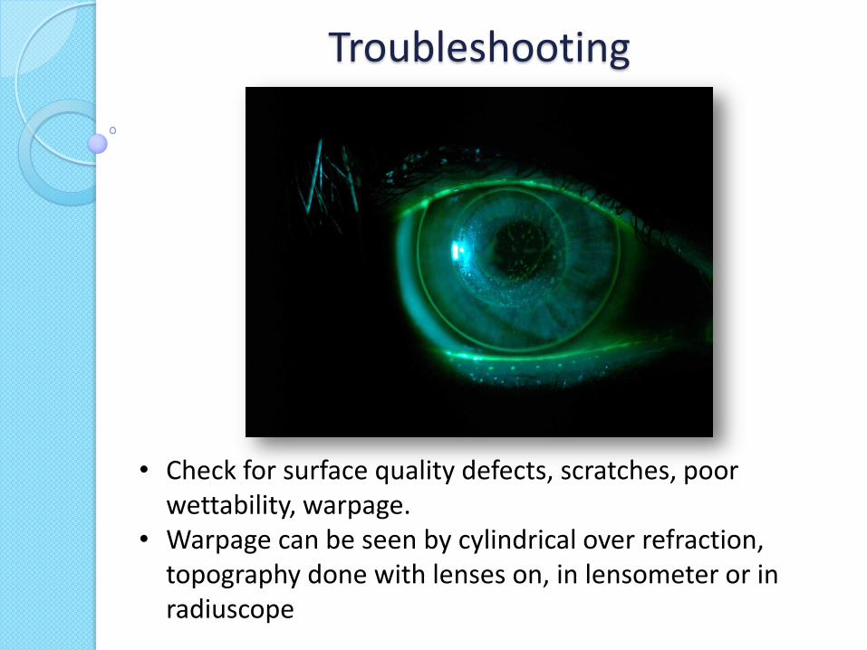
• Check for surface quality defects, scratches, poor wettability, warpage.
• Warpage can be seen by cylindrical over refraction, topography done with lenses on, in lensometer or in radiuscope
Troubleshooting

Troubleshooting Corneal Abrasion
• Dirty lens • Chipped/cracked lens • Rubbing eyes during lens wear • Most often handling difficulties,
insertion/removal • Treatment protocol well established • Depending on severity, may need to
postpone OK treatment until resolved

Troubleshooting
Dimple Veiling
• Not fluorescein staining, but pooling
• Often caused from bubbles in too steep or too wide RC which break down and froth
• Usually recovers in 1-2 hours
• If fit is good, filling lens with non-preserved sterile saline solution before insertion should help

Troubleshooting
Epithelial Iron Deposition
• Common • Typically occurs in the
same area as the reverse curve under the lens and coincides with the area of greatest corneal curvature change
• Can be incomplete or complete rings • Not pathological and requires no treatment • Easily viewed with blue filter (no NaFl)

Troubleshooting
Microbial Keratitis
• Obviously the one we never want to see • Low likelihood with good patient compliance,
proper aftercare • Patients must remove their lenses and seek urgent
medical attention • Only materials approved for overnight use should
be used

Troubleshooting Microbial Keratitis
“The Ohio State study found that “the risk of MK with overnight reshaping lenses is similar to other overnight modalities” (Bullimore, 2009). The researchers obtained data from 86 randomly selected practitioners and 1,317 patients fitted during 2005 and 2006. The patients contributed 2,593 patient years of wear divided almost evenly between adults (49 percent) and children. Fifty event forms were submitted with 11 reporting corneal infiltrates. Two of these were MK, resulting in an
estimated incidence of 7.7 per 10,000 years of wear. What does
this all mean? That in all likelihood your corneal reshaping patients have a slightly higher risk of developing MK than your daily wear soft contact lens wearers do. Also, the risk of MK in ortho-k wearers may be as high as that for silicone hydrogel lenses worn overnight (Schein, 2005). Additional studies are needed to more
completely address this issue.”
From Herzberg, C. “An Update on Orthokeratology” Contact Lenses Today 03/01/2010

Troubleshooting
Loss of Effect
• Often associated with heavy back surface deposits
on the lenses • Can be caused by lens warpage • Common if lenses are inadvertently put in the
wrong eyes (different color lenses can avoid this complication)

Troubleshooting
Glare/Photophobia
• Often occurs in patients with large pupils • More common when correcting high myopia as TxZ
diameter decreases • Tends to improve over time • May use Alphagan “off label” to produce some
miosis at night • May need to increase treatment zone diameter
(when appropriate or available) • Scleral Ortho-K? • Aspheric TxZ?
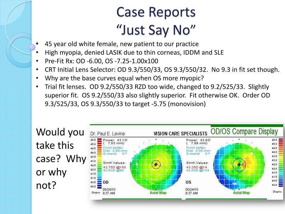
Case Reports “Just Say No”
• 45 year old white female, new patient to our practice • High myopia, denied LASIK due to thin corneas, IDDM and SLE • Pre-Fit Rx: OD -6.00, OS -7.25-1.00x100 • CRT Initial Lens Selector: OD 9.3/550/33, OS 9.3/550/32. No 9.3 in fit set though. • Why are the base curves equal when OS more myopic? • Trial fit lenses. OD 9.2/550/33 RZD too wide, changed to 9.2/525/33. Slightly
superior fit. OS 9.2/550/33 also slightly superior. Fit otherwise OK. Order OD 9.3/525/33, OS 9.3/550/33 to target -5.75 (monovision)
Would you take this case? Why or why not?

Case Reports “Just Say No”
• 1st morning: DVa sc OD 20/400, OS 20/400 • SRx OD: -4.50, OS -4.75-0.50x160 • Diffuse SPK OS • Plan: Continue lenses, DD for daily use, frequent artificial tears
How is the position?

Case Reports “Just Say No”
• 1 week: DVa sc OD 20/200, OS 20/400 • SRx OD: -2.25, OS -4.25 • Diffuse SPK OS with subepithelial infiltrates • Plan: Discontinue lenses, Vigamox QID OS, recheck in 1 week • Refit to improve centration
New parameters: CRT OD 9.2/575/33 OS 9.3/600/33 Why these changes?

Case Reports “Just Say No”
• 1 week later: Eyes feeling better, vision back to baseline • OS Cornea clear, but still scattered staining on cornea and conj • Plan: Start again with new lenses. Collagen plug placed in LLL to improve signs of
dryness. Recheck in 1 week
Are we starting to do too much?

Case Reports “Just Say No”
• 1 week later: DVa sc OD 20/100, OS 20/400 • SRx OD: -2.00-0.50x175, OS -3.50-.075x125 • 3+ SPK OS • Plan: We decided to discontinue due to chronic SPK • Refit to soft bifocal contacts
This looks pretty good though huh?
Case Reports “Just Say No”
• Where did we go wrong? • What were the main stumbling blocks? • Why did this case fail? • What would you have done?

Case Reports “Up and In”
Understanding Tear Layer Profiles
Wave®

Case Reports “Up and In”
• 13 year old white female, existing patient in our practice • Rx increased between ages 11 and 15 by -2.00D • Soft contact lens wearer 2 years prior • Interested in myopia control • Pre-fit Rx OD: -3.25 SPH, OS -3.25-0.50x175 • HVID 11.4

Case Reports “Up and In”
First Lens Designs (OD)

Case Reports “Up and In”
First Lens Designs (OS)
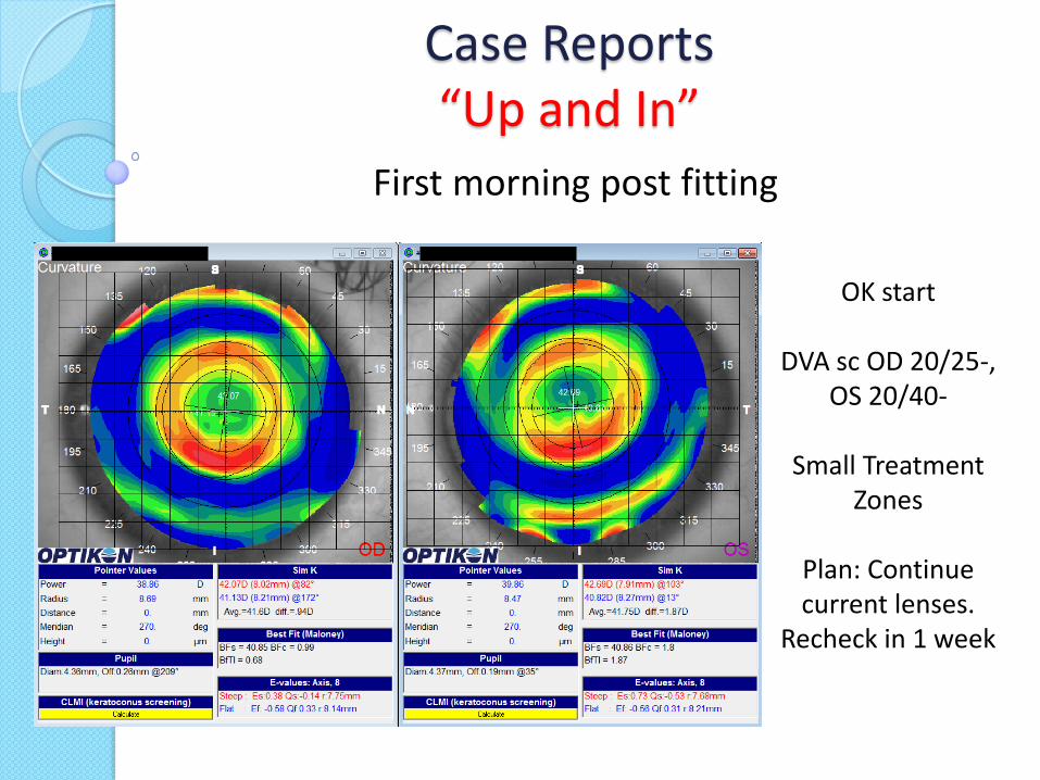
Case Reports “Up and In”
First morning post fitting
OK start
DVA sc OD 20/25-, OS 20/40-
Small Treatment
Zones
Plan: Continue current lenses.
Recheck in 1 week

Case Reports “Up and In”
Week 1
DVA sc OD 20/20-,
OS 20/20-
TxZ in and up, oval.
Patient happy, Doctor not.
Plan: Continue current lenses. Recheck in 2-3
weeks

Case Reports “Up and In”
Month 1
DVA sc OD 20/20-, OS 20/25+
TxZ still in and up,
oval.
Patient still happy, Doctor still not.
Plan: Continue current lenses.
Recheck in 3 months

Case Reports “Up and In”
Month 3
DVA sc OD 20/25-3, OS 20/25-3
TxZ still in and up,
oval.
Patient still happy, Doctor still not.
Plan: Redesign
lenses and schedule pickup

Case Reports “Up and In”
Second lens design to
improve TxZ position (OD)
Bigger, did not average
astigmatism, used minimal blend of reverse curves,
decreased TxZ size increased CT/ET

Case Reports “Up and In”
Second lens
design to improve
TxZ position
(OS)

Case Reports “Up and In”
Day 6 Post Dispense Patient not noticing much improvement.
DVA sc OD 20/20-
2, OS 20/20-
TxZ still in and up, oval. Minimal
change.
Patient still happy, Doctor still not.
Plan: Continue
current lenses and recheck 1 week

Case Reports “Up and In”
No change in acuity (still good) or Bull’s Eye Topography (still bad). Initiated 3rd redesign. Decreased OAD slightly, changed to Free Form, normal blend, increased TxZ diameter, decreased CT/ET

Case Reports “Up and In”
For comparison, kept minimal blend feature on for left eye

Case Reports “Up and In”
Week 1 Post Dispense Lens 3
DVA sc OD 20/20,
OS 20/25+
Much improvement OD, still in and up OS,
but less oval.
Patient still happy, Doctor almost
happy.
Plan: Remake OS with normal blend

Case Reports “Up and In”
Lens 4 OS Free Form with normal Blending

Case Reports “Up and In”
Month 1 Post Dispense OD Lens 3 Week 1 Post Dispense OS Lens 4
DVA sc OD 20/20-,
OS 20/20-
OD continues to look great, OS best
so far, small flat spot of red ring
superiorly.
Patient still happy, Doctor happy.
Plan: Continue
with current lenses.
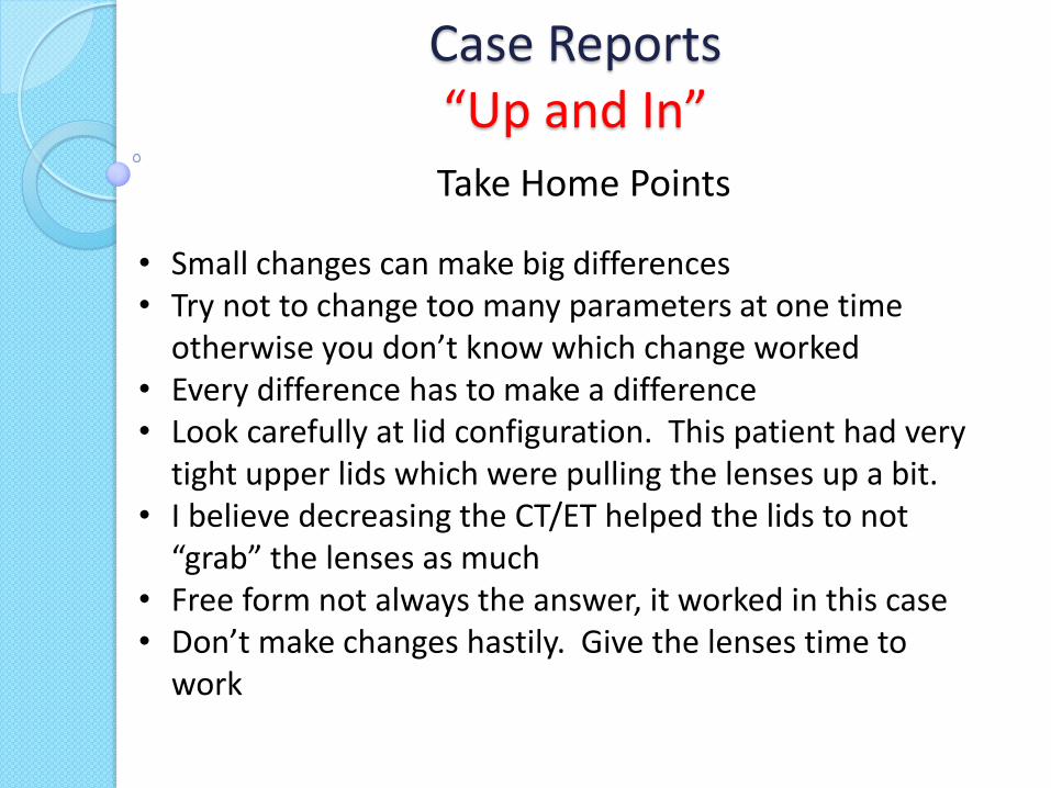
Case Reports “Up and In” Take Home Points
• Small changes can make big differences • Try not to change too many parameters at one time
otherwise you don’t know which change worked • Every difference has to make a difference • Look carefully at lid configuration. This patient had very
tight upper lids which were pulling the lenses up a bit. • I believe decreasing the CT/ET helped the lids to not
“grab” the lenses as much • Free form not always the answer, it worked in this case • Don’t make changes hastily. Give the lenses time to
work

Clinical Pearls
• Don’t say yes just because your patients do • Talk talk talk. Engage patient it the process • Provide sufficient written information • Make yourself available • Be careful delegating too much to staff • Do your own topos • Try not to squeeze in, give them sufficient time • Progent lenses every 6 months or more if needed • Reinforce care at each and every visit • Always have them bring their lenses to the office

Clinical Pearls • Don’t make hasty changes. Sometimes the process
needs time to work • Lenses fit differently during sleep than they do at
the slit lamp. Rely heavily on your topographies for position
• Need buy-in from staff • Charge for your time and expertise! • Ask for referrals and testimonials • CE!! Travel, take the time, learn all you possibly can • Under promise and over deliver • Make them feel special. These are not your average
lens wearers. These are your “A” patients

Links for Further Research
• Myopiaprevention.org • Orthokacademy.com • Orthokdoctors.com • Paragoncrt.com • www.bausch.com/en/ECP/Our-
Products/Orthokeratology/Vision-Shaping-Treatment
• govlenses.com • wavecontactlenses.com