Embed Size (px)
Citation preview

TREMATODES
http://cvet.tu.edu.iq

CHARACTERISTICS
Dorso-ventrally flattened
Unsegmented
Leaf-like
Hermaphroditic except blood flukes
Two radially striated suckers
Incomplete digestive tract
Adults are covered with spines, except
Incomplete digestive tract
Most of the body is occupied by reproductive organs
http://cvet.tu.edu.iq

http://cvet.tu.edu.iq

BLOOD FLUKES
http://cvet.tu.edu.iq

BLOOD FLUKES
Characteristics – Dioecious – Males are shorter and stouter than females – Lateral margins of males are folded ventrally to form a
gynecophoral canal in which females are received – Suckers are armed with delicate spines – There is no muscular pharynx – Eggs are non-operculated – Eggs are fully embryonated when laid – Embryonated eggs have a ciliated embryo called miracidium – Cercariae have bifid tails – There is no encysted metacerciarial stage – Infective Stage: cercaria penetrating the unbroken skin
http://cvet.tu.edu.iq

BLOOD FLUKES: Schistosomes
Schistosomes
– Schistosoma japonicum : Oriental blood fluke
– Schistosoma haematobium: Vesical blood fuke
– Schistosoma mansoni: Manson’s blood fluke
http://cvet.tu.edu.iq
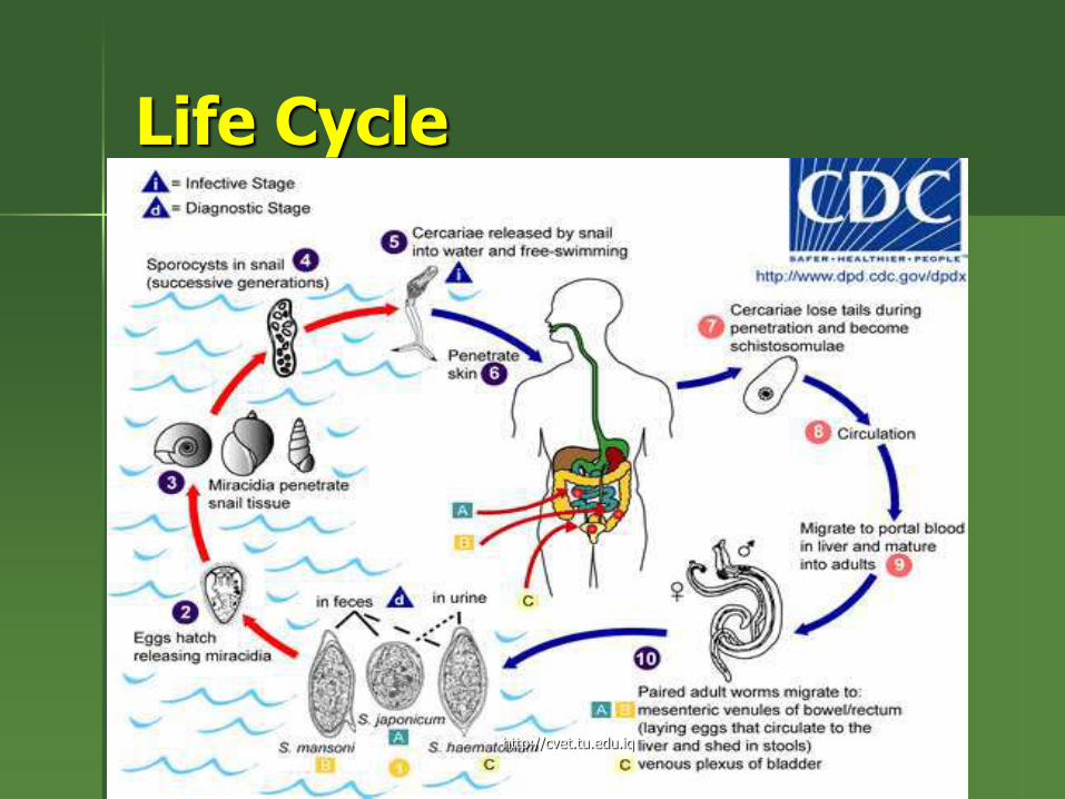
Life Cycle
http://cvet.tu.edu.iq
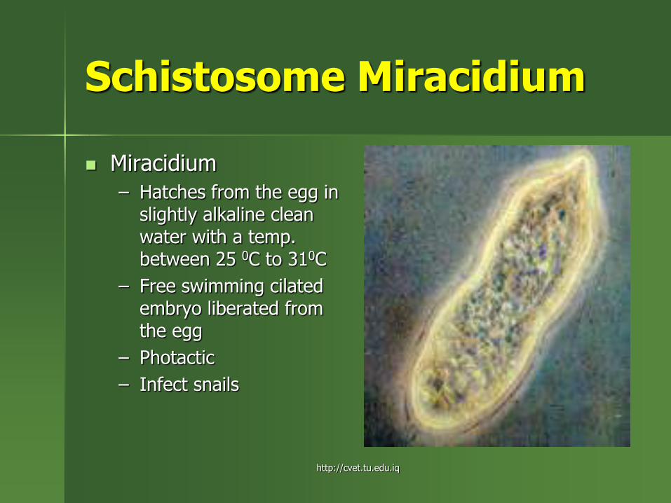
Schistosome Miracidium
Miracidium
– Hatches from the egg in slightly alkaline clean water with a temp. between 25 0C to 310C
– Free swimming cilated embryo liberated from the egg
– Photactic
– Infect snails
http://cvet.tu.edu.iq
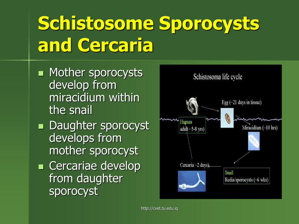
Schistosome Sporocysts and Cercaria
Mother sporocysts develop from miracidium within the snail
Daughter sporocyst develops from mother sporocyst
Cercariae develop from daughter sporocyst
http://cvet.tu.edu.iq

Schistosome Cercaria
Cercaria
– Emerges from daughter sporocysts
– Escapes from the snail
– Has a body and a forked tail
– Infects man by skin penetration
http://cvet.tu.edu.iq

Schistosomulae
Schistosomule
– Develops from cercaria after skin penetration
– Adapted to survive in serum or physiologic saline at 37 0C
– Enter the pleural cavity---diaphragm---peritoneal space---penetrate the liver to reach the intrahepatic portions of the portal vein
http://cvet.tu.edu.iq

Schistosoma japonicum
Schistosoma japonicum – Life cycle involves
alternating parasitic stages in mammalian hosts and free living stages Egg and miracidium First stage (mother)
sporocyst Second stage
(daughter) sporocyst cercaria Schistosomulum Adult schistosome
http://cvet.tu.edu.iq

Schistosoma japonicum
Schistosoma japonicum – Primarily parasites of
the portal vein and its branches
– Each female fluke deposits 500-2000 immature eggs/day
– Embryonation takes place within 10-12 days
– Eggs escape through ulcerations in the intestinal lumen and are passed out with the feces
http://cvet.tu.edu.iq

Schistosoma japonicum
Schistosoma japonicum in eternal copula
– Males have a gynecophoral canal which receives the female during copulation
http://cvet.tu.edu.iq

Schistosoma japonicum
Schistosoma japonicum Ova
– Ovoidal, rounded or pear-shaped
– Thin shell
– Pale yellow
– Curved hook or spine or lateral knob
– Laid in the multicellular stage and embryonte within 10-12 days
http://cvet.tu.edu.iq

Schistosoma mansoni
Schistosoma mansoni male and female
– Female inside the gynecophoral canal of male
http://cvet.tu.edu.iq

Schistosoma mansoni
Schistosoma mansoni male and female
– Female inside the gynecophoral canal of male
http://cvet.tu.edu.iq

Schistosoma haematobium
Schistosoma haematobium adult
http://cvet.tu.edu.iq

Schistosoma haematobium
Schistosoma haematobium ova
– Note the presence of terminal spine
http://cvet.tu.edu.iq
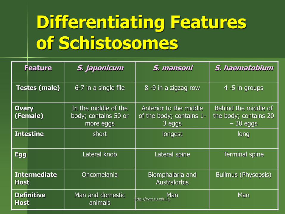
Differentiating Features of Schistosomes
Feature S. japonicum S. mansoni S. haematobium
Testes (male) 6-7 in a single file 8 -9 in a zigzag row 4 -5 in groups
Ovary (Female)
In the middle of the body; contains 50 or
more eggs
Anterior to the middle of the body; contains 1-
3 eggs
Behind the middle of the body; contains 20
– 30 eggs
Intestine short longest long
Egg Lateral knob Lateral spine Terminal spine
Intermediate Host
Oncomelania Biomphalaria and Australorbis
Bulimus (Physopsis)
Definitive Host
Man and domestic animals
Man Man http://cvet.tu.edu.iq
Blood Flukes: Pathogenesis and Clinical Manifestations
Schistosomiasis
Host granulomatous reaction to eggs
Pneumonitis due to schistosomula in the lungs
Hepatosplenic disease
Colonic schistosomiasis
Cerebral schistosomiasis
http://cvet.tu.edu.iq

Blood Flukes: Schistosomiasis
Distended belly is one of the symptoms of schistosomiasis http://cvet.tu.edu.iq

Blood Flukes: Diagnosis
Schistosomiasis
– Eggs may not be demonstrable in the feces
– Infections where there is scarring prevent passage of eggs into the intestinal lumen
http://cvet.tu.edu.iq

Blood Flukes: Diagnosis
Schistosomiasis – Stool Examination Techniques
Merthiolate-Iodine Formlin Concentration Technique (MIFC)
– Sensitive for moderate and heavy infections
– Not adequate for light infections (less than 10 eggs/gram of stool)
Kato KatzTechnique – For enumeration of eggs
– Most commonly used for evaluating epidemiology, effect of control measures, drug trials
http://cvet.tu.edu.iq
Blood Flukes: Diagnosis
Schistosomiasis Immunodiagnosis
– Intradermal tests for immediate cutaneous hypersensitivity using adult worm extracts
– Indirect hemagglutination using adult worm and egg antigens
– Circumoval precipitin test
– Enzyme-Linked Immunosorbent Assay (ELISA) using soluble antigens of adults and eggs
http://cvet.tu.edu.iq

Blood Flukes: Treatment
Treatment – Praziquantel
(heterocyclic prazinoisoquinolone compound)
– Single dose of 40-50 mg/kg
– 25 mg/kg in two doses
– 20 mg/kg in three doses
http://cvet.tu.edu.iq

Blood Flukes: Epidemiology
In the Philippines
– 24 endemic provinces
Sorsogon
Oriental Mindoro
Samar
Leyte
Bohol
All provinces in Mindanao except Misamis Oriental
http://cvet.tu.edu.iq

Lung Flukes
http://cvet.tu.edu.iq
Lung Flukes
Paragonimus westermani
Oriental Lung Fluke
Paragonimus philippinensis
Paragonimus siamensis (cats)
http://cvet.tu.edu.iq

Lung Flukes: Paragonimus westermani Paragonimus westermani
adult – Hermaphroditic
– Body covered with spines
– Reddish brown
– Measures 4-6 mm in width and 3.5-5 mm in thickness
– Resembles a coffee bean
– Adult worms are found in pairs or in threes in fibrotic capsules or cysts in the lungs
http://cvet.tu.edu.iq

Lung Flukes: Paragonimus westermani
Paragonimus westermani ova – Yellowish brown
– Thick-shelled
– Operculated with a thickened abopercular egg
– May be seen in the sputum or in feces if the sputum is swallowed
http://cvet.tu.edu.iq

Lung Flukes: Paragonimus westermani Life Cycle
http://cvet.tu.edu.iq

Lung Flukes: Epidemiology of Paragonimiasis
First Intermediate Host
– Brotia asperata (snail)
– Where miracidium develops into 1 sporocyst and 2 redial stages of development
http://cvet.tu.edu.iq

Lung Flukes: Epidemiology of Paragonimiasis
Second Intermediate Host
– Sundathelpusa philippina or Parathelpusa grapsoides (former name)
– Harbors the metacercaria that is infective to man
http://cvet.tu.edu.iq

Lung Flukes: Paragonimus westermani
Man gets infected after ingestion of raw or insufficiently cooked crabs harboring the metacercariae
http://cvet.tu.edu.iq

Lung Flukes: Pathogenesis and Clinical Manifestations
Paragonimiasis
– Cough
– Hemoptysis
– Symptoms consistent with pulmonary tuberculosis
– Misdiagnosed as PTB
http://cvet.tu.edu.iq

Lung Flukes: Diagnosis of Paragonimiasis
Radiographs aid in diagnosis
Definitive diagnosis is based on the finding of ova in the sputum, stool or less frequently in aspirated material from abscesses or pleural effusions
Multi-dot ELISA
http://cvet.tu.edu.iq

Lung Flukes: Treatment of Paragonimiasis
Praziquantel – Drug of choice
– 25 mg/kg body weight 3x a day for three day
Bithionol – 15 – 25 mg/kg / day
on alternate days for a total of 10-15 days
http://cvet.tu.edu.iq

Lung Flukes: Epidemiology of Paragonimiasis
Has a global distribution In the Philippines
– Leyte – Sorsogon – Mindoro – Camarines – Samar – Davao – Cotabato – Basilan
http://cvet.tu.edu.iq

Intestinal Flukes
http://cvet.tu.edu.iq
Intestinal Flukes: Fasciolopsis buski
Giant intestinal fluke of man
Parasite of the intestines of humans and pigs
Mode of transmission is by ingestion of encysted metacercariae on aquatic plants
The viable metacercariae excyst in the duodenum and becomes mature in about three months
http://cvet.tu.edu.iq

Intestinal Flukes: Fasciolopsis buski
Elongated
Oval
20 – 75 mm in length and 8 -20 mm in width
Covered with spines
No cephalic cone
Unbranched intestina caeca which reach up to the posterior end
http://cvet.tu.edu.iq

Intestinal Flukes: Life cycle of Fasciolopsis buski
http://cvet.tu.edu.iq
Intestinal Flukes: Pathogenesis of Fasciolopsis buski
Fasciolopsiasis
Pathological changes caused are:
Traumati c – Inflammation and ulceration
Obstructive – Intestinal obstruction due to heavy infection
Toxic – Due to absorption of worm metabolites by the
host
http://cvet.tu.edu.iq

Intestinal Flukes: Diagnosis Fasciolopsiasis
Detection of parasite eggs in stool
Resemble Fasciola eggs
Provided with an operculum
http://cvet.tu.edu.iq

Intestinal Flukes: Diagnosis Fasciolopsiasis
Detection of parasite eggs in stool – Provided with an
operculum
– Large
– Unembryonated when laid
Resemble Fasciola eggs
http://cvet.tu.edu.iq

Intestinal Flukes: Treatment of Fasciolopsiasis
Praziquantel
25 mg/kg for 3 doses for one day
Side effects:
– Diziness
– Drowsiness
– Epigastric pain
http://cvet.tu.edu.iq
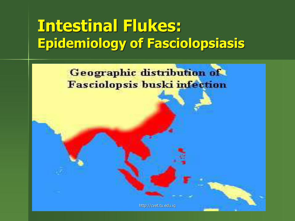
Intestinal Flukes: Epidemiology of Fasciolopsiasis
http://cvet.tu.edu.iq
Intestinal Flukes: Epidemiology of Fasciolopsiasis
Endemic in – Southeast Asia – China – Korea – India
Endemicity in the Philippines has not been demonstrated yet
Fasciolopsiasis in Filipinos are probably imported cases
http://cvet.tu.edu.iq

Intestinal Flukes: Echinostoma ilocanum
Garrison’s fluke
Echinostomid
There are several species that infect man
There are 2 identified echinostomids that infect man in the Philippines: – Echinostoma ilocanum
– Artyfechinostomum malayanum
http://cvet.tu.edu.iq

Intestinal Flukes: Echinostoma ilocanum
Adult
– Reddish gray
– Horse-shape collar of spines (circum oral disk) around the oral suckers
– 49-51 collar spines
– Integument is covered by plaque like scales
– Simple intestinal caeca
http://cvet.tu.edu.iq
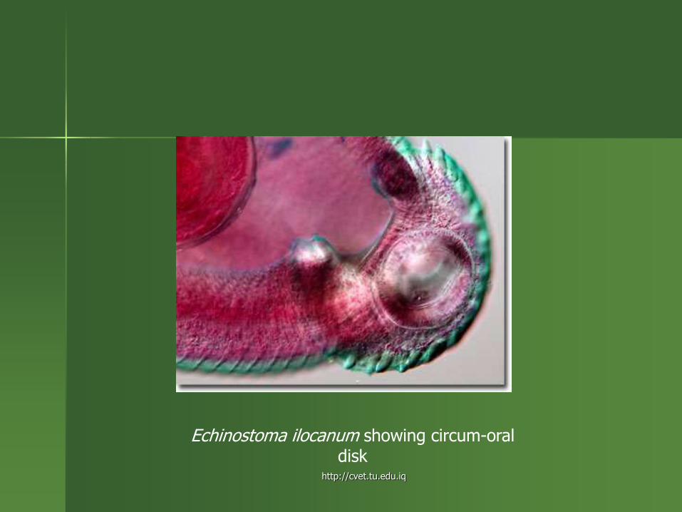
Echinostoma ilocanum showing circum-oral disk
http://cvet.tu.edu.iq

Intestinal Flukes: Echinostoma ilocanum
Echinostoma ilocanum Life cycle
http://cvet.tu.edu.iq

Intestinal Flukes: Echinostoma ilocanum
Echinostoma ilocanum Ova
– Straw-colored
– Operculated
– Ovoid
– Similar to Fasciola and Fasciolopsis buski ova
http://cvet.tu.edu.iq

Intestinal Flukes: Echinostoma ilocanum
First Intermediate Host:
– Gyraulus convexciusculus
– Hippeutis umbilicalis
http://cvet.tu.edu.iq

Intestinal Flukes: Echinostoma ilocanum
Second Intermediate Host:
– Pila luzonica (kuhol)
– Vivipara angularis (susong pampang)
Pila luzonica http://cvet.tu.edu.iq
Intestinal Flukes: Pathogenesis and Clinical Manifestations Echinostoma ilocanum
Man gets infected when metacercariae in the second intermediate hosts are ingested
Inflammation at the site of attachment of adults
Ulceration
Diarrhea (sometimes bloody)
Abdominal pains
General intoxication
http://cvet.tu.edu.iq

Intestinal Flukes: Diagnosis Echinostoma ilocanum
Detection of characteristic eggs in the stool
http://cvet.tu.edu.iq

Intestinal Flukes Treatment Echinostoma ilocanum
Praziquantel
– 25 mg/kg for 3 doses for one day
– No alcohol; no fats must be taken 24 hours before and after treatment
– Only water must be taken within 3 hours of medication
http://cvet.tu.edu.iq

Intestinal Flukes Epidemiology Echinostoma ilocanum
Echinostoma ilocanum is endemic in:
– Northern Luzon
– Leyte
– Samar
– Mindanao provinces
Artyfechinostomum malayanum
– First reported in 1987
– Northern and Central Luzon
http://cvet.tu.edu.iq

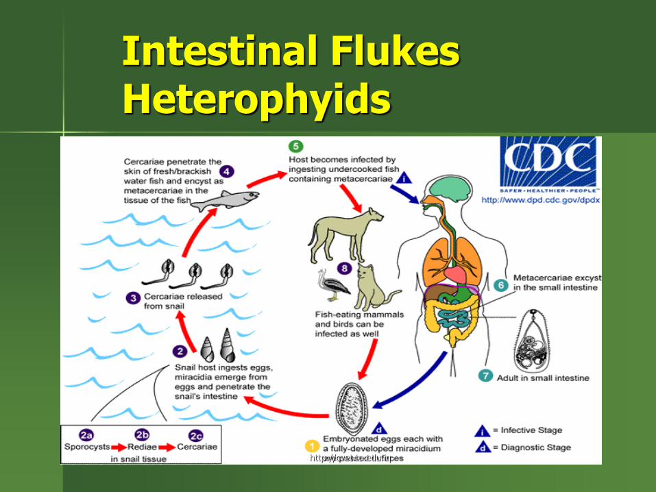
Intestinal Flukes Heterophyids
Many species live in the intestine of fish-eating hosts:
– Heterophys heterophyes
– Metagonimus yokogawai
– Haplorchis taichui
– Haplorchis yokogawai
http://cvet.tu.edu.iq

Intestinal Flukes Heterophyids
Mode of transmission is by ingestion of metacercariae encysted in fish
Metacercariae in the abdomen excysts, liberating a larva that attaches to the intestinal wall
http://cvet.tu.edu.iq
Intestinal Flukes Heterophyids
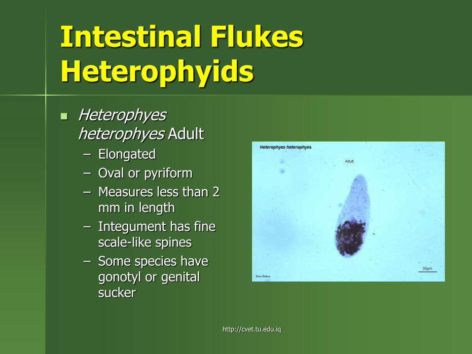
Heterophyes heterophyes Adult
– Elongated
– Oval or pyriform
– Measures less than 2 mm in length
– Integument has fine scale-like spines
– Some species have gonotyl or genital sucker
http://cvet.tu.edu.iq

Intestinal Flukes Heterophyids Heterophyes
heterophyes Ova – Light brown in color – Ovoid in shape – Operculated – A fully developed
symmetrical miracidium is already present
– Operculum fits into the egg smoothly
– No abopercular protuberance like that of Clonochis sinensis ovum
http://cvet.tu.edu.iq
Intestinal Flukes Heterophyids
http://cvet.tu.edu.iq

Intestinal Flukes Heterophyes heterophyes Pathogenesis and Clinical Manifestations
Heterophyiasis
Inflammation at the site of attachment
Manifesations are consistent with peptic ulcer (observed among infected individuals in Compostela Valley)
– Upper abdominal discomfort
– Gurgling abdomen
http://cvet.tu.edu.iq

Intestinal Flukes Diagnosis Pathogenesis and Clinical Manifestations
Detection of eggs in the stool using Kato Katz method
Care must be taken to distinguish them from Clonorchis and Opistorchis ova
http://cvet.tu.edu.iq

Intestinal Flukes Treatment Pathogenesis and Clinical Manifestations
Praziquantel
– 25 mg/kg in 3 doses for 1 day
http://cvet.tu.edu.iq
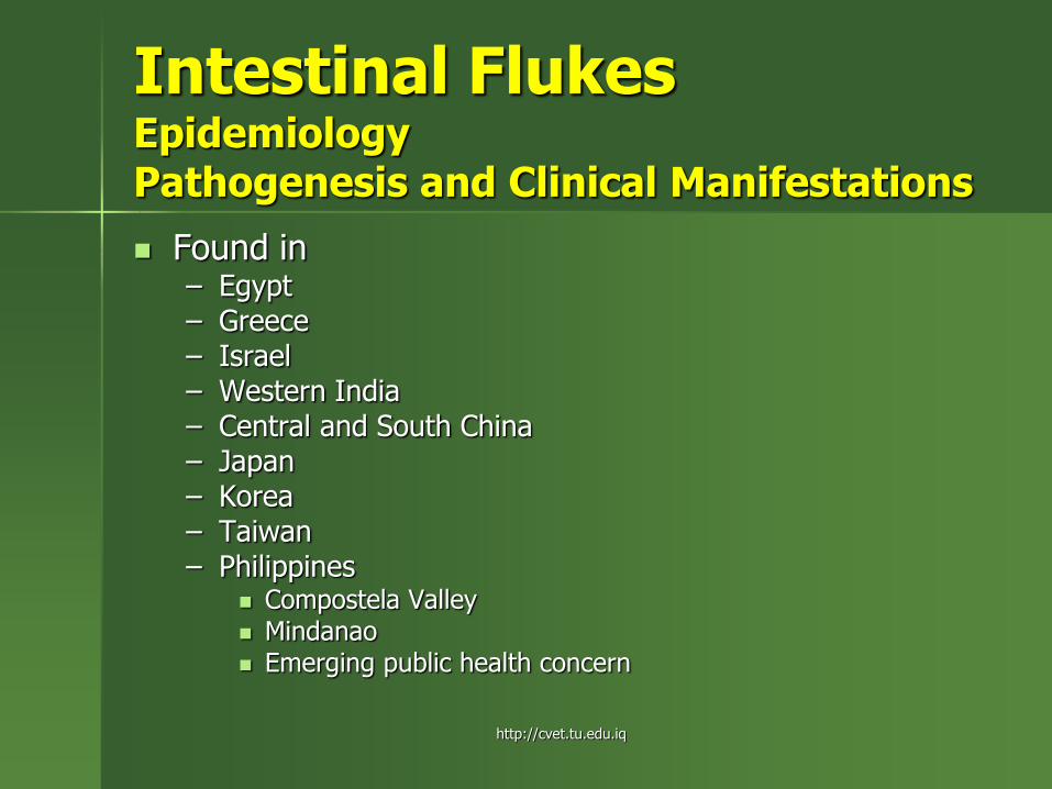
Intestinal Flukes Epidemiology Pathogenesis and Clinical Manifestations
Found in – Egypt – Greece – Israel – Western India – Central and South China – Japan – Korea – Taiwan – Philippines
Compostela Valley Mindanao Emerging public health concern
http://cvet.tu.edu.iq

Liver Flukes
http://cvet.tu.edu.iq
Liver Flukes Fasciola species
Found in the liver and biliary passages of humans and ruminants
Fasciola hepatica – Sheep liver fluke
– Temperate liver fluke
Fasciola gigantica – Giant liver fluke
– Tropical liver fluke
http://cvet.tu.edu.iq

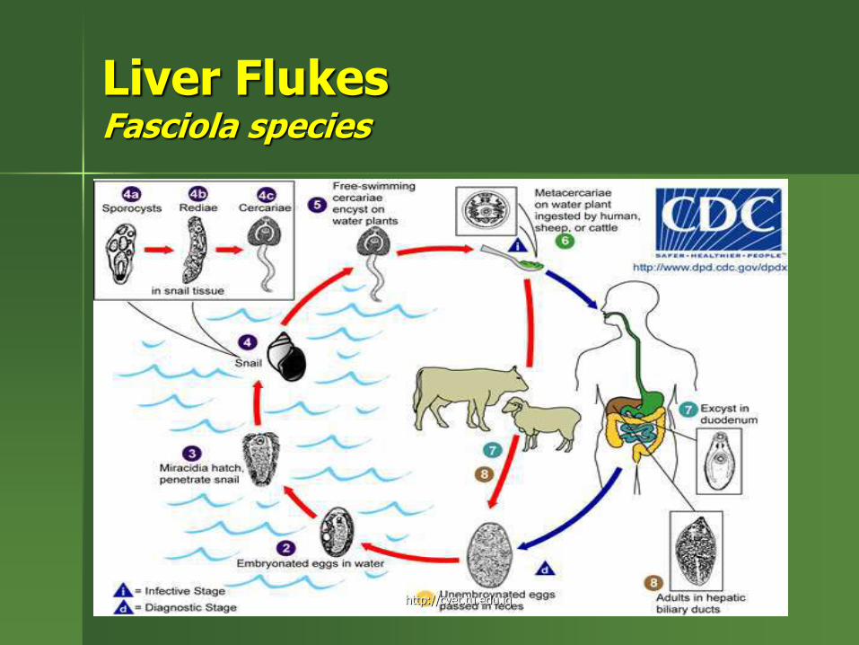
Liver Flukes Fasciola species
Mode of transmission is by ingestion of metacercariae found in edible aquatic plants or by drinking water with floating metacercariae
Metacercariae excsts in the duodenum or jejunum and liberate the juvenile fluke
Juvenile fluke penetrates the intestinal wall and reaches the liver capsule
The parasite burrows into the liver parenchyma where it grows and develops
It becomes sexually mature in the bile ducts
http://cvet.tu.edu.iq
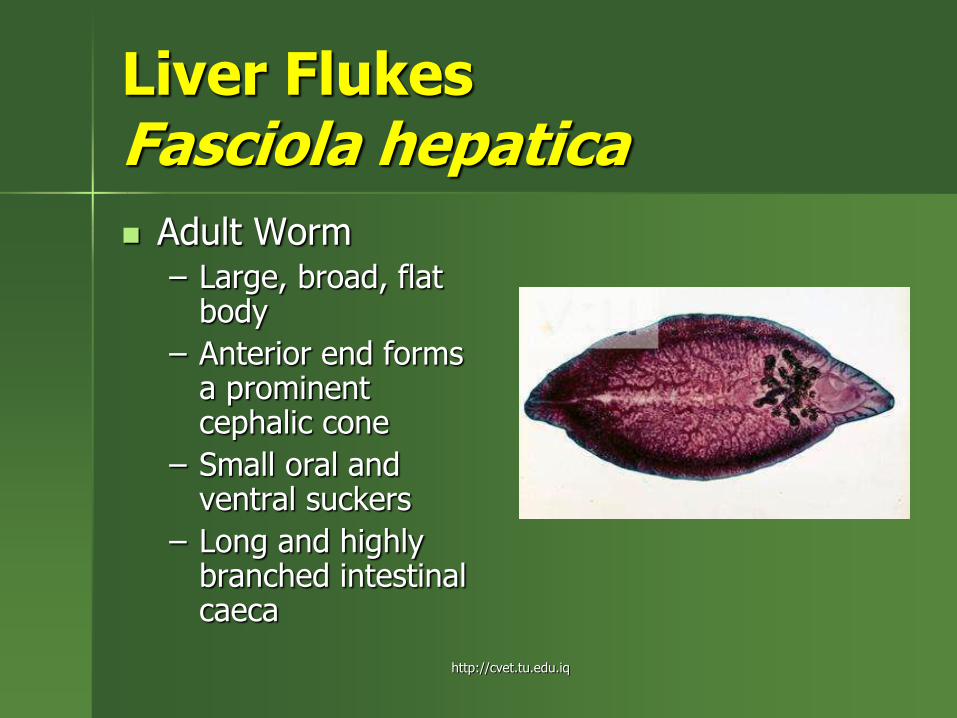
Liver Flukes Fasciola hepatica
Adult Worm – Large, broad, flat
body
– Anterior end forms a prominent cephalic cone
– Small oral and ventral suckers
– Long and highly branched intestinal caeca
http://cvet.tu.edu.iq

Liver Flukes Fasciola gigantica
Adult Worm
– Larger
– More lanceolate
– Less developed shoulders (shorter cephalic cone)
– Larger ventral sucker
http://cvet.tu.edu.iq
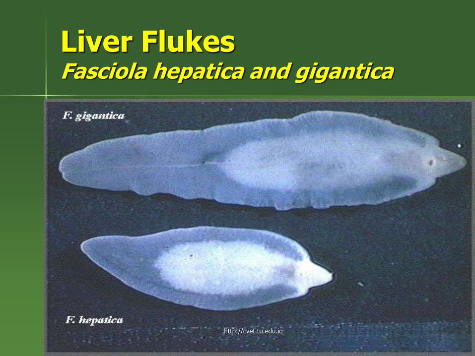
Liver Flukes Fasciola hepatica and gigantica
Adult Worm
http://cvet.tu.edu.iq

Liver Flukes Fasciola hepatica and gigantica
First Intermediate Host:
– Lymnea philippinensis
Second Intermediate Host
– Watercress
– grass
http://cvet.tu.edu.iq

Liver Flukes Fasciola hepatica
Fasciola hepatica Ova
– Large
– Ovoid
– Operculated
– Bile stained
– unsegmented
http://cvet.tu.edu.iq

Liver Flukes Fasciola hepatica
Fasciola gigantica Ova
– Larger but very similar to Fasciola hepatica ova
*Because of similarities, it is just safe to say Fasciola ova
http://cvet.tu.edu.iq

Liver Flukes Fasciola hepatica
Fasciola gigantica Ova
– Larger but very similar to Fasciola hepatica ova
*Because of similarities, it is just safe to say Fasciola ova
http://cvet.tu.edu.iq
Liver Flukes Fasciola species
http://cvet.tu.edu.iq

Liver Flukes Fasciola species Pathogenesis and Clinical Manifestations
Fascioliasis Asymptomatic Can produce fever Right upper quadrant abdominal pain Hypereosinophilia Acute or invasive phase
– Migration from intestine to liver – Traumatic and necrotic lesions in liver parenchyma
Chronic or latent phase – Asymptomatic – Parasite has reached the bile ducts – Obstruction – Stimulates inflammation in the biliary epithelium leading to
fibrosis
http://cvet.tu.edu.iq

Liver Flukes Fasciola species Diagnosis
Microscopy
Serologic tests
– Low specificity because of cross reactivity with antigens of other parasites
RFLP
– PCR restriction Fragment Length Polymorphism
http://cvet.tu.edu.iq

Liver Flukes Fasciola species Treatment
Bithionol – 20-50 mg/kg body weight on alternate days to
complete 10 to 5 doses
Triclabendazole – Also a recommended drug of choice due to:
Efficacy
Safety
Ease of use
http://cvet.tu.edu.iq
Liver Flukes Fasciola species Epidemiology
Worldwide distribution
Economic importance n livestock raising
In the Philippines, the dominant species is Fasciola gigantica affecting cattle and water buffalos
Few human cases are reported locally
http://cvet.tu.edu.iq

Liver Flukes Clonorchis sinensis
Chinese liver fluke
Oriental Liver Fluke
Distome of China
First intermediate Host: – Bulimus fuchsiana (snail not found in the
Philippines)
Second Intermediate Host: – Ctenopharyngondon idellus (fish)
http://cvet.tu.edu.iq

Liver Flukes Clonorchis sinensis
Adult Worm
– Narrow, oblong, flat worm
– Oral sucker is slightly larger than the ventral sucker
– Blind intestinal caeca are simple and extend to the caudal region
– Life span is 20-30 years
http://cvet.tu.edu.iq

Liver Flukes Clonorchis sinensis
Ova – Bile stained
– Flask-shaped
– Operculated
– Contains a miracidium when oviposited
– Does not hatch in water but is ingested with a molluscan host
– Has a terminal spine
– Electric bulb in shape
– Infective to snails only
http://cvet.tu.edu.iq

Liver Flukes Clonorchis sinensis Life Cycle
http://cvet.tu.edu.iq
Liver Flukes Clonorchis sinensis Pathogenesis and Clinical Manifestations
Clonorchiasis
Provokes intense proliferation of intestinal epithelium
Acute stage (less than 1 month of infection) – Chills
– Fever
Chronic stage – Cirrhosis
– Portal hypertension
http://cvet.tu.edu.iq
Liver Flukes Clonorchis sinensis Diagnosis
Detection of parasite egg in stool
Clonorchis, Opistorchis and Hetrophyid ova may not be differentiated under ordinary light microscope
ELISA with crude Clonorchis sinensis antigen
Enzyme immunoassay (EIA
Polymerase Chain Reactions
http://cvet.tu.edu.iq

Liver Flukes Clonorchis sinensis Treatment
Praziquantel
– 25 mg/kg three times a day for two days
– 60 mg/kg in three doses for one day
– May be used together with albendazole for light and moderate infections
http://cvet.tu.edu.iq
Liver Flukes Clonorchis sinensis Epidemiology
Transmission is due to consumption of raw, undercooked fish and salted and dried fish harboring the metacercariae
Over 30 million people are infected in Southeastern Asia
No reported cases in children below 10 years old
Endemic in: – China
– Japan
– Korea
– Vietnam
http://cvet.tu.edu.iq

Pancreatic Fluke
http://cvet.tu.edu.iq

Pancreatic Fluke Eurytrema pancreaticum
Pancreatic fluke
Stout worm with ruffled margins
oral sucker is larger than the ventral sucker
http://cvet.tu.edu.iq

Pancreatic Fluke Eurytrema pancreaticum
First Intermediate Host:
– Macrochlamys indica (snail)
Second intermediate Host:
– Technomyrmex deterquens (ant)
http://cvet.tu.edu.iq
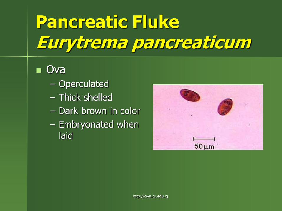
Pancreatic Fluke Eurytrema pancreaticum
Ova
– Operculated
– Thick shelled
– Dark brown in color
– Embryonated when laid
http://cvet.tu.edu.iq