Embed Size (px)
Citation preview

PEER-REVIEWED ARTICLE bioresources.com
Raymond et al. (2020). “Tree ID via extractives NMR,” BioResources 15(2), 2371-2384. 2371
Tree Species Identification via 1H NMR Fingerprinting of Supercritical Carbon Dioxide Wood Extractives Laura G. Raymond,a,* David Sandquist,b Stefan J. Hill,a Roger Meder,c,d and
Volker C. Behr e
Six tree species were examined using 1H NMR spectroscopy of sap extracted by supercritical CO2. A metabolomic approach was developed to evaluate the sap extracted from sapwood of Norway spruce (Picea abies), Sitka spruce (Picea sitchensis), radiata pine (Pinus radiata), macrocarpa (Cupressus macrocarpa), and two Eucalyptus species—shining gum and mountain ash (Eucalyptus nitens and Eucalyptus regnans. The sap extraction patterns in the different species were visualised using 1H magnetic resonance imaging. In softwoods with distinct annual rings, water was first removed from the latewood bands, and then gradually from the earlywood bands. In the case of the hardwood species an almost random water redistribution, rather than water expulsion, was observed. Analysis of the principal component analysis loading plots showed that the significant differences in the sap between each species were due to the carbohydrate region. Key discriminators were identified as pinitol, sucrose, glucose, and fructose.
Keywords: Wood metabolites; NMR fingerprinting; Magnetic resonance imaging; Wood anatomy;
Supercritical carbon dioxide
Contact information: a: Scion, Te Papa Tipu Innovation Park, 49 Sala St, Rotorua 3046, New Zealand;
b: VTT Technical Research Centre of Finland Ltd, P.O. Box 1000, 02044 VTT, Finland; c: Meder
Consulting, PO Box 3185, Bracken Ridge, QLD 4017, Australia; d: Forest Industries Research Centre,
University of the Sunshine Coast, Sippy Downs, QLD 4558, Australia; e: Experimental Physics 5,
University of Würzburg, Am Hubland, 97074 Würzburg, Germany;
* Corresponding author: [email protected]
INTRODUCTION
Nuclear magnetic resonance (NMR) based metabolomics research has rapidly
increased over the past decade into many diverse fields (Nicholson et al. 1999; Markley et
al. 2017; Pontes et al. 2017). It is a robust, high throughput technique that needs little or
no sample separation or derivatisation and is a recognised tool in a wide range of
metabolomics related applications (Ward et al. 2010), compared with classical approaches
such as microscopic identification. There has been significant research drive for NMR
metabolic profiling and fingerprinting of biofluids in the medical field, and the application
of metabolomics has been used to characterise biomarkers widely in disease diagnosis
(Emwas et al. 2013), with examples such as diabetes (Maher et al. 2009) and inborn errors
(Bamforth et al. 1999). These platforms aid in decision making within surgical and clinical
fields (Nicholson et al. 1999, 2012).
Metabolomic approaches have expanded to food science to establish food quality,
safety and nutrition, and in regulation of commodities such as wine (Hong 2011), juice
(Spraul et al. 2009), and coffee (Consonni et al. 2012). Numerous studies have been
dedicated to the assessment of the metabolomic response of plants to insects (Widarto et
PEER-REVIEWED ARTICLE bioresources.com
Raymond et al. (2020). “Tree ID via extractives NMR,” BioResources 15(2), 2371-2384. 2372
al. 2006), viruses (Choi et al. 2006), environmental effects (Bernillon et al. 2013), and at
developmental stages (Ali et al. 2011). Classification of different species and cultivars
using metabolic fingerprinting within the Edelweiss genus (Leontopodium spp.) has been
successful, where other morphological and molecular methods have been problematic
(Safer et al. 2011).
Comparatively little investigation has been undertaken in the study of metabolites
in the sapwood of trees. Recently, the metabolic differences of bark and sapwood in four
Sitka spruce clones after being challenged with a fungal pathogen were investigated using 1H NMR fingerprinting. Inoculated sapwood showed lower amounts of all metabolites,
while bark had increased signals in the aromatic region (Deflorio et al. 2012). However,
comparisons between the composition and behaviour of sapwood in different species using
magnetic resonance has not yet been considered.
This study demonstrates the identification of tree species based on a metabolomic
fingerprinting technique, for wood sap extraction under supercritical conditions based on 1H NMR spectroscopy and multivariate statistical analysis. Sap extraction of six tree
species (three softwoods and three hardwoods) was performed using Scion’s oscillating
supercritical carbon dioxide (scCO2) extraction technology for dewatering wood (Franich
et al. 2014). The scCO2 extraction approach is ideal to obtain bioactive compounds, as the
extract is conserved from contact with air and light, and uses moderate temperature (Khaw
et al. 2017). Both wood sap flow and wood impregnation using either fluids or supercritical
fluids are well known to be affected by wood anatomy (Schneider et al. 2006; Siau 1984).
Previous studies of the wood-water relationship using supercritical carbon dioxide have
investigated the movement and nature of water during the dewatering process of Pinus
radiata using 1H and/or 13C MR imaging (Behr et al. 2014; Meder et al. 2015; Newman et
al. 2016; Franich et al. 2019). The same approach was used in this study to visualise the
sap flow under scCO2 conditions for the six different wood anatomies investigated in this
study, using a high-pressure autoclave with a 1H/13C double-tuned resonator (Behr et al.
2013) to acquire 1H magnetic resonance (MR) images of the water during the extremes of
the pressure cycles.
EXPERIMENTAL
Materials Wood samples for sap and extracts collection
Wood core samples, 10 mm in diameter, were sourced in March (Autumn) from
around the Rotorua region, New Zealand (38.1 °S, 176.3 °E). The species analysed are
listed in Table 1. The cored samples were wrapped in plastic and stored at -20 °C for no
more than two days before scCO2 extraction.
Table 1. Wood Species Analysed
Common Name Latin Name Wood Type
Radiata pine Pinus radiata D. Don Softwood
Macrocarpa Cupressus macrocarpa Softwood
Norway spruce Picea abies Softwood
Sitka spruce Picea sitchensis Softwood
Shining gum Eucalyptus nitens Hardwood
Mountain ash Eucalyptus regnans Hardwood
PEER-REVIEWED ARTICLE bioresources.com
Raymond et al. (2020). “Tree ID via extractives NMR,” BioResources 15(2), 2371-2384. 2373
Wood samples for MR imaging
Green sapwood samples of all the species listed in Table 1, except Norway spruce,
were sourced using the green-chain of a local sawmill. Samples were cut to 15 mm (radial)
× 15 mm (tangential) × 95 mm (longitudinal) and edge-chamfered to achieve a tight fit in
the 20 mm internal diameter of the high-pressure MR cell (Behr et al. 2013). The samples
were packed with a wet tissue, sealed first in plastic, then in aluminium foil, and express
delivered from New Zealand to Würzburg, Germany in under five days. The green Norway
spruce samples were sourced in July (summer) from the region around Trento, Italy, and
cut to the same dimensions as noted above.
Methods Supercritical carbon dioxide sap extraction
The 10 mm cored green wood specimens were extracted sequentially in a 500 mL
high-pressure stainless-steel cylinder fitted with an inlet port for delivering liquid CO2 for
establishing the supercritical phase (Tcrit =31.1 °C, Pcrit = 7.38 MPa), an outlet port for
allowing exit of CO2 gas, and a separate lower port for recovery of wood sap.
Supercritical CO2 phase was generated using liquid CO2 (99.7% pure), which was
first chilled at ice-bath temperature to ensure maintenance of the liquid phase entering the
pump, and then delivered through a heated water bath maintained at 50 °C to establish the
supercritical phase at the target pressure of 20 MPa. A period at the target pressure, the
‘hold-time’ of the order of 10 minutes, was used to establish an equilibrium while the wood
specimen was held in contact with scCO2 at a constant pressure of 20 MPa and 50 °C. The
pressure was then decreased to around 4 MPa by pumping into the storage cylinder. The
lower valve was then opened to allow wood sap to flow out to be collected in a receiver.
The process was repeated as alternating pressure cycles until the sap flow
diminished or ceased. At the end of each experiment, the CO2 pressure was lowered to
atmospheric. The wood specimens were then removed from the cylinder and the resultant
aqueous sap samples were stored in 15 mL Falcon tubes at -80 °C until analysis.
Sample preparation for NMR analysis
The sample preparation and NMR experimental protocol were developed by
optimisation of several considerations, including water suppression parameters, pH, buffer
concentration, and ratio of buffer to sample. Sodium azide was added to suppress
microorganism activity, and the sodium salt of 3-(trimethylsilyl)-propionate acid-d4, (TSP)
was used as an internal NMR standard (0 ppm).
Frozen sap samples were thawed at room temperature and mixed on a vortex mixer
for one minute, after which 500 µL was transferred to a 2 mL microtube. A 500 µL aliquot
of a pH 7.00 phosphate buffer mixture was added to the aqueous sap samples. This gave a
final concentration of the phosphate buffer mixture of 0.1 mol L-1, which also contained
D2O (10% v/v), TSP (0.05% w/v), and sodium azide (0.05% w/v). The mixture was
homogenised by vortex mixing at room temperature for one minute before an aliquot of
600 µL was transferred into a 5 mm NMR tube for analysis.
1H NMR analysis
One-dimensional 1H NMR spectra of the sap samples were acquired on a Bruker
Avance III 400 MHz NMR spectrometer (Bruker, Rheinstetten, Germany) using the
noesygppr1d pulse sequence in the Bruker pulse sequence library, with a pre-saturation
power field of 33 Hz and transmitter frequency offset (O1) at 1886.70 Hz.
PEER-REVIEWED ARTICLE bioresources.com
Raymond et al. (2020). “Tree ID via extractives NMR,” BioResources 15(2), 2371-2384. 2374
The internal probe temperature was set to 300 K. The spectral data were obtained
with 65k data points, a relaxation delay of 8 s and 256 scans.
After Fourier transformation, NMR data were automatically phased, baseline
corrected and referenced to the TSP resonance at 0.00 ppm. The region from 4.57 to
5.20 ppm was excluded from analysis to remove variations in the suppression of the water
resonance, as was the residual acetone (from earlier cleaning cycles) signal in the region
from 2.20 to 2.30 ppm.
Functional group analysis was carried out by the integration of the following broad
proton chemical shift ranges; 0 to 3 ppm alkyl, 3 to 5.6 ppm carbohydrate / O-alkyl, and
5.6 to 8.5 ppm aromatic regions. Solvent peaks were excluded where required.
Multivariate statistical analysis was performed by bucketing spectra across the
region including 0.00 to 10.00 ppm using AMIX software (Bruker, Karlsruhe, Germany)
with a bucket size of 0.04 ppm. Bucketed data were evaluated using principal component
analysis (PCA) with a Pareto scaling method using SIMCA 14.1 software (Sartorius Stedim
Data Analytics AB, Göttingen, Germany).
1H MR imaging
The 1H MR imaging was performed on a Bruker Biospec 70/30 magnetic resonance
imaging system operating at 7.05 T (300 MHz 1H) with an Avance AV console, using
ParaVision v4 software for acquisition (Bruker, Ettlingen, Germany). The supercritical
extraction was performed using a purpose-built high-pressure polyetheretherketone
(PEEK) cell and birdcage radiofrequency coil (Behr et al. 2013). 1H MR images were acquired using a FLASH (Fast Low Angle SHot) sequence
(Haase et al. 1986) with a gradient echo time (TE) of 3.1 ms, a repetition time (TR) of 100
ms, and flip angle of α ≈ 20° optimised for T1(H) of a typical specimen (Ernst and Anderson
1966). Images used in the present paper were taken from the fifth of eight centrally located
slices, or from the third slice of four, with slice thickness = 1 mm and interslice distance =
3 mm. The field of view dimensions was 20 mm × 20 mm, with a matrix size of 128×128,
resulting in a voxel size of 1 mm × 156 μm × 156 μm presented as a pixel of size 156 μm
× 156 μm.
A total of 16 signals were averaged for each phase encoding step, requiring 3 min
of data acquisition. Profiles across rows of pixels or up columns of pixels were processed
in Prospa (Version 3.12, Magritek, Wellington, New Zealand). Because FLASH uses
gradient echoes rather than spin echoes, the signal strength is proportional to the
concentration of water molecules with T2*(H) > TE (Haase et al. 1986).
Image analysis
Greyscale images from the MR FLASH experiments were processed using Prospa.
The generated images were collected, and stack-aligned in ImageJ using the SIFT
algorithm (Lowe 2004), and then cropped to the sample outline.
Drying maps were calculated by comparing individual pixel values at a cut-off 80%
drop in intensity from the reference image and recording the cycle at which this took place.
A false colour image map was generated showing at which cycle the limit was reached.
PEER-REVIEWED ARTICLE bioresources.com
Raymond et al. (2020). “Tree ID via extractives NMR,” BioResources 15(2), 2371-2384. 2375
RESULTS AND DISCUSSION
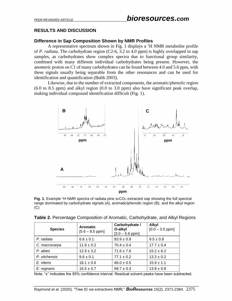
Difference in Sap Composition Shown by NMR Profiles A representative spectrum shown in Fig. 1 displays a 1H NMR metabolite profile
of P. radiata. The carbohydrate region (C2-6, 3.2 to 4.0 ppm) is highly overlapped in sap
samples, as carbohydrates show complex spectra due to functional group similarity,
combined with many different individual carbohydrates being present. However, the
anomeric proton on C1 of many carbohydrates can be found between 4.0 and 5.6 ppm, with
these signals usually being separable from the other resonances and can be used for
identification and quantification (Bubb 2003).
Likewise, due to the number of extracted components, the aromatic/phenolic region
(6.0 to 8.5 ppm) and alkyl region (0.0 to 3.0 ppm) also have significant peak overlap,
making individual compound identification difficult (Fig. 1).
Fig. 1. Example 1H NMR spectra of radiata pine scCO2 extracted sap showing the full spectral range dominated by carbohydrate signals (A), aromatic/phenolic region (B), and the alkyl region (C)
Table 2. Percentage Composition of Aromatic, Carbohydrate, and Alkyl Regions
Species Aromatic [5.6 – 8.5 ppm]
Carbohydrate / O-alkyl [3.0 – 5.6 ppm]
Alkyl [0.0 – 3.0 ppm]
P. radiata 6.6 ± 0.1 83.9 ± 0.9 9.5 ± 0.8
C. macrocarpa 11.9 ± 0.2 70.4 ± 0.4 17.7 ± 0.4
P. abies 12.9 ± 3.2 71.6 ± 7.6 15.2 ± 6.2
P. sitchensis 9.6 ± 0.1 77.1 ± 0.2 13.3 ± 0.2
E. nitens 18.1 ± 0.6 66.0 ± 0.5 15.9 ± 1.1
E. regnans 16.5 ± 0.7 69.7 ± 0.3 13.8 ± 0.8
Note: “±” indicates the 95% confidence interval. Residual solvent peaks have been subtracted.
ppm
ppm ppm
A
B C
PEER-REVIEWED ARTICLE bioresources.com
Raymond et al. (2020). “Tree ID via extractives NMR,” BioResources 15(2), 2371-2384. 2376
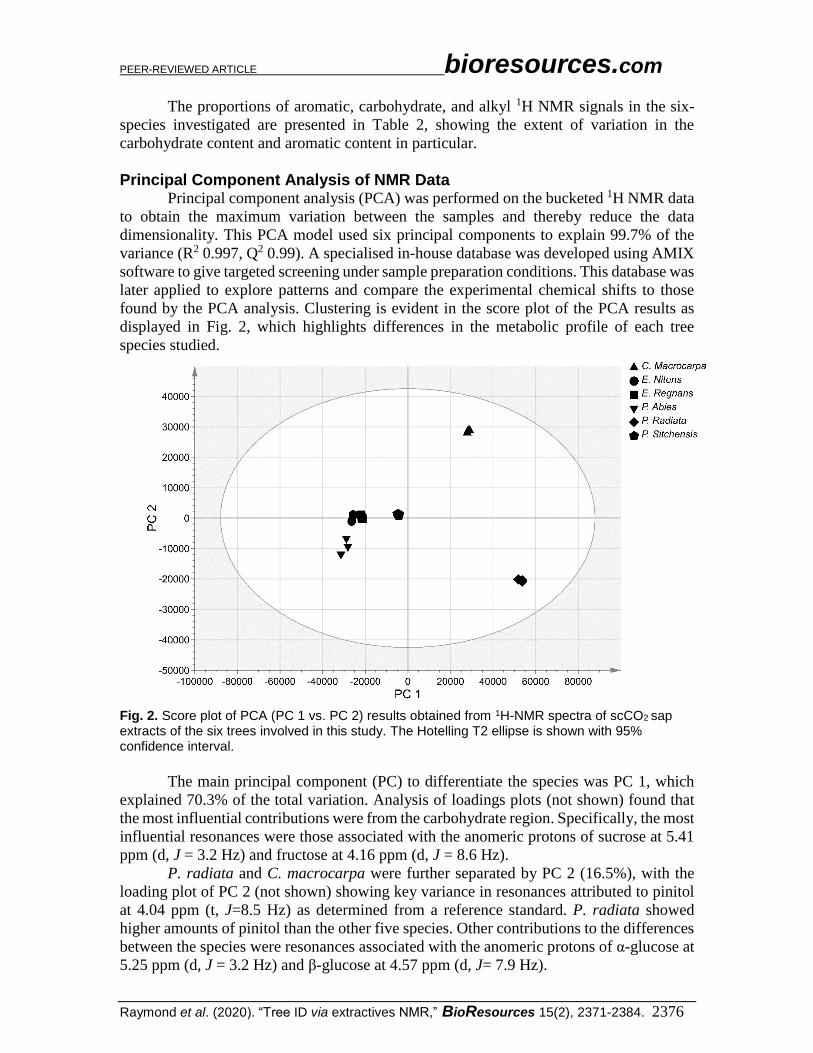
The proportions of aromatic, carbohydrate, and alkyl 1H NMR signals in the six-
species investigated are presented in Table 2, showing the extent of variation in the
carbohydrate content and aromatic content in particular.
Principal Component Analysis of NMR Data
Principal component analysis (PCA) was performed on the bucketed 1H NMR data
to obtain the maximum variation between the samples and thereby reduce the data
dimensionality. This PCA model used six principal components to explain 99.7% of the
variance (R2 0.997, Q2 0.99). A specialised in-house database was developed using AMIX
software to give targeted screening under sample preparation conditions. This database was
later applied to explore patterns and compare the experimental chemical shifts to those
found by the PCA analysis. Clustering is evident in the score plot of the PCA results as
displayed in Fig. 2, which highlights differences in the metabolic profile of each tree
species studied.
Fig. 2. Score plot of PCA (PC 1 vs. PC 2) results obtained from 1H-NMR spectra of scCO2 sap extracts of the six trees involved in this study. The Hotelling T2 ellipse is shown with 95% confidence interval.
The main principal component (PC) to differentiate the species was PC 1, which
explained 70.3% of the total variation. Analysis of loadings plots (not shown) found that
the most influential contributions were from the carbohydrate region. Specifically, the most
influential resonances were those associated with the anomeric protons of sucrose at 5.41
ppm (d, J = 3.2 Hz) and fructose at 4.16 ppm (d, J = 8.6 Hz).
P. radiata and C. macrocarpa were further separated by PC 2 (16.5%), with the
loading plot of PC 2 (not shown) showing key variance in resonances attributed to pinitol
at 4.04 ppm (t, J=8.5 Hz) as determined from a reference standard. P. radiata showed
higher amounts of pinitol than the other five species. Other contributions to the differences
between the species were resonances associated with the anomeric protons of α-glucose at
5.25 ppm (d, J = 3.2 Hz) and β-glucose at 4.57 ppm (d, J= 7.9 Hz).
PEER-REVIEWED ARTICLE bioresources.com
Raymond et al. (2020). “Tree ID via extractives NMR,” BioResources 15(2), 2371-2384. 2377
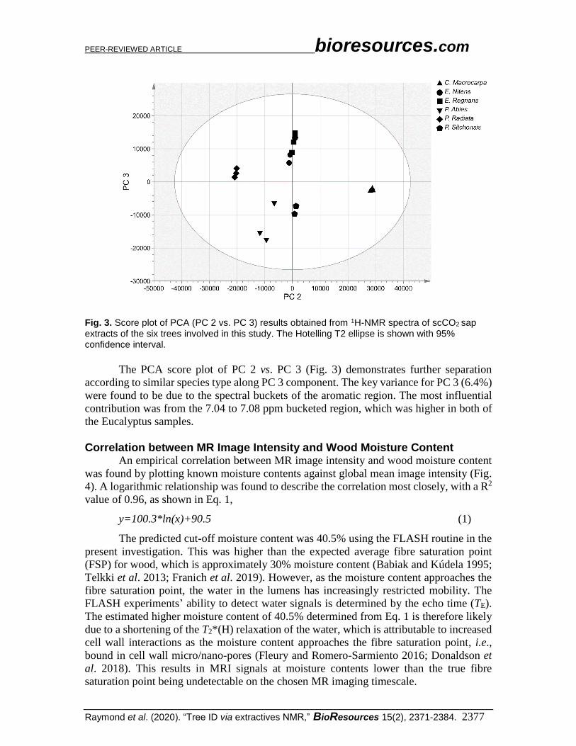
Fig. 3. Score plot of PCA (PC 2 vs. PC 3) results obtained from 1H-NMR spectra of scCO2 sap extracts of the six trees involved in this study. The Hotelling T2 ellipse is shown with 95% confidence interval.
The PCA score plot of PC 2 vs. PC 3 (Fig. 3) demonstrates further separation
according to similar species type along PC 3 component. The key variance for PC 3 (6.4%)
were found to be due to the spectral buckets of the aromatic region. The most influential
contribution was from the 7.04 to 7.08 ppm bucketed region, which was higher in both of
the Eucalyptus samples.
Correlation between MR Image Intensity and Wood Moisture Content An empirical correlation between MR image intensity and wood moisture content
was found by plotting known moisture contents against global mean image intensity (Fig.
4). A logarithmic relationship was found to describe the correlation most closely, with a R2
value of 0.96, as shown in Eq. 1,
y=100.3*ln(x)+90.5 (1)
The predicted cut-off moisture content was 40.5% using the FLASH routine in the
present investigation. This was higher than the expected average fibre saturation point
(FSP) for wood, which is approximately 30% moisture content (Babiak and Kúdela 1995;
Telkki et al. 2013; Franich et al. 2019). However, as the moisture content approaches the
fibre saturation point, the water in the lumens has increasingly restricted mobility. The
FLASH experiments’ ability to detect water signals is determined by the echo time (TE).
The estimated higher moisture content of 40.5% determined from Eq. 1 is therefore likely
due to a shortening of the T2*(H) relaxation of the water, which is attributable to increased
cell wall interactions as the moisture content approaches the fibre saturation point, i.e.,
bound in cell wall micro/nano-pores (Fleury and Romero-Sarmiento 2016; Donaldson et
al. 2018). This results in MRI signals at moisture contents lower than the true fibre
saturation point being undetectable on the chosen MR imaging timescale.

PEER-REVIEWED ARTICLE bioresources.com
Raymond et al. (2020). “Tree ID via extractives NMR,” BioResources 15(2), 2371-2384. 2378
Fig. 4. Correlation between MR image intensity and wood moisture content. Using a logarithmic regression, the empirical cut off for MR intensity in these experiments is at a 40.6% moisture content.
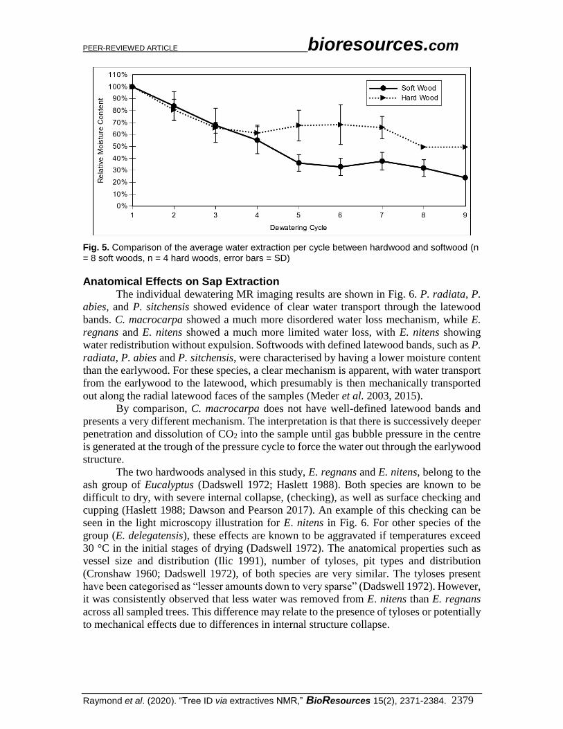
Comparing Sap Extraction for Softwoods and Hardwoods Softwoods and hardwoods have significantly different anatomy, with softwoods
consisting of fewer types of cells than hardwoods. In both hardwoods and softwoods, the
sapwood component of the trunk is the main conduit for water from the roots to the crown,
with a counter flow of sugar-rich sap from the crown to the rest of the tree in the cambium,
just below the bark (Dinwoodie 2002).
In softwoods, the tracheids are the main conduit of water, with some influence of
the rays (Siau 1984). In the case of mechanical or scCO2 assisted dewatering, the resin
canals, as well as effects such as earlywood/latewood transitions, also have a significant
contribution to the water removal pattern. The transition between adjacent fibres is
regulated by bordered pits.
In hardwoods, the vessels are the main conduit of water, with a more significant
contribution of the rays due to their increased diameter and number. In the case of
mechanical or scCO2 assisted dewatering, the rest of the anatomy can also transport water
to a lesser degree, including the fibre cells (Siau 1984).
In both hardwoods and softwoods, water flow between the different cell types is
regulated by pits, which differ in their complexity and diameter. On a structurally higher
level, the influence of the transition between earlywood and latewood also has a significant
impact on water flow (Siau 1984).
These differences are examined here at two levels, firstly to determine any overall
significant differences between the analysed hardwoods and softwoods as groups, and then
for any individual differences of note.
Comparing soft- and hardwood groups, a significant difference was observed
between the 4th and 5th dewatering cycle, as shown in Fig. 5. For hardwoods, there was a
relative drop of relative moisture content from 100 to 60%, where it began to plateau, while
for softwoods the moisture content continued to reduce to 40% before plateauing, as has
been previously observed (Dawson and Pearson 2017). In softwoods, this is attributed to
the sap removal via mechanical transportation (or redistribution) of water through the less
saturated latewood bands (Fig. 6). In hardwoods, on the other hand, vessels are the main
longitudinal conduit of water, with the pit structure limiting transverse transportation
(Cronshaw 1960). Considering this, most of the sap loss is attributed to longitudinal
removal, with only subsequent limited redistribution of water in the transverse plane.
PEER-REVIEWED ARTICLE bioresources.com
Raymond et al. (2020). “Tree ID via extractives NMR,” BioResources 15(2), 2371-2384. 2379
Fig. 5. Comparison of the average water extraction per cycle between hardwood and softwood (n = 8 soft woods, n = 4 hard woods, error bars = SD)
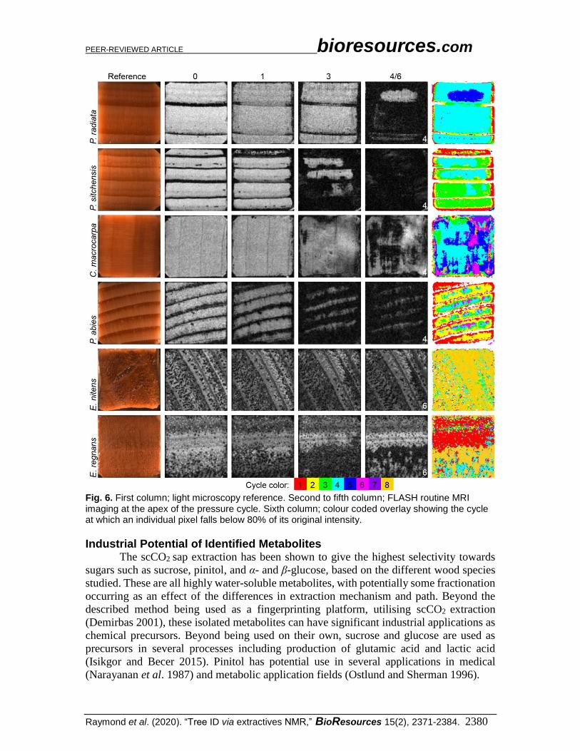
Anatomical Effects on Sap Extraction The individual dewatering MR imaging results are shown in Fig. 6. P. radiata, P.
abies, and P. sitchensis showed evidence of clear water transport through the latewood
bands. C. macrocarpa showed a much more disordered water loss mechanism, while E.
regnans and E. nitens showed a much more limited water loss, with E. nitens showing
water redistribution without expulsion. Softwoods with defined latewood bands, such as P.
radiata, P. abies and P. sitchensis, were characterised by having a lower moisture content
than the earlywood. For these species, a clear mechanism is apparent, with water transport
from the earlywood to the latewood, which presumably is then mechanically transported
out along the radial latewood faces of the samples (Meder et al. 2003, 2015).
By comparison, C. macrocarpa does not have well-defined latewood bands and
presents a very different mechanism. The interpretation is that there is successively deeper
penetration and dissolution of CO2 into the sample until gas bubble pressure in the centre
is generated at the trough of the pressure cycle to force the water out through the earlywood
structure.
The two hardwoods analysed in this study, E. regnans and E. nitens, belong to the
ash group of Eucalyptus (Dadswell 1972; Haslett 1988). Both species are known to be
difficult to dry, with severe internal collapse, (checking), as well as surface checking and
cupping (Haslett 1988; Dawson and Pearson 2017). An example of this checking can be
seen in the light microscopy illustration for E. nitens in Fig. 6. For other species of the
group (E. delegatensis), these effects are known to be aggravated if temperatures exceed
30 °C in the initial stages of drying (Dadswell 1972). The anatomical properties such as
vessel size and distribution (Ilic 1991), number of tyloses, pit types and distribution
(Cronshaw 1960; Dadswell 1972), of both species are very similar. The tyloses present
have been categorised as “lesser amounts down to very sparse” (Dadswell 1972). However,
it was consistently observed that less water was removed from E. nitens than E. regnans
across all sampled trees. This difference may relate to the presence of tyloses or potentially
to mechanical effects due to differences in internal structure collapse.
PEER-REVIEWED ARTICLE bioresources.com
Raymond et al. (2020). “Tree ID via extractives NMR,” BioResources 15(2), 2371-2384. 2380
Fig. 6. First column; light microscopy reference. Second to fifth column; FLASH routine MRI imaging at the apex of the pressure cycle. Sixth column; colour coded overlay showing the cycle at which an individual pixel falls below 80% of its original intensity.
Industrial Potential of Identified Metabolites The scCO2 sap extraction has been shown to give the highest selectivity towards
sugars such as sucrose, pinitol, and α- and β-glucose, based on the different wood species
studied. These are all highly water-soluble metabolites, with potentially some fractionation
occurring as an effect of the differences in extraction mechanism and path. Beyond the
described method being used as a fingerprinting platform, utilising scCO2 extraction
(Demirbas 2001), these isolated metabolites can have significant industrial applications as
chemical precursors. Beyond being used on their own, sucrose and glucose are used as
precursors in several processes including production of glutamic acid and lactic acid
(Isikgor and Becer 2015). Pinitol has potential use in several applications in medical
(Narayanan et al. 1987) and metabolic application fields (Ostlund and Sherman 1996).
PEER-REVIEWED ARTICLE bioresources.com
Raymond et al. (2020). “Tree ID via extractives NMR,” BioResources 15(2), 2371-2384. 2381
CONCLUSIONS
1. Tree species can be identified from wood sap isolated via scCO2 extraction with the
application of metabolomics in combination with multivariate statistical analysis.
2. Sap drainage patterns differ significantly between hard- and softwoods under scCO2
extraction, which may contribute to the identified differences in sap chemical
composition.
3. A plethora of aliphatic and aromatic compounds were extracted under mild
temperatures, that with separation and identification of individual compounds could
feed into high value chemical industries or act as potential biomarkers, opening the
possibility of employing them as a diagnostics tool.
ACKNOWLEDGMENTS
This work was funded by the Ministry of Business, Innovation and Employment,
through Crown Research Institute (CRI) Core Funding to Scion. The authors would like to
thank the technical assistance provided by Sheree Anderson (Scion), Hank Kroese (Scion),
Daniel Van De Pas (Scion), and Sabine Voll (Universität Würzburg) for help with both the
experimental setup and sourcing of the wood materials. The authors acknowledge Drs.
Jakub and Anna Sandak at IVALSA, Italy, for the supply of the Norway spruce sample to
the Universität Würzburg. Roger Meder would like to acknowledge travel assistance from
the JW Gottstein Memorial Trust and the Deutsche Akademischer Austauschdienst for
travel during the period 2011 to 2013.
REFERENCES CITED
Ali, K., Maltese, F., Fortes, A. M., Pais, M. S., Choi, Y. H., and Verpoorte, R. (2011).
“Monitoring biochemical changes during grape berry development in Portuguese
cultivars by NMR spectroscopy,” Food Chemistry 124(4), 1760-1769. DOI:
10.1016/j.foodchem.2010.08.015
Babiak, M., and Kúdela, J. (1995). “A contribution to the definition of the fiber saturation
point,” Wood Science and Technology 29(3). DOI: 10.1007/bf00204589
Bamforth, F., Dorian, V., Vallance, H., and Wishart, D. (1999). “Diagnosis of inborn
errors of metabolism using 1H NMR spectroscopic analysis of urine,” Journal of
Inherited Metabolic Disease 22(3), 297-301. DOI: 10.1023/A:1005531432766
Behr, V. C., Schmid, M. W., Franich, R. A., and Meder, R. (2013). “An advanced,
integrated large-volume high-pressure autoclave and 1H/13C double-tuned resonator
for chemistry and materials nuclear magnetic resonance spectroscopy and microscopy
investigations: NMR spectroscopy and microscopy at high pressures,” Concepts in
Magnetic Resonance Part B: Magnetic Resonance Engineering 43(2), 49-58. DOI:
10.1002/cmr.b.21233
Behr, V. C., Hill, S. J., Meder, R., Sandquist, D., Hindmarsh, J. P., Franich, R. A., and
Newman, R. H. (2014). “Carbon-13 NMR chemical-shift imaging study of
dewatering of green sapwood by cycling carbon dioxide between supercritical fluid
and gas phases,” The Journal of Supercritical Fluids, 95, 535-540. DOI:

PEER-REVIEWED ARTICLE bioresources.com
Raymond et al. (2020). “Tree ID via extractives NMR,” BioResources 15(2), 2371-2384. 2382
10.1016/j/supflu.2014.08.026
Bernillon, S., Biais, B., Deborde, C., Maucourt, M., Cabasson, C., Gibon, Y., Hansen, T.
H., Husted, S., de Vos, R. C., Mumm, R., and others. (2013). “Metabolomic and
elemental profiling of melon fruit quality as affected by genotype and environment,”
Metabolomics 9(1), 57-77. DOI: 10.1007/s11306-012-0429-1
Bubb, W. A. (2003). “NMR spectroscopy in the study of carbohydrates: Characterizing
the structural complexity,” Concepts in Magnetic Resonance 19A(1), 1-19. DOI:
10.1002/cmr.a.10080
Choi, Y. H., Kim, H. K., Linthorst, H. J. M., Hollander, J. G., Lefeber, A. W. M.,
Erkelens, C., Nuzillard, J.-M., and Verpoorte, R. (2006). “NMR metabolomics to
revisit the tobacco mosaic virus infection in Nicotiana tabacum leaves,” Journal of
Natural Products 69(5), 742-748. DOI: 10.1021/np050535b
Consonni, R., Cagliani, L. R., and Cogliati, C. (2012). “NMR based geographical
characterization of roasted coffee,” Talanta 88, 420-426. DOI:
10.1016/j.talanta.2011.11.010
Cronshaw, J. (1960). “The fine structure of the pits of Eucalyptus regnans (F. Muell.) and
their relation to the movement of liquids into the wood,” Australian Journal of
Botany 8(1), 51-57. DOI: 10.1071/BT9600051
Dadswell, H. E. (1972). The Anatomy of Eucalypt Woods (Technological Paper), Forest
Products Laboratory, Division of Applied Chemistry, Commonwealth Scientific and
Industrial Research Organization, Australia, Melbourne.
Dawson, B. S. W., and Pearson, H. (2017). “Effect of supercritical CO2 dewatering
followed by oven-drying of softwood and hardwood timbers,” Wood Science and
Technology 51(4), 771-784. DOI: 10.1007/s00226-017-0895-8
Deflorio, G., Horgan, G., Jaspars, M., and Woodward, S. (2012). “Defense response of
Sitka spruce before and after inoculation with Heterobasidion annosum: 1H NMR
fingerprinting of bark and sapwood metabolites,” Analytical and Bioanalytical
Chemistry 402(10), 3333-3344. DOI: 10.1007/s00216-011-5666-z
Demirbas, A. (2001). “Supercritical fluid extraction and chemicals from biomass with
supercritical fluids,” Energy Conversion & Management 42(3), 279-294. DOI:
10.1016/S0196-8904(00)00059-5
Dinwoodie, J. (2002). Timber: Its Nature and Behaviour, CRC Press, Boca Raton, FL,
USA.
Donaldson, L. A., Cairns, M., and Hill, S. J. (2018). “Comparison of micropore
distribution in cell walls of softwood and hardwood xylem,” Plant Physiology 178(3),
1142–53. DOI: 10.1104/pp.18.00883
Emwas, A.-H. M., Salek, R. M., Griffin, J. L., and Merzaban, J. (2013). “NMR-based
metabolomics in human disease diagnosis: applications, limitations, and recom-
mendations,” Metabolomics, 9(5), 1048-1072. DOI: 10.1007/s11306-013-0524-y
Fleury, M., and Romero-Sarmiento, M. (2016). “Characterization of shales using T1-T2
NMR maps,” Journal of Petroleum Science and Engineering 137, 55-62. DOI:
10.1016/j.petrol.2015.11.006
Franich, R. A., Gallagher, S., and Kroese, H. (2014). “Dewatering green sapwood using
carbon dioxide cycled between supercritical fluid and gas phase,” The Journal of
Supercritical Fluids 89, 113-118. DOI: 10.1016/j.supflu.2014.02.019
Franich, R. A., Meder, R., Falge, M., Fuchs, J., and Behr, V. C. (2019). “Uncovering
supercritical CO2 wood dewatering via interleaved 1H-imaging and 13C-spectroscopy
with real-time reconstruction,” The Journal of Supercritical Fluids 114, 56-62. DOI:

PEER-REVIEWED ARTICLE bioresources.com
Raymond et al. (2020). “Tree ID via extractives NMR,” BioResources 15(2), 2371-2384. 2383
10.1016/j.supflu.2018.10.006
Haslett, A. N. (1988). Properties and Utilisation of Exotic Speciality Timbers Grown in
New Zealand Part V: Ash Eucalypts and Eucalyptus Nitens (FRI Bulletin no. 119),
Scion, Rotorua, New Zealand.
Hong, Y.-S. (2011). “NMR-based metabolomics in wine science,” Magnetic Resonance
in Chemistry 49, S13-S21. DOI: 10.1002/mrc.2832
Ilic, J. (1991). CSIRO Atlas of Hardwoods, Springer-Verlag, Berlin.
Isikgor, F. H., and Becer, C. R. (2015). “Lignocellulosic biomass: A sustainable platform
for the production of bio-based chemicals and polymers,” Polymer Chemistry 6(25),
4497-4559. DOI: 10.1039/C5PY00263J
Khaw, K. Y., Parat, M. O., Shaw, P. N., and Falconer, J. R. (2017). “Solvent supercritical
fluid technologies to extract bioactive compounds from natural sources: A review,”
Molecules 22(7), 1186-1208. DOI:10.3390/molecules22071186
Lowe, D. G. (2004). “Distinctive image features from scale-invariant keypoints,”
International Journal of Computer Vision 60(2), 91-110. DOI:
10.1023/b:visi.0000029664.99615.94
Maher, A. D., Lindon, J. C., and Nicholson, J. K. (2009). “1H NMR-based metabonomics
for investigating diabetes,” Future Medicinal Chemistry 1(4), 737-747. DOI:
10.4155/fmc.09.54
Markley, J. L., Brüschweiler, R., Edison, A. S., Eghbalnia, H. R., Powers, R., Rafferty,
D., and Wishart, D. S. (2017). “The future of NMR-based metabolomics,” Current
Opinion in Biotechnology 43, 34-40. DOI: 10.1016/j.copbio.2016.08.001
Meder, R., Codd, S. L., Franich, R. A., Callaghan, P. T., and Pope, J. M. (2003).
“Observation of anisotropic water movement in Pinus radiata D. Don wood using
magnetic resonance micro-imaging,” Holz als Roh- und Werkstoff 61(4), 251-256.
DOI: 10.1007/s00107-003-0400-y
Meder, R., Franich, R. A., Callaghan, P. T., and Behr, V. C. (2015). “A comparative
study of dewatering of Pinus radiata sapwood using supercritical CO2 via in situ
magnetic resonance microimaging,” Holzforschung 69(9), 1137-1142. DOI:
10.1515/hf-2014-0134 Narayanan, C., Joshi, D., Mujumdar, A., and Dhekne, V. (1987). “Pinitol—A new anti-
diabetic compound from the leaves of Bougainvillea spectabilis,” Current Science
56(3), 139-141.
Newman, R. H., Franich, R. A., Meder, R., Hill, S. J., Kroese, H., Sandquist, D.,
Hindmarsh, J. P., Schmid, M. W., Fuchs, J. and Behr, V. C. (2016). “Proton magnetic
resonance imaging used to investigate dewatering of green sapwood by cycling
carbon dioxide between supercritical fluid and gas phases,” The Journal of
Supercritical Fluids 111, 36-42. DOI: 10.1016/j/supflu.2016.01.007
Nicholson, J. K., Holmes, E., Kinross, J. M., Darzi, A. W., Takats, Z., and Lindon, J. C.
(2012). “Metabolic phenotyping in clinical and surgical environments,” Nature,
491(7424), 384-392. DOI: 10.1038/nature11708
Nicholson, J. K., Lindon, J. C., and Holmes, E. (1999). “ ‘Metabonomics’: Understanding
the metabolic responses of living systems to pathophysiological stimuli via
multivariate statistical analysis of biological NMR spectroscopic data,” Xenobiotica,
29(11), 1181-1189. DOI: 10.1080/004982599238047
Ostlund, R. E., and Sherman, W. R. (1996). “Pinitol and derivatives thereof for the
treatment of metabolic disorders,” U. S. Patent No. US5827896A.
Pontes, J. G. M., Brasil, A. J. M., Cruz, G. C. F., de Souza, R. N., and Tasic, L. (2017)

PEER-REVIEWED ARTICLE bioresources.com
Raymond et al. (2020). “Tree ID via extractives NMR,” BioResources 15(2), 2371-2384. 2384
“NMR-based metabolomics strategies: Plants, animal and humans,” Analytical
Methods 9, 1078-1096. DOI: 10.1039/C6AY03102A
Safer, S., Cicek, S. S., Pieri, V., Schwaiger, S., Schneider, P., Wissemann, V., and
Stuppner, H. (2011). “Metabolic fingerprinting of Leontopodium species (Asteraceae)
by means of 1H NMR and HPLC–ESI-MS,” Phytochemistry 72(11–12), 1379-1389.
DOI: 10.1016/j.phytochem.2011.04.006
Schneider, P. F., Levien, K. L., and Morrell, J. J. (2006). “Effect of wood characteristics
on pressure responses during supercritical carbon dioxide treatment,” Wood and
Fiber Science 38(4), 660-671.
Siau, J. F. (1984). Transport Processes in Wood, Springer-Verlag, Berlin.
Spraul, M., Schütz, B., Humpfer, E., Mörtter, M., Schäfer, H., Koswig, S., and Rinke, P.
(2009). “Mixture analysis by NMR as applied to fruit juice quality control,” Magnetic
Resonance in Chemistry 47(S1), S130-S137. DOI: 10.1002/mrc.2528.
Telkki, V.-V., Yliniemi, M., and Jokisaari, J. (2013). “Moisture in softwoods: Fiber
saturation point, hydroxyl site content, and the amount of micropores as determined
from NMR relaxation time distributions,” Holzforschung 67(3), 291-300. DOI:
10.1515/hf-2012-0057
Ward, J. L., Baker, J. M., Miller, S. J., Deborde, C., Maucourt, M., Biais, B., Rolin, D.,
Moing, A., Moco, S., Vervoort, J., Lommen, A., Schäfer, H., Humpfer, E., and Beale,
M. H. (2010). “An inter-laboratory comparison demonstrates that 1H-NMR
metabolite fingerprinting is a robust technique for collaborative plant metabolomic
data collection,” Metabolomics 6(2), 263-273. DOI: 10.1007/s11306-010-0200-4
Widarto, H. T., Van Der Meijden, E., Lefeber, A. W. M., Erkelens, C., Kim, H. K., Choi,
Y. H., and Verpoorte, R. (2006). “Metabolomic differentiation of Brassica rapa
following herbivory by different insect instars using two-dimensional nuclear
magnetic resonance spectroscopy,” Journal of Chemical Ecology 32(11), 2417-2428.
DOI: 10.1007/s10886-006-9152-6
Article submitted: July 4, 2019; Peer review completed: December 7, 2019; Revised
version received and accepted: February 2, 2020; Published: February 11, 2020.
DOI: 10.15376/biores.15.2.2371-2384