Embed Size (px)
Citation preview
ORIGINAL ARTICLES
Treatment of Superficial Infantile Hemangiomas of the EyelidUsing the 595-nm Pulsed Dye Laser
CHRISTOPHER M. HUNZEKER, MD, AND ROY G. GERONEMUS, MD�
BACKGROUND Despite the proven effectiveness of the 595-nm pulsed dye laser (PDL) in treating su-perficial infantile hemangiomas, many physicians are reluctant to treat such lesions involving the eyelid.
OBJECTIVE To examine the safety and efficacy of the 595-nm PDL for the treatment of superficialinfantile hemangiomas of the eyelid.
MATERIALS & METHODS Records were reviewed for patients with superficial infantile hemangiomas ofthe eyelid treated with 595-nm PDL. Pre- and post-treatment photographs were compared. Reviewersrated the degree of improvement of the hemangioma as excellent (76–100%), good (51–75%), moderate(26–50%), or poor (0–25%) and indicated whether the hemangioma was 100% clear. Side effects ofscarring, atrophy, hyperpigmentation, and hypopigmentation were assessed.
RESULTS Twenty-two patients met the study criteria. Eight (36.4%) demonstrated complete clearanceof their hemangioma, 17 (77.3%) received an improvement rating of excellent, and five (22.7%) receiveda rating of good. No scarring, atrophy, or hypopigmentation was noted. Two patients (9.1%) were notedto have hyperpigmentation in the treated area.
CONCLUSION Early treatment with the 595-nm PDL can safely and effectively diminish proliferativegrowth and hasten resolution of superficial infantile hemangiomas of the eyelid.
Roy G. Geronemus, MD, is on the Medical Advisory Board for Candela Laser Corp.
Hemangiomas affect 2% to 3% of newborns
and up to 10% of infants within the first year
of life, making them the most common tumors of
infancy.1,2 Sixty percent of hemangiomas occur on
the head and neck, and approximately 16% of facial
hemangiomas involve the eyelid.3,4 Like elsewhere
on the body, hemangiomas of the eyelid can be
superficial, deep, or compound (having a superficial
and a deep component). Superficial lesions appear
bright red and can be flat patches or slightly elevated
plaques extending no deeper than the papillary der-
mis histologically. Deep hemangiomas involve the
reticular dermis to varying degrees and can protrude
into the subcutaneous tissue, appearing clinically as
skin-colored or bluish nodules.
Compound and deep eyelid hemangiomas warrant
special attention because of their potential to com-
promise developing vision because of amblyopia
resulting from anisometropia and, less commonly,
because of strabismus or obstruction of the visual
axis.5 These findings can be seen with small hem-
angiomas, so evaluation by a pediatric ophthalmolo-
gist is recommended with all compound and deep
periocular hemangiomas.6 In the case of rapidly
enlarging periocular hemangiomas, close monitoring
by an ophthalmologist is essential, because vision may
be permanently compromised in as little as 2 weeks.7
A majority of eyelid hemangiomas are superficial,
posing no significant threat to the vision. These
lesions are uncomplicated medically but can grow to
become extensive and disfiguring and can persist for
years. Hemangiomas typically undergo gradual
spontaneous involution at a rate of approximately
10% per year such that 50% of lesions fully involute
& 2010 by the American Society for Dermatologic Surgery, Inc. � Published by Wiley Periodicals, Inc. �ISSN: 1076-0512 � Dermatol Surg 2010;36:590–597 � DOI: 10.1111/j.1524-4725.2010.01511.x
5 9 0
�Both authors are affiliated with Laser and Skin Surgery Center of New York, NewYork, New York
by age 5, 70% by age 7, and so on, regardless of
their size or location.3,8 According to these esti-
mates, roughly half of hemangiomas will still be
apparent when the affected children begin school.
Treatment methods for eyelid hemangiomas are
numerous and should be chosen according to the
clinical characteristics of the lesion. Observation is
an acceptable treatment alternative for compound
and deep eyelid hemangiomas that are proliferating
slowly or stable in size and have been determined,
upon evaluation by a pediatric ophthalmologist, to
be no threat to the patient’s vision. For rapidly pro-
liferating deep or compound hemangiomas or those
threatening the infant’s vision, treatment alternatives
include surgical excision or debulking procedures,
intraarterial embolization, systemic medical therapy
(e.g., oral corticosteroids and propranolol), or
intralesional injections of corticosteroids or inter-
feron.9,10 Each of these therapies has its own set of
risks and side effects and, therefore, consultation
with a physician specializing in the treatment of
vascular tumors is recommended.
It can be difficult during the proliferative phase to
predict accurately whether a deep component will
arise within a seemingly superficial hemangioma.
For this reason, close monitoring during the prolif-
erative phase is recommended. The majority of
purely superficial eyelid hemangiomas have histori-
cally been managed with clinical observation.
Corticosteroids have been used topically, with
modest improvement.11 Corticosteroids are also
frequently administered orally or intralesionally,
with better results, for rapidly growing superficial
hemangiomas. Although somewhat controversial,
the pulsed dye laser (PDL) is being used with
increasing frequency and has become an accepted
treatment alternative for superficial hemangiomas.
PDL has been used for more than 25 years to treat
port-wine stains, telangiectases, and vascular neo-
plasms, including infantile hemangiomas. Several
studies have confirmed the effectiveness of PDL in
treating hemangiomas, most concluding that PDL is
more effective in the treatment of superficial hem-
angiomas than deep lesions because of the limited
depth of vascular injury.12–17 Some authors consider
PDL to be the treatment of choice for superficial
hemangiomas.15
Physicians who oppose treating superficial hem-
angiomas with PDL often cite a high incidence of
side effects resulting from treatment and argue that
uncomplicated hemangiomas are better managed
with clinical observation. In a prospective,
randomized controlled trial, Batta and colleagues18
demonstrated that superficial hemangiomas
treated with PDL showed significantly reduced red-
ness and a six times greater rate of complete clear-
ance at 1 year than lesions observed clinically. This
study also demonstrated a significant difference with
respect to the median change in surface area 1 year
from baseline, supporting the concept that early
treatment with PDL can slow or halt the proliferative
growth phase of the hemangioma.13 Despite their
findings, the authors concluded that there is no
benefit to treating superficial hemangiomas with
PDL and implied that early treatment with PDL is
inferior to observation because of the high incidence
of atrophy (28%) and hypopigmentation (45%).18
Reported side effects associated with PDL treatment
of hemangiomas include pigmentary alteration,
ulceration, atrophy, and scarring.19 The authors
of one report cite 12 cases at three tertiary referral
centers over a 5-year period, concluding that
significant complications from PDL treatment
of hemangiomas are rare.20 This paucity of cases
is consistent with the low rate of complications
reported in other studies.15,16,19 Pulsed dye laser
technology has advanced in recent years, incorpo-
rating epidermal cooling using a cryogen spray,
which protects the epidermis from thermal injury,
reducing the risk of side effectsfrom treatment.
The study performed by Batta and colleagues
excluded patients with periocular hemangiomas,
stating that these lesions have the highest risk
of psychosocial morbidity and complications.
3 6 : 5 : M AY 2 0 1 0 5 9 1
H U N Z E K E R A N D G E R O N E M U S
Clinically, we have found that superficial periocular
hemangiomas, and specifically superficial eyelid
hemangiomas, respond exceedingly well to
treatment with PDL. This retrospective study was
designed to examine the efficacy and safety of the
595-nm PDL for the treatment of superficial infantile
hemangiomas of the eyelid during the proliferative
growth phase.
Materials and Methods
Medical records were reviewed for all patients
with infantile hemangiomas of the eyelid treated
with the 595-nm PDL at the Laser and Skin Surgery
Center of New York from July 2004 to December
2008. With our intention to focus on hemangiomas
treated during the proliferative growth phase,
only patients with superficial eyelid hemangiomas
initiating treatment before 9 months of age were
included in the study. Patients with a deep
component to their hemangioma were excluded, as
were those receiving prior or concomitant medical
or surgical treatment for their hemangioma.
Additional exclusion criteria included inadequate
photographic documentation and a lapse in
follow-up exceeding 6 months.
Records for 39 patients with eyelid hemangiomas
were reviewed. Twenty-two patients, 15 girls and
seven boys, met the study criteria. Seventeen patients
were excluded; eight had compound hemangiomas,
four had received prior oral corticosteroid therapy,
two had previously undergone surgical debulking
procedures, two initiated treatment at an age older
than 9 months, and one was excluded because of a
7-month lapse in follow up.
Patients were treated with the 595-nm PDL (Vbeam
Perfecta, Candela Corp., Wayland, MA) with the
following parameters: energy fluence of 11.0 to
11.5 J/cm2, 7-mm spot size, and a pulse width of
0.45 ms or 1.5 ms. The dynamic cooling device set-
tings consisted of a 30-ms cryogen spray duration
with a 20-ms delay. The child was placed in the
supine position on the examining table and gently
held still by a nurse or parent. Tetracaine ophthalmic
solution 0.5% (Bausch & Lomb, Inc., Tampa, FL)
was administered before the insertion of a metal
corneal eye shield (Stefanovsky & Associates Inc.,
Willowich, OH) to protect the patient’s eye ipsilat-
eral to the hemangioma. A nurse covered the
contralateral eye with an external shield during the
treatment. A thin layer of Surgilube (E. Fougera
& Co., Melville, NY) was applied to the patient’s
eyebrows and eyelashes to prevent them from
singeing. Lesions were treated until a purpuric end
point was reached. This occasionally required dou-
ble pulsing of the laser, but pulse stacking was
avoided. In most cases, the entire treatment, includ-
ing insertion of the intraocular shield, was completed
in 1 to 2 minutes.
Three nontreating reviewers, all board-certified der-
matologists, compared pretreatment photographs
with photographs taken on the date of the patient’s
final laser treatment or on the date of the first visit
that complete clearance was documented and treat-
ment deemed unnecessary. Considering color and
elevation of the lesion, the reviewers rated the degree
of improvement of each hemangioma as excellent
(76–100%), good (51–75%), moderate (26–50%),
or poor (0–25%) and indicated whether the hem-
angioma was 100% clear. The reviewers also eval-
uated the photographs for the presence of side
effects, including scarring, atrophy, hyperpigmenta-
tion, and hypopigmentation, and the authors
reviewed the chart to note any occurrence of ulcer-
ation, infection, or other complications resulting
from treatment.
In the cases in which the reviewers’ ratings
were not unanimous, a majority rule was applied.
(The rating selected by two of three reviewers was
used.) The same majority rule was applied in deter-
mining whether there was 100% clearance of the
hemangioma or the presence of a treatment-related
side effect. There was one case in which all three
reviewers selected different degrees of improvement.
In this instance, the highest and lowest ratings were
disregarded and the middle rating was used.
D E R M AT O L O G I C S U R G E RY5 9 2
T R E AT M E N T O F E Y E L I D H E M A N G I O M A S W I T H P U L S E D D Y E L A S E R
Results
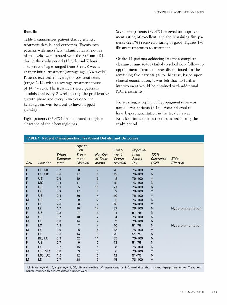
Table 1 summarizes patient characteristics,
treatment details, and outcomes. Twenty-two
patients with superficial infantile hemangiomas
of the eyelid were treated with the 595-nm PDL
during the study period (15 girls and 7 boys).
The patients’ ages ranged from 5 to 28 weeks
at their initial treatment (average age 13.8 weeks).
Patients received an average of 5.6 treatments
(range 2–14) with an average treatment course
of 14.9 weeks. The treatments were generally
administered every 2 weeks during the proliferative
growth phase and every 3 weeks once the
hemangioma was believed to have stopped
growing.
Eight patients (36.4%) demonstrated complete
clearance of their hemangiomas.
Seventeen patients (77.3%) received an improve-
ment rating of excellent, and the remaining five pa-
tients (22.7%) received a rating of good. Figures 1–5
illustrate responses to treatment.
Of the 14 patients achieving less than complete
clearance, nine (64%) failed to schedule a follow-up
appointment. Treatment was discontinued for the
remaining five patients (36%) because, based upon
clinical examination, it was felt that no further
improvement would be obtained with additional
PDL treatments.
No scarring, atrophy, or hypopigmentation was
noted. Two patients (9.1%) were believed to
have hyperpigmentation in the treated area.
No ulcerations or infections occurred during the
study period.
TABLE 1. Patient Characteristics, Treatment Details, and Outcomes
Sex Location
Widest
Diameter
(cm)
Age at
First
Treat-
ment
(Weeks)
Number
of Treat-
ments
Treat-
ment
Course
(Weeks)
Improve-
ment
Rating
(%)
100%
Clearance
(Y/N)
Side
Effect(s)
F LE, MC 1.2 8 7 20 76–100 Y
F LE, MC 3.6 27 4 13 76–100 N
F UE 0.6 19 3 8 76–100 Y
F MC 1.4 11 5 18 76–100 N
F UE 4.1 5 11 27 76–100 N
F LE 0.3 17 2 3 76–100 Y
F UE 0.4 26 4 10 76–100 Y
M UE 0.7 9 2 2 76–100 N
F LE 2.6 6 9 16 76–100 Y
M LE 1.7 15 14 57 76–100 N Hyperpigmentation
F UE 0.8 7 3 4 51–75 N
M UE 0.7 18 2 4 76–100 N
M LE 0.8 14 4 9 76–100 N
F LC 1.3 7 4 10 51–75 N Hyperpigmentation
M LE 1.0 5 6 13 76–100 Y
F LE 0.6 14 9 23 51–75 N
F BE, LC 3.3 22 11 35 76–100 N
F UE 0.7 9 7 13 51–75 N
F LE 0.7 15 5 8 76–100 N
M UE, MC 0.8 9 3 6 76–100 Y
F MC, UE 1.2 12 6 12 51–75 N
M LE 0.7 28 3 15 76–100 Y
�LE, lower eyelid; UE, upper eyelid; BE, bilateral eyelids; LC, lateral canthus; MC, medial canthus; Hyper, Hyperpigmentation. Treatment
course rounded to nearest whole number week.
3 6 : 5 : M AY 2 0 1 0 5 9 3
H U N Z E K E R A N D G E R O N E M U S

Conclusions
The results of this study demonstrate the safety and
efficacy of PDL for the treatment of superficial in-
fantile hemangiomas of the eyelid. With an average
of 5.6 treatments, 77.3% of patients experienced
excellent (76–100%) clearance of their hemangioma,
with 36.4% of patients experiencing complete
clearance. The average treatment course for all
patients in the study was 14.9 weeks, which is
dramatically shorter than the expected course of
spontaneous involution had the lesions been
observed clinically.
In addition to hastening the resolution of superficial
hemangiomas, this study suggests that early
treatment with PDL can slow and, in some cases,
halt the proliferative growth phase. All study
patients initiated treatment before 9 months of age;
the average age at the first treatment was roughly 3
months (13.8 weeks). The average treatment course
for our patients was 14.9 weeks. Thus, the average
patient age upon completion of treatment in this
study was roughly 7 months, well within the prolif-
erative growth phase that spans from birth to 12
months. By undergoing early treatment with PDL,
77.3% of the study patients experienced excellent
improvement in their hemangioma, thereby avoiding
potential years of observing the lesion had they not
intervened.
The accelerated clearance of hemangiomas with PDL
often has a significant psychological benefit. Facial
hemangiomas can negatively affect a child’s
confidence and self-image, especially if they are slow
to involute. In addition, these lesions can create
considerable emotional stress in parents, an
overwhelming majority of whom experience feelings
of anxiety, disbelief, panic, or fear when faced with
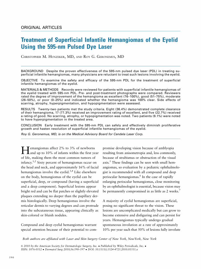
Figure 2. A 6-week-old infant shown before (A) and after (B) nine treatments over 16 weeks demonstrating complete (100%)clearance.
Figure 1. An 8-week-old infant shown before (A) and after (B) seven treatments over 20 weeks demonstrating complete(100%) clearance.
D E R M AT O L O G I C S U R G E RY5 9 4
T R E AT M E N T O F E Y E L I D H E M A N G I O M A S W I T H P U L S E D D Y E L A S E R
their child’s proliferating hemangioma.21,22 Public
reactions to their child’s hemangioma invoke feelings
of anger, hurt, frustration, and helplessness. It is
not uncommon for uninformed strangers or
acquaintances to accuse these parents of child abuse,
and some parents even cite their child’s hemangioma
as a reason for not taking them out in public. We
do not support the treatment of hemangiomas solely
to alleviate parental anxiety, but we recognize
that this is an important secondary benefit of
treatment.
There were no cases of scarring, atrophy, hypopig-
mentation, ulceration, or infection resulting from
PDL treatment in this study. The side effect profile is
comparable to the use of ultrapotent topical cor-
ticosteroids and considerably more favorable than
the results published by Batta and colleagues, with
atrophy and hypopigmentation seen in 28% and
45% of patients, respectively.11,18 The high inci-
dence of side effects seen in the Batta study are
attributable to the treatment parameters usedFhigh
energy settings with small spot sizesFand the lack
of epidermal cooling. To our knowledge, no other
study using PDL to treat hemangiomas has reported
such a high incidence of side effects.
Our results suggest that PDL results in a better final
cosmetic outcome than untreated hemangiomas that
involute spontaneously. Studies have shown that
textural changes in the skin result after spontaneous
involution of hemangiomas in up to 50% of cases.23
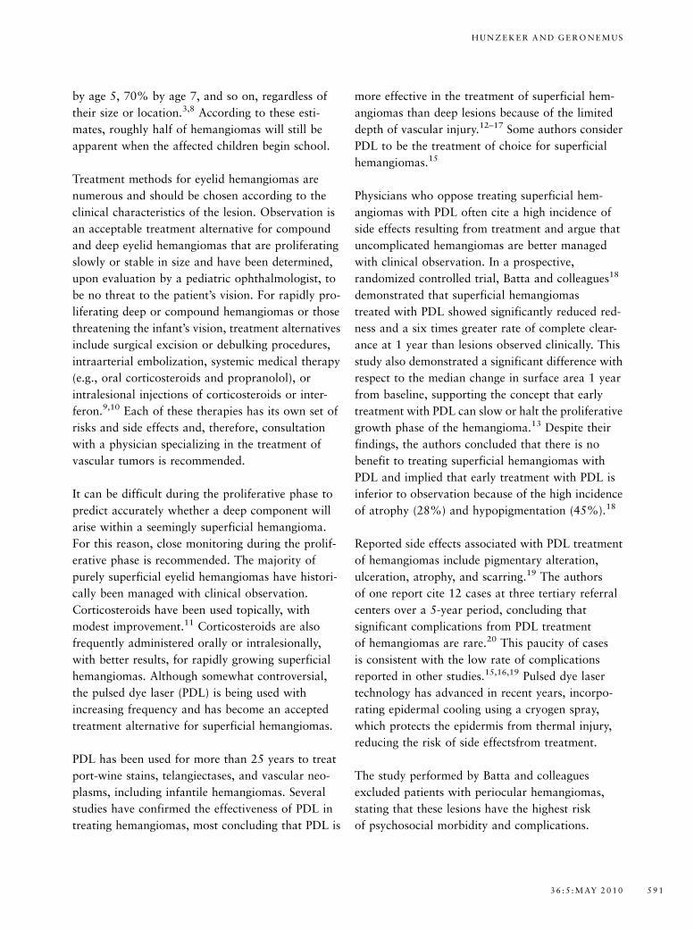
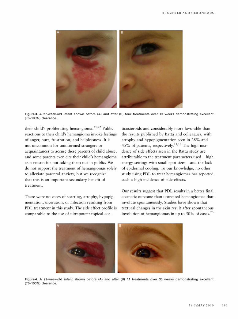
Figure 4. A 22-week-old infant shown before (A) and after (B) 11 treatments over 35 weeks demonstrating excellent(76–100%) clearance.
Figure 3. A 27-week-old infant shown before (A) and after (B) four treatments over 13 weeks demonstrating excellent(76–100%) clearance.
3 6 : 5 : M AY 2 0 1 0 5 9 5
H U N Z E K E R A N D G E R O N E M U S

Superficial hemangiomas predispose skin to atrophy
and telangiectasia, whereas untreated deep hem-
angiomas often leave behind residual fibrofatty
tissue. The absence of atrophy in our study provides
a strong argument in favor of early PDL treatment.
Early treatment minimizes the risk of atrophic skin
changes by stunting the proliferative growth of the
hemangioma and expediting its resolution. Addi-
tionally, PDL has been shown to increase the pro-
duction of dermal collagen and elastic fibers.24,25 It
is plausible that treatment with PDL simultaneously
shrinks the vascular component of the hemangioma
and increases collagen and elastic fiber production in
the superficial dermis, avoiding atrophy of the
treated skin.
Two patients in our study experienced hyperpig-
mentation of the treated skin. Because of the retro-
spective design of the study, long-term follow-up
photographs were not available to evaluate these
patients’ improvement over time. Most nonablative
laser treatment-induced hyperpigmentation is tran-
sient, resolving within 6 months. There is a high
likelihood that the hyperpigmentation that these two
patients experienced would have cleared over several
months, as has been reported in other studies using
PDL to treat hemangiomas.15
The risks of PDL are less than those associated
with systemic medical management or intralesional
therapy for eyelid hemangiomas. Oral corticoste-
roids are effective in decreasing the size of
hemangiomas, but potential side effects include
adrenal suppression, growth delay, irritability, and
gastritis.5,26 Oral corticosteroid therapy also
requires delaying the recommended immunization
schedule, placing the incompletely immunized infant
at risk for infection. Propranolol was recently
reported for the treatment of severe and disfiguring
hemangiomas, but it can cause bradycardia and
hypotension.27 Intralesional corticosteroid therapy
carries a risk of atrophy and depigmentation of
treated skin, adrenal suppression, failure to thrive,
and retinal artery occlusion leading to blindness.5,28
Meanwhile, intralesional interferon 2-a is reserved
for sight-threatening hemangiomas refractory to oral
corticosteroids because of its associated 20%
incidence of the development of spastic diplegia.29
All of these therapies are effective in decreasing the
size of hemangiomas, but because of their side-effect
profiles, they are primarily indicated in cases of
vision-threatening hemangiomas and should not be
employed as first-line therapy for proliferating
superficial periocular hemangiomas.
Ultimately, treatment must be tailored to fit the
hemangioma. Early treatment of superficial eyelid
hemangiomas with PDL reduces the lesion size,
shortens the duration of the proliferative growth
phase, and expedites resolution of the hemangioma.
PDL should be considered as an alternative to
‘‘active nonintervention’’ in the treatment of super-
ficial eyelid hemangiomas because of the lower risk
of textural change to the skin and the potential to
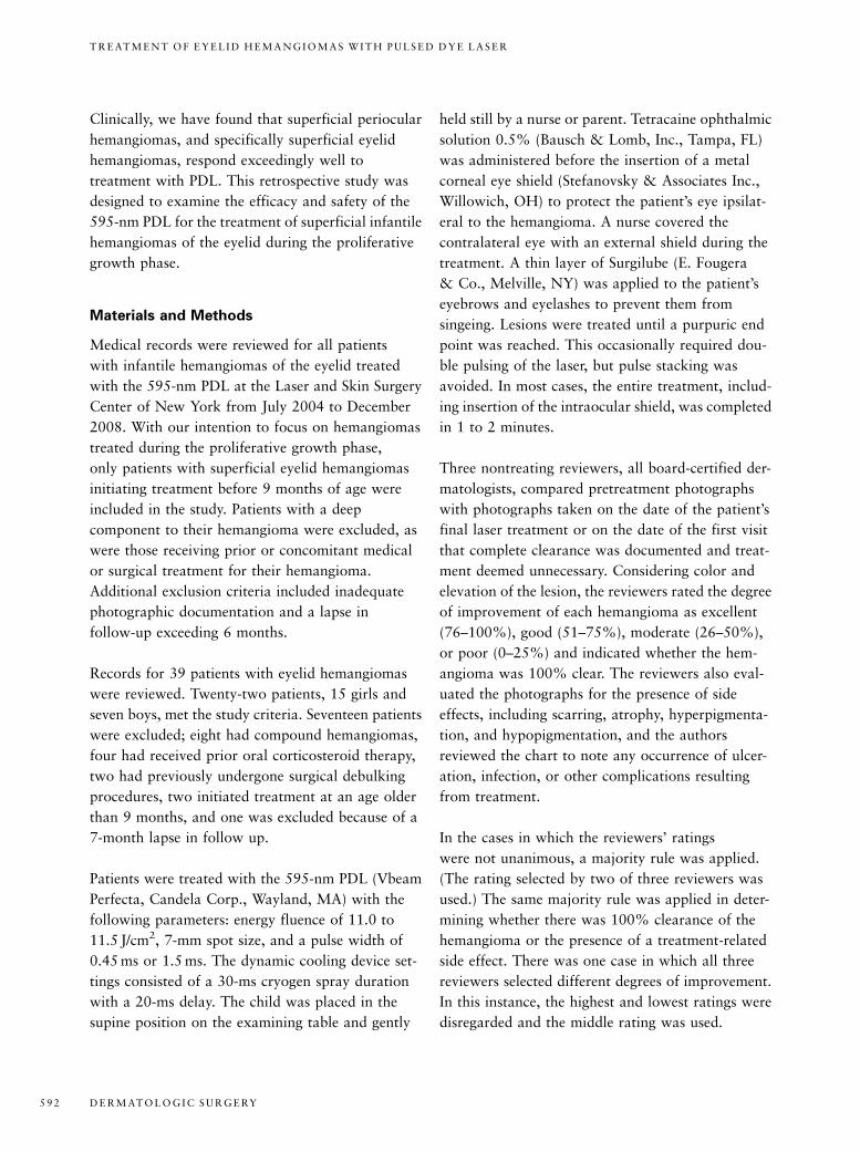
Figure 5. An 11-week-old infant shown before (A) and (B) after five treatments over 17 weeks demonstrating excellent(76–100%) clearance.
D E R M AT O L O G I C S U R G E RY5 9 6
T R E AT M E N T O F E Y E L I D H E M A N G I O M A S W I T H P U L S E D D Y E L A S E R

mitigate psychosocial stresses associated with facial
hemangiomas. The decision to ‘‘actively’’ not inter-
vene should be a decision made by the infant’s par-
ents after a complete discussion of available
treatment options with their consulting physician.
We encourage prospective studies comparing PDL
with observation for the treatment of superficial
hemangiomas of the eyelid as well as other sites on
the face, but we feel there is sufficient evidence to
support PDL as an effective and safe therapeutic
option in the management of superficial infantile
hemangiomas of the eyelid.
References
1. Jacobs AH, Walton RG. The incidence of birthmarks in the
neonate. Pediatrics 1976;58:218–22.
2. Jacobs AH. Strawberry hemangiomas: the natural history of the
untreated lesion. Calif Med 1957;86:8–10.
3. Finn MC, Glowacki J, Mulliken JB. Congenital vascular lesions:
clinical application of a new classification. J Ped Surg
1983;18:894–900.
4. Waner M, North PE, Scherer KA, et al. The nonrandom distri-
bution of facial hemangiomas. Arch Dermatol 2003;139:869–75.
5. Ceisler E, Blei F. Ophthalmic issues in hemangiomas of infancy.
Lymph Res Biol 2003;1:321–30.
6. Dubois J, Milot J, Jaeger BI, et al. Orbit and eyelid hemangiomas:
is there a relationship between location and ocular problems?
J Am Acad Dermatol 2006;55:614–9.
7. Smolinski KN, Yan AC. Hemangiomas of infancy: clinical and
biological characteristics. Clin Pediatr 2005;44:747–66.
8. Bowers RE, Graham EA, Tomlinson KM. The natural history of
the strawberry nevus. Arch Dermatol 1960;82:667–80.
9. Haik BG, Karcioglu ZA, Gordon RA, Pechous BP. Capillary
hemangioma (infantile periocular hemangioma). Surv Ophthal-
mol 1994;38:399–426.
10. Leaute-Labreze C, Dumas de la Roque E, Hubiche T, Boralevi F.
Propranolol for severe hemangiomas of infancy. N Engl J Med
2008;358:2649–51.
11. Garzon MC, Lucky AW, Hawrot A, Frieden I. Ultrapotent topical
corticosteroid treatment of hemangiomas of infancy. J Am Acad
Dermatol 2005;52:281–6.
12. Ashinoff R, Geronemus RG. Capillary hemangiomas and treat-
ment with the flash lamp-pumped pulsed dye laser. Arch Dermatol
1991;127:202–5.
13. Garden JM, Bakus AD, Paller AS. Treatment of cutaneous hem-
angiomas by the flashlamp-pumped pulsed dye laser: prospective
analysis. J Pediatr 1992;120(4 Pt 1):555–60.
14. Landthaler M, Hohenleutner U, el-Raheem TA. Laser therapy of
childhood haemangiomas. Br J Dermatol 1995;133:275–81.
15. Poetke M, Philipp C, Berlien HP. Flashlamp-pumped pulsed dye
laser for hemangiomas in infancy. Arch Dermatol 2000;136:
628–32.
16. Kono T, Hiroyuki S, William FG, et al. Comparison study of a
traditional pulsed dye laser versus a long-pulsed dye laser in the
treatment of early childhood hemangiomas. Lasers Surg Med
2005;38:112–5.
17. Rizzo C, Brightman L, Chapas AM, et al. Outcomes of childhood
hemangiomas treated with the pulsed-dye laser: a retrospective
chart analysis. Dermatol Surg 2009;35:1947–54.
18. Batta K, Goodyear HM, Moss C, et al. Randomised controlled
study of early pulsed dye laser treatment of uncomplicated
childhood haemangiomas: results of a 1-year analysis. Lancet
2002;360:521–7.
19. Levine VJ, Geronemus RG. Adverse effects associated with the
577- and 585-nanometer pulsed dye laser in the treatment of
cutaneous vascular lesions: a study of 500 patients. J Am Acad
Dermatol 1995;32:613–7.
20. Witman PM, Wagner AM, Scherer K, et al. Complications
following pulsed dye laser treatment of superficial hemangiomas.
Lasers Surg Med 2006;38:116–23.
21. Tanner JL, Dechert MP, Frieden IJ. Growing up with a facial
hemangioma: parent and child coping and adaptation. Pediatrics
1998;101:446–52.
22. Williams III EF, Marcelo H, Bret JR, et al. A psychological profile
of children with hemangiomas and their families. Arch Facial
Plast Surg 2003;5:229–34.
23. Mulliken JB, Fishman SJ, Burrows PE. Vascular anomalies.
Curr Probl Surg 2000;37:517–84.
24. Zelickson BD, Kilmer SL, Bernstein E, et al. Pulsed dye laser
therapy for sun damaged skin. Lasers Surg Med 1999;25:
229–36.
25. Moody BR, McCarthy JE, Hruza GJ. Collagen remodeling after
585-nm pulsed dye laser irradiation: an ultrasonographic analysis.
Dermatol Surg 2003;29:997–9.
26. Boon LM, MacDonald DM, Mulliken JB. Complications of
systemic corticosteroid therapy for problematic hemangioma.
Plast Reconstr Surg 1999;104:1616–23.
27. Siegfried EC, Keenan WJ, Al-Jureidini S. More on propranolol for
hemangiomas of infancy. N Engl J Med 2008;259:2846.
28. Goyal R, Watts P. Adrenal suppression and failure to thrive after
steroid injections for periocular hemangioma. Ophthalmology
2004;111:389–95.
29. Barlow CF, Cedric J, Priebe MD, et al. Spastic diplegia as a
complication of interferon Alfa-2a treatment of hemangiomas
of infancy. J Pediatr 1998;132:527–30.
Address correspondence and reprint requests to: Roy G.Geronemus, MD, Director, Laser & Skin Surgery Center ofNew York, 317 East 34th Street, 11th floor, New York, NY10016, or e-mail: [email protected]
3 6 : 5 : M AY 2 0 1 0 5 9 7
H U N Z E K E R A N D G E R O N E M U S
![Expression of HES and HEY genes in infantile hemangiomas · HemSC and HemEC isolation was conducted as previouslydescribed.[22]Briefly,thehemangioma samples were minced into small](https://img.dokumen.tips/doc/110x75/5f2831ab2645680e7c5f14f5/expression-of-hes-and-hey-genes-in-infantile-hemangiomas-hemsc-and-hemec-isolation.jpg)


![Vascular tumors in infants and adolescents · havior after birth [10]. Most importantly, infantile hemangiomas rarely require imaging for correct diagnosis and similarly important,](https://img.dokumen.tips/doc/110x75/5fb80e4757ad3030fd742786/vascular-tumors-in-infants-and-adolescents-havior-after-birth-10-most-importantly.jpg)