Embed Size (px)
Citation preview

RESEARCH ARTICLE Open Access
Treatment of kyphosis in ankylosingspondylitis by osteotomy throughthe gap of a pathological fracture:a retrospective studyHongqi Zhang*, Zhenhai Zhou, Chaofeng Guo, Yuxiang Wang, Honggui Yu and Longjie Wang
Abstract
Background: Surgical interventions are commonly advocated for correcting kyphotic deformities and relievingsevere back pain in ankylosing spondylitis (AS) patients. The aim of this study was to evaluate the clinical outcomeof osteotomy performed through the gap of a pathological fracture for the treatment of kyphosis in ankylosingspondylitis and to introduce the key points of this novel surgical approach.
Methods: From January 1, 2010, to December 31, 2014, 13 consecutive AS patients who were treated with osteotomythrough the fracture gap were retrospectively reviewed. Patients underwent the radiographic assessment of sagittalbalance parameters. Visual analog scale (VAS) scores were used to assess improvement in back pain.
Results: The average follow-up time was 2 years and 1 month. The median operation time was 280 min(range, 220–460 min). The mean blood loss was 1100 mL (range, 820–1300 mL). No major acute complications suchas death or complete paralysis occurred. There were no neurologic complications or cerebrospinal fluid leaks in anypatient. One patient had postoperative wound infection, which subsided after a switch of antibiotics. The globalkyphosis Cobb angle of patients decreased from the preoperative 55.8° ± 11.0° to 23.2° ± 6.7° (P < 0.001) after surgery.The C7 plumb line was used to assess global balance; its relationship with the posterosuperior corner of the sacrumdecreased from 166 ± 37 mm to 111 ± 20 mm (P < 0.001). The thoracolumbar kyphosis Cobb angle decreased from51.0° ± 9.9° to 21.6° ± 11.0° (P < 0.001). VAS scores for back pain decreased from 7.2 ± 1.2 to 2.1 ± 1.1 (P < 0.001).Lumbar lordosis increased from 5.7° ± 23.2° to 10.5° ± 29.2° (P = 0.001).
Conclusions: Osteotomy through the pathological fracture gap is a safe and effective surgical procedure forkyphosis correction and improvement of back pain in AS patients with pathological fractures. A significantkyphosis correction and improvement of back pain can be achieved with this surgical procedure.
Keywords: Ankylosing spondylitis, Kyphosis, Pathological fracture, Osteotomy
BackgroundAnkylosing spondylitis (AS) is a chronic inflammatorydisease. It always affects the axial skeleton, oftenstarting from the sacroiliac joints and then extendingto the upper spine [1]. The interaction betweenchronic inflammation and the spine is primarily char-acterized by progressive ossification of the spinal liga-ments and facet joints, eventually leading to a fixed
and stiff spine [2]. AS is also associated with vertebralosteoporosis [3, 4]. Because of sagittal imbalance ofthe spine and osteoporosis, pathological fractures canoccur, with the mechanism being similar to Chancefracture and seat belt injury [5]. A pathological frac-ture can occur with minor trauma or even withoutany trauma, which is different from a general spinefracture, and is most likely in the thoracolumbarjunction, a region where tremendous stress is concen-trated [6]; the fracture is usually located at the disclevel or adjacent to the disc [7].
* Correspondence: [email protected] of Spine Surgery, Xiangya Hospital of Central South University,Xiangya Road 87, Changsha, Hunan 410008, China
© The Author(s). 2016 Open Access This article is distributed under the terms of the Creative Commons Attribution 4.0International License (http://creativecommons.org/licenses/by/4.0/), which permits unrestricted use, distribution, andreproduction in any medium, provided you give appropriate credit to the original author(s) and the source, provide a link tothe Creative Commons license, and indicate if changes were made. The Creative Commons Public Domain Dedication waiver(http://creativecommons.org/publicdomain/zero/1.0/) applies to the data made available in this article, unless otherwise stated.
Zhang et al. Journal of Orthopaedic Surgery and Research (2016) 11:136 DOI 10.1186/s13018-016-0469-8
A pseudoarthrosis usually forms at the fracture sitewhen there is abnormal movement and repeated inflam-matory stimuli [8]. Pathological fracture and formationof a pseudoarthrosis progressively increase the kyphoticdeformity, with the patient suffering from severe backpain and, in some cases, nerve dysfunction [9]. A pro-gressive kyphosis makes it difficult for the patient to liedown or gaze forward, which can interfere with the per-formance of daily activities and adversely impact thequality of life. Surgical treatment is the only way tosimultaneously relieve back pain and correct kyphosis inAS patients with pathological fractures.Surgical treatments, including Smith-Petersen osteot-
omy (SPO or SPOs), pedicle subtraction osteotomy(PSO), vertebral column resection (VCR), polysegmentalosteotomy (PO), or any combination of these, are com-monly advocated for correcting kyphotic deformitiessecondary to AS [10, 11]. Presently, three surgical strat-egies are available for kyphosis correction in AS patientswith a pathological fracture or pseudoarthrosis: (1)anterior debridement only; (2) PSO, bone graft fusion,and internal fixation; and (3) a combination of the anter-ior and posterior approaches [12]. Selection of the oper-ation depends on the extent of ossification of theanterior column and intervertebral disc and the severityof anterior spinal cord compression, apart from manyother factors [6]. Despite their advantages, these strat-egies have concerns due to risky or difficult operation,limited correction angle, and/or increased complicationsand economic burden.In our clinical practice, we have developed a new
surgical procedure—a posterior osteotomy throughthe gap of the pathological fracture—that can relieveback pain and correct the kyphotic deformity in pa-tients with AS. In this study, we evaluate the clinicaloutcomes and correction results in patients undergo-ing this procedure and discuss the salient features ofthis novel procedure.
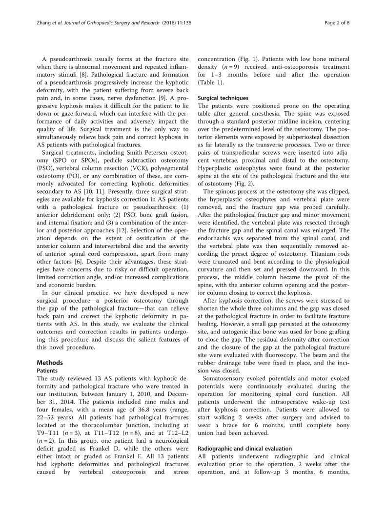
MethodsPatientsThe study reviewed 13 AS patients with kyphotic de-formity and pathological fracture who were treated inour institution, between January 1, 2010, and Decem-ber 31, 2014. The patients included nine males andfour females, with a mean age of 36.8 years (range,22–52 years). All patients had pathological fractureslocated at the thoracolumbar junction, including atT9–T11 (n = 3), at T11–T12 (n = 8), and at T12–L2(n = 2). In this group, one patient had a neurologicaldeficit graded as Frankel D, while the others wereeither intact or graded as Frankel E. All 13 patientshad kyphotic deformities and pathological fracturescaused by vertebral osteoporosis and stress
concentration (Fig. 1). Patients with low bone mineraldensity (n = 9) received anti-osteoporosis treatmentfor 1–3 months before and after the operation(Table 1).
Surgical techniquesThe patients were positioned prone on the operatingtable after general anesthesia. The spine was exposedthrough a standard posterior midline incision, centeringover the predetermined level of the osteotomy. The pos-terior elements were exposed by subperiosteal dissectionas far laterally as the transverse processes. Two or threepairs of transpedicular screws were inserted into adja-cent vertebrae, proximal and distal to the osteotomy.Hyperplastic osteophytes were found at the posteriorspine at the site of the pathological fracture and the siteof osteotomy (Fig. 2).The spinous process at the osteotomy site was clipped,
the hyperplastic osteophytes and vertebral plate wereremoved, and the fracture gap was probed carefully.After the pathological fracture gap and minor movementwere identified, the vertebral plate was resected throughthe fracture gap and the spinal canal was enlarged. Theendorhachis was separated from the spinal canal, andthe vertebral plate was then sequentially removed ac-cording the preset degree of osteotomy. Titanium rodswere truncated and bent according to the physiologicalcurvature and then set and pressed downward. In thisprocess, the middle column became the pivot of thespine, with the anterior column opening and the poster-ior column closing to correct the kyphosis.After kyphosis correction, the screws were stressed to
shorten the whole three columns and the gap was closedat the pathological fracture in order to facilitate fracturehealing. However, a small gap persisted at the osteotomysite, and autogenic iliac bone was used for bone graftingto close the gap. The residual deformity after correctionand the closure of the gap at the pathological fracturesite were evaluated with fluoroscopy. The beam and therubber drainage tube were fixed in place, and the inci-sion was closed.Somatosensory evoked potentials and motor evoked
potentials were continuously evaluated during theoperation for monitoring spinal cord function. Allpatients underwent the intraoperative wake-up testafter kyphosis correction. Patients were allowed tostart walking 2 weeks after surgery and advised towear a brace for 6 months, until complete bonyunion had been achieved.
Radiographic and clinical evaluationAll patients underwent radiographic and clinicalevaluation prior to the operation, 2 weeks after theoperation, and at follow-up 3 months, 6 months,
Zhang et al. Journal of Orthopaedic Surgery and Research (2016) 11:136 Page 2 of 8

1 year, and 2 years after the operation. Radiographicassessment of sagittal balance parameters was per-formed by standing lateral radiography of the wholespine. Sagittal balance parameters included globalkyphosis (GK), lumbar lordosis (LL), thoracolumbarlordosis (TLK), and sagittal vertical axis (SVA; thehorizontal distance from a vertical plumb line cen-tered in the middle of the C7 vertebral body to theposterosuperior corner of the S1 endplate). The clin-ical results were assessed with the visual analog scale(VAS) score.
Statistical analysisStatistical analysis was performed using SPSS 17.0(SPSS Inc., Chicago, IL, USA). All results werereported as means ± standard deviation (SD). Thepaired sample t test was used to compare the pre-operative, postoperative, and final follow-up clinicaland radiographic data. Statistical significance was setat P ≤ 0.05.
ResultsOperative resultsOsteotomy through the fracture gap was successfullyperformed in all 13 patients. The average follow-up timewas 2 years and 1 month. The median operation timewas 280 min (range, 220–460 min). The mean blood losswas 1100 mL (range, 820–1300 mL). No major acutecomplications such as death or complete paralysis oc-curred. There were no neurologic complications or cere-brospinal fluid leaks in any patient. One patient hadwound infection after the operation, which subsidedafter a switch of antibiotics.
Table 1 Basic characteristics of patients with ankylosingspondylitis
Mean age (years) 36.8 (22–52)
Male/female 9/4
T9–T11 (n) 3
T11–T12 (n) 8
T12–L2 (n) 2
Low bone mineral density (n) 9
Average follow-up time (months) 25 (3–52)
Fig. 1 Preoperative imaging findings of a 47-year-old female patient with ankylosing spondylitis. a, b Photographs show a kyphotic deformity,with the patient having difficulty in holding the head up straight. c, d Radiographs show the thoracolumbar kyphotic deformity, with a pathologicalfracture located at L1. e MRI image shows destruction of bone and compression of spinal cord. f, g CT images show the pathological fracture at L1
Zhang et al. Journal of Orthopaedic Surgery and Research (2016) 11:136 Page 3 of 8

Radiological resultsSatisfactory correction of kyphotic deformity wasachieved in all patients. In addition, the pathologicalfracture was healing in all patients at the final follow-up.There were no cases of pseudoarthrosis formation at theosteotomy site or instrumentation failure. No internalfixation loosening, fracture, or correction loss occurred(Figs. 2 and 3).Radiographic results showed that the postoperative
and final follow-up levels of GK decreased significantlycompared with the preoperative results (23.2° ± 6.7° and26.4° ± 9.4° vs. 55.8° ± 11.0°; P < 0.001). Similar decreaseswere found in TLK (21.6° ± 11.0° and 24° ± 8.4° vs. 51.0°;P < 0.001). SVA showed smaller decreases (111 ± 20 mmand 87 ± 29 mm vs. 166 ± 37 mm; P < 0.001), whereasthe opposite changes were observed in LL (10.5° ± 29.0°and 18.8° ± 21.6° vs. 5.7° ± 23.2°; P < 0.05; Table 2).
Clinical resultsBack pain was obviously improved in all 13 patients.The improvement of VAS scores is shown in Table 2.The postoperative VAS score was markedly lower thanthe preoperative score (2.1 ± 1.1 vs. 7.2 ± 1.2; P < 0.001)(7.2 ± 1.2 vs. 2.1 ± 1.1; P < 0.001). Similarly, the finalfollow-up VAS scores were significantly lower than thepreoperative VAS score (1.9 ± 1.4 vs. 7.2 ± 1.2; P < 0.001).
DiscussionAdvantages and disadvantages of various surgicalapproachesSPO, PSO, VCR, and PO, or any combination of these, arestandardized surgical procedures for correcting kyphoticdeformities in AS patients [13, 14]. Because of the increas-ing vertebral osteoporosis and bony brittleness [15], theankylosed spine is prone to fracture even after a minor
Fig. 2 Surgical procedure of osteotomy through the fracture gap in a female patient with ankylosing spondylitis. a Photograph shows theposition of the patient on the operating table. The patient was flexed in a reverse V shape to accommodate the kyphotic spine and adaptsimultaneously to the correction of kyphotic deformity during operation. b Photograph shows a hyperplastic osteophyte located at theoseteotomy site. c, d Intraoperative X-ray fluoroscope was used after inserting the pedicle screws and correcting the kyphotic deformity
Fig. 3 Result of osteotomy through the fracture gap in the female patient with ankylosing spondylitis. a, b Photographs show satisfactorycorrection achieved via osteotomy through the pathological fracture gap. c Radiograph shows stable internal fixation (without displacement)and corrected kyphosis. d Radiograph at follow-up after 1 year shows the closed fracture gap and stable bone fusion achieved at the posterior column
Zhang et al. Journal of Orthopaedic Surgery and Research (2016) 11:136 Page 4 of 8

trauma, which is a two- to eightfold increase as comparedto non-AS patients [16, 17]. Additionally, the continuedmovement at the fracture site eventually contributes tothe development of pseudoarthrosis [18]. There are threesurgical strategies for kyphosis in AS patients with patho-logical fractures or pseudoarthroses, and the advantagesand disadvantages are summarized below:
1) Anterior debridement only: Anterior debridementis especially suitable for eliminating the compressionin front of the spine as, for example, in spinaltuberculosis and spinal metastatic carcinoma [19].The surgeon could have an ideal biomechanicalenvironment via anterior approach [20], and thesurgical procedure to eliminate the stress in thefront of the spine is much easier [21, 22]. However,a kyphosis correction is difficult to achieve viaanterior approach only [23]. Moreover, bloodvessels and tissue in front of the spine are easilyinjured due to ossification of the tissues andligaments [23, 24].
2) PSO, bone graft fusion, and internal fixation:PSO is modified to treat some fixed sagittalplane deformities in various disease states,including tuberculosis, trauma, and postsurgicalconditions [7, 25]. In AS patients, a pseudoarthrosisis liable to be formed at the pathological fracturesite. Kyphosis correction and spinal canaldecompression can be achieved at the sametime by PSO [26]. Nevertheless, limited correctionangle is a problem [27]. Additionally, PSO isassociated with high risk, and the proceduremay not be sufficient to eliminate bonecompression in front of the spine [2].
3) Combined anterior and posterior approach:In recent studies, surgeons have demonstratedthe value of a combined anterior and posteriorapproach for kyphosis correction in AS patientswith pseudoarthrosis [8, 28, 29]. PSO or SPO(SPOs), bone graft fusion, and internal fixationwere adopted in the first stage, followed byanterior debridement in the second stage. Thisapproach could correct the kyphosis and improvesymptoms such as back pain and neurologic
deficits simultaneously. However, this approachis associated with higher costs, longer hospitalizationtime, greater operative risks, and more postoperativecomplications than the one-stage posterior surgicalprocedure.
Moreover, whether an anterior bone graft is actuallyneeded is still a controversial topic in the field of spinalsurgery [10, 30]. Some surgeons deem that the necessityof supplemental anterior fusion for pseudoarthrosis fol-lowing PSO depends on the extent of the osteotomy clos-ure and the anterior column defect. A pseudoarthrosis iscompletely cleared after PSO. If the osteotomy site can becompletely closed, there is no need to perform an anteriorinterbody fusion. On the other hand, if the postoperativeradiograph demonstrates an anterior column defect with awide opening at the level of the pseudoarthrosis followingPSO, a supplemental anterior fusion must be considered[10, 29]. Qian et al. [29] performed PSO with a supple-mental anterior fusion through the pseudoarthrosis inseven AS patients with severe kyphotic deformities. Aftera mean follow-up of 3 years and 7 months, they reportedthat the outcome of correction was satisfactory and backpain was obviously improved in all seven patients. How-ever, Chang et al. pointed out that posterior correctionand fixation without an anterior support is an effectivemethod for kyphosis correction in AS with pseudoarthro-sis. They believed in the superior fusion capacity of AS[31]. In the current study, we have paid more attention tothe site of the osteotomy, which is the pathological frac-ture site. We are also concerned more about theimprovement of back pain, fracture healing, and kyphosiscorrection. SPO, PSO and any kinds of these can bechosen for kyphosis correction according to the prede-signed correction angle. Additionally, all the patientsunderwent one-stage posterior kyphosis correction with-out an anterior column support, and complete bone fu-sion and fracture healing were achieved at follow-up. Ourdata further suggested that a complete bone fusion couldbe achieved via posterior approach only.
Key points of the new surgical procedureIn our group of AS patients with kyphotic deformity, thepathological fracture site was chosen as the osteotomy
Table 2 Radiological assessment of sagittal balance parameters and clinical assessment of preoperation (Pre-OP), postoperation(Post-OP), and at final follow-up (mean ± SD; n = 13)
Parameter Pre-OP Post-OP t value P value Final follow-up t value P value
GK (°) 55.8 ± 11.0 23.2 ± 6.7 11.398 <0.001 26.4 ± 9.4 8.733 <0.001
SVA (mm) 166 ± 37 111 ± 20 7.197 <0.001 87 ± 29 8.616 <0.001
TLK (°) 51.0 ± 9.9 21.6 ± 11.0 6.911 <0.001 24 ± 8.4 7.911 <0.001
LL (°) 5.7 ± 23.2 10.5 ± 29.0 −4.674 0.001 18.8 ± 21.6 −2.578 0.024
VAS 7.2 ± 1.2 2.1 ± 1.1 11.813 <0.001 1.9 ± 1.4 12.086 <0.001
Zhang et al. Journal of Orthopaedic Surgery and Research (2016) 11:136 Page 5 of 8
site. Kyphosis correction and improvement of back painwere achieved after the operation, in addition to fracturehealing and bone fusion at follow-up. The main featuresof this operation are as follows:
1) Internal fixation of the spine: Fixed segmentsprovide a stable mechanical environment whichis essential for proper correction.
2) Finding the fracture gap: A pseudoarthrosis is alwaysformed in AS patients at the site of a pathologicalfracture, with compensatory hyperplasia at thefracture site and vertebrae. Therefore, finding thepathological fracture gap is a vital step in thisprocedure. The main process of finding the fracturegap is to eliminate the hyperplasia of osteophytes inthe posterior column and to probe the fracture gap,where a minor movement can be found. Then thefracture gap is enlarged, and dural adhesion isreleased.
3) Kyphotic deformity correction: After enlarging thefracture gap, the screw–rod system is used to openthe anterior spine while closing the posterior spineat the fracture site. This process must be operatedslowly and progressively to protect the ossific vesselsand tissues in the front of the spine.
4) The screw–rod system for pressing vertebral bodies:In order to decrease the fracture gap, the screw–rodsystem is used to keep the upper and lower vertebralbodies pressed together, closing the pathologicalfracture gap. In this process, the middle columnbecomes the pivot of the spine and the threecolumns are shortened concurrently. The posteriorcolumn is shortened more than the other twocolumns, thereby avoiding sharp angulation in thesagittal plane and spinal cord shrinkage andpreventing excessive opening of the anterior column,which may cause an injury of vessels and tissues inthe front of the spine.
5) Autologous iliac crest bone graft: A small gapremains after the kyphosis correction. It is necessaryto make the upper and lower vertebral plates coarse
for the autologous iliac bone graft. This processcontributes to the closure of the fracture gap, whichprovides support for bone fusion.
Factors promoting pathological fracture healingThe obvious advantage of this approach is that the frac-ture can be healed in a short time after the kyphosiscorrection, without a second-stage anterior bone graft.There are some factors that can promote pathologicalfracture healing.
1) Fixation: Adjacent segments are fixed by a screw–rod system, and partial stabilization of the spine isincreased. Abnormal movement is also decreasedat the site of the pseudoarthrosis and fracture.These are necessary conditions for bone healing.
2) Closure of the fracture gap: The fracture gap isdecreased with the use of an internal fixation systemthat stresses the upper and lower vertebral bodies,which is pivotal for bone healing.
3) Change of spine stress line: Before kyphosiscorrection, shearing force and traction areconcentrated at the site of the pathological fractureand the separation traction is mainly concentratedin the posterior column. The spine stress line isimproved after a kyphosis correction, and thetraction force transforms into stress. In accordancewith Wolff ’s law, the stress of the fracture siteincreases and the regional osteogenesis increases[32], which is another factor that promotes bonehealing.
4) Autologous iliac bone graft: After kyphosiscorrection, the autologous iliac bone graftaccelerates bone fusion of the residual fracture gap,stabilizes the spine, and provides stable supportfor the middle and anterior column bone fusion.
Evaluation of this surgical approachIn the 13 AS patients included in this study, the averagekyphosis correction was 31°, which is comparable to thatobtained with several other surgical techniques (Table 3)
Table 3 Results of studies (including ours) that have used osteotomies for correcting kyphosis in ankylosing spondylitis patientswith pathological fracture (or pseudoarthrosis)
Author (year) No. of cases Surgical method Single-segment correction (°) Perioperative complications
Chang (2010) [34] 30 OWO 38 Postoperative pneumonia in 1 patient
Superficial infection in 1 patient
Kim (2007) [20] 12 SPO + AF or PSO + AF 24 and 31 Intraoperative dural tears in 3 patients
Leg pain with paresthesia in 2 patients
Qian (2012) [29] 7 PSO through pseudoarthrosis + AF 45 No complications
Our study 13 Osteotomy through pathologicalfracture gap without AF
31 Superficial infection in 1 patient
OWO posterior opening-wedge osteotomy, SPO Smith-Petersen osteotomy, AF anterior fusion, PSO pedicle subtraction osteotomy
Zhang et al. Journal of Orthopaedic Surgery and Research (2016) 11:136 Page 6 of 8

[29, 33, 34]. The correction result was satisfactory, andthere was no correction loss during follow-up. At thefinal follow-up, complete bone fusion had been achievedin all patients. This approach also has its limitations.The degree of kyphosis correction in our study waslower than that which has been achieved with PSOthrough pseudoarthrosis (Table 3). This procedure isespecially applicable in AS patients with a pathologicalfracture and severe back pain but relatively moderatekyphosis. For AS patients with severe kyphosis, anadditional osteotomy, including SPOs or a two-levelPSO, is necessary to achieve better correction. A weak-ness of our study is that it included only a small numberof patients. A large randomized controlled study isnecessary to accurately evaluate the feasibility, reliability,and complications of this method.
ConclusionsOsteotomy through a pathological fracture gap is a noveland feasible procedure for kyphosis correction in ASpatients with pathological fractures. With this method,satisfactory kyphotic deformity correction, successfulbone fusion, and obvious improvement of back pain canbe achieved simultaneously, without any neurologicalcomplications. This surgical procedure is a safe andeffective approach for the treatment of AS with patho-logical fracture and can significantly improve the qualityof life.
AbbreviationsAF: Anterior fusion; AS: Ankylosing spondylitis; GK: Global kyphosis; LL: Lumbarlordosis; OWO: Posterior opening-wedge osteotomy; PO: Polysegmentalosteotomy; Post-OP: Postoperation; Pre-OP: Preoperation; PSO: Pediclesubtraction osteotomy; SPO: Smith-Petersen osteotomy; SVA: Sagittal verticalaxis; TLK: Thoracolumbar lordosis; VAS: Visual analog scale; VCR: Vertebralcolumn resection
FundingThis work was financially supported by the National Natural ScienceFoundation of China (CN) (81472145).
Availability of data and materialsThis article is a clinical retrospective study, and the available data, includingimageology findings, and photographic documentation were collected from13 AS patients and have been listed in the table of the manuscript. Thestatistical data is strongly supporting the conclusion of our study. However,for a further study of surgical treatment for AS in the future, we do not wishto share the raw data now.
Authors’ contributionsHZ and ZZ contributed to the conception. ZZ and CG contributed to thedesigns and the draft of the work and revised it critically for importantintellectual content. YW, HY, and LW did the acquisition, analysis, orinterpretation of the data of the work. HZ and CG approved the versionto be published. HZ agreed to be accountable for all aspects of the workin ensuring that questions related to the accuracy or integrity of any partof the work are appropriately investigated and resolved. All authors readand approved the final manuscript.
Competing interestsThe authors declare that they have no competing interests.
Consent for publicationWritten informed consent was acquired from each of the patients toauthorize treatment, imageology findings, and photographic documentation.The patients consented to the publication of their pictures as well as theiranonymous and clustered data.
Ethics approval and consent to participateThis study was approved by the Ethics Committee of Xiangya Hospital,Central South University. The patients received a thorough explanation ofthis study and gave their oral and written informed consent to be includedin this study.
Received: 26 May 2016 Accepted: 14 September 2016
References1. Zhang HQ, Huang J, Guo CF, Liu SH, Tang MX. Two-level pedicle
subtraction osteotomy for severe thoracolumbar kyphotic deformity inankylosing spondylitis. Eur Spine J. 2014;23(1):234–41.
2. Zhao Y, Wang Y, Wang Z, Zhang X, Mao K, Zhang Y. Effect and strategy ofone-stage interrupted two-level transpedicular wedge osteotomy forcorrecting severe kyphotic deformities in ankylosing spondylitis. Clin SpineSurg. 2016. [Epub ahead of print].
3. Ardizzone M, Javier RM, Kuntz JL. Ankylosing spondylitis and osteoporosis.La Revue de medecine interne / fondee par la Societe nationale francaisede medecine interne. 2006;27(5):392–9.
4. Briot K, Roux C. Inflammation, bone loss and fracture risk in spondyloarthritis.RMD Open. 2015;1(1):e000052.
5. Mulpuri K, Jawadi A, Perdios A, Choit RL, Tredwell SJ, Reilly CW. Outcomeanalysis of chance fractures of the skeletally immature spine. Spine. 2007;32(24):E702–7.
6. Arun R, Dabke HV, Mehdian H. Comparison of three types of lumbarosteotomy for ankylosing spondylitis: a case series and evolution of a safetechnique for instrumented reduction. Eur Spine J. 2011;20(12):2252–60.
7. Barrey C, Perrin G, Michel F, Vital JM, Obeid I. Pedicle subtraction osteotomyin the lumbar spine: indications, technical aspects, results andcomplications. Eur J Orthop Surg Traumatol. 2014;24 Suppl 1:S21–30.
8. Qian BP, Qiu Y, Ji ML, Wang B, Yu Y, Zhu ZZ, Jiang J. Osteotomy for severethoracolumbar kyphosis in advanced ankylosing spondylitis: skipping two-level pedicle subtraction osteotomy. Zhonghua yi xue za zhi. 2013;93(7):491–5.
9. Zhao Y, Xu H, Zhang Y, Wang Z, Zhang X, Wang Y. Comparison of twosurgeries in treatment of severe kyphotic deformity caused by ankylosingspondylitis: transpedicular bivertebrae wedge osteotomy versus one-stageinterrupted two-level transpedicular wedge osteotomy. Clin NeurolNeurosurg. 2015;139:252–7.
10. Kim KT, Jo DJ, Lee SH, Park KJ, Sin JH. Does it need to perform anteriorcolumn support after Smith-Petersen osteotomy for ankylosing spondylitis?Eur Spine J. 2012;21(5):985–91.
11. Ravinsky RA, Ouellet JA, Brodt ED, Dettori JR. Vertebral osteotomies inankylosing spondylitis-comparison of outcomes following closing wedgeosteotomy versus opening wedge osteotomy: a systematic review.Evidence-based Spine-care J. 2013;4(1):18–29.
12. Schwab F, Blondel B, Chay E, Demakakos J, Lenke L, Tropiano P, Ames C,Smith JS, Shaffrey CI, Glassman S, et al. The comprehensive anatomicalspinal osteotomy classification. Neurosurgery. 2015;76 Suppl 1:S33–41.discussion S41.
13. Zheng GQ, Zhang YG, Chen JY, Wang Y. Decision making regarding spinalosteotomy and total hip replacement for ankylosing spondylitis: experiencewith 28 patients. Bone Joint J. 2014;96-B(3):360–5.
14. Hyun SJ, Lenke LG, Kim YC, Koester LA, Blanke KM. Long-term radiographicoutcomes of a central hook-rod construct for osteotomy closure: minimum5-year follow-up. Spine. 2015;40(7):E428–32.
15. Ma J, Wang C, Zhou X, Zhou S, Jia L. Surgical therapy of cervical spinefracture in patients with ankylosing spondylitis. Medicine. 2015;94(44):e1663.
16. Boonen A, van der Linden SM. The burden of ankylosing spondylitis.J Rheumatol Suppl. 2006;78:4–11.
17. Maas F, Spoorenberg A, van der Slik BP, van der Veer E, Brouwer E, Bootsma H,Bos R, Wink FR, Arends S. Clinical risk factors for the presence anddevelopment of vertebral fractures in patients with ankylosing spondylitis.Arthritis Care Res. 2016. [Epub ahead of print].
Zhang et al. Journal of Orthopaedic Surgery and Research (2016) 11:136 Page 7 of 8

18. Bron JL, de Vries MK, Snieders MN, van der Horst-Bruinsma IE, van Royen BJ.Discovertebral (Andersson) lesions of the spine in ankylosing spondylitisrevisited. Clin Rheumatol. 2009;28(8):883–92.
19. Liu ZD, Li XF, Zang WP, Wang ZY, Wu LM. Combined pedicle subtractionosteotomy and polysegmental closing wedge osteotomy for correction ofthe severe thoracolumbar kyphotic deformity in ankylosing spondylitis.Zhonghua Wai Ke Za Zhi. 2009;47(9):681–4.
20. Kim KT, Lee SH, Suk KS, Lee JH, Im YJ. Spinal pseudarthrosis in advancedankylosing spondylitis with sagittal plane deformity: clinical characteristicsand outcome analysis. Spine. 2007;32(15):1641–7.
21. Benli IT, Alanay A, Akalin S, Kis M, Acaroglu E, Ates B, Aydin E. Comparisonof anterior instrumentation systems and the results of minimum 5 yearsfollow-up in the treatment of tuberculosis spondylitis. Kobe J Med Sci. 2004;50(5–6):167–80.
22. Chen LH, Kao FC, Niu CC, Lai PL, Fu TS, Chen WJ. Surgical treatment ofspinal pseudoarthrosis in ankylosing spondylitis. Chang Gung Med J. 2005;28(9):621–8.
23. Qian BP, Qiu Y, Wang B, Yu Y, Zhu ZZ. Clinical features and strategies fortreatment of spinal fracture complicating ankylosing spondylitis. Zhonghuayi xue za zhi. 2007;87(41):2893–8.
24. Wang T, Wang D, Cong Y, Yin C, Li S, Chen X. Evaluating a posteriorapproach for surgical treatment of thoracolumbar pseudarthrosis inankylosing spondylitis. Clin Spine Surg. 2016. [Epub ahead of print].
25. Liu C, Zheng G, Zhang Y, Tang X, Song K, Fu J, Wang Z, Cui G, Wang Y.The radiologic, clinical results and digestive function improvement inpatients with ankylosing spondylitis kyphosis after pedicle subtractionosteotomy. Spine J. 2015;15(9):1988–93.
26. Liu Z, Qiu Y, Zhu ZZ, Qian BP, Qiao J, Jiang L, Wang B, Zhu F. Pediclesubtraction osteotomy for correction of severe thoracolumbar kyphosis inankylosing spondylitis. Orthop Surg. 2014;6(3):257–8.
27. Zheng GQ, Song K, Zhang YG, Wang Y, Huang P, Zhang XS, Wang Z,Mao KY, Cui G. Two-level spinal osteotomy for severe thoracolumbarkyphosis in ankylosing spondylitis. Experience with 48 patients. Spine.2014;39(13):1055–8.
28. Qian BP, Jiang J, Qiu Y, Wang B, Yu Y, Zhu ZZ. Radiographical predictors forpostoperative sagittal imbalance in patients with thoracolumbar kyphosissecondary to ankylosing spondylitis after lumbar pedicle subtractionosteotomy. Spine. 2013;38(26):E1669–75.
29. Qian BP, Qiu Y, Wang B, Sun X, Zhu ZZ, Jiang J, Ji ML. Pedicle subtractionosteotomy through pseudarthrosis to correct thoracolumbar kyphoticdeformity in advanced ankylosing spondylitis. Eur Spine J. 2012;21(4):711–8.
30. Zhu Z, Wang X, Qian B, Wang B, Yu Y, Zhao Q, Qiu Y. Loss of correctionin the treatment of thoracolumbar kyphosis secondary to ankylosingspondylitis: a comparison between Smith-Petersen osteotomies and pediclesubtraction osteotomy. J Spinal Disord Tech. 2012;25(7):383–390.
31. Chang KW, Tu MY, Huang HH, Chen HC, Chen YY, Lin CC. Posteriorcorrection and fixation without anterior fusion for pseudoarthrosis withkyphotic deformity in ankylosing spondylitis. Spine. 2006;31(13):E408–13.
32. Adams MA, Dolan P. Biomechanics of vertebral compression fractures andclinical application. Arch Orthop Trauma Surg. 2011;131(12):1703–10.
33. Kim KT, Park KJ, Lee JH. Osteotomy of the spine to correct the spinaldeformity. Asian Spine J. 2009;3(2):113–23.
34. Chang KW. Closing-opening wedge osteotomy for kyphotic deformity dueto ankylosing spondylitis. Zhonghua Wai Ke Za Zhi. 2010;48(22):1683–6.
• We accept pre-submission inquiries
• Our selector tool helps you to find the most relevant journal
• We provide round the clock customer support
• Convenient online submission
• Thorough peer review
• Inclusion in PubMed and all major indexing services
• Maximum visibility for your research
Submit your manuscript atwww.biomedcentral.com/submit
Submit your next manuscript to BioMed Central and we will help you at every step:
Zhang et al. Journal of Orthopaedic Surgery and Research (2016) 11:136 Page 8 of 8
![Ankylosing spondylitis and related conditions - NHS Wales1].pdf · Condition Ankylosing spondylitis Ankylosing spondylitis and related conditions This booklet provides information](https://img.dokumen.tips/doc/110x75/5d53eb2788c993a4728b841d/ankylosing-spondylitis-and-related-conditions-nhs-1pdf-condition-ankylosing.jpg)