Embed Size (px)
Citation preview
Treatment of Chronic Deep Vein Thrombosis Using Ultrasound AcceleratedCatheter-directed Thrombolysis
M. Dumantepe *, I.A. Tarhan, A. Ozler
Department of Cardiovascular Surgery, Memorial Atasehir Hospital, Istanbul, Turkey
* CorKat: 1E-ma1078
Surgeryhttp:
WHAT THIS PAPER ADDS
Few data are available concerning the feasibility, safety, and efficacy of ultrasound-accelerated, catheter-directed thrombolysis (UACDT) in “chronic” lower extremity deep vein thrombosis. Our study illustrates thebeneficial role of UACDT using the EkoSonic system. Importantly, the study showed a higher treatment successrate with significant reduction in thrombolytic duration, thrombolytic dosage, and hemorrhagic complications.Additional clinical studies are necessary to validate the benefit and corroborate our results.Objective: To evaluate the feasibility, efficacy and safety of ultrasound-accelerated catheter-directedthrombolysis (UACDT) in the delayed treatment of lower extremity deep venous thrombosis (DVT).Design: Twelve patients with unilateral iliofemoral or femoropopliteal DVT (mean symptom duration 92 � 44days) were prospectively investigated.Method: UACDT was performed using recombinant human tissue plasminogen activator delivered using the EKOSEkoSonic system. Stents were deployed if indicated by post-procedure venography. Follow-up comprised weeklyduplex ultrasound for 1 month and monthly thereafter.Results: Successful thrombolysis occurred in 11/12 limbs (92%; complete 6/12, partial 5/12) after a meaninfusion time of 26 � 7 hours. 2/12 patients required angioplasty and stent insertion. At a mean follow-up of 9(6e15) months, 10/11 (91%) veins were patent whereas 1/11 re-occluded at 2 months (patient with protein-Cdeficiency). 2/11 limbs developed symptoms/signs of post-thrombotic syndrome and 3/11 had developed deepvein reflux (duplex ultrasound). 2/12 patients experienced peri-catheter bleeding but no major hemorrhage orsymptomatic pulmonary embolism occurred.Conclusions: This preliminary evidence suggests that UACDT may be a safe and effective option for the delayedtreatment of lower limb DVT.� 2013 European Society for Vascular Surgery. Published by Elsevier Ltd. All rights reserved.Article history: Received 4 July 2012, Accepted 28 May 2013, Available online 25 June 2013Keywords: Catheter-directed thrombolysis, Chronic deep vein thrombosis, Post-thrombotic syndrome,
Ultrasound
INTRODUCTION
Deep venous thrombosis (DVT) of the lower extremity isrecognized as a cause of both pulmonary embolism (PE) andthe post-thrombotic syndrome (PTS).1 Although anti-coagulation is currently considered the standard of care toprevent PE and recurrent DVT, it remains ineffective inremoving thrombus burden and consequently does notprevent PTS, which can appear months to years after anacute DVT.2
A novel technique, ultrasound-accelerated catheter-directed thrombolysis (UACDT), has been developed torapidly and completely lyse existing thrombus, thereby
responding author. M. Dumantepe, Merit Life Park Sitesi, B Blok,Daire: 5, Serifali/Umraniye 34660, _Istanbul, Turkey.il address: [email protected] (M. Dumantepe).-5884/$ e see front matter � 2013 European Society for Vascular. Published by Elsevier Ltd. All rights reserved.//dx.doi.org/10.1016/j.ejvs.2013.05.019
decreasing the potential risk of PTS.3,4 The mechanismproposed is that this technique integrates high-frequency,low-intensity ultrasound (US) with standard catheter-directed thrombolysis (CDT) in order to accelerate clotdissolution, reducing treatment time, drug dosage, and theincidence of thrombolysis-related complications. US wavesare known to increase permeability of biological structuresincluding tissue, vessel walls, and thrombus. This increasedpermeability combined with the pressure gradient associ-ated with an acoustic field is thought to facilitate delivery oftherapeutic agents into, and past, older organized thrombusinto regions where dissolvable clot remains.5,6
Several studies demonstrate evidence of safety andeffectiveness in removing acute venous thrombosis usingEKOS,7e9 but limited data are available supporting its usefor chronic DVT, particularly in patients with long-standingsymptoms. Treatment of chronic DVT has long been a clin-ical problem as the thrombus has a tendency to adhere andorganize after the acute stage. Therefore, the aim of this
M. Dumantepe et al. 367
study is to examine the feasibility of using UACDT as a safeand effective option to treat patients with chronic DVTdefined as having symptoms for greater than 28 days.
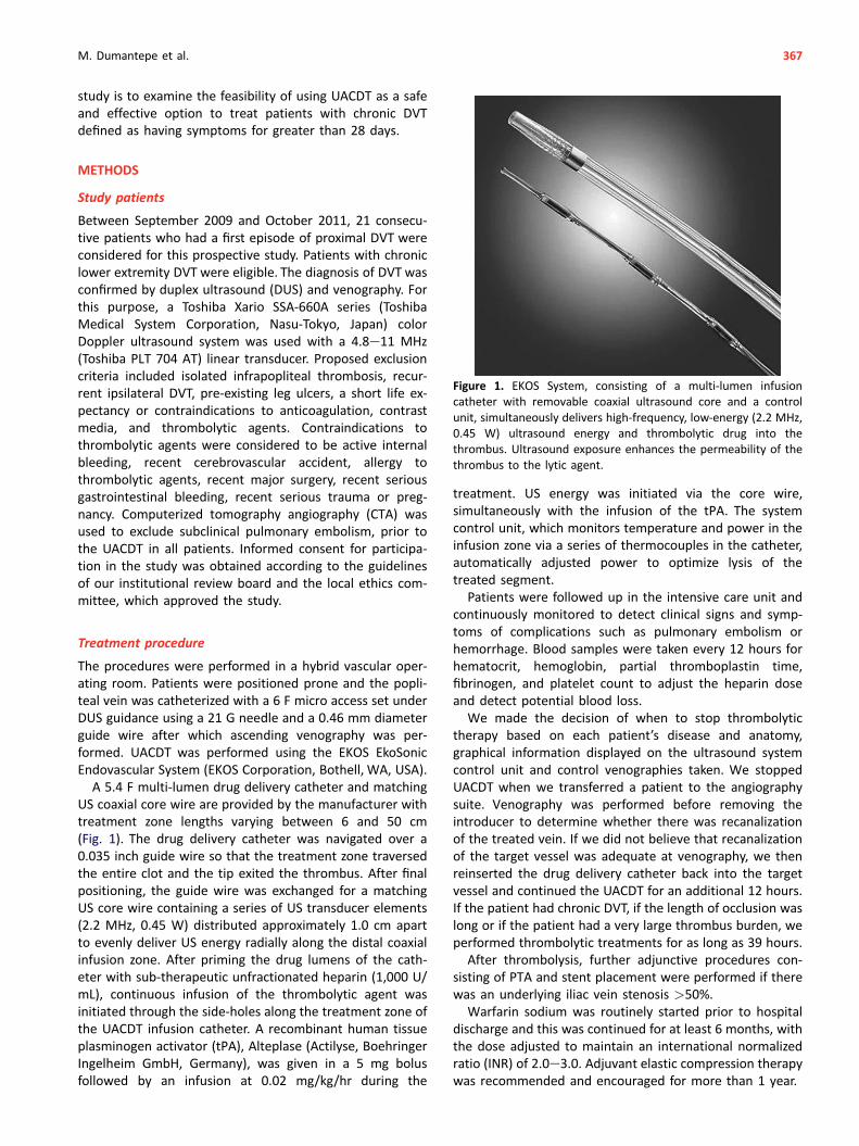
Figure 1. EKOS System, consisting of a multi-lumen infusioncatheter with removable coaxial ultrasound core and a controlunit, simultaneously delivers high-frequency, low-energy (2.2 MHz,0.45 W) ultrasound energy and thrombolytic drug into thethrombus. Ultrasound exposure enhances the permeability of thethrombus to the lytic agent.
METHODS
Study patients
Between September 2009 and October 2011, 21 consecu-tive patients who had a first episode of proximal DVT wereconsidered for this prospective study. Patients with chroniclower extremity DVT were eligible. The diagnosis of DVT wasconfirmed by duplex ultrasound (DUS) and venography. Forthis purpose, a Toshiba Xario SSA-660A series (ToshibaMedical System Corporation, Nasu-Tokyo, Japan) colorDoppler ultrasound system was used with a 4.8e11 MHz(Toshiba PLT 704 AT) linear transducer. Proposed exclusioncriteria included isolated infrapopliteal thrombosis, recur-rent ipsilateral DVT, pre-existing leg ulcers, a short life ex-pectancy or contraindications to anticoagulation, contrastmedia, and thrombolytic agents. Contraindications tothrombolytic agents were considered to be active internalbleeding, recent cerebrovascular accident, allergy tothrombolytic agents, recent major surgery, recent seriousgastrointestinal bleeding, recent serious trauma or preg-nancy. Computerized tomography angiography (CTA) wasused to exclude subclinical pulmonary embolism, prior tothe UACDT in all patients. Informed consent for participa-tion in the study was obtained according to the guidelinesof our institutional review board and the local ethics com-mittee, which approved the study.
Treatment procedure
The procedures were performed in a hybrid vascular oper-ating room. Patients were positioned prone and the popli-teal vein was catheterized with a 6 F micro access set underDUS guidance using a 21 G needle and a 0.46 mm diameterguide wire after which ascending venography was per-formed. UACDT was performed using the EKOS EkoSonicEndovascular System (EKOS Corporation, Bothell, WA, USA).
A 5.4 F multi-lumen drug delivery catheter and matchingUS coaxial core wire are provided by the manufacturer withtreatment zone lengths varying between 6 and 50 cm(Fig. 1). The drug delivery catheter was navigated over a0.035 inch guide wire so that the treatment zone traversedthe entire clot and the tip exited the thrombus. After finalpositioning, the guide wire was exchanged for a matchingUS core wire containing a series of US transducer elements(2.2 MHz, 0.45 W) distributed approximately 1.0 cm apartto evenly deliver US energy radially along the distal coaxialinfusion zone. After priming the drug lumens of the cath-eter with sub-therapeutic unfractionated heparin (1,000 U/mL), continuous infusion of the thrombolytic agent wasinitiated through the side-holes along the treatment zone ofthe UACDT infusion catheter. A recombinant human tissueplasminogen activator (tPA), Alteplase (Actilyse, BoehringerIngelheim GmbH, Germany), was given in a 5 mg bolusfollowed by an infusion at 0.02 mg/kg/hr during the
treatment. US energy was initiated via the core wire,simultaneously with the infusion of the tPA. The systemcontrol unit, which monitors temperature and power in theinfusion zone via a series of thermocouples in the catheter,automatically adjusted power to optimize lysis of thetreated segment.
Patients were followed up in the intensive care unit andcontinuously monitored to detect clinical signs and symp-toms of complications such as pulmonary embolism orhemorrhage. Blood samples were taken every 12 hours forhematocrit, hemoglobin, partial thromboplastin time,fibrinogen, and platelet count to adjust the heparin doseand detect potential blood loss.
We made the decision of when to stop thrombolytictherapy based on each patient’s disease and anatomy,graphical information displayed on the ultrasound systemcontrol unit and control venographies taken. We stoppedUACDT when we transferred a patient to the angiographysuite. Venography was performed before removing theintroducer to determine whether there was recanalizationof the treated vein. If we did not believe that recanalizationof the target vessel was adequate at venography, we thenreinserted the drug delivery catheter back into the targetvessel and continued the UACDT for an additional 12 hours.If the patient had chronic DVT, if the length of occlusion waslong or if the patient had a very large thrombus burden, weperformed thrombolytic treatments for as long as 39 hours.
After thrombolysis, further adjunctive procedures con-sisting of PTA and stent placement were performed if therewas an underlying iliac vein stenosis >50%.
Warfarin sodium was routinely started prior to hospitaldischarge and this was continued for at least 6 months, withthe dose adjusted to maintain an international normalizedratio (INR) of 2.0e3.0. Adjuvant elastic compression therapywas recommended and encouraged for more than 1 year.
368 European Journal of Vascular and Endovascular Surgery Volume 46 Issue 3 September/2013
Assessment of venous recanalization and symptomsresolution
Venous recanalization after completion of treatment wasdetermined by comparing the thrombus scores3 of the pre-and post-treatment venograms, which was categorized as“complete” recanalization for 95e100% restoration ofpatency, “partial” for 50e95% and “minimal” for less than50% as a result of residual stenosis or organizedthrombus.10,11 The final score was calculated as the per-centage difference between pre- and post-treatmentvenograms. Relief of symptoms (pain and swelling) at thecompletion of treatment was rated as (i) absent, (ii) poor,(iii) significant, or (iv) complete. Clinical success was definedas achieving at least 50% thrombus resolution and imme-diate clinical improvement of symptoms defined as resolu-tion of pain or swelling following thrombolysis.
Table 1. Patients’ baseline characteristics.
VariableAge (year, range) 46.1 (33e62)Gender (female/male) 7/5Reported symptom duration (days, range) 92.4 (34e183)Affected limb (left/right) 8/4
Follow-up
After hospital discharge, all patients were followed in theoutpatient clinic, weekly during the first month and then atmonthly intervals. At each visit, every patient underwent aclinical evaluation according to a modified Villalta scale12
and a DUS assessment of the affected lower limb forpatency of the deep venous system and presence orabsence of residual thrombus in the treated veins as well asvenous reflux. The presence of five leg symptoms (pain,cramps, heaviness, pruritus, and paresthesia) and sixobjective signs (pretibial edema, skin induration, hyperpig-mentation, new venous ectasia, redness, and pain duringcalf compression) were scored. Each of the symptoms andsigns were rated as 0 (absent), 1 (mild), 2 (moderate) or(severe). Clinical evaluation outcomes were classified asfollows: a total score greater than 14 points or a venousulcer was defined as severe post-thrombotic syndrome(PTS); 5e14 points was considered to indicate mild PTS, andless than 5 points was given for no PTS. Post-treatment DUSassessment of the affected lower limb was also performedat weekly intervals for 1 month, and monthly thereafter.Valvular reflux was defined as greater than 0.5 seconds forthe valve closure time after distal compression and releaseusing an ultrasonic probe with the patient standing andnon-weight bearing on the treated limb.13 Primary patencywas defined as confirmed patency and <50% restenosis atthe sixth month as documented by DUS.14 We recordedlimb circumferences in the thigh at 15 cm above the kneejoint and in the calf at 10 cm below the tibial tuberosityprior to treatment and at the 6-month post-treatmentreview.
Thrombosis locationIliofemoral 5Femoropopliteal 7
Risk factorsPostpartum 3Postoperation 1Trauma 2Immobilization 4Malignancy 2
Complications
Complications were classified according to scoring system ofthe Society of Interventional Radiology.15 Complications notrequiring therapy and presenting without consequence orcomplications requiring nominal therapy and without aconsequence, including an overnight admission for obser-vation, were considered “minor”.
RESULTS
Patients
Of the 21 patients evaluated, nine were excluded becauseof previous ipsilateral DVT (n ¼ 3), pre-existing leg ulcers orvenous insufficiency (n ¼ 2), poor life expectancy (n ¼ 1),recent cerebrovascular accident (n ¼ 1), or inability toparticipate at follow-up visits (n ¼ 2). The remaining 12patients were enrolled in this study. Their baseline charac-teristics are shown in Table 1. Mean total alteplase dosewas 42.4 � 7.9 (range 30e54) mg and the mean infusiontime was 26.2 � 7.0 (range 16e39) hours. The location ofthrombosis was in the iliofemoral segment in five patientsand the femoropopliteal segment in seven patients. Thedrug delivery catheter was successfully positioned withinthe occlusions and UACDT was successfully performed in allpatients. There were no device failures or adverse eventsassociated with the EKOS system. Thrombolysis was suc-cessful in 11 of 12 patients (92%) with complete clot lysis(>95% restored patency) in six patients and partial clot lysis(50e95% restored patency) in five patients. In one patientpresenting with 154 days since onset of symptoms,thrombolysis was not successful with only minimal clot lysis(<50%). Figs. 2 and 3 illustrate representative cases.A statistically significant correlation was found between thepercentage of thrombus removal and degree of clinicalimprovement (p < .01, Table 2). Two patients treated foriliofemoral DVT showed symptoms of associated iliac veinstenosis and were further treated to improve flow withballoon angioplasty and stent implantation (60/16 mmWallstent, 40/10 mm Nitinol stent and 100/14 mm Wall-stent, respectively) immediately after control venography(Fig. 3).
Immediate clinical improvement was observed in 11 of12 patients, with significant or complete clinical improve-ment occurring in 10. These patients also exhibited over50% of thrombus clearance. All together, clinical successwas observed in 10 patients (83%). In the follow-up period,thigh circumference of the affected limb decreased by3.9 � 1.7 cm, and calf circumference decreased by3.3 � 1.5 cm in the first month (Table 3).
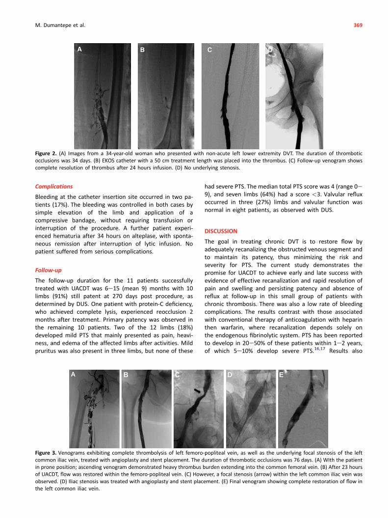
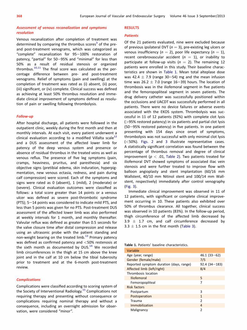
Figure 2. (A) Images from a 34-year-old woman who presented with non-acute left lower extremity DVT. The duration of thromboticocclusions was 34 days. (B) EKOS catheter with a 50 cm treatment length was placed into the thrombus. (C) Follow-up venogram showscomplete resolution of thrombus after 24 hours infusion. (D) No underlying stenosis.
M. Dumantepe et al. 369
Complications
Bleeding at the catheter insertion site occurred in two pa-tients (17%). The bleeding was controlled in both cases bysimple elevation of the limb and application of acompressive bandage, without requiring transfusion orinterruption of the procedure. A further patient experi-enced hematuria after 34 hours on alteplase, with sponta-neous remission after interruption of lytic infusion. Nopatient suffered from serious complications.
Follow-up
The follow-up duration for the 11 patients successfullytreated with UACDT was 6e15 (mean 9) months with 10limbs (91%) still patent at 270 days post procedure, asdetermined by DUS. One patient with protein-C deficiency,who achieved complete lysis, experienced reocclusion 2months after treatment. Primary patency was observed inthe remaining 10 patients. Two of the 12 limbs (18%)developed mild PTS that mainly presented as pain, heavi-ness, and edema of the affected limbs after activities. Mildpruritus was also present in three limbs, but none of these
Figure 3. Venograms exhibiting complete thrombolysis of left femoro-common iliac vein, treated with angioplasty and stent placement. The din prone position; ascending venogram demonstrated heavy thrombus bof UACDT, flow was restored within the femoro-popliteal vein. (C) Howobserved. (D) Iliac stenosis was treated with angioplasty and stent placthe left common iliac vein.
had severe PTS. The median total PTS score was 4 (range 0e9), and seven limbs (64%) had a score <3. Valvular refluxoccurred in three (27%) limbs and valvular function wasnormal in eight patients, as observed with DUS.
DISCUSSION
The goal in treating chronic DVT is to restore flow byadequately recanalizing the obstructed venous segment andto maintain its patency, thus minimizing the risk andseverity for PTS. The current study demonstrates thepromise for UACDT to achieve early and late success withevidence of effective recanalization and rapid resolution ofpain and swelling and persisting patency and absence ofreflux at follow-up in this small group of patients withchronic thrombosis. There was also a low rate of bleedingcomplications. The results contrast with those associatedwith conventional therapy of anticoagulation with heparinthen warfarin, where recanalization depends solely onthe endogenous fibrinolytic system. PTS has been reportedto develop in 20e50% of these patients within 1e2 years,of which 5e10% develop severe PTS.16,17 Results also
popliteal vein, as well as the underlying focal stenosis of the lefturation of thrombotic occlusions was 76 days. (A) With the patienturden extending into the common femoral vein. (B) After 23 hoursever, a focal stenosis (arrow) within the left common iliac vein wasement. (E) Final venogram showing complete restoration of flow in
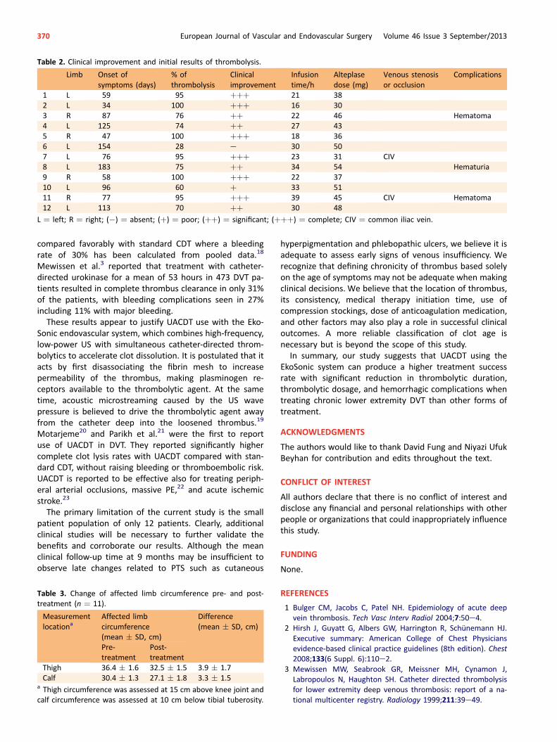
Table 2. Clinical improvement and initial results of thrombolysis.
Limb Onset ofsymptoms (days)
% ofthrombolysis
Clinicalimprovement
Infusiontime/h
Alteplasedose (mg)
Venous stenosisor occlusion
Complications
1 L 59 95 þþþ 21 382 L 34 100 þþþ 16 303 R 87 76 þþ 22 46 Hematoma4 L 125 74 þþ 27 435 R 47 100 þþþ 18 366 L 154 28 e 30 507 L 76 95 þþþ 23 31 CIV8 L 183 75 þþ 34 54 Hematuria9 R 58 100 þþþ 22 3710 L 96 60 þ 33 5111 R 77 95 þþþ 39 45 CIV Hematoma12 L 113 70 þþ 30 48
L ¼ left; R ¼ right; (�) ¼ absent; (þ) ¼ poor; (þþ) ¼ significant; (þþþ) ¼ complete; CIV ¼ common iliac vein.
370 European Journal of Vascular and Endovascular Surgery Volume 46 Issue 3 September/2013
compared favorably with standard CDT where a bleedingrate of 30% has been calculated from pooled data.18
Mewissen et al.3 reported that treatment with catheter-directed urokinase for a mean of 53 hours in 473 DVT pa-tients resulted in complete thrombus clearance in only 31%of the patients, with bleeding complications seen in 27%including 11% with major bleeding.
These results appear to justify UACDT use with the Eko-Sonic endovascular system, which combines high-frequency,low-power US with simultaneous catheter-directed throm-bolytics to accelerate clot dissolution. It is postulated that itacts by first disassociating the fibrin mesh to increasepermeability of the thrombus, making plasminogen re-ceptors available to the thrombolytic agent. At the sametime, acoustic microstreaming caused by the US wavepressure is believed to drive the thrombolytic agent awayfrom the catheter deep into the loosened thrombus.19
Motarjeme20 and Parikh et al.21 were the first to reportuse of UACDT in DVT. They reported significantly highercomplete clot lysis rates with UACDT compared with stan-dard CDT, without raising bleeding or thromboembolic risk.UACDT is reported to be effective also for treating periph-eral arterial occlusions, massive PE,22 and acute ischemicstroke.23
The primary limitation of the current study is the smallpatient population of only 12 patients. Clearly, additionalclinical studies will be necessary to further validate thebenefits and corroborate our results. Although the meanclinical follow-up time at 9 months may be insufficient toobserve late changes related to PTS such as cutaneous
Table 3. Change of affected limb circumference pre- and post-treatment (n ¼ 11).
Measurementlocationa
Affected limbcircumference(mean � SD, cm)
Difference(mean � SD, cm)
Pre-treatment
Post-treatment
Thigh 36.4 � 1.6 32.5 � 1.5 3.9 � 1.7Calf 30.4 � 1.3 27.1 � 1.8 3.3 � 1.5
a Thigh circumference was assessed at 15 cm above knee joint andcalf circumference was assessed at 10 cm below tibial tuberosity.
hyperpigmentation and phlebopathic ulcers, we believe it isadequate to assess early signs of venous insufficiency. Werecognize that defining chronicity of thrombus based solelyon the age of symptoms may not be adequate when makingclinical decisions. We believe that the location of thrombus,its consistency, medical therapy initiation time, use ofcompression stockings, dose of anticoagulation medication,and other factors may also play a role in successful clinicaloutcomes. A more reliable classification of clot age isnecessary but is beyond the scope of this study.
In summary, our study suggests that UACDT using theEkoSonic system can produce a higher treatment successrate with significant reduction in thrombolytic duration,thrombolytic dosage, and hemorrhagic complications whentreating chronic lower extremity DVT than other forms oftreatment.
ACKNOWLEDGMENTS
The authors would like to thank David Fung and Niyazi UfukBeyhan for contribution and edits throughout the text.
CONFLICT OF INTEREST
All authors declare that there is no conflict of interest anddisclose any financial and personal relationships with otherpeople or organizations that could inappropriately influencethis study.
FUNDING
None.
REFERENCES
1 Bulger CM, Jacobs C, Patel NH. Epidemiology of acute deepvein thrombosis. Tech Vasc Interv Radiol 2004;7:50e4.
2 Hirsh J, Guyatt G, Albers GW, Harrington R, Schünemann HJ.Executive summary: American College of Chest Physiciansevidence-based clinical practice guidelines (8th edition). Chest2008;133(6 Suppl. 6):110e2.
3 Mewissen MW, Seabrook GR, Meissner MH, Cynamon J,Labropoulos N, Haughton SH. Catheter directed thrombolysisfor lower extremity deep venous thrombosis: report of a na-tional multicenter registry. Radiology 1999;211:39e49.
M. Dumantepe et al. 371
4 Sharafuddin MJ, Sun S, Hoballah JJ, Youness FM, Sharp WJ,Roh BS. Endovascular management of venous thrombotic andocclusive disease of the lower extremities. J Vasc Interv Radiol2003;13:405e23.
5 Francis CW, Blinc A, Lee S, Cox C. Ultrasound acceleratestransport of recombinant tissue plasminogen activator intoclots. Ultrasound Med Biol 1995;21:419e24.
6 Siddiqi F, Blinc A, Braaten J, Francis CW. Ultrasound increasesflow through fibrin gels. Thromb Haemost 1995;73:495e8.
7 Dumantepe M, Tarhan A, Yurdakul I, Ozler A. US-acceleratedcatheter-directed thrombolysis for the treatment of deepvenous thrombosis. Diagn Interv Radiol 2012 Dec 27. http://dx.doi.org/10.5152/dir.2012.004.
8 Grommes J, Strijkers R, Greiner A, Mahnken AH, Wittens CH.Safety and feasibility of ultrasound-accelerated catheter-directed thrombolysis in deep vein thrombosis. Eur J VascEndovasc Surg 2011;41(4):526e32.
9 Lin PH, Ochoa LN, Duffy P. Catheter-directed thrombectomyand thrombolysis for symptomatic lower-extremity deep veinthrombosis: review of current interventional treatment stra-tegies. Perspect Vasc Surg Endovasc Ther 2010;22(3):152e63.
10 Vedantham S, Thorpe PE, Cardella JF, Grassi CJ, Patel NH,Ferral H, et al. Quality improvement guidelines for the treat-ment of lower extremity deep vein thrombosis with use ofendovascular thrombus removal. J Vasc Interv Radiol 2009;20(7 Suppl):227e39.
11 Park YJ, Choi JY, Min SK, Lee T, Jung IM, Chung JK, et al.Restoration of patency in iliofemoral deep vein thrombosis withcatheter-directed thrombolysis does not always prevent post-thrombotic damage. Eur J Vasc Endovasc Surg 2008;36:725e30.
12 Kahn SR, Partsch H, Vedantham S, Prandoni P,Kearon CSubcommittee on Control of Anticoagulation of theScientific and Standardization Committee of the InternationalSociety on Thrombosis and Haemostasis. Definition of post-thrombotic syndrome of the leg for use in clinical in-vestigations: a recommendation for standardization. J ThrombHaemost 2009;7(5):879e83. [Epub 2009 Jan].
13 Mattos MA, Sumner DS. Direct noninvasive tests (duplexscan) for the evaluation of chronic venous obstruction and
valvular incompetence. In: Gloviczki P, Yao JS, editors. Hand-book of venous disorders. 2nd ed. New York, NY: Arnold; 2001.p. 120e31.
14 Kölbel T, Lindh M, Akesson M, Wasselius J, Gottsater A,Ivancev K. Chronic iliac vein occlusion: midterm results ofendovascular recanalization. J Endovasc Ther 2009;16:483e91.
15 Vedantham S, Grassi CJ, Ferral H, Patel NH, Thorpe PE,Antonacci VP, et al. Reporting standards for endovasculartreatment of lower extremity deep vein thrombosis. J VascInterv Radiol 2006;17(3):417e34.
16 Kahn SR, Ginsberg JS. Relationship between deep venousthrombosis and the postthrombotic syndrome. Arch InternMed 2004;164:17e26.
17 Kahn SR. The post-thrombotic syndrome: the forgottenmorbidity of deep venous thrombosis. J Thromb Thrombolysis2006;21:41e8.
18 Alesh I, Kayali F, Stein PD. Catheter-directed thrombolysis(intrathrombus injection) in treatment of deep venousthrombosis: a systematic review. Catheter Cardiovasc Interv2007;70:143e8.
19 Atar S, Rosenschein U. Perspectives on the role of ultrasonicdevices in thrombolysis. J Thromb Thrombolysis 2004;17:107e14.
20 Motarjeme A. Ultrasound-enhanced thrombolysis. J EndovascTher 2007;14(2):251e6.
21 Parikh S, Motarjeme A, McNamara T, Raabe R, Hagspiel K,Benenati JF, et al. Ultrasound-accelerated thrombolysis for thetreatment of deep vein thrombosis: initial clinical experience.J Vasc Interv Radiol 2008;19(4):521e8.
22 Chamsuddin A, Nazzal L, Kang B, Best I, Peters G, Panah S, et al.Catheter-directed thrombolysis with the Endowave system inthe treatment of acute massive pulmonary embolism: aretrospective multicenter case series. J Vasc Interv Radiol2008;19(3):372e6.
23 Tsivgoulis G, Eggers J, Ribo M, Perren F, Saqqur M, Rubiera M,et al. Safety and efficacy of ultrasound-enhanced thrombolysis:a comprehensive review and meta-analysis of randomized andnonrandomized studies. Stroke 2010;41(2):280e7.