Embed Size (px)
Citation preview
Case ReportTreatment of Cervical Pregnancy with Ultrasound-GuidedIntragestational Injection of Methotrexate: A Case Report
Angeliki Rouvali,1 Panagiotis Vlastarakos ,1 Sofoklis Stavros,1 Maria Giourga,1
Kalliopi Pappa,1 Angeliki Gerede,2 Alexandros Rodolakis,1 and Ekaterini Domali 1
11st Department of Obstetrics and Gynaecology, Alexandra Hospital, National and Kapodistrian University of Athens,Athens, Greece2Department of Obstetrics and Gynecology Democritus, University of Thrace Alexandroupolis, Greece
Correspondence should be addressed to Panagiotis Vlastarakos; [email protected]
Received 30 August 2021; Accepted 19 October 2021; Published 10 November 2021
Academic Editor: Maria Grazia Piccioni
Copyright © 2021 Angeliki Rouvali et al. This is an open access article distributed under the Creative Commons Attribution License,which permits unrestricted use, distribution, and reproduction in any medium, provided the original work is properly cited.
This study is aimed at describing a noninvasive conservative strategy to the treatment of cervical pregnancy and highlighting thesuccess of ultrasound-guided therapeutic techniques. A 43-year-old woman with a history of one previous cesarean sectionpresented in our unit with vaginal spotting and a positive urine pregnancy test. She was diagnosed with a cervical pregnancy,and she was successfully treated conservatively with the administration of intragestational sac methotrexate under ultrasoundguidance. Cervical pregnancy is a rare form of ectopic pregnancy that results from conceptus implantation in the cervicalcanal. The main concern is the associated life-threatening hemorrhage and subsequent need for urgent hysterectomy. Theevolution of ultrasound over the past decades has enabled early diagnosis and has shifted the management from a radicalsurgical approach towards a stepwise conservative therapeutic approach, when possible.
1. Introduction
A cervical ectopic pregnancy occurs when a blastocyst implantin the endocervical canal. Cervical pregnancies are relativelyuncommon, and it is estimated that they account for less thanone percent of all ectopic pregnancies [1]. Although this condi-tion is rare, it has been associated with high morbidity due tothe risk of major fatal hemorrhage. Early diagnosis of cervicalimplantation is crucial for the subsequent management thatshould minimize the risk of hemorrhage, eliminate the gesta-tional tissue, and spare fertility. Nowadays, transvaginal ultra-sound allows the diagnosis of cervical ectopic pregnancies atearly stages, thus increasing the chances of success withmedicaltreatment.
Although there is no consensus about the appropriatetreatment of cervical pregnancies, conservative managementis preferred [2, 3] in asymptomatic or minimally symptomaticand hemodynamically stable patients, leaving more radicalsurgical approaches such as hysterectomy for unstable patients
with life-threatening symptoms and/or failure of a conserva-tive approach. In this paper, we describe a case of cervicalpregnancy which was successfully treated with ultrasound-guided intragestational sac methotrexate injection in our unit.
2. Case Presentation
A 43-year-old woman (gravida 2, para 1, cesarean section 1)presented in the Early Pregnancy Unit (EPU) with nauseaand one-day history of painless vaginal spotting. Her last men-strual period was five weeks before admission, and she had apositive urine pregnancy test. She reported regular menstrualcycles, and she was not using any contraception. Her pastmedical history was unremarkable, and she had a cesareansection 13 years ago from her surgical history. She was a non-smoker, and her body mass index was (BMI) was 20.9kg/m2. Upon arrival, she was hemodynamically stable withhemoglobin of 11.2 g/dL. White blood count, platelets, liver,
HindawiCase Reports in Obstetrics and GynecologyVolume 2021, Article ID 6601461, 5 pageshttps://doi.org/10.1155/2021/6601461
and renal function tests were all normal. The initial serum betachorionic gonadotropin (β-hCG) level was 25.270 mIU/mL.
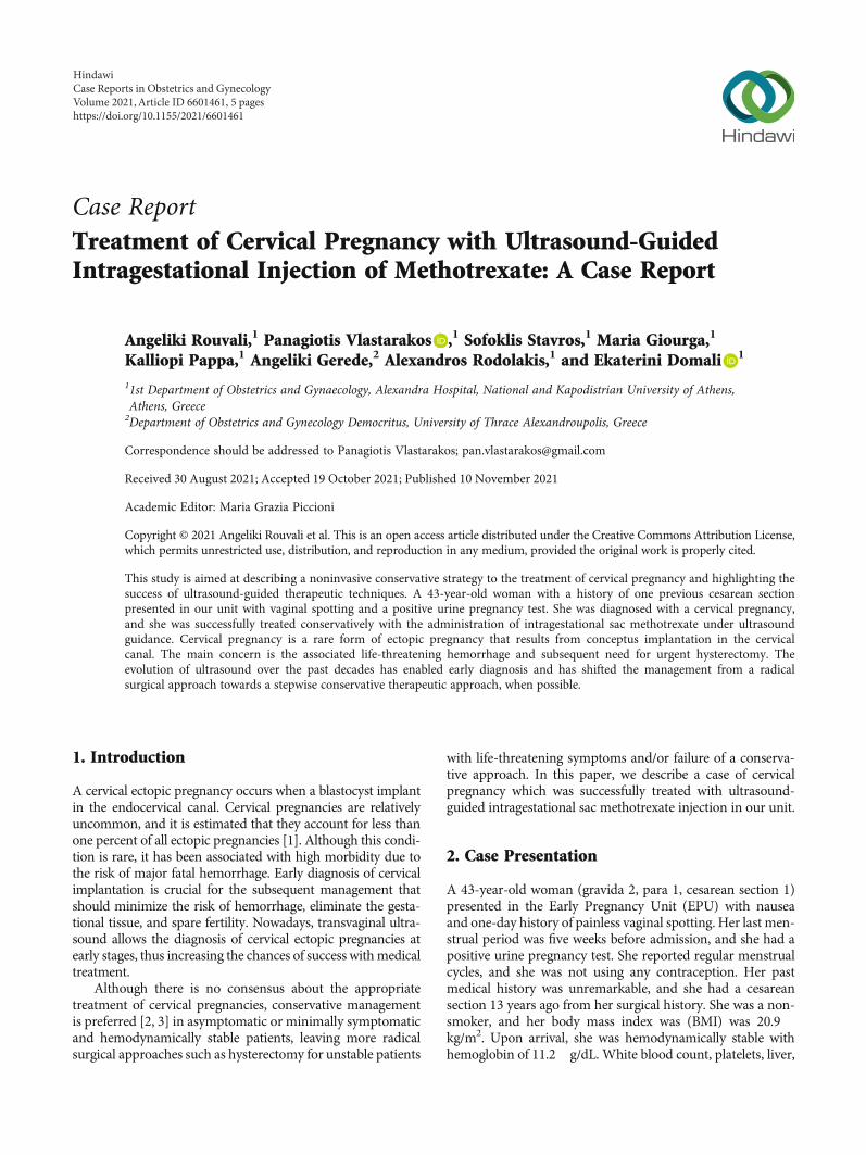
On physical examination, her abdomen was soft and non-tender. A gentle speculum examination revealed a hyperemiccervix with gestational tissue on the anterior lip of the externalcervical os and no active bleeding. Transvaginal ultrasoundshowed an empty uterine cavity with thick endometrial liningand a ballooned cervical canal with a gestational sac contain-ing a yolk sac in the lower portion of the cervix and an absent“sliding sign” (Figure 1(a)). The use of Color Doppler studies
showed extensive blood flow to the gestational sac(Figure 1(b)). Both ovaries were normal, and there was no evi-dence of free fluid. Thus, a diagnosis of cervical pregnancy wasestablished.
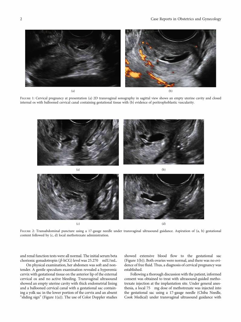
Following a thorough discussion with the patient, informedconsent was obtained to treat with ultrasound-guided metho-trexate injection at the implantation site. Under general anes-thesia, a local 75 mg dose of methotrexate was injected intothe gestational sac using a 17-gauge needle (Chiba Needle,Cook Medical) under transvaginal ultrasound guidance with
(a) (b)
Figure 1: Cervical pregnancy at presentation (a) 2D transvaginal sonography in sagittal view shows an empty uterine cavity and closedinternal os with ballooned cervical canal containing gestational tissue with (b) evidence of peritrophoblastic vascularity.
(a) (b)
(c) (d)
Figure 2: Transabdominal puncture using a 17-gauge needle under transvaginal ultrasound guidance. Aspiration of (a, b) gestationalcontent followed by (c, d) local methotrexate administration.
2 Case Reports in Obstetrics and Gynecology
a prior aspiration of the gestational sac’s fluid content to reducepregnancy volume (Figure 2).
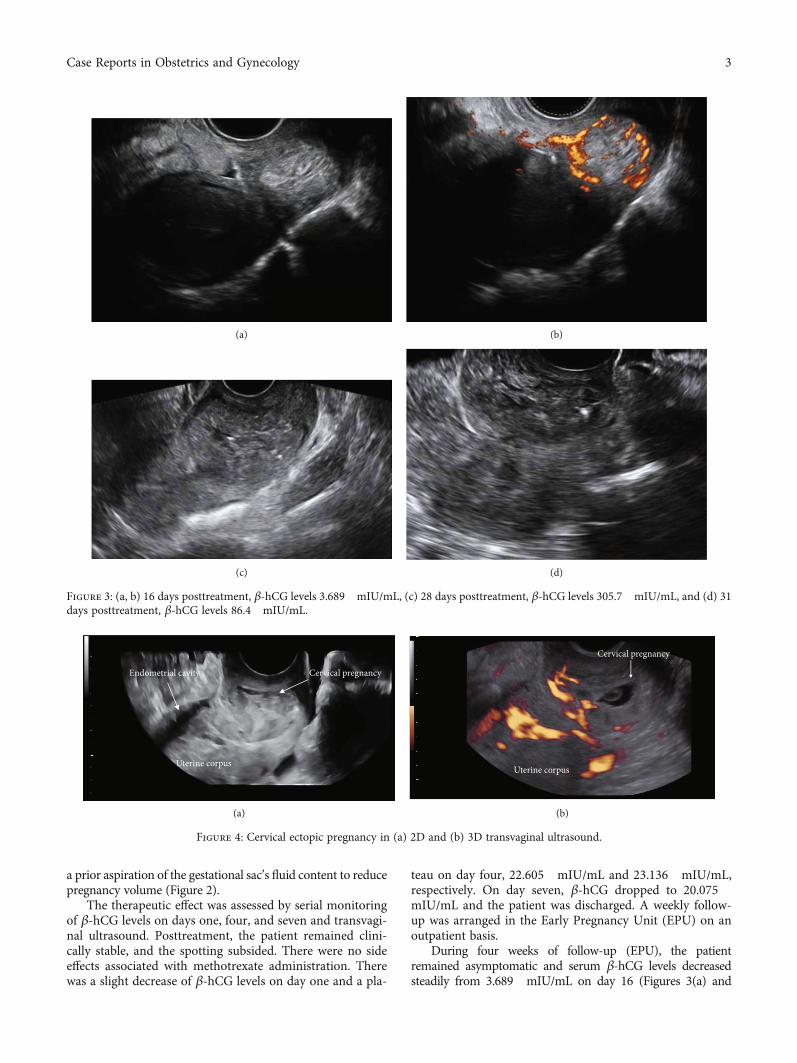
The therapeutic effect was assessed by serial monitoringof β-hCG levels on days one, four, and seven and transvagi-nal ultrasound. Posttreatment, the patient remained clini-cally stable, and the spotting subsided. There were no sideeffects associated with methotrexate administration. Therewas a slight decrease of β-hCG levels on day one and a pla-
teau on day four, 22.605 mIU/mL and 23.136 mIU/mL,respectively. On day seven, β-hCG dropped to 20.075mIU/mL and the patient was discharged. A weekly follow-up was arranged in the Early Pregnancy Unit (EPU) on anoutpatient basis.
During four weeks of follow-up (EPU), the patientremained asymptomatic and serum β-hCG levels decreasedsteadily from 3.689 mIU/mL on day 16 (Figures 3(a) and
(a) (b)
(c) (d)
Figure 3: (a, b) 16 days posttreatment, β-hCG levels 3.689 mIU/mL, (c) 28 days posttreatment, β-hCG levels 305.7 mIU/mL, and (d) 31days posttreatment, β-hCG levels 86.4 mIU/mL.
Cervical pregnancyEndometrial cavity
Uterine corpus
(a)
Cervical pregnancy
Uterine corpus
(b)
Figure 4: Cervical ectopic pregnancy in (a) 2D and (b) 3D transvaginal ultrasound.
3Case Reports in Obstetrics and Gynecology

3(b)) to 4mIU/mL on day 46. Transvaginal ultrasound findingswere suggestive of gestational sac regression (Figure 3).
3. Discussion
Cervical ectopic pregnancy is a rare entity that accounts for asmall proportion of all ectopic pregnancies, and the reportedincidence is approximately one per 10.000 live births [4]. Itoccurs when a fertilized ovum implants in the lining of theendocervix below the internal os level. Cervical pregnancycan be a life-threatening condition, and early diagnosis andtreatment are essential to preserve fertility and avoid theneed for hysterectomy.
The pathogenesis of ectopic pregnancy remains unclear.Prior dilatation and curettage and cesarean section have beenreported as predisposing factors, possibly causing damage tothe endometrial lining and the cervix [4]. Another theorysuggests a rapid crossing of the fertilized ovum to the cervicalcanal before it is capable of implantation [5]. Cervical ectopicpregnancies have also been associated with assisted reproduc-tive techniques [6].
Most commonly, patients with cervical ectopic preg-nancy present with painless vaginal bleeding, and less thanone-third of them experience lower abdominal pain [6].
Diagnosis of cervical ectopic pregnancy is based on clinicalexamination and ultrasound findings in a patient with a posi-tive pregnancy test. Differential diagnosis, especially from anincomplete miscarriage or a cesarean section scar pregnancy,can be challenging. Ultrasound diagnostic criteria include (a)empty uterine cavity, (b) hourglass uterine shape with bal-looned cervical canal, (c) the presence of gestational sac orplacental tissue within the cervical canal, (d) absent “slidingsign,” and (e) high peritrophoblastic vascularity on Dopplerexamination [7].
Also, the use of three-dimensional (3D) ultrasound imag-ing in addition to two-dimensional (2D) scan (Figure 4) mayprovide additional information from the coronal section andhelp towards the correct diagnosis of cervical pregnancy [8, 9].
There is a wide array of therapeutic options for cervicalectopic pregnancy, varying from conservative drug therapiesto radical surgical procedures. The optimal treatment methoddepends on the gestational age at the time of diagnosis, theclinical manifestation and its severity, the presence of coexist-ing viable intrauterine pregnancy, and clinician’s experience.
Among the conservative management options, the use ofmethotrexate has revolutionized the treatment of cervicalectopic pregnancies. In cervical pregnancies, methotrexatecan be administered systemically (single- or multidose regi-men) with high treatment success; however, if gestationalage is >9weeks, β − hCG levels > 10:000 IU/L, fetal cardiacactivity is present, and failure rates are higher [10]. Localmethotrexate injection has also been proved successful andis the proposed first-line option in cervical ectopic pregnan-cies with fetal cardiac activity [2]. Alternatively, potassiumchloride (KCL) injection can be used as a first step [11].Some authors suggest that curettage is necessary to reducesevere hemorrhage associated with trophoblastic sheddingfrom the atonic cervix that occurs as a metabolic effect ofmethotrexate [10, 12].
Other approaches that can be used in conjunction withdrug therapies or if medical treatments fail to reduce the riskof bleeding include uterine artery embolization, Foley catheterballoon tamponade [13], suction evacuation, high cervicalcerclage, dilatation and curettage, and ligation of cervicovagi-nal branches of the uterine arteries at third and ninth o’clockpositions of the cervix [14].
4. Conclusion
Although cervical ectopic pregnancy is a rare event, delay indiagnosis or misdiagnosis can be detrimental for the patientwith life-threatening consequences. The introduction of EPUsand the widespread use of transvaginal ultrasound means thatrecognizing this condition is feasible early in pregnancy. Earlydetection enables the use of conservative therapeutic options.Intragestational injection of methotrexate with posttreatmentsurveillance in clinically stable patients is an effective and safeapproach that preserves fertility.
Consent
Written informed consent was obtained from the patient forpublication of this case report and accompanying images. Acopy of the written consent is available for review by theEditor-in-Chief of this journal on request.
Conflicts of Interest
The authors have no conflicts of interest to declare.
References
[1] J. Bouyer, J. Coste, H. Fernandez, J. L. Pouly, and N. Job-Spira,“Sites of ectopic pregnancy: a 10 year population-based studyof 1800 cases,” Human Reproduction, vol. 17, no. 12,pp. 3224–3230, 2002.
[2] E. Kirk, G. Condous, Z. Haider, A. Syed, K. Ojha, andT. Bourne, “The conservative management of cervical ectopicpregnancies,” Ultrasound in Obstetrics & Gynecology, vol. 27,no. 4, pp. 430–437, 2006.
[3] S. H. Bakour, P. K. Thompson, and K. S. Khan, “Successfulconservative management of cervical ectopic pregnancy withcombination of methotrexate, mifepristone, surgical evacua-tion and tamponade using a double balloon three-way cathe-ter,” Journal of Obstetrics and Gynaecology, vol. 25, no. 6,pp. 616–618, 2005.
[4] G. Vela and T. Tulandi, “Cervical pregnancy: the importanceof early diagnosis and treatment,” Journal of Minimally Inva-sive Gynecology, vol. 14, no. 4, pp. 481–484, 2007.
[5] W. Studdiford, “Cervical pregnancy: a partial review of the lit-erature and a report of two probable cases,” American Journalof Obstetrics and Gynecology, vol. 49, no. 2, pp. 169–185, 1945.
[6] E. S. Ginsburg, M. C. Frates, M. S. Rein, J. H. Fox, M. D. Horn-stein, and A. J. Friedman, “Early diagnosis and treatment ofcervical pregnancy in an in vitro fertilization program,” Fertil-ity and Sterility, vol. 61, no. 5, pp. 966–969, 1994.
[7] D. Jurkovic, E. Hacket, and S. Campbell, “Diagnosis and treat-ment of early cervical pregnancy: a review and a report of twocases treated conservatively,”Ultrasound in Obstetrics & Gyne-cology, vol. 8, no. 6, pp. 373–380, 1996.
4 Case Reports in Obstetrics and Gynecology

[8] R. Ruano, F. Reya, O. Picone et al., “Three-dimensional ultra-sonographic diagnosis of a cervical pregnancy,” Clinics (SãoPaulo, Brazil), vol. 61, no. 4, pp. 355–358, 2006.
[9] D. Jurkovic, A. Geipel, K. Gruboeck, E. Jauniaux, M. Natucci,and S. Campbell, “Three-dimensional ultrasound for theassessment of uterine anatomy and detection of congenitalanomalies: a comparison with hysterosalpingography andtwo-dimensional sonography,” Ultrasound in Obstetrics &Gynecology, vol. 5, no. 4, pp. 233–237, 1995.
[10] T. H. Hung, W. Y. Shau, T. T. Hsieh, J. J. Hsu, Y. K. Soong, andC. J. Jeng, “Prognostic factors for an unsatisfactory primarymethotrexate treatment of cervical pregnancy: a quantitativereview,” Human Reproduction, vol. 13, no. 9, pp. 2636–2642,1998.
[11] C. J. Jeng, M. L. Ko, and J. Shen, “Transvaginal ultrasound-guided treatment of cervical pregnancy,” Obstetrics and Gyne-cology, vol. 109, no. 5, pp. 1076–1082, 2007.
[12] S. Mesogitis, A. Pilalis, G. Daskalakis, N. Papantoniou, andA. Antsaklis, “Management of early viable cervical pregnancy,”BJOG : An International Journal of Obstetrics and Gynaecol-ogy, vol. 112, no. 4, pp. 409–411, 2005.
[13] D. L. Fylstra, “Cervical pregnancy: 13 cases treated with suc-tion curettage and balloon tamponade,” American Journal ofObstetrics and Gynecology, vol. 210, no. 6, pp. 581.e1–581.e5,2014.
[14] E. S. Saygili Yilmaz, D. Aydin, and Z. Yilmaz, “Conservativetreatment of cervical pregnancy by evacuation after transvagi-nal suture ligation of the cervicovaginal branches of uterinearteries,” Acta Obstetricia et Gynecologica Scandinavica,vol. 81, no. 10, pp. 988–990, 2002.
5Case Reports in Obstetrics and Gynecology