Embed Size (px)
Citation preview

OsteoArthritis and Cartilage (2007) 15, 226e231
ª 2006 Osteoarthritis Research Society International. Published by Elsevier Ltd. All rights reserved.doi:10.1016/j.joca.2006.08.008
Brief reportTreatment of a full-thickness articular cartilage defect in the femoralcondyle of an athlete with autologous bone-marrow stromal cells1
R. Kuroda M.D.y*, K. Ishida M.D.y, T. Matsumoto M.D.y, T. Akisue M.D.y, H. Fujioka M.D.y,K. Mizuno M.D.y, H. Ohgushi M.D.z, S. Wakitani M.D.x and M. Kurosaka M.D.yyDepartment of Orthopaedic Surgery, Kobe University Graduate School of Medicine, JapanzResearch Institute for Cell Engineering (RICE), National Institute of Advanced IndustrialScience and Technology (AIST), JapanxDepartment of Orthopaedic Surgery, Osaka City University Graduate School of Medicine, Japan
Summary
Objectives: Human bone-marrow stromal cells are believed to be multipotent even in adults. This study assessed the effectiveness of autol-ogous bone-marrow stromal cells, which were embedded within a collagen scaffold, to repair a full-thickness articular cartilage defect in themedial femoral condyle of an athlete.
Patient and methods: A 31-year-old male judo player suffering from pain in the right knee was reviewed. A 20� 30-mm full-thickness cartilagedefect (International Cartilage Repair Society classification (ICRS) grade IV) was revealed in the weight-bearing area of the medial femoralcondyle. With the informed consent of the patient, the defect was treated with autologous bone-marrow stromal cells. Bone marrow was as-pirated from the iliac crest of the patient 4 weeks before surgery. After removing the erythrocytes, the remaining cells were expanded in cul-ture. Adherent cells were collected and embedded within a collagen gel, which was transferred to the articular cartilage defect in the medialfemoral condyle. The implant was covered with an autologous periosteal flap.
Results: Seven months after surgery, arthroscopy revealed the defect to be covered with smooth tissues. Histologically, the defect was filledwith a hyaline-like type of cartilage tissue which stained positively with Safranin-O. One year after surgery, the clinical symptoms had improvedsignificantly. The patient had reattained his previous activity level and experienced neither pain nor other complications.
Conclusions: Our findings indicate that the transplantation of autologous bone-marrow stromal cells can promote the repair of large focal ar-ticular cartilage defects in young, active patients.ª 2006 Osteoarthritis Research Society International. Published by Elsevier Ltd. All rights reserved.
Key words: Bone-marrow stromal cell, Autologous cell implantation, Athlete, Cartilage repair, Hyaline cartilage.
InternationalCartilageRepairSociety
Introduction
Articular cartilage defects have a poor capacity to undergoself-repair, owing to the low mitotic potential of chondro-cytes in vivo1. Since articular cartilage defects can progressto osteoarthritis in some patients, they need to be repairedeven though their exact natural course remains obscure2e4.Strategies that have been instigated to repair articular carti-lage defects include microfractuing5 and mosaicplasty6,7.However, these procedures are limited to small- and me-dium-sized focal chondral and osteochondral defects. Mo-saicplasty is also limited by the need to create defects atdonor sites, by an insufficient repair result between thegrafts, and by the technical difficulties experienced in resur-facing the original curvature of the joint6,7. The autologous
1No benefits in any form have been received or will be receivedfrom a commercial party related directly or indirectly to the subjectof this article.
*Address correspondence and reprint requests to: RyosukeKuroda, M.D., Department of Orthopaedic Surgery, KobeUniversity Graduate School of Medicine, 7-5-1 Kusunoki-cho,Chuo-ku, Kobe 650-0017, Japan. Tel: 81-78-382-5987; Fax: 81-78-351-6944; E-mail: [email protected]
Received 11 May 2006; revision accepted 12 August 2006.
2
chondrocyte implantation (ACI) technique was first per-formed by Peterson et al.8 in 1994. This was the first appli-cation of a cell-engineering strategy in orthopaedic surgery.However, several problems are associated with the proce-dure. These include difficulties in obtaining a sufficient num-ber of chondrocytes for autotransplantation, the necessity ofcreating donor-site defects within autologous cartilage, andpoor histological repair1,6.
Mesenchymal stromal cells within the adult bone marroware multipotent, being capable of forming bone, cartilageand other connective tissues9. It has been suggested thatthese cells may be used effectively for the repair of cartilagetissue. We report on a case of a focal articular cartilage in-jury in an athlete. This study assessed the effectiveness ofautologous bone-marrow stromal cells to repair a full-thick-ness articular cartilage defect in the medial femoral condyleof a 31-year-old male judo player.
Case report
In 1999, a 31-year-old male judo player injured his rightknee whilst playing judo, and he underwent medial menis-cectomy at another hospital. He resumed his judo activities
26

227Osteoarthritis and Cartilage Vol. 15, No. 2
and experienced no pain. However, in May 2004, he twistedhis knee whilst playing judo. He complained of pain in theright knee and was referred to our hospital. His right kneewas remarkably swollen, and the active range of motionwas limited to extension. There were no signs of collateralor cruciate ligament instability. X-radiography of theweight-bearing knee in the standing position disclosed a ra-diolucent area in the medial femoral condyle [Fig. 1(A)], butthe limb alignment was normal. Magnetic resonance imag-ing (MRI) revealed a cartilage defect within the medial fem-oral condyle, oedema of the subchondral bone, anddegeneration of the posterior part of the medial meniscus[Fig. 2(A, B)]. Laboratory tests disclosed no abnormalities.
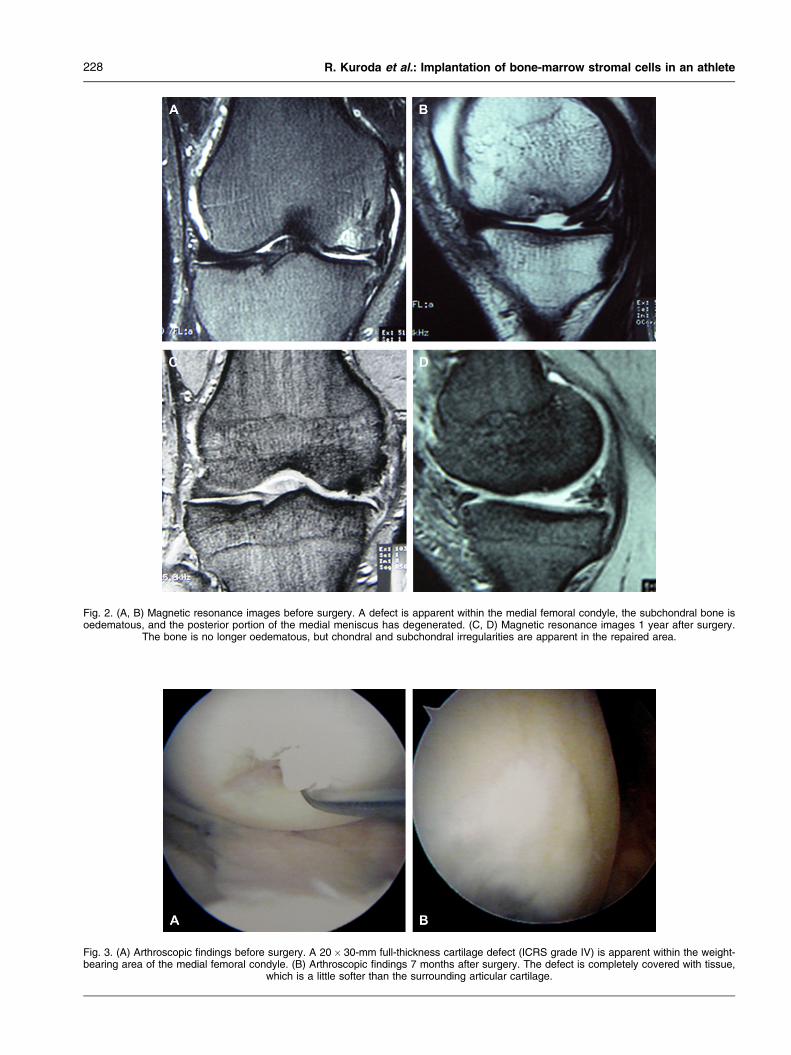
Arthroscopy revealed a 20� 30-mm full-thickness carti-lage defect (International Cartilage Repair Society classifi-cation (ICRS) grade IV) within the weight-bearing area ofthe medial femoral condyle [Fig. 3(A)] and a macroscopictear in the middle third of the medial meniscus. A partial me-niscectomy was performed, but the cartilage defect was leftuntreated. In an attempt to repair the articular cartilage de-fect, we decided to perform an autologous transplantation ofbone-marrow cells, with the informed consent of the patient.Fifteen millilitres of bone marrow were aspirated from theleft iliac crest and transferred to tubes containing heparin-ized phosphate-buffered saline. Four hundred millilitres ofthe patient’s blood were collected. After centrifugation,180 ml of serum were obtained. The cells derived from3 ml of fresh bone marrow were transferred to two 75-cm2
plastic culture flasks, within which they were maintainedfor 4 weeks, with changes of medium (alpha minimal essen-tial medium supplemented with 15% autologous serum)three times per week. At the time when the medium waschanged, non-adherent haematopoietic cells were re-moved. After about 10 days, the number of adherent cellshad increased to several million. The cells were collectedafter trypsinization (first passage) and further cultured (sub-cultured) in other flasks for about 10 days. At this stage,they had a fibroblast-like appearance. They were negativefor markers of haematopoietic cells (CD14, CD34) and forHLA-DR, but positive for markers of mesenchymal ones(CD73, CD90, CD105). These findings indicate that the ad-herent cultured cells were of the mesenchymal type10. The
subcultured cells were collected and suspended withina 1% acid-soluble solution of porcine tendon type-I collagen(final cell density: 5� 106 cells/ml). The collagenous cellsuspension was placed on a sheet of collagen (Gunze,Kyoto, Japan), which acted as a support, and gelled at37�C. This gelecell composite was further cultured fora couple of days. A small aliquot of medium collected atthe time of the last medium change was used to check forbacterial and fungal contamination. These tests were nega-tive. Hence, the composite was transplanted. The entire cul-turing procedure was conducted at the Cell ProcessingCenter (CPC) of the National Institute of Advanced Indus-trial Science and Technology (AIST). The CPC is anISO13485 certified facility.
Transplantation surgery was performed in September2004. Following a medial para-patellar approach, all fi-brous tissue covering the surface of the defect was re-moved. The subchondral bone was not stimulated. Thegelecell composite was introduced into the defect andcovered with an autologous periosteal flap (harvestedfrom the anterior surface of the tibia), with the cambiallayer facing the bone marrow. The autologous periostealpatch was affixed to the surrounding rim of the normal car-tilage with interrupted absorbable sutures. The knee wasimmobilised for 10 days with a knee brace. Continuouspassive motion was initiated 11 days after surgery. Partialweight-bearing was instigated 4 weeks, and full weight-bearing 8 weeks after surgery.
Arthroscopy was performed 7 months after surgery. Thedefect was completely covered with smooth tissues[Fig. 3(B)], which appeared to have a firm consistency onprobing. Histologically, the defect was filled with three distinctlayers of repaired tissue. The first (superficial) layer consistedof fibrous tissue and was presumably the periosteal patch.The second (middle) layer was composed of a hyaline-liketype of cartilage tissue, which stained positively with bothSafranin-O and Toluidine Blue [Fig. 4(A, B)]. The third (lower)layer was subchondral bone. Imaging at higher magnificationrevealed cells within the middle layer to have a chondrocyte-like appearance [Fig. 4(C)]. Immunohistochemistry for type-II collagen revealed a positive reaction [Fig. 4(D)]. One yearafter surgery, X-radiography disclosed no radiolucent area
Fig. 1. (A) Anterioreposterior X-radiographic view of the weight-bearing knee before surgery. The image reveals a radiolucent area (*) withinthe medial femoral condyle. (B) Anterioreposterior X-radiographic view of the weight-bearing knee 1 year after surgery. No radiolucent area is
now apparent within the medial femoral condyle.
228 R. Kuroda et al.: Implantation of bone-marrow stromal cells in an athlete
Fig. 2. (A, B) Magnetic resonance images before surgery. A defect is apparent within the medial femoral condyle, the subchondral bone isoedematous, and the posterior portion of the medial meniscus has degenerated. (C, D) Magnetic resonance images 1 year after surgery.
The bone is no longer oedematous, but chondral and subchondral irregularities are apparent in the repaired area.
Fig. 3. (A) Arthroscopic findings before surgery. A 20� 30-mm full-thickness cartilage defect (ICRS grade IV) is apparent within the weight-bearing area of the medial femoral condyle. (B) Arthroscopic findings 7 months after surgery. The defect is completely covered with tissue,
which is a little softer than the surrounding articular cartilage.

229Osteoarthritis and Cartilage Vol. 15, No. 2
Fig. 4. High-resolution imaging of the defect area 1 year after surgery. (A) Appearance of the tissue after staining with Safranin-O. Three dis-tinct layers are apparent. The first (upper) layer consists of fibrous tissue, and is presumably the periosteal patch. The second (middle) layer iscomposed of a hyaline-like type of cartilage tissue, which stains positively with Safranin-O. The third (lower) layer is subchondral bone. (B)After treatment with Toluidine Blue, the middle layer stains metachromatically. (C) Higher-magnification view of the Toluidine-Blue-stainedmiddle layer of tissue revealing the chondrocyte-like appearance of the cells and an abundant extracellular matrix. (D) Immunohistochemical
staining of the tissue for type-II collagen reveals a positive reaction within the middle layer.
[Fig. 1(B)]. However, MRI revealed focal chondral and sub-chondral irregularities within the repaired area [Fig. 2(C,D)]. Clinical symptoms had improved significantly. Thepatient had resumed his previous activity level and experi-enced neither pain nor other complications.
Discussion
Articular cartilage has a poor intrinsic capacity for re-pair11. Even a small defect caused by mechanical damagewill fail to heal. Indeed, it undergoes further degenerationwith time, often progressing to the debilitating condition ofosteoarthritis12,13.
Human bone-marrow stromal cells are believed to bemultipotent, and even in adults they have the potential to dif-ferentiate into bone, cartilage, fat, tendon and muscle9.When these cells are implanted within such defects, they
appear to be capable both of differentiating into articularcartilage and of forming subchondral bone.
Defects that are confined to the articular cartilage layer donot heal spontaneously. The usual inflammatory response istriggered, but an inferior, fibrocartilaginous type of tissue isformed. Techniques that have been instigated to treat arthri-tis and chondral damage include abrasion14, bone-marrowstimulation5, autografting, allografting and cell transplanta-tion1. Recently, the ACI procedure was introduced1, andhas since been widely performed. However, the outcomeof this surgical treatment strategy is still controversial. Al-though clinical symptoms improve, histological analyseshave revealed the repaired tissue to be insufficient1,6.
The transplantation of culture-expanded bone-marrowstromal cells has the advantage of not requiring an addi-tional arthroscopic procedure to harvest articular cartilage,and of being suitable for large focal cartilage defects. This

230 R. Kuroda et al.: Implantation of bone-marrow stromal cells in an athlete
procedure is clinically much easier to perform, since it in-volves no cell collection. The cells are collected underconditions of local anaesthesia at an outpatient clinic.Furthermore, neither bone nor cartilage defects are createdduring the collection of the autologous bone-marrow stro-mal cells, and these can proliferate without loosing theircapacity to differentiate15. Wakitani et al., who were the firstto report good results after the transplantation of autologousbone-marrow stromal cells in an animal model15, initiatedthe procedure in humans. The reported cases include thetreatment of patellar cartilage defects16 and of osteoarthriticknees undergoing high tibial osteotomy17. Histological anal-yses revealed the defects to be repaired with fibrocartilagi-nous tissue16. In the present study, we report for the firsttime on the implantation of autologous bone-marrow stro-mal cells within a focal cartilage defect of the weight-bearingarea of the medial femoral condyle in an athlete. The patienthas expressed his satisfaction with the outcome. Sevenmonths after transplantation, arthroscopy revealed the de-fect to be completely filled with cartilaginous tissue and tobe covered with a smooth surface. At no location was thesubchondral bone exposed. The histological analysis re-vealed the defect to be repaired with a hyaline-like type ofcartilage tissue which was rich in extracellular matrix. Thebony compartment of the defect was also repaired. We pre-sume that the transplanted bone-marrow stromal cellsdifferentiated into both cartilage and bone. The resultachieved is notably better than the outcome reported byother investigators who have applied the same techniquein humans16,17. After ACI, the repair tissue formed in the pa-tella has been reported to be substantially inferior to thatlaid down in the femoral condyle1. We consider that the re-parative capacity of a chondral defect is linked to its topo-graphic location. Appropriate mechanical conditions maybe necessary to provide a suitable environment for thesynthesis of an extracellular-matrix-rich cartilage16. Further-more, it has been postulated that the formation of fibrocar-tilage may be inhibited by preventing bleeding from thesubchondral bone. Most mesenchymal tissues derive theirnutrients from a well-developed network of capillaries. Butcartilage is normally devoid of capillary networks and,except during endochondral bone formation, is resistant tovascular invasion from surrounding tissues18. Since thevascular barrier is broken down in full-thickness cartilagedefects, various cells and cytokines, including fibroblasts,haematopoietic stem cells and angiogenic growth factors,would invade the damaged cartilage if the subchondralbone were stimulated. In our case, the subchondral bonewas not stimulated. Hence, the environment favoured carti-lage regeneration. In previous reports, the tissue derivedfrom implanted chondrocytes was not invaded by vesselsor replaced by subchondral bone, and the repair cartilagemaintained its thickness throughout the depth of the originaldefect19,20. These findings may also reflect an absence ofbone stimulation.
The standard set for full healing is high, and has not yetbeen achieved. Promising approaches could involve thecombination of bone-marrow stromal cells with various scaf-folds and growth factors, using either recombinant proteinsor the gene-therapy approach. Although further studies witha large number of patients and longer follow-up periods arerequired to investigate the long-term efficacy of this proce-dure, the conclusion of our study is that the transplantationof autologous bone-marrow stromal cells is an acceptableprocedure to treat full-thickness focal chondral defects ofcritical size (2e10 cm2) in young (15e50 years of age), ac-tive patients, including athletes.
References
1. Brittberg M, Lindahl A, Nilsson A, Ohlsson C,Isaksson O, Peterson L. Treatment of deep cartilagedefects in the knee with autologous chondrocyte trans-plantation. N Engl J Med 1994;331:889e95.
2. Hjelle K, Solheim E, Strand T, Muri R, Brittberg M. Artic-ular cartilage defects in 1,000 knee arthroscopies.Arthroscopy 2002;18:730e4.
3. Hunziker EB. Articular cartilage repair: basic scienceand clinical progress. A review of the current statusand prospects. Osteoarthritis Cartilage 2002;10:432e63.
4. Mankin HJ. The response of articular cartilage to me-chanical injury. J Bone Joint Surg Am 1982;64:460e6.
5. Steadman JR, Briggs KK, Rodrigo JJ, Kocher MS,Gill TJ, Rodkey WG. Outcomes of microfracture fortraumatic chondral defects of the knee: average 11-year follow-up. Arthroscopy 2003;19:477e84.
6. Horas U, Pelinkovic D, Herr G, Aigner T, Schnettler R.Autologous chondrocyte implantation and osteochon-dral cylinder transplantation in cartilage repair of theknee joint. A prospective, comparative trial. J BoneJoint Surg Am 2003;85A:185e92.
7. Matsusue Y, Yamamuro T, Hama H. Arthroscopic mul-tiple osteochondral transplantation to the chondral de-fect in the knee associated with anterior cruciateligament disruption. Arthroscopy 1993;9:318e21.
8. Peterson L, Minas T, Brittberg M, Lindahl A. Treatment ofosteochondritis dissecans of the knee with autologouschondrocyte transplantation: results at two to ten years.J Bone Joint Surg Am 2003;85A(Suppl 2):17e24.
9. Pittenger MF, Mackay AM, Beck SC, Jaiswal RK,Douglas R, Mosca JD, et al. Multilineage potential ofadult human mesenchymal stem cells. Science1999;284:143e7.
10. Kotobuki N, Hirose M, Takakura Y, Ohgushi H. Culturedautologous human cells for hard tissue regeneration:preparation and characterization of mesenchymalstem cells from bone marrow. Artif Organs 2004;28:33e9.
11. Buckwalter JA, Mankin HJ. Articular cartilage repair andtransplantation. Arthritis Rheum 1998;41:1331e42.
12. Fuller JA, Ghadially FN. Ultrastructural observations onsurgically produced partial-thickness defects in articu-lar cartilage. Clin Orthop 1972;86:193e205.
13. Ghadially FN, Fuller JA, Kirkaldy-Willis WH. Ultrastruc-ture of full-thickness defects in articular cartilage. ArchPathol 1971;92:356e69.
14. Friedman MJ, Berasi CC, Fox JM, Del Pizzo W,Snyder SJ, Ferkel RD. Preliminary results with abra-sion arthroplasty in the osteoarthritic knee. Clin Orthop1984;182:200e5.
15. Wakitani S, Goto T, Pineda SJ, Young RG,Mansour JM, Caplan AI, et al. Mesenchymal cell-based repair of large, full-thickness defects of articularcartilage. J Bone Joint Surg Am 1994;76:579e92.
16. Wakitani S, Mitsuoka T, Nakamura N, Toritsuka Y,Nakamura Y, Horibe S. Autologous bone marrow stro-mal cell transplantation for repair of full-thickness artic-ular cartilage defects in human patellae: two casereports. Cell Transplant 2004;13:595e600.
17. Wakitani S, Imoto K, Yamamoto T, Saito M, Murata N,Yoneda M. Human autologous culture expanded bonemarrow mesenchymal cell transplantation for repair ofcartilage defects in osteoarthritic knees. OsteoarthritisCartilage 2002;10:199e206.

231Osteoarthritis and Cartilage Vol. 15, No. 2
18. Shukunami C, Oshima Y, Hiraki Y. Chondromodulin-I andtenomodulin: a new class of tissue-specific angiogene-sis inhibitors found in hypovascular connective tissues.Biochem Biophys Res Commun 2005;333:299e307.
19. Wakitani S, Goto T, Young RG, Mansour JM,Goldberg VM, Caplan AI. Repair of large full-thicknessarticular cartilage defects with allograft articular
chondrocytes embedded in a collagen gel. TissueEng 1998;4:429e44.
20. Kawamura S, Wakitani S, Kimura T, Maeda A,Caplan AI, Shino K, et al. Articular cartilage repair.Rabbit experiments with a collagen gel-biomatrix andchondrocytes cultured in it. Acta Orthop Scand 1998;69:56e62.