Embed Size (px)
Citation preview

travismulthaupt.com
Chapter 48
Nervous SystemsNervous Systems

travismulthaupt.com
Nerve Systems
A neuron is a nerve cell, and there are 100 billion in the brain.
Except for sponges, all animals have some type of nervous system. The thing that sets them apart is their organization.

travismulthaupt.com
Nerve Systems Simple animals
have nerve systems classified in nerve nets-very diffuse organization. Example:
Cnidarian
QuickTime™ and aTIFF (Uncompressed) decompressor
are needed to see this picture.
Copyright ©2005 Pearson Education, Inc. Publishing as Pearson Benjamin Cummings. All rights reserved.
travismulthaupt.com
Nerve Systems
Increasing in their complexity, nerve nets are also associated with nerves.
These assist with more complex movements.
Example: Sea stars
QuickTime™ and aTIFF (Uncompressed) decompressor
are needed to see this picture.
Copyright ©2005 Pearson Education, Inc. Publishing as Pearson Benjamin Cummings. All rights reserved.

travismulthaupt.com
Nerve Systems Nerve systems with
greater complexity involve cephalization.
This included the clustering of neurons in the head and bilaterally symmetrical bodies. These are simple CNS’s. Example: Planarians
QuickTime™ and aTIFF (Uncompressed) decompressor
are needed to see this picture.
Copyright ©2005 Pearson Education, Inc. Publishing as Pearson Benjamin Cummings. All rights reserved.

travismulthaupt.com
Nerve Systems The more complex
brains as well as ventral nerve cords and clusters of nerve cells called ganglia are seen in more complex invertebrates.
These systems have a peripheral nervous system that connects with the CNS.
Example: Annelids
QuickTime™ and aTIFF (Uncompressed) decompressor
are needed to see this picture.
Copyright ©2005 Pearson Education, Inc. Publishing as Pearson Benjamin Cummings. All rights reserved.
travismulthaupt.com
Nerve Systems The structure of
nerve system organization is closely related to function.
For example: molluscs are slow moving and don’t have a very highly organized nervous system. Example: Clams and
Chitons
QuickTime™ and aTIFF (Uncompressed) decompressor
are needed to see this picture.
Copyright ©2005 Pearson Education, Inc. Publishing as Pearson Benjamin Cummings. All rights reserved.

travismulthaupt.com
Nerve Systems
Fast moving molluscs such as the cephalopods have more highly organized nervous systems. Example: Squids
and Octupi
QuickTime™ and aTIFF (Uncompressed) decompressor
are needed to see this picture.
Copyright ©2005 Pearson Education, Inc. Publishing as Pearson Benjamin Cummings. All rights reserved.
travismulthaupt.com
Nerve Systems
Vertebrates have a CNS consisting of a brain and spinal cord running along the dorsal side of the body, along with nerves and ganglia comprising the PNS. Example:
Salamander
QuickTime™ and aTIFF (Uncompressed) decompressor
are needed to see this picture.
Copyright ©2005 Pearson Education, Inc. Publishing as Pearson Benjamin Cummings. All rights reserved.

travismulthaupt.com
Nerve Systems
Information processing by the nervous system consisting of 3 stages: 1. Sensory input 2. Integration 3. Motor output

travismulthaupt.com
Nerve Systems
These three stages are handled by specialized neurons. 1. Sensory neurons transmit
information from sensors that detect external stimuli and internal conditions.
2. Interneurons integrate and analyze sensory input.
3. Motor output leaves the CNS via motor neurons which communicate with effector cells eliciting a change.

travismulthaupt.com
Form Fitting Function The organelles of a neuron are
located in the cell body. Two extensions arise from the cell body: 1. Axons--longer, transmit signals. 2. Dendrites--highly branched, receive
signals.

travismulthaupt.com
Form Fitting Function
Near its end, an axon divides into several branches, each ending in a synaptic terminal.
QuickTime™ and aTIFF (Uncompressed) decompressor
are needed to see this picture.
Copyright ©2005 Pearson Education, Inc. Publishing as Pearson Benjamin Cummings. All rights reserved.

travismulthaupt.com
Form Fitting Function
A synapse is the site of communication between one synaptic terminal and another.
Neurotransmitters transmit the signal from a pre-synaptic cell to a post-synaptic cell.
QuickTime™ and aTIFF (Uncompressed) decompressor
are needed to see this picture.
http://biologyclass.neurobio.arizona.edu/images/synapse2.jpg

travismulthaupt.com
Supporting Cells of the Nervous System Glia are the supporting cells of the
nervous system. There are several different types,
among them are: 1. Schwaan cells 2. Oligodendrocytes 3. Radial glia 4. Astrocytes.

travismulthaupt.com
1. Schwaan Cells
Schwaan cells are associated with the PNS as are glia, and they form myelin sheaths around the axons of many vertebrate neurons.

travismulthaupt.com
2. Oligodendrocytes
Oligodendrocytes are associated with the CNS and do the same thing as Schwaan cells.
The myelin sheath generated by these cells forms an insulation blanket. This aids in nerve conduction.
QuickTime™ and aTIFF (Uncompressed) decompressor
are needed to see this picture.
travismulthaupt.com
3. Radial Glia
In an embryo, radial glia form tracks along which newly formed neurons migrate from the neural tube during development.
Radial glia and astrocytes act as stem cells and give rise to new neurons and glia.

travismulthaupt.com
4. Astrocytes These provide structural
support, regulate extracellular ion concentrations and neurotransmitter concentrations.
They are involved in dilating blood vessels, increasing blood flow to neurons, and they facilitate information transfer.
They induce tight junction formation in the course of development of the CNS helping form the blood-brain barrier.

travismulthaupt.com
Potential Difference A typical cell has a potential difference
across the membrane of -60 to -80mV. This is the resting membrane potential.
The membrane voltage at equilibrium is calculated using the Nernst equation. It is called the equilibrium potential, (Eion).
Eion = 62mV(log([ion]outside/[ion]inside))
travismulthaupt.com
The Nernst Equation
Eion = 62mV(log([ion]outside/[ion]inside)) This equation applies to any
membrane that is permeable to a single type of ion.
All you need to know is the ion concentration inside and outside of the membrane.
A minus sign indicates the inside is more negative than the outside.
travismulthaupt.com
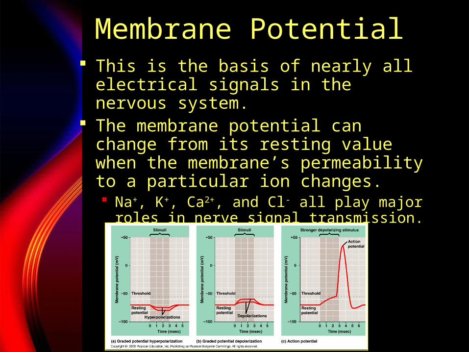
Membrane Potential This is the basis of nearly all
electrical signals in the nervous system.
The membrane potential can change from its resting value when the membrane’s permeability to a particular ion changes. Na+, K+, Ca2+, and Cl- all play major roles
in nerve signal transmission.
travismulthaupt.com
Ion Channels
When ion channels are always open, they are said to be ungated.
Gated ion channels switch open and closed to one of three kinds of stimuli: Stretch gated ion channels sense stretch. Ligand gated ion channels open and close
in response to specific signals. Voltage gated ion channels open and
close due to changes in membrane potential.

travismulthaupt.com
Ion Channel Stimulation
Stimulating gated ion channels can trigger hyperpolarization or depolarization.

travismulthaupt.com
Ion Channel Stimulation
Hyperpolarization results in an increased magnitude of membrane potential--The inside of the membrane becomes more negative.
QuickTime™ and aTIFF (Uncompressed) decompressor
are needed to see this picture.
Copyright ©2005 Pearson Education, Inc. Publishing as Pearson Benjamin Cummings. All rights reserved.

travismulthaupt.com
Ion Channel Stimulation
Depolarization reduces the magnitude of the membrane potential--the inside becomes less negative.
QuickTime™ and aTIFF (Uncompressed) decompressor
are needed to see this picture.
Copyright ©2005 Pearson Education, Inc. Publishing as Pearson Benjamin Cummings. All rights reserved.
travismulthaupt.com
Ion Channel Stimulation
In most neurons, depolarizations are graded up to a certain threshold.
Once a stimulus has reached a threshold, an action potential is triggered.
QuickTime™ and aTIFF (Uncompressed) decompressor
are needed to see this picture.
Copyright ©2005 Pearson Education, Inc. Publishing as Pearson Benjamin Cummings. All rights reserved.

travismulthaupt.com
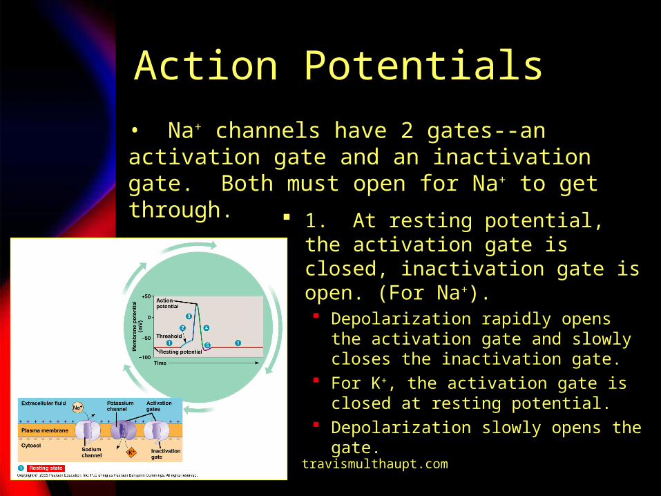
Action Potentials Action potentials are all or none.
They carry signals over a long distance along axons. They are very brief, and can thus be generated at a high frequency.
Both Na+ and K+ voltage-gated ion channels are involved in the production of an action potential.
Both open by depolarization of the membrane. Na+ opens 1st, K+ 2nd.
travismulthaupt.com
Action Potentials
1. At resting potential, the activation gate is closed, inactivation gate is open. (For Na+). Depolarization rapidly opens the
activation gate and slowly closes the inactivation gate.
For K+, the activation gate is closed at resting potential.
Depolarization slowly opens the gate.
• Na+ channels have 2 gates--an activation gate and an inactivation gate. Both must open for Na+ to get through.

travismulthaupt.com
Action Potentials
2. When a stimulus depolarizes the membrane, the activation gates open on some channels allowing some Na+ in. Na+ influx causes
depolarization opening more activation gates and so on (positive feedback).

travismulthaupt.com
Action Potentials
3. When the threshold is crossed, this positive feedback cycle brings the membrane potential close to ENa (equilibrium potential) during the rising phase.

travismulthaupt.com
Action Potentials
4. ENa is not reached: -Activation gates
close most Na+ channels halting Na+ influx.
-K+ activation gates open causing efflux of K+ decreasing the membrane potential.

travismulthaupt.com
Action Potentials
5. Undershoot occurs as too much K+ leaves the cell. Eventually, K+ activation gates close and the membrane returns to its membrane resting potential.

travismulthaupt.com
Action Potentials
The refractory period occurs when the Na+ channels remain closed and prevent the triggering of another action potential. This is what prevents the backflow of a
stimulus.

travismulthaupt.com
Action Potentials
Myelinated axons help to increase the diameter of the nerve and thereby increase the speed at which the impulse is propagated.
It also contributes to saltatory conduction which is where the action potential appears to jump from node to node along the axon.

travismulthaupt.com
Action Potentials--Synapses When action potentials reach the
ends of axons, they contribute one of 2 general mechanisms of information transfer. 1. Electrical synapse. 2. Chemical synapse.

travismulthaupt.com
Synapses--Electrical
1. Electrical synapses contain gap junctions which allow electric current to flow from cell to cell.

travismulthaupt.com
Synapses--Chemical 2. Chemical
synapses make up the vast majority of synapses. They involve the
release of chemical neurotransmitters from the pre-synaptic neurons via synaptic vesicles.
The synaptic vesicles interact with the dendrites of a post-synaptic neuron.

travismulthaupt.com
Action Potentials
The diffusion of neurotransmitter through the synaptic cleft has a change on the post-synaptic neuron, either direct or indirect.

travismulthaupt.com
Action Potentials When the neurotransmitter binds
directly to the post-synaptic membrane and opens a channel, ions can diffuse across the membrane in a process called direct synaptic transmission.

travismulthaupt.com
Action Potentials In indirect synaptic transmission, a
neurotransmitter binds to a receptor that is not part of an ion channel.
travismulthaupt.com
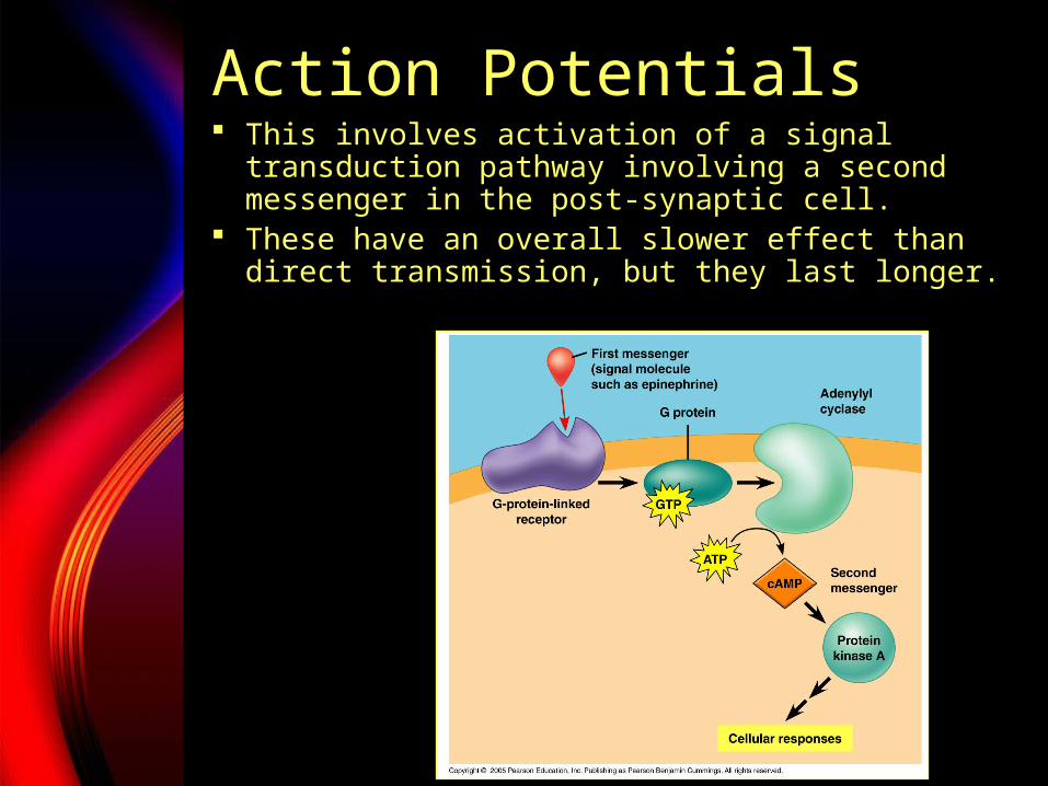
Action Potentials This involves activation of a signal
transduction pathway involving a second messenger in the post-synaptic cell.
These have an overall slower effect than direct transmission, but they last longer.