Embed Size (px)
Citation preview
TRANSVAGINAL SONOGRAPHY IN EARLY HUMAN PREGNANCY
(Transvaginale echoscopie in de vroege humane graviditeit)
PROEFSCHRIFT
TER VERKRIJGING VAN DE GRAAD VAN DOCTOR
AAN DE ERASMUS UNIVERSITEIT ROTTERDAM
OP GEZAG VAN DE RECTOR MAGNIFICUS
PROF. DR. C.J. RIJNVOS
EN VOLGENS BESLUIT VAN HET COLLEGE VAN DEKANEN.
DE OPENBARE VERDEDIGING ZAL PLAATSVINDEN OP
WOENSDAG 30 JANUARI 1991 OM 15.45 UUR
DOOR
ROELOF SCHATS
GEBOREN TE LEIDEN

PROMOTIECOMMISSIE
PROMOTOR: Prof. Jhr. Dr. J.W. Wladimiroff
CO-PROMOTOR: Dr. C.A.M. Jansen
OVERIGE LEDEN: Prof. Dr. Ir. N. Born Prof. Dr. H.P. van Geijn Prof. Dr. J. Voogd

Voor Wilma, Rachel & Tamar

PASMANS OFFSETDRUKKERJJ B.V., 's-GRAVENHAGE
CONTENTS
Page
CHAPTER 1
GENERAL INTRODUCTION AND DEFINITION
OF THE STUDY OBJECTIVES ................................. 7
1.1. Introduction ............................................. 7
1.2. Definition of the study objectives ............................ 8
1.3. References .............................................. 9
CHAPTER 2
TRANSVAGINAL SONOGRAPHY: technical and
methodological aspects ........................................ 11
2.1. Introduction ............................................. 11
2.2. Technical aspects ......................................... 11
2.3. Acceptability ............................................ 13
2.4. The examination ......................................... 14
2.5. Transvaginal ultrasound equipment used in the present study ........ 20
2.6. References ............................................. 21
CHAPTER 3
THE SAFETY OF DIAGNOSTIC ULTRASOUND
WITH PARTICULAR REFERENCE TO
THE TRANSVAGINAL APPLICATION ........................... 23
3.1. Introduction ............................................ 23
3.2. Diagnostic applications ..................................... 23
1
Page
3.3. The biological effects ...................................... 25
3.3.1. The generation of heat ................................. 25
3.3.2. Radiation force ...................................... 26
3.3.3. Cavitation .......................................... 26
3.3.4. Radiation torque ..................................... 26
3.4. Combined thermal and non-thermal effects on tissues .............. 27
3.5. Experimental results and diagnostic conditions ................... 27
3.6. The problem stated ........................................ 28
3.7. References .............................................. 33
CHAPTER 4
MORPHOLOGICAL AND BIOMETRICAL ASPECTS OF
NORMAL EARLY PREGNANCY DEVELOPMENT .................. 35
Introductory remarks .......................................... 35
4.1. Normal morphological development in early pregnancy ............ 37
4.1.1. Introduction ....................................... .37
4.1.2. Subjects and methods ................................ .37
4.1.3. Embryonic development .............................. .38
4.1.4. Sonographic findings ................................. .44
4.1.5. Discussion .......................................... 50
4.1.6. References ......................................... 52
4.2. The crown-rump length in early human pregnancy: a reappraisal. .... 55
4.2.1. Summary .......................................... 55
4.2.2. Introduction ........................................ 56
4.2.3. Subjects and methods ................................ .56
4.2.4. Results ............................................ 57
2
Page
4.2.5. Discussion .......................................... 58
4.2.6. References ......................................... 59
CHAPTER 5
DIAGNOSTIC, MORPHOLOGICAL AND THERAPEUTIC
ASPECTS OF ABNORMAL EARLY
PREGNANCY DEVELOPMENT ................................. 61
Introductory remarks .......................................... 61
5.1. Morphological aspects of abnormal embryonic development ......... 63
5.1.1. Introduction ........................................ 63
5.1.2. Subjects and methods ................................. 63
5.1.3. Results ............................................ 64
5.1.3.1. Gestational sac ................................ 65
5.1.3.2. Yolk sac ..................................... 66
5.1.3.3. Crown-rump length ............................ 68
5.1.3.4. Cardiac activity ............................... 69
5.1.3.5. Pregnancy outcome ............................. 70
5.1.4. Discussion ......................................... 70
5.1.5. References ......................................... 75
5.2. The role of transvaginal sonography in diagnosis and
management of ectopic pregnancy ............................ 79
5.2.1. Introduction ........................................ 79
5.2.2. Subjects and methods ................................. 80
5.2.3. Results ................................ · ............ 83
5.2.4. Discussion ........................................•. 92
5.2.5. References .•....................................... 96
3

Page
CHAPTER 6
FUNCTIONAL ASPECTS OF THE DEVELOPING EMBRYO .......... 101
Introductory remarks ......................................... 101
6.1. Embryonic cardiac activity: appearance and development
in early human pregnancy .................................. 103
6.1.1. Summary .......................................... 103
6.1.2. Introduction ....................................... 104
6.1.3. Subjects and methods ................................ 104
6.1.4. Results ........................................... 106
6.1.5. Discussion ......................................... 111
6.1.6. References ........................................ 113
6.2. Asynchronous appearance of embryonic cardiac activity
in multiple pregnancies following in-vitro fertilisation ............ 115
6.2.1. Introduction ....................................... 115
6.2.2. Subjects and methods ................................ 116
6.2.3. Results ........................................... 117
6.2.4. Discussion ......................................... 118
6.2.5. References ........................................ 120
6.3. Abnormal embryonic heart rate pattern in
early pregnancy associated with Down's syndrome ............... 121
6.3.1. Summary .......................................... 121
6.3.2. Introduction ....................................... 122
6.3.3. Case history ....................................... 122
6.3.4. Discussion ......................................... 124
4

Page
6.3.5. References ........................................ 125
CHAPTER 7
CONCLUSIONS ............................................ 127
SUMMARY ................................................ 129
SAMENVATTING ........................................... 132
ACKNOWLEDGEMENTS ..................................... 135
CURRICULUM VITAE. ...................................... 136
5

6
Transvaginal sonography in early human pregnancy
CHAPTER 1
GENERAL INTRODUCTION AND DEFINITION
OF THE STUDY OBJECTIVES
1.1. INTRODUCTION
Efforts to employ transvaginal sonography as a method to visualise the
internal genitalia and their contents already date from the late 1960's, when
it was used to detect the embryonic heart beat and to study the female genital
tract1• 2• It was reported that embryonic cardiac activity could be detected as
early as 46 days menstrual age or 32 days after ovulation, which is much
earlier than by then available conventional abdominal ultrasound techniques.
However, the equipment was bulky and consisted of a large device producing
A-mode images. Creation of two-dimensional images was extremely difficult
and soon the method was forgotten 3•
It was only after the introduction of the grey scale technique and of real
time imaging in the mid-seventies that transvaginal sonography became
feasible again. However, it took a considerable time before its value was
rediscovered. This was probably due more to apprehension on behalf of the
investigator than to lack of acceptance by the patient.
Although the significance of the transvaginal approach was recognised in
the early eighties, notably in the German speaking countries and the United
States, the major breakthrough came from IVF centres where it was first
employed for the puncture of follicles and later for routine monitoring of
induction of follicular growth4• 10. It soon became clear that transvaginal
sonography could give more detailed information in the field of early
embryonic development and gynaecological disease. Recently, a host of data
on the first topic has been reported9 "17. Its role in late pregnancy is mainly
determined by its accuracy in diagnosing placenta praevia18 • 19•
Transvaginal sonography, because of its superior resolution, has changed
our concepts about normal and abnormal early pregnancy. More detailed
evaluation of normal and abnormal early pregnancy first became possible
7
General introduction
with the introduction of sensitive radio-immuno assays for human chorionic
gonadotropin (hCG). Transvaginal sonography has opened the possibility of
more accurate differentiation between early failure of intra-uterine gestation
and ectopic pregnancy. More insight is needed into the synergistic role of
transvaginal sonography and serial hCG determinations in the diagnosis and
subsequent management of early pregnancy pathology.
The improved visualisation of the developing embryo permits imaging of
organs shortly after their development. Its clinical application may, therefore,
be determined by more accurate pregnancy dating and the detection of gross
structural anomalies20. It is suggested that the neural tube can be reliably
traced as early as eight weeks menstrual age21 . Accurate information on renal
size can now be obtained as early as 11 - 12 weeks menstrual age22 .
Transvaginal sonography has also been claimed to improve the procedure
of chorionic villus sampling using an automatic puncturing device23 .Its role
as a routine application has not been determined yet.
Recently, the introduction of traditional pulsed Doppler and colour
Doppler facilities in transvaginal sonographic equipment has provided the
option of studying early fetal cardiovascular dynamics24 • 25 •
1.2. DEFINITION OF THE STUDY OBJECTIVES
In this thesis attention will be focused on the role of transvaginal sonography
in early pregnancy development. The objectives of the present study were:
a. to determine the safety of diagnostic ultrasound with particular reference
to the transvaginal application,
b. to define the role of transvaginal sonography in:
(i) the visualisation of normal early pregnancy development with emphasis
on embryonic anatomy and biometrical aspects,
(ii) the recognition, diagnosis and management of abnormal development,
in particular missed abortion and ectopic pregnancy,
(iii) the functional development of early pregnancy with special reference
to the appearance and development of embryonic cardiac activity in single
and multiple pregnancies under physiological and pathophysiological
8
Transvaginal sonography in early human pregnancy
circumstances.
1.3. REFERENCES
1. Kratochwil A, Eisenhut L. Der fruheste Nachweis der fetalen Herzaktion
durch Ultraschall. Geburtshilfe Frauenheilkd 1967; 27: 176-80.
2. Kratochwil A. Ein neues vaginales Ultraschall-Schnittbild-verfahren.
Geburtshilfe Frauenheilkd 1969; 29: 379-84.
3. Bernaschek G. Vorteile der endosonographischen Diagnostik in
Gynacologie und Geburtshilfe. Geburtshilfe Frauenheilkd 1987; 47:471-6.
4. Feichtinger W, Kemeter P. Transvaginal sector scan sonography for needle
guided transvaginal follicle aspiration and other applications in
gynaecologic routine and research. Fertil Steril1986; 45: 722-5.
5. Meldrum D, Chetkowski RJ, Steingold KA, Randle D. Transvaginal
ultrasound scanning of ovarian follicles. Fertil Steri11984; 42: 803-5.
6. Popp LW, Lueken RP, Muller-Holve W, Lindemann HJ. Gynakologischen
Endosonographie: erste Erfahrungen. Ultraschal11983; 4: 92-8.
7. Schwimer SR, Lebovic J. Transvaginal pelvic ultrasonography. J
Ultrasound Med 1984; 3: 381-4.
8. Schwimer SR, Lebovic J. Transvaginal pelvic ultrasonography: accuracy
in follicle and cyst size determination. J Ultrasound Med 1985; 4: 61-2.
9. Deutinger J, Reinthaller A, Bernaschek G, Chaischek P, Fischl F, Muller
Tyl E. Comparison of the results of vaginal and abdominal follicle scans.
Arch Gynecol Obstet 1987; 241: 171-6.
10. Deutinger J, Reinthaller A, Riss P, Bernaschek G, Csaicsich P, Muller
Tyl E, Fischl F, Janisch H. Vergleich von vaginosonographischer,
transa bdomineller sonogra phischer und la paroskopischer F ollikel punktion
zur Gewinning von Eizellen in Rahmen eines In-vitro
Fertilizierungsprogrammes. Wien Med Wochenschr 1987; 137: 108-12.
11. DeCrespigny LCh, Early diagnosis of pregnancy failure with transvaginal
ultrasound. Am J Obstet Gynecol1988; 159: 408-9.
12. Fossum GT, Davajan V, Kletzky OA. Early detection of pregnancy with
transvaginal ultrasound. Fertil Steril1988; 49: 788-91.
9
General introduction
13. Endovaginal Ultrasound. S.R. Goldstein (ed) A.R. Liss, New York 1988.
14. Jain KA, Hamper UM, Sanders RC. Comparison of transvaginal and
transabdominal sonography in the detection of early pregnancy and its
complications. AJR 1988; 151: 1139-43.
15. Pennell RG, Baltarowich OH, Kurtz AB, Vilaro MM, Rifkin MD,
Needleman L, Mitchell DG, Mervis SA, Goldberg BB. Complicated first
trimester pregnancies: evaluation with endovaginal US versus
transabdominal technique. Radiology 1987; 165: 79-83.
16. Timor-Tritsch IE, Farine D, Rosen MG. A close look at early embryonic
development with the high-frequency transvaginal transducer. Am J
Obstet Gynecol 1988; 159: 676-81.
17. Transvaginal Sonography.I.E. Timor-Tritsch & S. Rottem (eds),
Heinemann Medical Books, London, 1988.
18. Farine D, Peisner DB, Timor-Tritsch IE. Placenta previa- is the
traditional diagnostic approach satisfactory ? J Clin Ultrasound 1990; 18:
328-30.
19. Leerentveld RA, Gilberts ECAM, Arnold MJCJW, Wladimiroff JW.
Placental localization by transvaginal sonography. submitted
20. Rottem S, Bronshtein M. Transvaginal sonographic diagnosis of congenital
anomalies between 9 weeks and 16 weeks, menstrual age. J Clin
Ultrasound 1990; 18: 307-14.
21. Timor-Tritsch IE, Peisner DB, Raju S. Sonoembryology: an organ
oriented approach using a high-frequency vaginal probe. J Clin
Ultrasound 1990; 18: 286-98.
22. Bronshtein M, Kushnir 0, Ben-Rafael Z, Shalev E, Nebel L, Mashiach
S, Shalev J. Transvaginal sonographic measurements of fetal kidneys in
the first trimester of pregnancy. J Clin Ultrasound 1990; 18: 299-301.
23. Popp LW, Ghirardini G. The role of transvaginal sonography in chorionic
villi sampling. J Clin Ultrasound 1990; 18: 315-22.
24. Kurjak A, Jurkovic D, Akfirevic Z, Zalud I. Transvaginal color Doppler
imaging. J Clin Ultrasound 1990; 18: 227-34.
25. Wladimiroff JW, Huisman TWA, Stewart PA. Cardiac flow velocities in
the late first trimester fetus; a transvaginal Doppler study. submitted.
10
Transvaginal sonography in early human pregnancy
CHAPTER 2
TRANSVAGINAL SONOGRAPHY:
Technical and Methodological aspects
2.1. INTRODUCTION
Ultrasonography is a technology which is gaining rapid acceptance by
clinicians. The development of transducers for vaginal use was the result of
the discontent with the information that could be obtained with the
traditional abdominal scanning techniques leading to the manufacturing of
transducers so small that they could be inserted into the vagina. This chapter
deals with the technical and methodological aspects of transvaginal
sonography and its patients' acceptability.
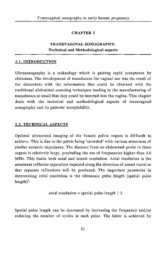
2.2. TECHNICAL ASPECTS
Optimal ultrasound imaging of the female pelvic organs is difficult to
achieve. This is due to the pelvis being "crowded" with various structures of
similar acoustic impedance. The distance from an abdominal probe to these
organs is relatively large, precluding the use of frequencies higher than 5.0
MHz. This limits both axial and lateral resolution. Axial resolution is the
minimum reflector separation required along the direction of sound travel so
that separate reflections will be produced. The important parameter in
determining axial resolution is the ultrasonic pulse length (spatial pulse
length)1:
axial resolution= spatial pulse length I 2
Spatial pulse length can be decreased by increasing the frequency and/or
reducing the number of cycles in each pulse. The latter is achieved by
11
Technical and Methodological aspects
increasing the transducer damping. When damping is reduced to a minimum
(e.g. 2- 3 cycles per pulse), the only way to improve axial resolution is to
increase frequency. Lateral resolution is the minimum separation in the
direction perpendicular to the course of the ultrasonic beam. This is the
minimal distance between two reflectors that will produce two separate
reflections when the beam is scanned across them. Lateral resolution is
directly proportional to the beam diameter or width. Increasing the frequency
reduces the beam diameter. A method of improving image quality is taking
pelvic scans while the bladder is full. By introduction of a fluid-filled space,
it is possible to observe more clearly some of the pelvic organs. However, it
1.4 ..L22.4
1-0.3 (:ll /1) -1 4 7.5 MHz 1
0.2
3
1-0.6
2__L.4
1.5' 7
6.5 MHz 1 0.3
Resolution Focal Length ._
6 Focal Reg ion (mm)
4~7 /
Lateral -1.5
2 12 Axial
5.0 MHz 1 0.4
6_Ls 1-2.0
p11 II) 5 • 1117
3.5 MHz I 0.6
0 1 2 3 4 5 6 7 8 91011121314151617181920
CENTIMETRES
Figure 2.1. A representation of the physical properties (axial and lateral resolution, focal length and focal region) of transducers with different emission frequencies.
12

Transvaginal sonography in early human pregnancy
appears that a full bladder distorts the anatomy2 and the problems discussed
above are still present, so many fine details cannot be visualised. The concept
of the vaginal transducer solved many of these problems and made it possible
to obtain high-quality images of the pelvic anatomy. The main improvement
is achieved by placing the ultrasonic probe closer to the pelvic structures. The
relevant anatomical structures for transvaginal imaging are almost always
within 9 em of the vaginal fornices. The closer the object of interest, the
higher the frequency that can be utilised and, therefore, the better the axial
resolution. It is possible to increase the transducer frequency up to 7.5 MHz,
while attenuation is still acceptable. Axial resolution is improved by 40-50%
compared with the resolutions obtained with the conventional2.5 to 5.0 MHz
transducers used in abdominal scanning. Lateral resolution is also improved
with the use of higher frequency, and stronger focusing is made possible by
the proximity of the scanning head to the pelvic structures. A schematic
representation of the physical properties of transducers with different
emission frequencies is provided in Figure 2.1 3• Transvaginal sonography is
preferably performed with an empty bladder. A full bladder may displace
most of the pelvic organs beyond the reach of the focal zone of the
transducer. Moreover it will distort pelvic anatomy.
2.3. ACCEPT ABILITY
The advantages mentioned above completely cover the objectives of the
examiner in trying to arrive at a more accurate diagnosis. Before embarking
on a new technique the question of patients' acceptability has to be answered.
With the introduction of transvaginal sonography an inquiry was conducted
into the acceptance of this new method. A total of 100 patients in which both
transabdominal and transvaginal sonography was planned was fully informed
about both sonographic techniques. It is noted that at that time most patients
do not associate ultrasound scanning with a vaginal examination. The
similarity between the vaginal transducer and a speculum was mentioned.
Only three patients experienced slight discomfort while introducing the probe
into the vagina. In general, transvaginal sonography was far preferred to
13
Technical and Methodological aspects
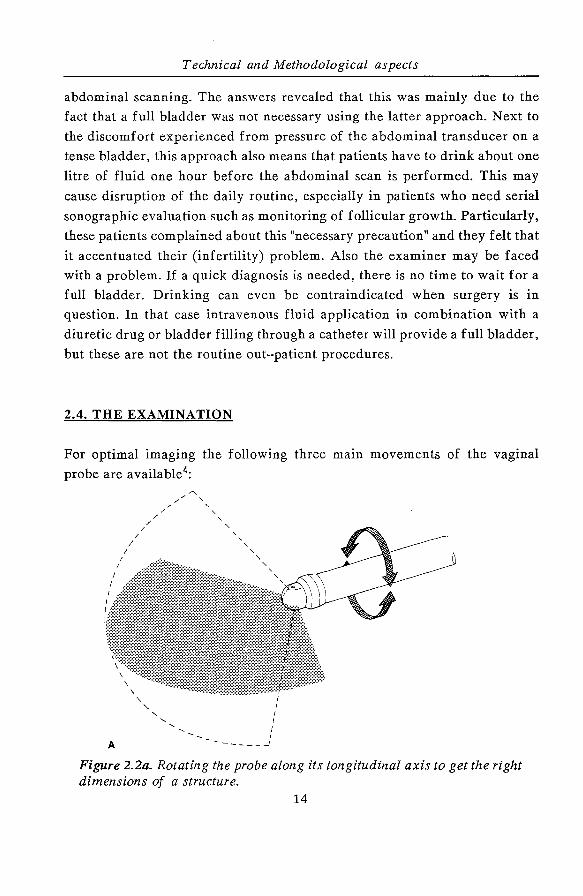
abdominal scanning. The answers revealed that this was mainly due to the
fact that a full bladder was not necessary using the latter approach. Next to
the discomfort experienced from pressure of the abdominal transducer on a
tense bladder, this approach also means that patients have to drink about one
litre of fluid one hour before the abdominal scan is performed. This may
cause disruption of the daily routine, especially in patients who need serial
sonographic evaluation such as monitoring of follicular growth. Particularly,
these patients complained about this "necessary precaution" and they felt that
it accentuated their (infertility) problem. Also the examiner may be faced
with a problem. If a quick diagnosis is needed, there is no time to wait for a
full bladder. Drinking can even be contraindicated when surgery is in
question. In that case intravenous fluid application in combination with a
diuretic drug or bladder filling through a catheter will provide a full bladder,
but these are not the routine out-patient procedures.
2.4. THE EXAMINATION
For optimal imaging the following three main movements of the vaginal
probe are available4:
I
I I·
I
A
I I
I
/ /
/
/ /
/
.... ....
' ' ' ....
' ' ' ' .... ....
I I I I I ______ _)
Figure 2.2a. Rotating the probe along its longitudinal axis to get the right dimensions of a structure.
14
Transvaginal sonography in early human pregnancy
1. rotating the handle slowly along the longitudinal axis of the probe to
change the scanning plane along a 360 degrees range (Figure 2.2a),
\ \
8
\ \
\
' ' ' ' ' '
I I
I -_;
I I
I I
I I
I
I I
I
' \ 1
I
>--------
Figure 2.2b. Angling or tilting of the probe to point at the structure of interest.
2. tilting or angling the shaft by its handle so as to point the tip of the probe
in any direction in the pelvis (Figure 2.2b ),
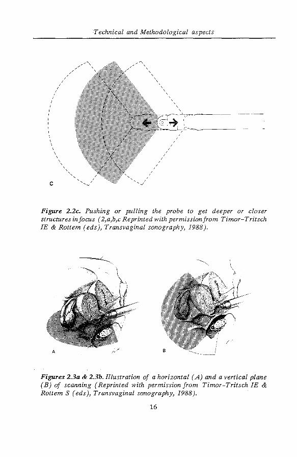
3. pushing- pulling the entire probe to bring a deeper or a closer situated
organ or structure into the focal region (Figure 2.2c).
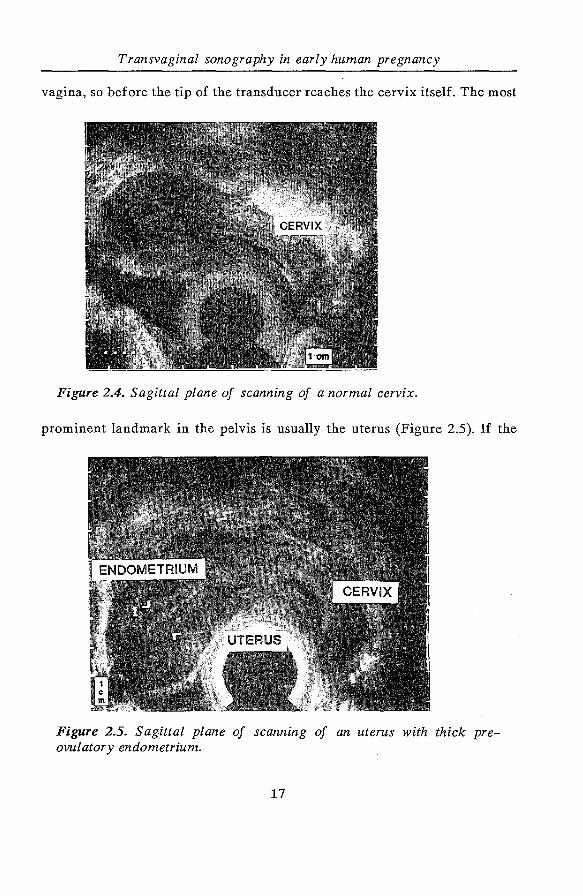
Pelvic structures should always be studied in a horizontal and vertical plane
(Figure 2.3a and 2.3b ). The first structure on the path of the vaginal probe to
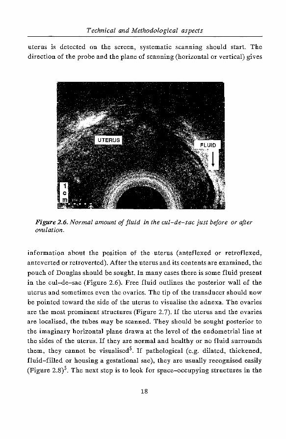
its final position for scanning the pelvis is the cervix (Figure 2.4). If the
cervix has to be scanned it may be done as the probe penetrates 3 em into the
15
I I
I
/
/
I
I I
I
I
I I
I I \
c
\ \
\ \
\
' \
'
'Technical and Methodological aspects
/-'"\
' ' ' '
/ I
I I
'-.J
\ \
\
I I
I
\
I
'
/ /
I
\
\
\
\
Figure 2.2c. Pushing or pulling the probe to get deeper or closer structures in focus ( 2,a,b,c Reprinted with permission from Timor-Tritsch IE & Rottem (eds), Transvaginal sonography, 1988).
Figures 2.3a & 2.3b. Illustration of a horizontal (A) and a vertical plane (B) of scanning (Reprinted with permission from Timor-Tritsch IE & Rottem S ( eds ), Transvaginal sonography, 1988 ).
16
Transvaginal sonography in early human pregnancy
vagina, so before the tip of the transducer reaches the cervix itself. The most
Figure 2.4. Sagittal plane of scanning of a normal cervix.
prominent landmark in the pelvis is usually the uterus (Figure 2.5). If the
Figure 2.5. Sagittal plane of scanning of an uterus with thick preovulatory endometrium.
17
Technical and Methodological aspects
uterus is detected on the screen, systematic scanning should start. The
direction of the probe and the plane of scanning (horizontal or vertical) gives
Figure 2.6. Normal amount of fluid in the cul-de-sac just before or after ovulation.
information about the position of the uterus (anteflexed or retroflexed,
anteverted or retroverted). After the uterus and its contents are examined, the
pouch of Douglas should be sought. In many cases there is some fluid present
in the cul-de-sac (Figure 2.6). Free fluid outlines the posterior wall of the
uterus and sometimes even the ovaries. The tip of the transducer should now
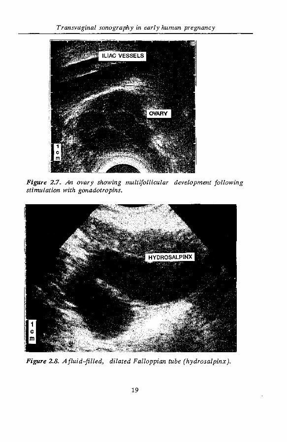
be pointed toward the side of the uterus to visualise the adnexa. The ovaries
are the most prominent structures (Figure 2. 7). If the uterus and the ovaries
are localised, the tubes may be scanned. They should be sought posterior to
the imaginary horizontal plane drawn at the level of the endometrial line at
the sides of the uterus. If they are normal and healthy or no fluid surrounds
them, they cannot be visualised 5• If pathological (e.g. dilated, thickened,
fluid-filled or housing a gestational sac), they are usually recognised easily
(Figure 2.8)5. The next step is to look for space-occupying structures in the
18
Transvaginal sonography in early human pregnancy
Figure 2.7. An ovary showing multifollicular development following stimulation with gonadotropins.
Figure 2.8. Afluid-filled, dilated Falloppian tube (hydrosalpinx).
19
Technical and Methodological aspects
pelvis. The entire pelvis must be systematically "covered" by horizontal and
vertical sonographic planes, and a thorough search should be made along
them. The effective focal zone of high frequency transducers ends at 7 - 8
em. One may see structures further away than 8 em, but they appear "blurred"
and are irregularly outlined. A fairly large mass may be missed by vaginal
scanning if, because of its size, it does extend out of the reach of the vaginal
transducer. If a pelvic mass has to be ruled out or is suspected, an additional
transabdominal scan should be performed. Some special manoeuvres can
improve the diagnostic potential of the technique6: The examiner may place
his/her free hand on the lower abdomen to bring pelvic structures closer to
the tip of the probe, as in a regular bimanual pelvic-abdominal examination.
In case of pelvic pain, localisation of the point of maximal intensity may be
attempted under direct vision and gentle pressure with the tip of the
transducer. Diagnosis of pelvic adhesions becomes possible by the "sliding
organs sign": the transducer tip is pointed at the uterus, ovaries, or any pelvic
finding (e.g. ovarian mass, tuba-ovarian complex), and a gentle push-pull
movement of several centimetres is started. If no adhesions are present, the
organs will move freely in the pelvis. This displacement of organs is
perceived on the screen as a sliding movement. One may, for instance,
observe the free sliding of an ovarian mass over the lateral pelvic wall, which
of course is static. In the case of a tuba-ovarian complex, the relative
locations of the uterus, tube, and ovary will not change under the pushing
motion of the probe, because of extensive adhesions preventing normal and
physiological sliding of these organs.
2.5. TRANSVAGINAL ULTRASOUND EQUIPMENT
USED IN THE PRESENT STUDY
1. Diasonics DRF 400 equipped with a 5.0 and a 7.5 MHz mechanical sector
scan transducer. Ispta = 2.0 mW/cm2 at a depth of 4.5 em for both
frequencies.
20
Transvaginal sonography in early human pregnancy
2. Toshiba SAL 77B equipped with a 5.0 MHz electronic convex sector scan
transducer. Ispta = 8.0 mW/cm2 at a depth of 4.5 em.
3. Kretz Com bison 310 equipped with a 5.0 and a 7.5 MHz mechanical sector
scan transducer. Ispta = 7.8 mW/cm2, depth not specified for both
frequencies.
The ultrasound equipment mentioned under points 1 and 2 was used in the
morphological, biometrical and dynamic studies, while the last apparatus was
used in a few patients described in chapter 5.2. as well as for producing some
photographic illustrations.
2.6. REFERENCES
1. Nyborg WL, Biophysical mechanisms of ultrasound. In: Essentials of
medical ultrasound. M.H. Repacholi & D.A. Benwell (eds), Humana Press,
Clifton 1982.
2. Timor-Tritsch IE, Bar-Yam Y, Elgali S, Rottem S. The technique of
transvaginal sonography with the use of a 6.5 MHz probe. Am J Obstet
Gynecol 1988; 158: 1019-24.
3. Thaler I, Manor D. Transvaginal imaging: applied physical principles and
terms. J Clin Ultrasound 1990; 18: 235-8.
4. Thaler I, Bruck A, Rottem S. The vaginal probe- physical considerations.
In: Transvaginal Sonography. I.E. Timor-Tritsch & S. Rottem (eds),
Heinemann Medical Books, London, 1988: 1-13.
5. Timor-Tritsch IE, Rottem S. Transvaginal ultrasonographic study of the
fallopian tube. Obstet Gynecol1987; 70: 424-8.
6. Timor-Tritsch IE, Rottem S, Elgali S. How transvaginal sonography is
done. In: Transvaginal Sonography. I.E. Timor-Tritsch & S. Rottem (eds),
Heinemann Medical Books, London, 1988: 15-25.
21

22
Transvaginal sonography in early human pregnancy
CHAPTER 3
THE SAFETY OF DIAGNOSTIC ULTRASOUND
WITH PARTICULAR REFERENCE TO
THE TRANSVAGINAL APPLICATION
3.1. INTRODUCTION
Ultrasound is a mechanical wave phenomenon: by definition it is made up of
sound waves with a frequency beyond the upper physiological auditory limit
(20.000 Hz). Generally, medical ultrasound is performed with frequencies
higher than 1.0 MHz. During propagation of the ultrasound pulse the
structures of the tissues are made to vibrate on both the molecular and the
cellular level. In addition to density (specific mass), the elastic properties of
the tissues determine the propagation of ultrasound and its biological effects.
These effects differ from those caused by ionising radiation (X-rays, gamnia
rays etc.). Ultrasound is neither ionising nor are its effects cumulative. The
fact that it lacks mutagenic effects means that the cells suffer from reversible
damage or are destroyed. Destruction is directly related to the radiation dose.
Hence the biological effect of ultrasound may be viewed as an "all or none"
phenomenon 1. In general, the physical phenomena induced by ultrasound may
be separated into thermal (heat generation) and non-thermal effects (radiation
force, cavitation and microstreaming). A direct relationship between sound
intensity and biological effects is not always present, moreover an increase in
frequency does not always influence all effects uniformly. Consequently it is
important to study the threshold values for the individual biological effects
in order to draw up potential guidelines for safe use.
3.2. DIAGNOSTIC APPLICATIONS
The application of ultrasound in medical diagnosis is generally called
echography. This comprises the imaging of anatomical structures and tissues
23
The safety of diagnostic transvaginal ultrasound
on a screen ( echoscopy) as well as detecting the movement of structures and
measuring flow in blood vessels (Doppler technique). A wide variety of
equipment has bef?n developed for both fields of application and various
techniques are available for ultrasound imaging:
A-Mode: a one-dimensional image of the echo magnitude as a function of
depth,
M-Mode: a one-dimensional image of the localisations of structures as a
function of time,
B-Mode: a two-dimensional image of a section of the body through which the
magnitude of the echo is transformed into luminosity on the screen.
There are two types of B mode equipment:
1. Static B Scanners: the transducer is manually moved over the patient's
body. The image on the monitor lasts one or more
seconds. These scanners have been used in the early
years of ultrasound in medicine and they are completely
replaced by real time scanners,
2. Real-time B scanners: the image is obtained in a short time, so that one to
50 images per second are produced and the
echoscopist is continuously able to follow all
changes caused by movements of the transducer or
patient (fetus, embryo).
Real-time B scanners are also of two types based on the way in which the
movement of the sound beam is obtained:
A. Mechanical real-time scanners:
the transducer makes a tilting movement or is rotated by means of an electric
motor. These scanners provide a sector image,
24

Transvaginal sonography in early human pregnancy
B. Electronic real-time scanners:
they operate according to the "linear array" or "phased array" principle. The
transducer consists of a series of small elements transmitting and receiving,
either one after the other or in subgroups (linear array) or phased (phased
array), resulting in a rectangular and a sector image respectively.
The Doppler apparatus for velocity measurements can be either of the
"continuous wave" (CW) or the "pulsed Doppler" (PD) type of equipment. The
latter can also be used for two-dimensional velocity imaging in the colour
Doppler mode. We did not use Doppler equipment in our study. Therefore
we will not discuss the specific fields of application.
3.3. THE BIOLOGICAL EFFECTS: physical aspects.
3.3.1. The generation of heat
The conversion of ultrasound energy into heat due to absorption may lead to
a rise in temperature in the tissues. The absorption increases with a rise in
frequency as well as a rise in ultrasound pressure (=intensity). This ratio is as
follows 2:
8W = 0.23 a (f) I,
where 8 W =local generation of heat per second in a volume of 1 em 3
(J.cm· 3.s),
and
a (f)= absorption coefficient in decibels per centimetre (dB/em); in
most tissues the coefficient is proportional to the frequency,
I= local average intensity per time unit in W/cm 2•
Biological effects may be expected when the temperature in the tissues
exceeds 42 ° C. Absorption in various tissues differs considerably, so that heat
generation resulting from a specific intensity is also affected by the location
and direction of the ultrasound beam. Since the intensity decreases
25
The safety of diagnostic transvaginal ultrasound
exponentially with penetration due to absorption for non-focusing
transducers, the heat effect will be greatest in those tissues which lie closest
to the transducer. Finally, the rise in temperature due to absorption also
depends on the blood flow and on the property of the tissues to conduct heat
(heat release). Experimental studies revealed that tissues in mammals exposed
to diagnostic ultrasound intensity levels show no significant rise in
temperature3• 4.
3.3.2. Radiation force
Ultrasound generates a force on the surface between two tissues which may
produce a biological effect. This radiation force may also cause effects in cell
suspensions such as stasis and aggregation 5• These phenomena usually occur
when standing waves are generated, for example at the point of transition
from soft tissue to bone.
3.3.3. Cavitation
Cavitation may occur during the use of a continuous wave (=CW) or pulsed
wave (=PW). It can occur only if microscopically small gas bubbles are present
in the tissue or blood. With CW ultrasound these bubbles may enlarge as a
result of the radiation to a size that will cause resonance (stable cavitation).
However, when pulsed ultrasound is used with a relatively high intensity the
gas bubble may implode, resulting in locally very high temperatures
(formation of radicals) and pressures (shock wave). This phenomenon is called
transient cavitation. Some authors are of the opinion that, if the peak
intensity of the ultrasound is less than 1500 WI em 2 transient cavitation does
not occur in-vivo6• 7• However, with a continuous wave intensity of 150 to
500 mW/cm2 stable cavitation may occur in-vivo 5• 8•
3.3.4. Radiation torque
A mechanical effect that causes microstreaming is the so-called radiation
torque, which produces a rotating movement of cells and even intracellular
structures. Consequently microstreaming will be present around the cells and
the cell wall may well be damaged by hydrodynamic shear stresses. There are
indications that an oblique sound beam when directed at blood vessel walls
26
Transvaginal sonography in early human pregnancy
may produce microstreaming. A third cause of microstreaming is stable
cavitation (also see 3.3.3.). High velocity gradients are established around the
oscillating gas bubbles which may damage structures like blood cells. No
direct evidence exists that microstreaming is of any importance in the range
of intensities used in diagnostic ultrasound for in-vivo diagnosis9.
3.4. COMBINED THERMAL AND NON-THERMAL
EFFECTS ON TISSUES
Krizian has shown that the processes mentioned above when combined may
influence each other10. When the temperature is raised, red blood cells are
more prone to dal;Ilage due to mechanical forces. In high intensity non
homogeneous sound fields the chance of red blood cells being damaged from
the combined effects of elevated temperature due to absorption and
mechanical forces provoked by the presence of pressure gradients, is much
higher than when only one of these two processes influences the cells.
3.5. EXPERIMENTAL RESULTS AND DIAGNOSTIC CONDITIONS
The effects of ultrasound on biological systems have been described in
approximately 800 publications. The American Institute of Ultrasound in
Medicine (AlUM) Bioeffects Committee reviews periodically the literature
concerning this topic and formulates guidelines for safe use11 . The following
facts should be taken into account:
A. The occurrence of a marked rise in temperature, stable cavitation,
radiation force and (micro-) streaming has been observed only when
continuous wave (Doppler) ultrasound of high intensities (~ 100
mW/cm2) 11 or pulsed ultrasound of long pulse lengths(~ 1 microsecond)
with high intensities(~ 100 mW/cm2) was used11 . Thus it is reassuring to
realise that the pulsed ultrasound used in clinical situations is emitted in
pulses of one microsecond followed by a pause of one millisecond. In an
27
The safety of diagnostic transvaginal ultrasound
examination lasting 15 minutes, the tissues are effectively exposed to
ultrasound to a maximum of only one second. Therefore, medical
diagnostic equipment should conform to the requirement that the intensity
(Ispta) emitted does not exceed 100 mW/cm2 as is recommended in a report
of a committee of the Dutch Health Council12 .
B. The biological effects of ultrasound depend on local conditions during
insonation. Certain cell types used in in-vitro experiments may be
specifically sensitive to certain ultrasound effects.
C. Extrapolation of the results of in-vitro studies to cover clinical results
possesses questionable validity. In-vitro studies may produce various
artificial situations, such as very high intensities in the tissues closest to
the transducer due to non-uniformity of the sound beam, and the
development of static waves and focusing due to ultrasound reflection
from curved structures.
D. A major problem arises if clinical studies are compared with animal
studies. In the mouse an ultrasound intensity of 20 m WI em 2 at the level of
the maternal abdomen results in a virtually unchanged intensity at the
level of the fetus. In humans, by contrast, a threefold reduction in
intensity takes place between the maternal abdominal wall and the fetus 12.
In addition, the animal will lie absolutely still during the examination and
the fetus will be fully insonated, whereas in humans the transducer is
usually repositioned repeatedly.
3.6. THE PROBLEM STATED
The introduction of diagnostic ultrasound may be viewed as one of the most
important developments in medical diagnosis during the past· 25 years.
Extensive studies have been carried out to evaluate the potential biological
effects of ultrasound. Extrapolation of the results of animal studies or tissue
culture experiments into the possible effects of echoscopy in pregnancy
28
Transvaginal sonography in early human pregnancy
presents a problem. In most publications no obvious evidence of any harmful
effect of diagnostic imaging in pregnancy is provided, but it is still premature
to state that it is harmless. There are no data available on possible deleterious
effects of the transvaginal application of diagnostic ultrasound in early
pregnancy. Before the start of our project we compared abdominal and
vaginal real-time B mode transducers for the factors influencing the sound
intensity (I) at the level of the embryo. The following factors have to be taken
into account:
1. I t , I t , I t , duration of the pulse and the pulse frequency. sa a spa sp p
2. Distance between transducer and embryo, frequency of the transducer
(MHz) and the absorption of ultrasound in the different tissues.
Ad 1. There are, as already has been mentioned, different ultrasound systems
with their own characteristics. The acoustic dosage is for example
dependent on which area in a certain period of time is covered.
Furthermore, intensity is determined by the pulse frequency and pulse
duration. The intensity will have a peak value during the pulse, while
the mean value in time is much lower. Three definitions are often used
to indicate and define characteristics of transducers:
SATA Intensity: Temporal mean intensity averaged over the beam
cross-sectional area in Watt/ em 2• This intensity is
often reported by manufacturers (SATA: ~patial
Average, Temporal Average).
SPTA Intensity: The local maximal and temporal average intensity in
Watt/cm2 (SPTA: ~atial f.eak, Temporal Average).
SPTP Intensity: The local maximal and tern poral peak intensity in Watt/
em 2• This intensity is of course higher than the SPT A
or SATA value (SPTP: ~patial f.eak, Temporal f.eak).
29
The safety of diagnostic transvaginal ultrasound
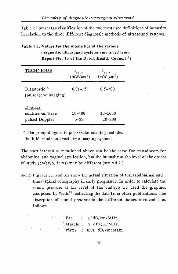
Table 2.I presents a classification of the two most used definitions of intensity
in relation to the three different diagnostic methods of ultrasound systems.
Table 2.1. Values for the intensities of the various
diagnostic ultrasound systems (modified from
Report No. 13 of the Dutch Health Counci112)
TECHNIQUE
Diagnostic * (pulse/ echo imaging)
Doppler
continuous wave
pulsed Doppler
Isata (mW/cm2)
0.01-15
10-400
5-32
0.5-200
10-1000
20-290
* The group diagnostic pulse/ echo imaging includes
both M-mode and real-time imaging systems.
The start intensities mentioned above can be the same for transducers for
abdominal and vaginal application, but the intensity at the level of the object
of study (embryo, fetus) may be different (see Ad 2.).
Ad 2. Figures 3.1 and 3.2 show the actual situation of transabdominal and
transvaginal echography in early pregnancy. In order to calculate the
sound pressure at the level of the embryo we used the graphics
composed by Wells13 , collecting the data from other publications. The
absorption of sound pressure in the different tissues involved is as
follows:
Pat
Muscle
Water
1 ·dB/ cm/MHz,
2 dB/cm/MHz,
0.01 dB/cm/MHz.
30
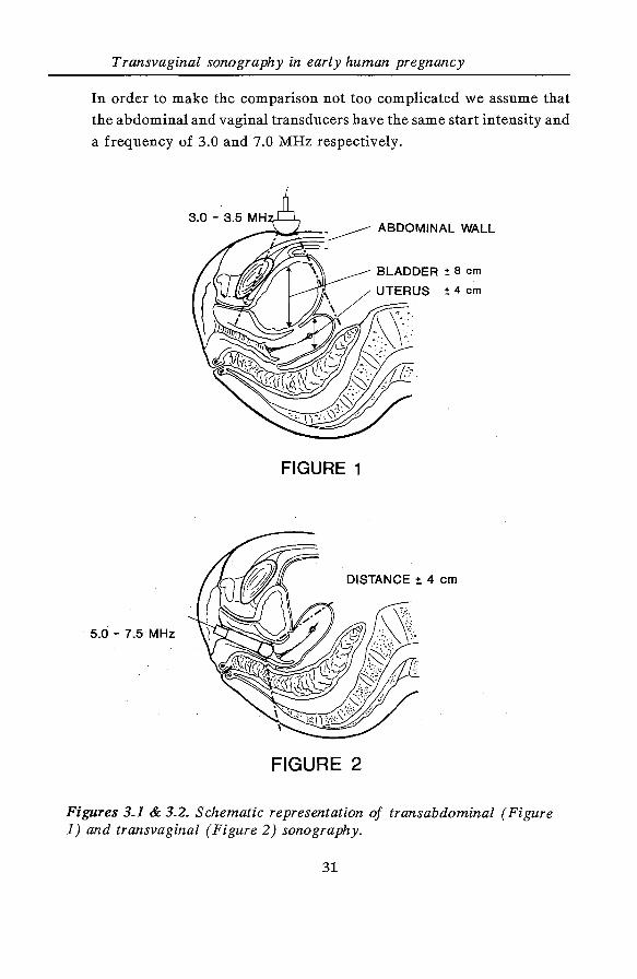
Transvaginal sonography in early human pregnancy
In order to make the comparison not too complicated we assume that
the abdominal and vaginal transducers have the same start intensity and
a frequency of 3.0 and 7.0 MHz respectively.
BLADDER :!: 8 em
:!: 4 em
FIGURE 1
DISTANCE :t 4 em
5.0- 7.5 MHz
FIGURE 2
Figures 3.1 & 3.2. Schematic representation of transabdominal (Figure 1) and transvaginal (Figure 2) sonography.
31
The safety of diagnostic transvaginal ultrasound
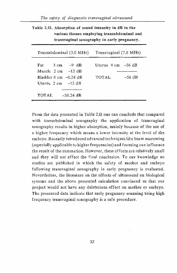
Table 2.11. Absorption of sound intensity in dB in the
various tissues employing transabdominal and
transvaginal sonography in early pregnancy.
Transabdominal (3.0 MHz)
Fat 3 em -9 dB
Muscle 2 em -12 dB
Bladder 8 em -0.24 dB
Uterus 2 em -12 dB
TOTAL -30.24 dB
Transvaginal (7.0 MHz)
Uterus 4 em -56 dB
TOTAL -56 dB
From the data presented in Table 2.II one can conclude that compared
with transabdominal sonography the application of transvaginal
sonography results in higher absorption, mainly because of the use of
a higher frequency which means a lower intensity at the level of the
embryo. Recently introduced advanced techniques like beam narrowing
(especially applicable to higher frequencies) and focusing can influence
the result of the summation. However, these effects are relatively small
and they will not affect the final conclusion. To our knowledge no
studies are published in which the safety of mother and embryo
following transvaginal sonography in early pregnancy is evaluated.
Nevertheless, the literature on the effects of ultrasound on biological
systems and the above presented calculation convinced us that our
project would not have any deleterious effect on mother or embryo.
The presented data indicate that early pregnancy scanning using high
frequency transvaginal sonography is a safe procedure.
32
Transvaginal sonography in early human pregnancy
3.7. REFERENCES
1. Williams AR. Ultrasound: biological effects and potential hazards.
Academic Press, London, 1983.
2. Nyborg WL, Ziskin MC. Biological effects of ultrasound. In: Clinics in
Diagnostic Ultrasound. W.L. Nyborg & M.C. Ziskin (eds), Churchill
Livingstone, New York, 1985.
3. Lele PP. An overview of ultrasound theory: measurement, medical
applications and biological effects. U.S. Department of health and human
service, 1982; 48 (HHS publication FDA 82 - 8190).
4. Nyborg WL et al. Biological effects of ultrasound: mechanisms and clinical
implications, 1983, NCRP report No 74.
5. Dyson M, Woodward B, Pond JB. Flow of red blood cells stopped by
ultrasound. Nature 1979; 232: 572-3.
6. Lele PP. Cavitation and its effects on organized mammalian tissues- a
summary. In: Ultrasound: its applications in Medicine and Biology. C.J.
Fry (ed), Elsevier, Amsterdam, 1978; 178: 737-41.
7. Gros DR, Miller DL, Williams AR. A search for ultrasonic cavitation
within the canine cardiovascular system. Ultrasound Med Biol 1985; 11:
85-97.
8. TerHaar G, Daniels S, Eastaugh K, Hill CR. Ultrasonically induced
cavitation in-vivo. Br J Cancer 1982; 45: 151-5.
9. Kremkau FW. Biological effects and possible hazards. In: Ultrasound in
Obstetrics and Gynaecology; recent advances. S. Campbell (ed), Saunders
Company Ltd, London, 1983.
10. Krizian JE, Williams AR, Biological membrane rupture and a phase
transition model. Nature New Biol1973; 246: 121.
11. American Institute of Ultrasound in Medicine Bioeffects Committee.
Bioeffects considerations for the safety of diagnostic ultrasound. J
Ultrasound Med 1988; 7: Suppl, whole issue.
12. Ultrasound in Medicine, Recommendations on the subject of Ultrasound
by a committee of the Health Council of the Netherlands, The Hague,
1986; Report No. 13.
13. Wells PNT. Biomedical Ultrasonics, Academic Press, London, 1977.
33

34
Transvaginal sonography in early human pregnancy
CHAPTER 4
MORPHOLOGICAL AND BIOMETRICAL ASPECTS OF
NORMAL EARLY PREGNANCY DEVELOPMENT
INTRODUCTORY REMARKS
Information on early morphological development of the embryo has been
based on transabdominal ultrasound studies. Transvaginal sonography can be
used earlier and a more detailed analysis of the embryo and its surroundings
is possible. This is of clinical importance when a pathological development of
pregnancy is suspected. This chapter consists of two parts. In the first part
( 4.1.) the role of transvaginal sonography in visualising embryonic structures
as they develop during the first six weeks following conception (eight weeks
menstrual age) will be determined. This will be preceded by an overview of
what is known about early human embryonic morphology from animal and
human post-mortem specimens. In the second part (4.2.), there will be a
reappraisal of the embryonic crown-rump length measurement in early
pregnancy using transvaginal sonography.
35

36
Transvaginal sonography in early human pregnancy
4.1. NORMAL MORPHOLOGICAL DEVELOPMENT
IN EARLY PREGNANCY
4.1.1. INTRODUCTION
Transabdominal sonography has been used as an effective diagnostic and
research tool in obstetrics. Its application is mainly in the second and third
trimester of pregnancy. Its use in the first trimester is relatively limited and
mainly diagnostic in nature. The introduction of the vaginal transducer which
allows higher emission frequencies, leading to a better resolution has opened
new possibilities in the study of early gestation. The present study describes
in detail the information which can be gathered on embryonic morphology
using transvaginal sonography in the first six weeks following conception.
4.1.2. SUBJECTS AND METHODS
Singleton and mult:jple pregnancies of 62 women (83 embryos) were studied.
All patients became pregnant by means of IVF treatment; the duration of
pregnancy was therefore exactly known. Twenty-three patients (28 gestational
sacs, 28 embryos) were scanned from day 17 following follicle aspiration (FA)
at two or three days intervals until day 23 after FA, then every day until the
detection of embryonic cardiac activity. The other 39 patients (55 embryos)
were examined daily from day 25 after FA until the appearance of embryonic
cardiac activity, thereafter they were followed at intervals of two or three
days until 42 days after FA. All examinations were performed using the
Diasonics DRF 400 and the Toshiba SAL 77B, the former equipped with a 5.0
and a 7.5 MHz mechanical sector scan transducers and the latter with a 5.0
MHz electronic sector scan probe.
37
Aspects of normal early pregnancy development
4.1.3. EMBRYONIC DEVELOPMENT
Before providing a detailed description of the morphological features of early
pregnancy as visualised by transvaginal sonography an overview of the
highlights of embryonic development during the first six weeks following
conception will be presented 1•
WEEKS 1 and 2: fertilisation and implantation
Spermatozoa deposited in the vagina pass through the cervical canal, the
uterine cavity and along the Falloppian tube to the ampulla, where
fertilisation usually occurs. As the zygote passes down the Falloppian tube,
B.
--endometrial stroma
cytotrophoblast
embryonic
~endoderm
uterine cavity
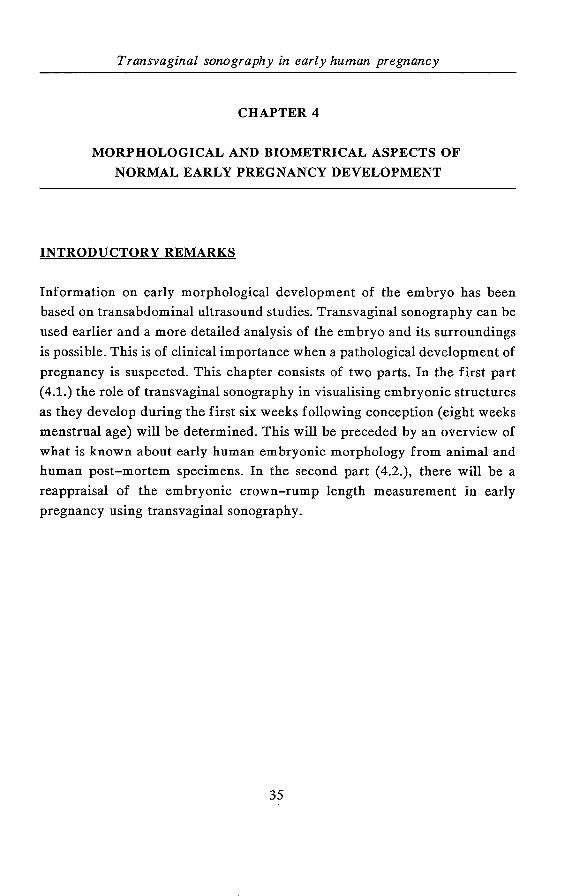
Figure 4.1.1. Blastocysts containing the embryoblast, a blastocyst cavity and the trophoblast (Reprinted with permission from Moore KL ( ed ), The developing human, 1973).
38
Transvaginal sonography in early human pregnancy
it undergoes cleavage in a number of small blastomeres. About 3 to 5 days
after fertilisation, a ball of approximately 16 blastomeres, called the morula,
enters the uterus. Soon a cavity is formed within the morula converting it into
a blastocyst consisting of: 1) an inner cell mass (embryoblast), which gives
rise to the embryo; 2) a blastocyst cavity and 3) an outer layer of cells
(trophoblast) which encloses the inner cell mass and blastocyst (Figure 4.1.1).
Implantation of the blastocyst begins at the end of the first week and ends
during the second week. The entire process may be summarised as follows:
1. Zona pellucida disappears (days 4 - 5), 2. Blastocyst attaches to endometrial epithelium (day 6),
3. Trophoblast erodes epithelium and endometrial stroma (day 7),
4. Trophoblast differentiates into cytotrophoblastic and
syncytiotrophoblastic layers (days 7- 8),
5. Lacunae appear in syncytiotrophoblast (days 8- 9),
6. Blastocyst sinks beneath surface of the endometrial epithelium (days 9 -
10),
7. Lacunar networks are formed by fusion of adjacent lacunae (days 10 -11),
8. Trophoblast invades endometrial sinusoids, allowing maternal blood to seep
into the lacunar networks and establishes the uteroplacental circulation
(days 11- 12),
9. Endometrial epithelium completely re-forms over the implanted
blastocyst (days 12 - 13),
10.Marked decidual reaction occurs in the endometrium around the
conceptus.
References to days are given only as guidelines and thus are approximations
of the truth because early stages of implantation of the human blastocyst have
not been observed. Most knowledge about early implantation is based on
studies in the Rhesus monkey, but the process is thought to be essentially
similar in man 2•
WEEK 3:
During this period rapid embryonic development coincides with the first
39
Aspects of normal early pregnancy development
missed menstrual period. Major changes occur as the bilaminar embryonic
disc is converted into a trilaminar embryo composed of three primary germ
layers. The most important features of the third week are:
1. Formation of the primitive streak and the intra-embryonic mesoderm,
2. Notochord formation,
3. Neural tube formation,
4. Somite formation: division of the paraxial mesoderm into pairs of somites
starts cranially by the end of the third week.
5. Intra-embryonic coelom formation: the coelomic spaces subsequently
coalesce to form a single horseshoe-shaped cavity which eventually gives
rise to body cavities.
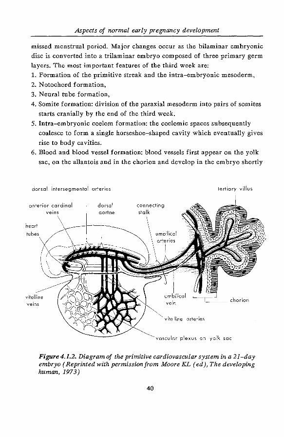
6. Blood and blood vessel formation: blood vessels first appear on the yolk
sac, on the allantois and in the chorion and develop in the embryo shortly
dorsal intersegmental arteries
anterior cardinal
heart
tubes
vitelline
veins
dorsal
aortae
connecting
stalk
umbilical
vein
vitelline arteries
vascular plexus on yolk sac
Figure 4.1.2. Diagram of the primitive cardiovascular system in a 21-day embryo (Reprinted with permission from Moore KL ( ed ), The developing human, 1973)
40
Transvaginal sonography in early human pregnancy
thereafter. At the end of the third week, the heart is represented by paired
heart tubes which are joined to blood vessels in the embryo and in the
extra-embryonic membranes (Figure 4.1.2).
7. Villi formation: primary villi become secondary villi as they acquire
mesenchymal cores. Before the end of the third week, capillaries develop
in the villi; this transforms them into tertiary villi.
WEEK4:
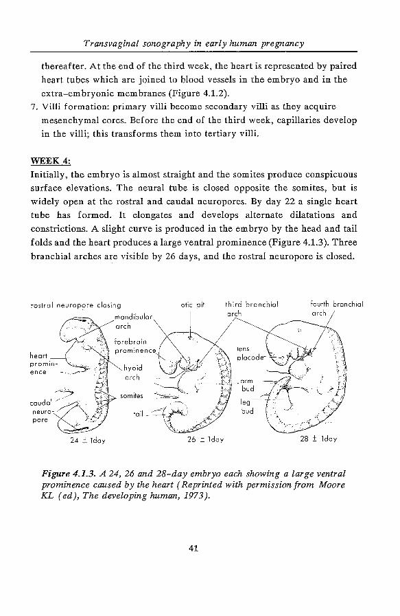
Initially, the embryo is almost straight and the somites produce conspicuous
surface elevations. The neural tube is closed opposite the somites, but is
widely open at the rostral and caudal neuropores. By day 22 a single heart
tube has formed. It elongates and develops alternate dilatations and
constrictions. A slight curve is produced in the embryo by the head and tail
folds and the heart produces a large ventral prominence (Figure 4.1.3). Three
branchial arches are visible by 26 days, and the rostral neuropore is closed.
so mites
tail
24 ±!day 26 ± lday
third branchial arch
lens placode
fourth branchial arch
28 ± Jday
Figure 4.1.3. A 24, 26 and 28-day embryo each showing a large ventral prominence caused by the heart (Reprinted with permission from Moore KL (ed), The developing human, 1973).
41
Aspects of normal early pregnancy development
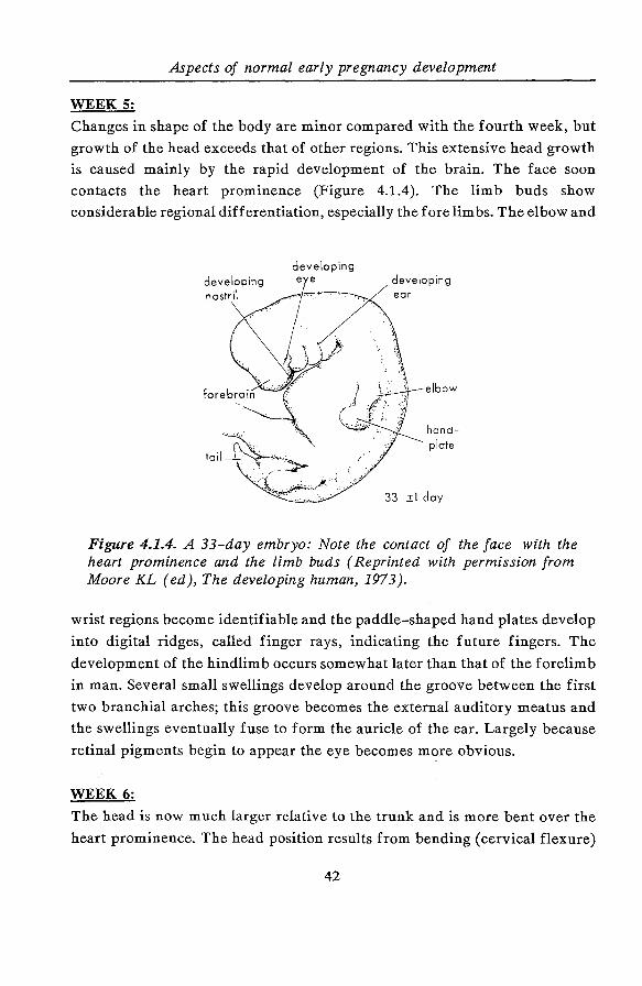
WEEK 5:
Changes in shape of the body are minor compared with the fourth week, but
growth of the head exceeds that of other regions. This extensive head growth
is caused mainly by the rapid development of the brain. The face soon
contacts the heart prominence (Figure 4.1.4). The limb buds show
considerable regional differentiation, especially the forelimbs. The elbow and
33 ±1 day
Figure 4.1.4. A 33-day embryo: Note the contact of the face with the heart prominence and the limb buds (Reprinted with permission from Moore KL (ed), The developing human, 1973).
wrist regions become identifiable and the paddle-shaped hand plates develop
into digital ridges, called finger rays, indicating the future fingers. The
development of the hindlimb occurs somewhat later than that of the forelimb
in man. Several small swellings develop around the groove between the first
two branchial arches; this groove becomes the external auditory meatus and
the swellings eventually fuse to form the auricle of the ear. Largely because
retinal pigments begin to appear the eye becomes more obvious.
WEEK 6:
The head is now much larger relative to the trunk and is more bent over the
heart prominence. The head position results from bending (cervical flexure)
42
Transvaginal sonography in early human pregnancy
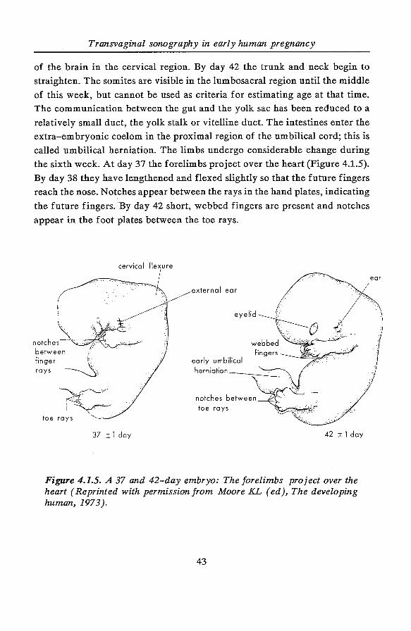
of the brain in the cervical region. By day 42 the trunk and neck begin to
straighten. The somites are visible in the lumbosacral region until the middle
of this week, but cannot be used as criteria for estimating age at that time.
The communication between the gut and the yolk sac has been reduced to a
relatively small duct, the yolk stalk or vitelline duct. The intestines enter the
extra-embryonic coelom in the proximal region of the umbilical cord; this is
called umbilical herniation. The limbs undergo considerable change during
the sixth week. At day 37 the forelimbs project over the heart (Figure 4.1.5).
By day 38 they have lengthened and flexed slightly so that the future fingers
reach the nose. Not.ches appear between the rays in the hand plates, indicating
the future fingers. By day 42 short, webbed fingers are present and notches
appear in the foot plates between the toe rays.
cervical fle~x· u .. re
' •.
( . . . ~
.. . . ·:-.-~
·.· .. ,_
~ '.' .
external ear
oo"h"'"'~t -between 1 ,:. ' •
eyelid
~i:~:r ~: early umbilical herniation ___ _
toe rays
37 _± 1 day 42 ± 1 day
Figure 4.1.5. A 37 and 42-day embryo: The forelimbs project over the heart (Reprinted with permission from Moore KL ( ed), The developing human, 1973).
43
ear
Aspects of normal early pregnancy development
4.1.4. SONOGRAPHIC FINDINGS
It will be obvious that no sonographic data of the first two weeks of
embryonic development are available. The resolution properties of ultrasound
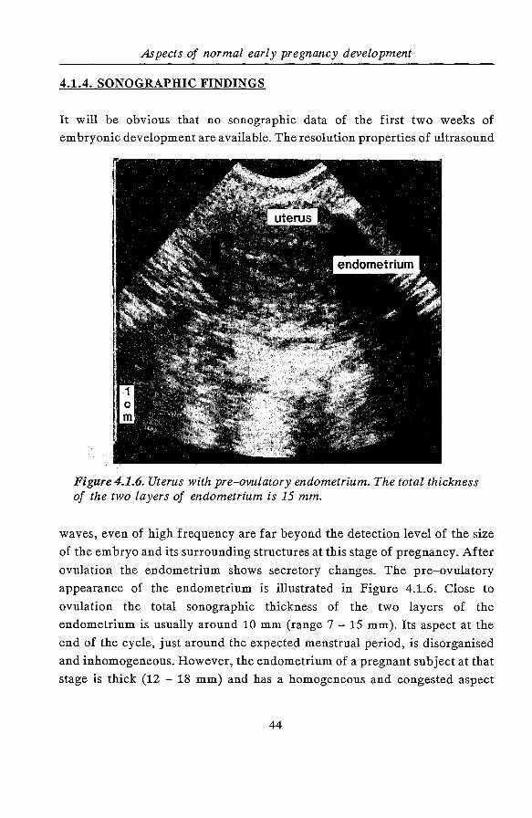
Figure 4.1.6. Uterus with pre-ovulatory endometrium. The total thickness of the two layers of endometrium is 15 mm.
waves, even of high frequency are far beyond the detection level of the size
of the embryo and its surrounding structures at this stage of pregnancy. After
ovulation the endometrium shows secretory changes. The pre-ovulatory
appearance of the endometrium is illustrated in Figure 4.1.6. Close to
ovulation the total sonographic thickness of the two layers of the
endometrium is usually around 10 mm (range 7 - 15 mm). Its aspect at the
end of the cycle, just around the expected menstrual period, is disorganised
and inhomogeneous. However, the endometrium of a pregnant subject at that
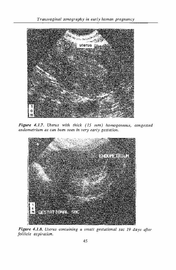
stage is thick (12 - 18 mm) and has a homogeneous and congested aspect
44
Transvaginal sonography in early human pregnancy
Figure 4.1.7. Uterus with thick ( 15 mm) homogeneous, congested endometrium as can been seen in very early gestation.
Figure 4.1.8. Uterus containing a small gestational sac 19 days after follicle aspiration.
45
Aspects of normal early pregnancy development
(Figure 4.1.7). Soon after the menstrual period is missed i.e. between 16 and
19 days after follicle aspiration (median 17 days) a gestational sac can be
observed (Figure 4.1.8). Between 20 and 23 days after follicle aspiration
(median 21 days) the yolk sac can be identified, but the embryonic pole is not
discernible yet. The size of the gestational sac varies between 7.5 and 9.2 mm
(median 8.1 mm) at that stage. The embryonic pole becomes visible between
24 and 29 days (median 26 days) after follicle aspiration. The most striking
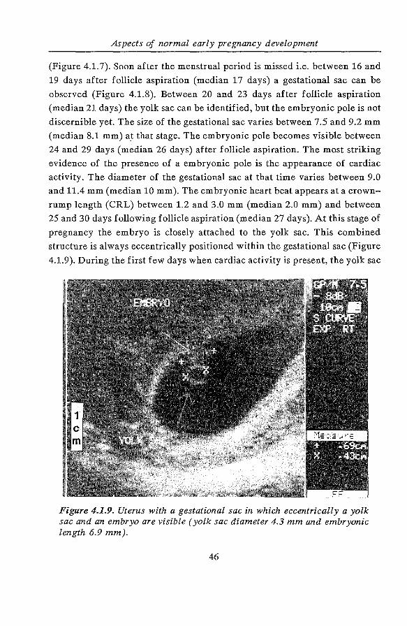
evidence of the presence of a embryonic pole is the appearance of cardiac
activity. The diameter of the gestational sac at that time varies between 9.0
and 11.4 mm (median 10 mm). The embryonic heart beat appears at a crown
rump length (CRL) between 1.2 and 3.0 mm (median 2.0 mm) and between
25 and 30 days following follicle aspiration (median 27 days). At this stage of
pregnancy the embryo is closely attached to the yolk sac. This combined
structure is always eccentrically positioned within the gestational sac (Figure
4.1.9). During the first few days when cardiac activity is present, the yolk sac
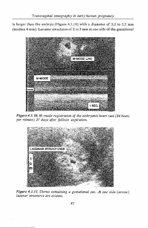
Figure 4.1.9. Uterus with a gestational sac in which eccentrically a yolk sac and an embryo are visible (yolk sac diameter 4.3 mm and embryonic length 6.9 mm).
46
Transvaginal sonography in early human pregnancy
is larger than the embryo (Figure 4.1.10) with a diameter of 3.2 to 5.2 mm
(median 4 mm). Lacunar structures of 2 to 3 mm at one side of the gestational
Figure 4.1.10. M-mode registration of the embryonic heart rate ( 84 beats per minute) 27 days after follicle aspiration.
Figure 4.1.11. Uterus containing a gestational sac. At one side (arrow) lacunar structures are evident.
47
Aspects of normal early pregnancy development
sac may be seen and the blood flow in these lacunar structures may be spotted
suggesting syncytiotrophoblastic invasion of maternal vessels (Figure 4.1.11).
There is only a slight increase in the size of the yolk sac during the first six
weeks following conception (0.5 to 1.5 mm; median 0.9 mm). The distance
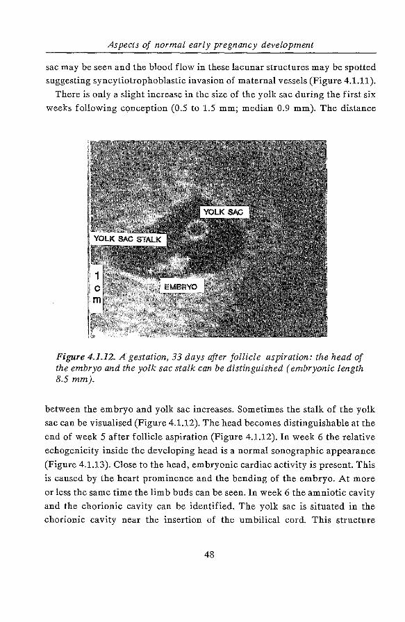
Figure 4.1.12. A· gestation, 33 days after follicle aspiration: the head of the embryo and the yolk sac stalk can be distinguished (embryonic length 8.5 mm).
between the embryo and yolk sac increases. Sometimes the stalk of the yolk
sac can be visualised (Figure 4.1.12). The head becomes distinguishable at the
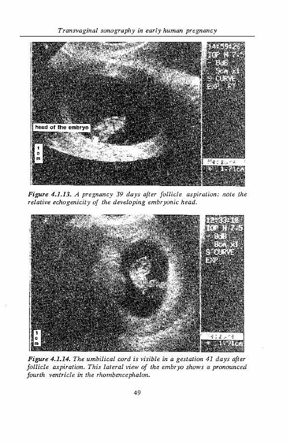
end of week 5 after follicle aspiration (Figure 4.1.12). In week 6 the relative
echogenicity inside the developing head is a normal sonographic appearance
(Figure 4.1.13). Close to the head, embryonic cardiac activity is present. This
is caused by the heart prominence and the bending of the embryo. At more
or less the same time the limb buds can be seen. In week 6 the amniotic cavity
and the chorionic cavity can be identified. The yolk sac is situated in the
chorionic cavity near the insertion of the umbilical cord. This structure
48
Transvaginal sonography in early human pregnancy
Figure 4.1.13. A pregnancy 39 days after follicle aspiration: note the relative echogenicity of the developing embryonic head.
Figure 4.1.14. The umbilical cord is visible in a gestation 41 days after follicle aspiration. This lateral view of the embryo shows a pronounced fourth ventricle in the rhombencephalon.
49
Aspects of normal early pregnancy development
becomes also visible at that time (Figure 4.1.14). A lateral view of the embryo
sometimes clearly displays the rhombencephalon, with a relatively large
cavity that later on becomes the fourth ventricle (Figure 4.1.14). In week 6
gross body movements can be identified for the first time. However,
quantitative and qualitative assessment are not yet feasible.
4.1.5. DISCUSSION
Transvaginal sonography provides detailed information on early human
pregnancy. We were able to examine a group of patients in which the
duration of pregnancy was exactly known, since all gestations emerged from
IVF treatment. The question as to whether these pregnancies can be
considered as normal and the results can be extrapolated to in-vivo fertilised
oocytes remains open for discussion. In this chapter the development of the
embryo and its surrounding structures is described in a semi-quantitative
way. We were able to visualise by means of transvaginal sonography relevant
embryonic features like chorionic cavity, yolk sac, cardiac activity, the head
and limbs about one week earlier than with the conventional transabdominal
scanning technique. Due to the resolution properties if high frequency vaginal
transducers, a gestational sac in-utero can be seen almost immediately after
the menstrual period has been missed. This implicates that this method of
scanning is almost as sensitive as the modern commercially available
pregnancy tests. There is a similarity between embryonic and follicular
development, in that the oocyte as well as the embryo develop in a fluid
filled compartment. The resulting echogenicity allows detection of these
structures as soon as their size equals the resolution properties of the vaginal
probe, which is between 1 and 2 mm3• 6•
Another cyst-like structure which becomes visible is the yolk sac.
Although the human secondary yolk sac is non-functional as far as yolk
storage is concerned (it would be better to speak of urn bilical vesicle), its
development is essential for several reasons. One assumes that it plays a role
in the transport of nutrients during the second and third week while the
utero-placental circulation is established 1. Therefore, it acts as a primitive
50
Transvaginal sonography in early human pregnancy
placenta. From the third until the sixth week blood cells are formed in the
yolk sac wall. Primitive germ cells appear in its wall early in the third week
and subsequently migrate to the developing gonads, where they become
spermatogonia or oogonia1. An embryo can not develop without a structure
supplying the necessary nutrients in the early stages of development.
Therefore, presence of the yolk sac is of crucial importance6• It grows slowly
and the size of the yolk sac is not very useful in pregnancy dating6· 9.
Embryonic abnormalities may coincide with abnormal development or
absence of this structure9 -ll (see chapter 5.1. & 6.3. ).
One of the first signs of a viable pregnancy is the appearance of embryonic
cardiac activity. S~veral investigators have shown the value of transvaginal
sonography detecting the early heart beat12 - 14 . Their data are in accordance
with our findings (between 25 and 30 days after ovulation (median 27 days)).
Only two authors reported embryonic cardiac activity as early as 21 15 and
2216 days after conception. However, no mention was made as to how the date
of conception was documented. The variability in the pre-ovulatory phase
was not taken into account. As patients visited the clinic after the diagnosis
of pregnancy was made, this early detection could well be due to early
ovulation. Moreover, the size of the embryo is beyond the resolution
properties of vaginal transducers and it can, therefore, be questioned if the
heart tube already has started to contract at this stage of development1.
DeCrespigny 12 examining 353 patients with a 5.0 MHz vaginal probe was
always able to demonstrate cardiac activity if the mean diameter of the
gestational sac exceeded 12 mm. Levi et al 13 always identified a yolk sac in
gestational sacs measuring 8 mm or greater in diameter and cardiac activity
in gestational sacs measuring over 16 mm in diameter. Bree et al 14 correlated
size of the gestational sac with structures within that sac. A yolk sac was first
seen in a gestational sac measuring between 6 and 9 mm and cardiac activity
was seen in each patient with a gestational sac of 2' 9 mm. The results of a
study dealing with the dynamics of embryonic cardiac activity in relation to
gestational age are presented in Chapter 6.
The results of our study indicate that transvaginal sonography is superior
to transabdominal scanning techniques when the development of early
pregnancy is studied. We were able to visualise embryonic structures in more
51
Aspects of normal early pregnancy development
detail and about one week earlier in comparison with transabdominal
sonogra ph y.
4.1.6. REFERENCES
1. Moore KL. The developing human- Clinically oriented embryology. WB
Saunders Co., Philadelphia, 1973.
2. Benirschke K. Principles and management of human reproduction. WB
Saunders Co., Philadelphia, 1972; 179-96.
3. Fossum GT, Davajan V, Kletzky OA. Early detection of pregnancy with
transvaginal ultrasound. Fertil Steril 1988; 49: 788-91.
4. Endovaginal Ultrasound. S.R. Goldstein (ed), A.R. Liss, New York, 1988.
5. Timor-Tritsch IE, Farine D, Rosen MG. A close look at early embryonic
development with the high-frequency transvaginal transducer. Am J
Obstet Gynecol 1988; 159: 676-81.
6. Nyberg DA, Mack LA, Harvey D, Wang K, Value of the yolk sac in
evaluating early pregnancies. J Ultrasound Med 1988; 7: 129-35.
7. Blumenfeld Z, Rottem S, Elgali S, Timor-Tritsch IE. Transvaginal
sonographic assessment of early embryological development. In:
Transvaginal Sonography. I.E. Timor-Tritsch & S. Rottem (eds),
Heinemann Medical Books, London, 1988; 87-108.
8. Crooij MJ, Westhuis M, Schoemaker J, Exalto N. Ultrasonographic
measurement of the yolk sac. Br J Obstet Gynaecol1982; 89, 931-4.
9. Reece EA, Scioscia AL, Pinter A, Hobbins JC, Green J, Mahoney MJ,
Naftolin F. Prognostic significance of the yolk sac assessed by
ultrasonography. Am J Obstet Gynecol1988; 159: 1191-4.
10. Ferrazzi E, Bnimbati B, Lanzani A, Oldrini A, Stipparo L, Guerneri S,
Makowski L. The yolk sac in early pregnancy failure. Am J Obstet
Gynecol 1988; 158: 137-42.
11. Rempen A. The embryonal yolk sac in disordered early pregnancy.
Geburtshilfe Frauenheilkd 1988; 48: 804-8.
12. DeCrespigny LCh, Early diagnosis of pregnancy failure with transvaginal
ultrasound. Am J Obstet Gynecol 1988; 159: 408-9.
52

Transvaginal sonography in early human pregnancy
13. Levi CS, Lyons EA, Lindsay DJ. Early diagnosis of nonviable pregnancy
with endovaginal US. Radiology 1988; 167: 383-85.
14. Bree RL, Edwards M, Bohm-Velez M, Beyler S, Mendelson EB.
Transvaginal sonography in the evaluation of normal early pregnancy:
correlation with HCG level. AJR 1989; 153: 75-9.
15. Degenhardt F. Kontrolle von Friihschwangerschaften durch
Vaginalsonographie. Z Geburtshilfe Perinatal 1987; 191: 96-8.
16. Haid C, Zech H, Martin J. Verbesserte Friihdiagnose der intrauterinen
Sch wangerschaft d urch Ultraschall-Vaginalsonde. Ge burtshilf e
Frauenheilk 1985; 45: 371-4.
53

54
Transvaginal sonography in early human pregnancy
4.2. THE CROWN-RUMP LENGTH IN EARLY HUMAN PREGNANCY:
A REAPPRAISAL.
R. Schats@, H.C. van Os '"', C.A.M. Jansen'"' and J.W. Wladimiroff+
@ Department of VEVO I IVF,
Academisch Ziekenhuis Vrije Universiteit
Amsterdam, The Netherlands.
* Department of Obstetrics and Gynaecology I IVF,
Diaconessenhuis Voorburg, The Netherlands.
+ Department of Obstetrics and Gynaecology,
Academic Hospital Dijkzigt, Rotterdam,
The Netherlands.
Accepted for publication in:
The British Journal of Obstetrics and Gynaecology
4.2.1. SUMMARY
The crown-rump length (CRL) was measured by means of transvaginal
sonography in 41 pregnancies resulting from in-vitro fertilisation (IVF ). The
embryonic crown-rump length could be determined as early as 25 days after
follicle aspiration. A reference chart was constructed relating the CRL to the
number of days following follicle aspiration. Comparison with the current
CRL chart by Robinson and Fleming1• 2 revealed that transvaginal sonography
allows earlier and more accurate measurement of embryonic CRL.
55
The Crown-rump length in early human pregnancy
4.2.2. INTRODUCTION
The reference curve currently in use for crown-rump length (CRL)
measurements was derived from data collected with compound B-scan
equipment1• 2• Transvaginal sonography with its high resolution imaging
might enable more accurate measurement of the CRL at an earlier stage of
pregnancy.
The object of the present study was to establish whether transvaginal
measurement of CRL could provide a more accurate reference chart for this
dimension.
4.2.3. SUBJECTS and METHODS
Forty-one women who were pregnant following in-vitro fertilisation agreed
to participate in the study. The IVF procedure provides exact information on
the time of ovulation and embryo transfer and, therefore, the duration of
pregnancy. The study was approved by the local ethics review committee.
Mean maternal age was 34 years (range 24- 40 years). A pilot-study in 23
women showed that the CRL could not be measured until day 25 after follicle
aspiration. Therefore, CRL measurements commenced on day 25 in the
present study. CRL was measured daily until embryonic cardiac activity
appeared, then at 2 or 3 day intervals, depending on patients' availability and
travelling distance, until day 42.
All patients were scanned between 10.00 and 12.00 a.m. using Diasonics
DRF 400 and Toshiba SAL 77B transvaginal probes. The former was
equipped with a 5.0 and a 7.5 MHz mechanical sector scan transducer and the
latter with a 5.0 MHz electronic sector scan transducer. Scans were recorded
by R.S. and C.A.M.J ..
A reference chart (mean +/-1 SD) was constructed of all the longitudinal
CRL data. In 11 normal subjects with spontaneous pregnancies (15 embryos)
four consecutive blind determinations of the CRL were obtained to establish
the intra-observer variability of these measurements. Inter-observer variation
was tested in 12 other normal subjects with single, spontaneous pregnancies.
56
Transvaginal sonography in early human pregnancy
Intra-observer variability was determined by the coefficient of variation; the
inter-observer variation was established by the Wilcoxon two-tailed signed
ranks test.
4.2.4. RESULTS
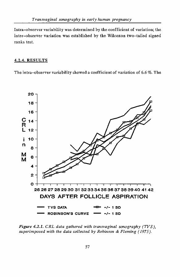
The intra-observer variability showed a coefficient of variation of 6.6 %. The
c R L
i n
M M
20
18
16
14
12
10
8
6
4
2
0 2526 27 282930 31323334363637 383940 4142
DAYS AFTER FOLLICLE ASPIRATION
TVS DATA -8- +/- 1 SD
ROBINSON'S CURVE +/- 1 SD
Figure 4.2.1. CRL data gathered with transvaginal sonography (TVS ), superimposed with the data collected by Robinson & Fleming ( 1975 ).
57
The Crown-rump length in early human pregnancy
inter-observer differences were not statistically significant (p = 0.38).
In eight out of 41 women we were unable to time the onset of embryonic
cardiac activity and therefore we also missed the first CRL measurements,
due to personal factors such as non-availability of the women when required.
Therefore, incomplete data from eight women (five singletons and three
twins) and the complete data from 33 women (21 singletons, nine twins, three
triplets) were available for further analysis. In total 293 CRL measurements
were obtained from 59 embryos. The number of CRL measurements varied
between four and eight per embryo (median six) and between 10 and 30 per
day of gestation (median 17). Figure 4.2.1 depicts the CRL reference chart
derived from all the transvaginally recorded CRL measurements, compared
with the reference chart by Robinson and Fleming2•
4.2.5. DISCUSSION
Our transvaginal CRL measurements demonstrate an acceptable intra- and
inter-observer variability. Particularly in the very early weeks of gestation
there is a discrepancy between our data and those compiled by Robinson and
Fleming2. We have referred, for comparison, to their original data instead of
the chart constructed from linear regression analysis. Inclusion of part of the
yolk sac may have been responsible for the larger CRL measurements in the
beginning of the reference chart obtained by compound B-scan equipment.
Degenhardt et al3 . have also reported initially smaller CRL values using
transvaginal sonography, though the number of CRL measurements per day
of gestation was considerably smaller than that in our study: 225
measurements in 209 subjects between 6 and 13 weeks menstrual age. The
smaller standard deviation displayed in our chart can be explained by (i) the
high resolution capability of modern transvaginal transducers allowing more
detailed identification of embryonic structures, (ii) the larger number of CRL
measurements per pregnancy and (iii) knowledge of the exact duration of the
pregnancy. The advantage of studying IVF pregnancies is that the precise
time of ovulation (follicle aspiration) and embryo transfer are known.
Whether the results can be extrapolated to spontaneous pregnancies is
58
Transvaginal sonography in early human pregnancy
uncertain. We included multiple pregnancies because the embryos behave like
separate individuals with their own growth profile and developmental
potential; in a number of cases we even observed a difference in the time of appearance of embryonic cardiac activity and in subsequent CRL values4• Of
interest is that transvaginal CRL measurements were obtained approximately
4 days earlier than reported previously, which could allow earlier pregnancy
dating in the event of an unreliable menstrual history2• We conclude from the
present study that transvaginal sonography allows more detailed recognition
of embryonic anatomy and, therefore, earlier and more accurate
measurements of embryonic CRL. A larger transverse study, preferably in
women with spontaneous pregnancies, should be initiated to construct a new
reference curve for this scanning technique.
4.2.6. REFERENCES
1. Robinson HP. Sonar measurements of fetal crown-rump length as means
of assessing maturity in first trimester of pregnancy. Br Med J 1973; 4:
28-31.
2. Robinson HP, Fleming JEE. A critical evaluation of sonar "crown-rump"
length measurements. Br J Obstet Gynaecol 1975; 82: 702-10.
3. Degenhardt F, Bohmer S, Behrens 0, Muhlhaus K. Transvaginale
Ultraschallbiometrie der Scheitel-SteiB-Lange im ersten Trimenon. Z
Geburtshilfe Perinatol1988; 192: 249-52.
4. Schats R, Brandsma G, Cleveringa LM, Lankhorst PFC, Vroegop IS,
Jansen CAM. Evidence of asynchronous implantation in IVF multiple
pregnancies. Hum Reprod 1988; 3: Suppl: Abstract 53, p 17.
59

60
Transvaginal sonography in early human pregnancy
CHAPTER 5
DIAGNOSTIC, MORPHOLOGICAL AND THERAPEUTIC ASPECTS OF
ABNORMAL EARLY PREGNANCY DEVELOPMENT
INTRODUCTORY REMARKS
In this chapter the value of transvaginal sonography as a diagnostic tool in
abnormal development of early pregnancy will be described. This chapter
consists of two parts. In the first part (5.1.) clinical signs and symptoms of
threatening abortion in relation to transvaginal sonographic findings in intra
uterine pregnancies are presented. Results will be compared with those
reported in literature. In the second part of this chapter (5.2.) the role of
transvaginal sonography in the diagnosis and management of ectopic
pregnancies will be discussed. The combination of sensitive hCG
determinations and transvaginal sonography allows to the identification of
ectopic pregnancy at a stage when it still may be asymptomatic. This may lead
to more conservative modes of management.
61

62
Transvaginal sonography in early human pregnancy
5.1. MORPHOLOGICAL ASPECTS OF ABNORMAL
EMBRYONIC DEVELOPMENT
5.1.1. INTRODUCTION
Approximately 25% of women experience vaginal bleeding during early
pregnancy. About half of these pregnancies will end in a spontaneous
abortion 1• Despite the fact that non-viable gestations ultimately abort, it is
important to identify correctly these pregnancies before they lead to
prolonged vaginal bleeding, infection and/ or patient anxiety and to determine
which patients merit dilatation and curettage1. Theoretically, the application
of vaginal transducers with high emission frequencies and therefore high
resolution imaging properties, should allow earlier and more detailed
visualisation of normal and abnormal embryonic development than the
standard transabdominal scanning techniques. Some authors compared the use
of transabdominal and transvaginal sonography in normal and abnormal early
gestation2•4. Others have published data concerning the transvaginal
sonographic features in complicated first trimester pregnancies5 · 9• The results
of these studies together with our own sonographic findings in 54 subjects
who became pregnant following long-term infertility will be discussed.
5.1.2. SUBJECTS AND METHODS
Between April 1987 and February 1988 234 transvaginal sonographic
examinations were performed in 54 pregnant women with vaginal bleeding
and a closed cervical os indicating threatening abortion. Three groups could
be recognised: (i) women who had become pregnant following IVF treatment
(Group 1; N = 19); (ii) women with well-documented duration of pregnancy
(Group 2; N = 21); and (iii) pregnant women with an unreliable menstrual
history (Group 3; N = 14). In each woman four parameters were determined:
a) the size of the gestational sac (mm) (=mean of longitudinal, transverse and
63
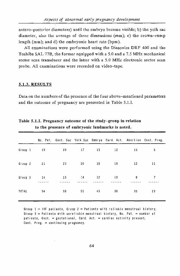
Aspects of abnormal early pregnancy development
antero-posterior diameters) until the embryo became visible; b) the yolk sac
diameter, also the average of three dimensions (mm); c) the crown-rump
length (mm); and d) the embryonic heart rate (bpm).
All examinations were performed using the Diasonics DRF 400 and the
Toshiba SAL 77B, the former equipped with a 5.0 and a 7.5 MHz mechanical
sector scan transducer and the latter with a 5.0 MHz electronic sector scan
probe. All examinations were recorded on video-tape.
5.1.3. RESULTS
Data on the numbers of the presence of the four above-mentioned parameters
and the outcome of pregnancy are presented in Table 5.1.1.
Table 5.1.1. Pregnancy outcome of the study-group in relation
to the presence of embryonic landmarks is noted.
No. Pat. Gest. Sac Yolk Sac Embryo Card. Act. Abortion Cont. Preg.
Group 1 19 20 17 15 12 15 5
Group 2 21 23 20 18 16 12 11
Group 3 14 15 14 12 10 8 7
TOTAL 54 58 51 45 38 35 23
Group 1 = IVF patients, Group 2 =Patients with reliable menstrual history, Group 3 =Patients with unreliable menstrual history, No. Pat. =number of patients, Gest. =gestational, Card. Act. =cardiac activity present, Cont. Preg. =continuing pregnancy.
64
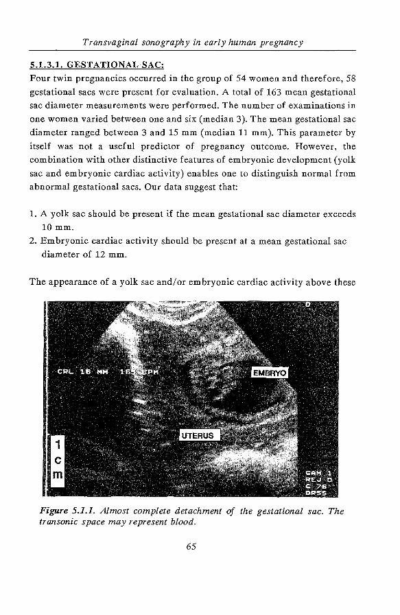
Transvaginal sonography in early human pregnancy
5.1.3.1. GESTATIONAL SAC:
Four twin pregnancies occurred in the group of 54 women and therefore, 58
gestational sacs were present for evaluation. A total of 163 mean gestational
sac diameter measurements were performed. The number of examinations in
one women varied between one and six (median 3). The mean gestational sac
diameter ranged between 3 and 15 mm (median 11 mm). This parameter by
itself was not a useful predictor of pregnancy outcome. However, the
combination with other distinctive features of embryonic development (yolk
sac and embryonic cardiac activity) enables one to distinguish normal from
abnormal gestational sacs. Our data suggest that:
1. A yolk sac should be present if the mean gestational sac diameter exceeds
10 mm.
2. Embryonic cardiac activity should be present at a mean gestational sac
diameter of 12 mm.
The appearance of a yolk sac and/or embryonic cardiac activity above these
Figure 5.1.1. Almost complete detachment of the gestational sac. The transonic space may represent blood.
65
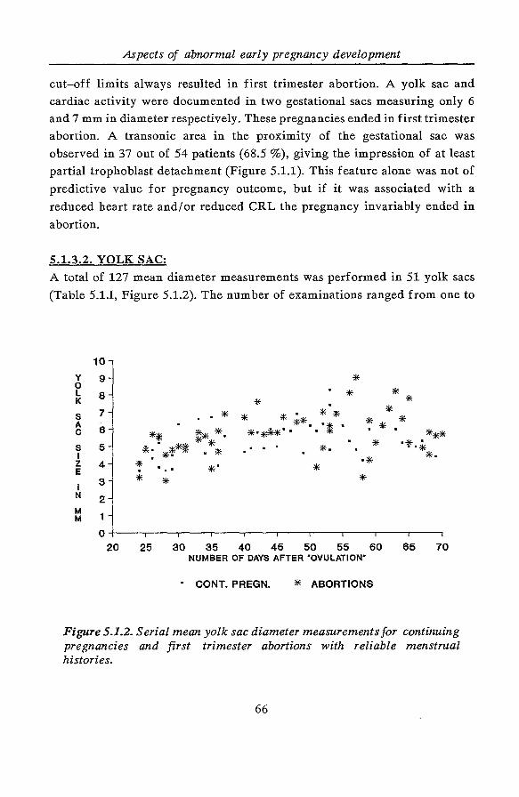
Aspects of abnormal early pregnancy development
cut-off limits always resulted in first trimester abortion. A yolk sac and
cardiac activity were documented in two gestational sacs measuring only 6
and 7 mm in diameter respectively. These pregnancies ended in first trimester
abortion. A transonic area in the proximity of the gestational sac was
observed in 37 out of 54 patients (68.5 %), giving the impression of at least
partial trophoblast detachment (Figure 5.1.1). This feature alone was not of
predictive value for pregnancy outcome, but if it was associated with a
reduced heart rate and/or reduced CRL the pregnancy invariably ended in
abortion.
5.1.3.2. YOLK SAC:
A total of 127 mean diameter measurements was performed in 51 yolk sacs
(Table 5.1.1, Figure 5.1.2). The number of examinations ranged from one to
10
y 0
9 L 8 K
s 7 A -
c 6
s 5 I z 4 E
I 3 N 2 M
1 M
0 20
* . *
* * * .. * ** :* *· . ~ *
* * . * * **· "* • *"*~·. . * ~ * *
~- *'f''* . * * . . . *· *
. . .. *" * * *
25
*
30 35 40 45 50 55 60 NUMBER OF DAYS AFTER "OVULATION"
• CONT. PREGN. * ABORTIONS
65 70
Figure 5.1.2. Serial mean yolk sac diameter measurements for continuing pregnancies and first trimester abortions with reliable menstrual histories.
66
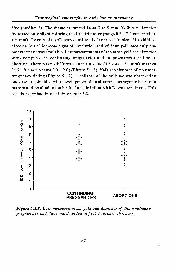
Transvaginal sonography in early human pregnancy
five (median 3). The diameter ranged from 3 to 9 mm. Yolk sac diameter
increased only slightly during the first trimester (range 0.5-3.2 mm, median
1.8 mm). Twenty-six yolk sacs consistently increased in size, 21 exhibited
after an initial increase signs of involution and of four yolk sacs only one
measurement was available. Last measurements of the mean yolk sac diameter
were compared in continuing pregnancies and in pregnancies ending in
abortion. There was no difference in mean value (5.3 versus 5.4 mm) or range
(3.6- 8.3 mm versus 3.0- 9.0) (Figure 5.1.3). Yolk sac size was of no use in
pregnancy dating (Figure 5.1.2). A collapse of the yolk sac was observed in
one case. It coincided with development of an abnormal embryonic heart rate
pattern and resulted in the birth of a male infant with Down's syndrome. This
case is described in detail in chapter 6.3.
10
y 9
0 8 L
K 7 .
s ... A 6 • I • c • I •
s 6 • I • I I z 4
... • I • I E . .
I 3 N
2 M M 1
0 CONTINUING ABORTIONS PREGNANCIES
Figure 5.1.3. Last measured mean yolk sac diameter of the continuing pregnancies and these which ended in first trimester abortions.
67
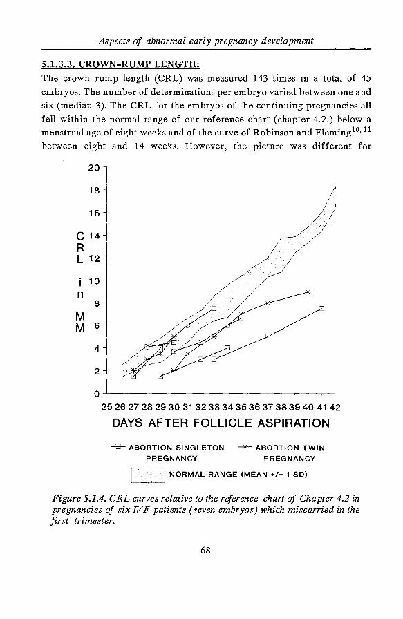
Aspects of abnormal early pregnancy development
5.1.3.3. CROWN-RUMP LENGTH:
The crown-rump length (CRL) was measured 143 times in a total of 45
embryos. The number of determinations per embryo varied between one and
six (median 3). The CRL for the embryos of the continuing pregnancies all
fell within the normal range of our reference chart (chapter 4.2.) below a
menstrual age of eight weeks and of the curve of Robinson and Fleming10• 11
between eight and 14 weeks. However, the picture was different for
20
18
16
c 14
R L 12
10
n 8
M M 6
4
2
0
25 26 27 28 29 30 31 32 33 34 35 36 37 38 39 40 41 42
DAYS AFTER FOLLICLE ASPIRATION
-B- ABORTION SINGLETON """*'-ABORTION TWIN
PREGNANCY PREGNANCY
I NORMAL RANGE (MEAN +/- 1 SO) '----------'-'
Figure 5.1.4. CRL curves relative to the reference chart of Chapter 4.2 in pregnancies of six IVF patients (seven embryos) which miscarried in the first trimester.
68
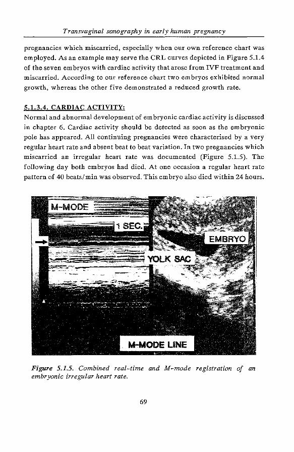
Transvaginal sonography in early human pregnancy
pregnancies which miscarried, especially when our own reference chart was
employed. As an example may serve the CRL curves depicted in Figure 5.1.4
of the seven embryos with cardiac activity that arose from IVF treatment and
miscarried. According to our reference chart two embryos exhibited normal
growth, whereas the other five demonstrated a reduced growth rate.
5.1.3.4. CARDIAC ACTIVITY:
Normal and abnormal development of embryonic cardiac activity is discussed
in chapter 6. Cardiac activity should be detected as soon as the embryonic
pole has appeared. All continuing pregnancies were characterised by a very
regular heart rate and absent beat to beat variation. In two pregnancies which
miscarried an irregular heart rate was documented (Figure 5.1.5). The
following day both embryos had died. At one occasion a regular heart rate
pattern of 40 beats/min was observed. This embryo also died within 24 hours.
Figure 5.1.5. Combined real-time and M-mode registration of an embryonic irregular heart rate.
69
Aspects of abnormal early pregnancy development
5.1.3.5. PREGNANCY OUTCOME:
Six of the IVF patients (Group 1, seven embryos) aborted after embryonic
cardiac activity was established and eight IVF patients aborted without
demonstration of this viability sign. In three of those eight women only a
gestational sac could be detected, in the other five women a gestational sac
as well as a yolk sac could be visualised, but no embryonic pole. Five patients
had vaginal bleeding but the pregnancy went on uneventfully. Seventeen of
the 35 remaining patients (Group 2 & 3) eventually aborted (18 gestational
sacs, 14 yolk sacs; 10 embryos of which in six cases at least once cardiac
activity was established). In two women vaginal bleeding was explained by
the so-called "vanishing twin". At the time of the sonographic examination
cardiac activity was (already) absent in the second gestational sac in these
cases. The growth profile of the remaining embryo was normal in both
instances. In 16 women the pregnancy proceeded without any further
complications.
5.1.4. DISCUSSION
Vaginal bleeding in the first trimester of pregnancy is a common clinical
feature. Also in our highly selected group of patients it was associated with
a high abortion rate (60%).1t is logical to assume that transvaginal sonography
is superior to transabdominal scanning in detecting abnormal early embryonic
development on the basis of the proximity to the object of scanning and the
application of high emission frequencies leading to a better image resolution.
The superiority of transvaginal sonography in the early and detailed
visualisation of embryonic structures and in biometry has been well
documented (Table 5.1.11).
The normal sequence in appearance of the distinctive features of
embryonic development (gestational sac, yolk sac, embryonic pole and cardiac
activity) as established by transvaginal sonography is described in chapter 4.1.
Deviations of this rather fixed developmental pattern may have considerable
implications for fetal outcome. If at a particular gestational age a certain stage
of development has not been reached, the pregnancy probably ends in an
70
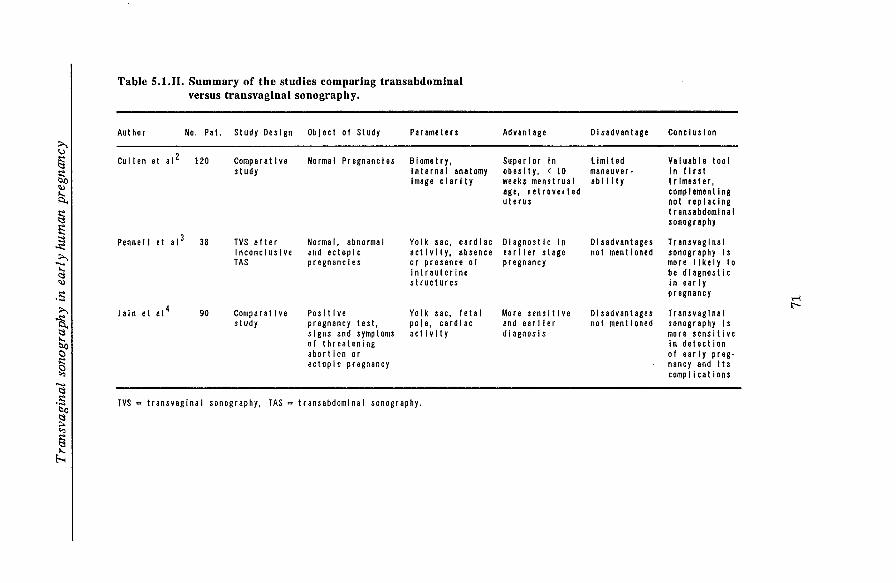
G' § So ~
~ § f2 ~ ~ ;;:.., ..... ..... ~ ~
.s ;;:..,
~ ~
~ C)
~ ..... ~ .s b() ~
~ § ~
Table S.l.II. Summary of the studies comparing transabdominal versus transvaginal sonography.
Author No. Pa I. Study Design Object of Study Par a meters
Cullen et al 2 120 Comparative Normal Pregnancies BIometry, study Internal anatomy
Image clarity
Pennell et ai 3 38 TVS after Norma I, abnormal Yolk sac, cardiac Inconclusive and ectopic activity, absence TAS pregnancies or presence of
intrauterine structures
Jain et a1 4 90 Comparative Positive Yolk sac, I eta I study pregnancy test, poI e, cardiac
signs and symptoms actIvIty of threatening abortion or ectopic pregnancy
TVS =transvaginal sonography, TAS =transabdominal sonography.
Advantage Disadvantage
Superior In L iml ted obesity, < 10 maneuver· weeks menstrual ab Ill t y age, retroverted uterus
Diagnostic In Disadvantages earlier stage not mentioned pregnancy
More sensitive Disadvantages and earlier not mentioned diagnosis
Conclusion
Valuable tool In I I r st trimester, complementing not replacing transabdominal sonography
Transvaginal sonography Is more likely to be diagnostic In ear I y pregnancy
Transvaginal sonography Is more sensitive In detection of early preg· nancy and its complications
T-i 1:"--
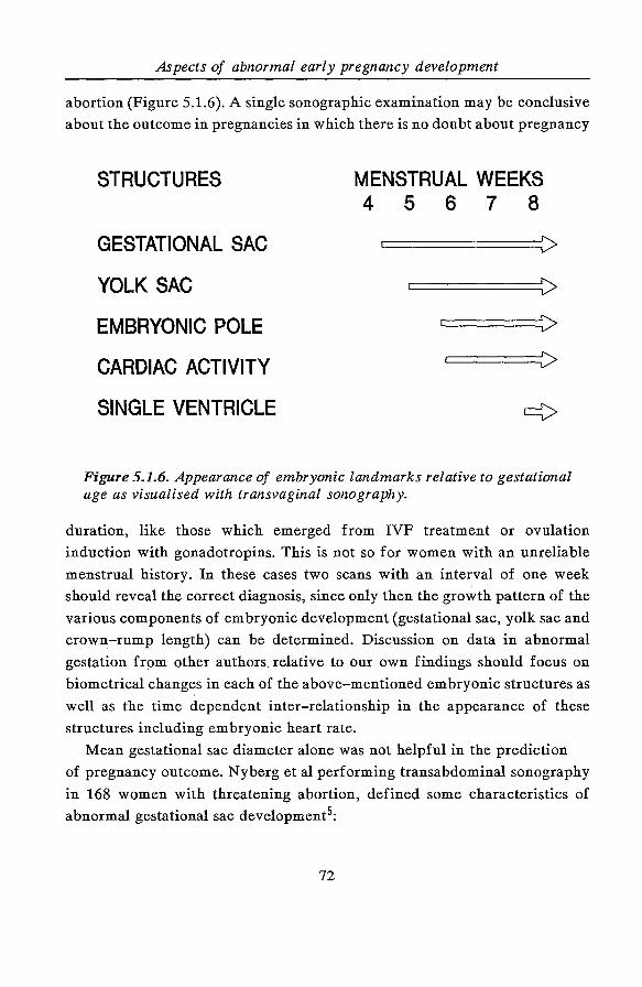
Aspects of abnormal early pregnancy development
abortion (Figure 5.1.6). A single sonographic examination may be conclusive
about the outcome in pregnancies in which there is no doubt about pregnancy
STRUCTURES
GESTATIONAL SAC
YOLK SAC
EMBRYONIC POLE
CARDIAC ACTIVITY
SINGLE VENTRICLE
MENSTRUAL WEEKS 4 5 6 7 8
c:::=:========V>
c::::::======V>
Figure 5.1.6. Appearance of embryonic landmarks relative to gestational age as visualised with transvaginal sonography.
duration, like those which emerged from IVF treatment or ovulation
induction with gonadotropins. This is not so for women with an unreliable
menstrual history. In these cases two scans with an interval of one week
should reveal the correct diagnosis, since only then the growth pattern of the
various components of embryonic development (gestational sac, yolk sac and
crown-rump length) can be determined. Discussion on data in abnormal
gestation from other authors relative to our own findings should focus on
biometrical changes in each of the above-mentioned embryonic structures as
well as the time dependent inter-relationship in the appearance of these
structures including embryonic heart rate.
Mean gestational sac diameter alone was not helpful in the prediction
of pregnancy outcome. Nyberg et al performing transabdominal sonography
in 168 women with threatening abortion, defined some characteristics of
abnormal gestational sac development5:
72
Transvaginal sonography in early human pregnancy
1. a gestational sac diameter~ 25 mm without an embryo;
2. absence of yolk sac at a gestational sac diameter ~ 20 mm;
3. distorted shape of the sac;
4. thin (~ 2 mm) and slightly echogenic or irregular choriodecidual reaction;
5. the typical "double sac" sign is lacking, at a sac diameter
> 10 mm;
6. gestational sac positioned in lower uterine region.
Our own data demonstrate that some of the characteristics mentioned above
should be redefined when transvaginal sonography is employed. Absence of
the yolk sac at a mean gestational sac diameter of ~ 10 mm and I or absence
of embryonic cardiac activity at a gestational diameter of ~ 12 mm indicates
an abnormal development of pregnancy. Appearance of a yolk sac or cardiac
activity above these limits is associated with a poor prognosis. The shape of
the gestational sac and thickness of choriodecidual reaction are less important
characteristics when transvaginal sonography is used, since they are regressive
and consequently late signs of abnormal gestational development. A fluid
filled compartment with partial or complete detachment of the decidual ring
may be seen and may actually reflect bleeding9.
The present study shows that yolk sac size relative to gestational age is
extremely variable. Increase in size was observed during the first trimester
but a common pattern could not be detected. This is at variance with data
reported by Reece et al 12 who assumed a curvilinear relationship with
gestational age. They found a weak correlation (r = 0.39) between gestational
age and size of the yolk sac. However, analysis of their data shows that the
number of measurements are not equally divided over the entire period of
study. Furthermore the study did not include observations before eight weeks
of gestational age. The absence of a yolk sac has been observed in a
pregnancy which resulted in embryonic death at 10 weeks menstrual age; the
embryo exhibited spina bifida13 . In our study, we did not observe any
difference in yolk ·Sac size between continuing and aborting pregnancies. In
the light of these findings yolk sac size does not appear to be a sensitive
predictor of embryonic integrity and pregnancy outcome. Too small as well
as too large yolk sacs have been associated with impending spontaneous
73
Aspects of abnormal early pregnancy development
abortion and karyotypic abnormalities12 • 14 . Rem pen 15 did find yolk sacs with
diameters exceedi~g 7 mm only in pregnancies with a pathological course.
A different picture emerged from embryonic CRL measurements. Here,
reduced growth rate was highly associated with poor pregnancy outcome.
Especially the combination of abnormal heart rate and reduced CRL relative
to gestational age was a very strong predictor of abnormal pregnancy
outcome.
Reduced heart rate may herald embryonic demise. The detection and
development of normal and abnormal embryonic cardiac activity is discussed
and described in detail in chapter 6.
Robinson16 was the first who used transabdominal (compound B-scan)
ultrasound in the diagnosis of early pregnancy failure. Our own data and
reports of other investigators suggest that transvaginal real-time sonography
allows one to recognise normal and abnormal morphological and functional
development about one week earlier than previously has been described using
the transabdominal approach6•8• 17 · 20 . A single transvaginal sonographic
examination can reveal a considerable amount of information on the
prognosis and may be even conclusive when the exact gestational age is
known and all available sonographic parameters are evaluated. The sequence
in appearance of the relevant structures of early gestation should be followed.
If, for example, the yolk sac is not present at day 25 following ovulation or
not present in a gestational sac of which the mean diameter exceeds 10 mm,
it will not appear any more and the pregnancy will end in abortion.
Correlation of sonographic findings with simultaneously determined
maternal hCG levels has been found to be a useful method for evaluating
early pregnancy complications. Nyberg et al21 found that the serum hCG
levels strongly correlated with gestational sac size up to a mean diameter of
25 mm. A disproportionately low serum hCG level relative to gestational sac
size indicates abnormal development of pregnancy21 • 22 • Nyberg et al23
performed transvaginal sonography in 84 women with suspected early
pregnancy complications and determined hCG levels quantitatively. Their
findings indicate that an intra-uterine gestational sac should normally be
visualised when the hCG level (Second International Standard) exceeds 1000
IU /L. Bree et al8 were able to detect a gestational sac by transvaginal
74
Transvaginal sonography in early human pregnancy
sonography in all patients with a hCG levels of 1000 IU /L (First International
Reference Preparation). A level of 7200 IU /L was necessary before the yolk
sac could be visualised and 10800 IU /L for the detection of an embryo
exhibiting cardiac activity. This implicates that there should be concordance
between the transvaginal sonographic findings and the hCG level.
It can be concluded from the results of the present study that transvaginal
sonography allows the diagnosis of pregnancy failure by about one week
earlier than with transabdominal sonography. Furthermore, disturbance of the
sequence in appearance of the relevant structures of early gestation is
indicative of poor pregnancy outcome.
5.1.5. REFERENCES
1. Pantel AG, Shepard TH. Basic aspects of early (first trimester) abortion.
In: Principles and practice of obstetrics and perinatology. L. Iffy & H.A.
Kaminetzky (eds), J. Wiley & Sons, New York 1981; Vol1: 553-63.
2. Cullen MT, Green JJ, Reece EA, Hobbins JC. A comparison of
transvaginal and abdominal ultrasound in visualizing the first trimester
conceptus. J Ultrasound Med 1989; 8: 565-9.
3. Pennell RG, Baltarowich OH, Kurtz AB, Vilaro MM, Rifkin MD,
Needleman L, Mitchell DG, Mervis SA, Goldberg BB. Complicated first
trimester pregnancies: evaluation with endovaginal US versus
transabdominal technique. Radiology 1987; 165:79-83.
4. Jain KA, Hamper UM, Sanders RC. Comparison of transvaginal and
transabdominal sonography in the detection of early pregnancy and its
complications. AJR 1988; 151: 1139-43.
5. Nyberg DA, Laing PC, Filly RA. Threatened abortion: sonographic
distinction of normal and abnormal gestation sacs. Radiology 1986; 158:
397-400.
6. DeCrespigny LCh. Early diagnosis of pregnancy failure with transvaginal
ultrasound. Ani J Obstet Gynecol1988; 159: 408-9.
7. Levi CS, Lyons EA, Lindsay DJ. Early diagnosis of nonviable pregnancy
with endovaginal US. Radiology 1988; 167: 383-85.
75
Aspects of abnormal early pregnancy development
8. Bree RL, Edwards M, Bohm-Velez M, Beyler S, Mendelson EB.
Transvaginal sonography in the evaluation of normal early pregnancy:
correlation with HCG leveL AJR 1989; 153: 75-9.
9. Timor-Tritsch IE, Rottem S, Blumenfeld Z. Pathology of the early
intrauterine pregnancy. In: Transvaginal Sonography. I.E. Timor-Tritsch
& S. Rottem (eds) Heinemann Medical Books, London, 1988; 109-23.
10. Robinson HP. Sonar measurements of fetal crown-rump length as means
of assessing maturity in first trimester of pregnancy. Br Med J 1973; 4:
28-31.
11. Robinson HP, Fleming JEE. A critical evaluation of sonar "crown-rump"
length measurements. Br J Obstet Gynaecol1975; 82: 702-10.
12. Reece EA, Scioscia AL, Pinter A, Hobbins JC, Green J, Mahoney MJ,
Naftolin F. Prognostic significance of the yolk sac assessed by
ultrasonography. Am J Obstet Gynecol1988; 159: 1191-4.
13. Crooij MJ, Westhuis M, Schoemaker J, Exalto N. Ultrasonographic
measurement of the yolk sac. Br J Obstet Gynecol1982; 89, 931-4.
14. Ferrazzi E, Brambati B, Lanzani A, Oldrini A, Stipparo L, Guerneri S,
Makowski L. The yolk sac in early pregnancy failure. Am J Obstet
Gynecol1988; 158: 137-42.
15. Rempen A. The embryonal yolk sac in disordered early pregnancy.
Geburtshilfe Frauenheilkd 1988: 48; 804-8.
16. Robinson HP. The diagnosis of early pregnancy failure by sonar. Br J
Obstet Gynaecol1975; 82: 849-56.
17. Fossum GT, Davajan V, Kletzky DA. Early detection of pregnancy with
transvaginal ultrasound. Fertil Steril 1988; 49: 788-91.
18. Blumenfeld Z, Rottem S, Elgali S, Timor-Tritsch IE. Transvaginal
assessment of early embryological development. In: Transvaginal
Sonography. I.E. Timor-Tritsch & S. Rottem (eds), Heinemann Medical
Books, London, 1988; 87-108.
19. Warren WB, Timor-Tritsch IE, Peisner DB, Raju S, Rosen MG. Dating
the early pregnancy by sequential appearance of embryonic structures.
Am J Obstet Gynecol1989; 161: 747-53.
76
Transvaginal sonography in early human pregnancy
20. Timor-Tritsch IE, Farine D, Rosen MG. A close look at early embryonic
development with the high-frequency transvaginal transducer. Am J
Obstet Gynecol 1988; 159: 676-81.
21. Nyberg DA, Mack LA, Laing PC, Jeffrey RB. Early pregnancy
complications: endovaginal sonographic findings correlated with human
chorionic gonadotropin levels. Radiology 1988; 167; 619-22.
22. Nyberg DA, Mack LA, Laing PC, Patten RM. Distinguishing normal from
abnormal gestation sac growth in early pregnancy. J Ultrasound Med
1987; 6: 23-7.
23. Nyberg DA, Filly RA, Duarte Filho DL, Laing PC, Mahony BS.
Abnormal pregnancy: early diagnosis by US and serum chorionic
gonadotropin levels. Radiology 1986; 158: 393-6.
77

78
Transvaginal sonography in early human pregnancy
5.2. THE ROLE OF TRANSVAGINAL SONOGRAPHY IN
DIAGNOSIS AND MANAGEMENT OF ECTOPIC PREGNANCY
5.2.1. INTRODUCTION
Ectopic pregnancy remains a diagnostic challenge to the gynaecologist. The
clinal signs and symptoms are diverse. The fear of a potentially life
threatening condition as well as the diagnostic problems have led to the fact
that the diagnosis of ectopic pregnancy is made more frequently than it
actually occurs1. Obviously there is an urge to diagnose an ectopic pregnancy
as soon as possible to prevent calamities. Another advantage of early
recognition is that if damage to the tubes and other structures can be
minimised, the preservation of future fertility can be optimised and
conservative modes of management can be pursued. In the past an ectopic
pregnancy could only occasionally be diagnosed by transabdominal
ultrasound. The introduction of transvaginal sonography has led to improved
visualisation of ectopic pregnancy, even when it is asymptomatic2•4. Several
comparative studies have been published showing the superiority of
transvaginal to transabdominal scanning in diagnosing this entity5 · 7• In most
cases ectopic pregnancy can be ruled out by demonstrating an intra-uterine
pregnancy. An exception to the rule may be a pregnancy which emerged from
IVF treatment. The combined presence of both intra-uterine and ectopic
pregnancy may be seen following the transfer of more than one embryo8• 9•
The recent introduction of sensitive serum and urine hCG determinations
with commercially available kits, makes it possible to diagnose the state of
pregnant at a very early stage. Serum hCG levels have been found to correlate
with transvaginal sonographic findings in normal pregnancy and as such these
techniques can be used in evaluating early pregnancy complications. Several
authors have tried to find discriminatory levels for the various embryonic
landmarks10 · 18. A problem in evaluating these studies is the application of
two different WHO standards for determining hCG levels: the first
international reference preparation (IRP) and the second international
79
Transvaginal sonography and ectopic pregnancy
standard. Most investigators assume that the first IRP is equivalent to twice
the value of the second international standard19. However, Fossum et al17
found in their study only 34% higher values for the first IRP. An explanation
for this finding has not been provided. Blumenfeld et al 18 report a lowest
value of 420 IU /L at which a gestational sac could be visualised.
Unfortunately they do not specify which standard preparation was used. If
all the values for hCG are corrected to the first IRP the following
discriminatory zones can be defined for transvaginal sonography10 · 18: a
gestational sac should be identified when the serum hCG level exceeds 1000
IU /L, a yolk sac at a level that is equal or greater than 7000 IU /L, an
embryonic pole above a level of 8500 IU /L and embryonic cardiac activity
over 11000 IU /L. This implicates that there should be concordance between
the transvaginal sonographic findings and the hCG level. If, for example a
intra-uterine gestational sac can not be seen at a serum hCG level of 3000
IU /L an ectopic pregnancy is very likely to be present. This should not lead
to immediate surgery, instead this early diagnosis should give one time to
consider the best possible management. The combination of these sensitive
diagnostic tools makes it possible to diagnose an ectopic pregnancy more
often at a stage that it is still asymptomatic2· 4• It is also possible that ectopic
pregnancies are diagnosed that would have been missed altogether in the past,
and would have resolved spontaneously. Many ectopic pregnancies turn out
to be non-viable. Either this may be due to the quality of the trophoblast
itself or to the inadequate site of nidation. However, viable ectopic
pregnancies occur more often after IVF treatment, most likely because the
mechanism of occurrence is different.
The scope of this chapter will be to present the results of a pilot study and
to illustrate with case histories new management options in case of ectopic
pregnancy with special reference to the advent of transvaginal sonography as
a diagnostic tool and its use in the treatment of ectopic pregnancy.
5.2.2. SUBJECTS AND METHODS
Ectopic pregnancy was diagnosed by transvaginal sonography in 14 patients
80
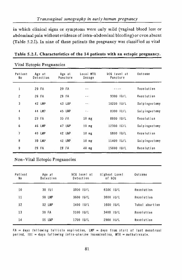
Transvaginal sonography in early human pregnancy
in which clinical signs or symptoms were only mild (vaginal blood loss or
abdominal pain without evidence of intra-abdominal bleeding) or even absent
(Table 5.2.!). In nine of these patients the pregnancy was classified as vital
Table 5.2.1. Characteristics of the 14 patients with an ectopic pregnancy.
Vital Ectopic Pregnancies
Patient Age at Age at Local MTX hCG level at Outcome No Detection Puncture Dosage Puncture
29 FA 29 FA Resolution
2 26 FA 28 FA 9300 I U/L Resolution
42 LMP 43 LMP 10200 I U/L Salpingectomy
4 44 LMP 45 LMP 8300 IU/L Salpingectomy
29 FA 30 FA 10 mg 8800 IU/L Resolution
6 46 LMP 47 LMP 10 mg 12300 I U/L Salpingectomy
7 40 LMP 42 LMP 10 mg 6800 I U/L Resolution
8 39 LMP 42 LMP 10 mg 11400 IU/L Salpingectomy
9 28 FA 28 FA 40 mg 15600 I U/L Resolution
Non-Vital Ectopic Pregnancies
Patient Age .at hCG level at Highest Leve I Outcome No Detection Detection of hCG
10 38 lUI 5000 I U/L 6500 I U/L Resolution
11 so LMP 3600 IU/L 3600 IU/L Resolution
12 52 LMP 1400 I U/L 1600 I U/L Tubal abortion
13 30 FA 3100 IU/L 3400 IU/L Resolution
14 55 LMP 1700 IU/L 2900 I U/L Resolution
FA = days following follicle aspiration, LMP days from start of I ast menstrua I period, lUI =days following intra-uterine insemination, MTX =methotrexate.
81
Transvaginal sonography and ectopic pregnancy
according to the following criteria: the presence of a gestational sac
containing a yolk sac and embryo exhibiting cardiac activity outside the
uterine cavity associated with rising hCG levels. Eight ectopic pregnancies
were situated within the Falloppian tube and one in the intramural part of the
right tube (No. 9, Table 5.2.I). In the remaining five patients the ectopic
pregnancy was classified as non-vital because of the absence of embryonic
cardiac activity and hCG levels below the lower limit for gestational age20.
In one patient a vital intra-uterine pregnancy as well as a vital ectopic
pregnancy was present (No.1, Table 5.2.I). A total of five ectopic pregnancies
occurred after IVJ.:i treatment, one following intra-uterine insemination of
capacitated spermatozoa and eight spontaneously of which three during an
infertility work-up.
According to the classification of ectopic pregnancies in vital and non
vital, transvaginal puncture was carried out or the natural course was
monitored with transvaginal sonography and hCG determinations. In the vital
ectopic pregnancies, except for patient No. 1, hCG determinations were
carried out at least once before and at two or three day intervals in
conjunction with transvaginal sonography after the transvaginal puncture.
The hCG determination and scanning rate reduced as soon as the hCG level
fell according to the half life time. In patients with a non-vital ectopic
pregnancy the same follow-up scheme was employed. Three months after the
puncture a hysterosalpingogram (HSG) was performed when suitable. In the
hCG assays the first international reference preparation (IRP 75/537) was
used as a standard.
All examinations were performed using the Diasonics DRF 400 the
Toshiba SAL 77B and the Kretz Combison 310 equipped with a 5.0 and a 7.5
MHz mechanical sector scan transducer, a 5.0 MHz electronic sector scan
probe and a 5.0 and a 7.5 MHz mechanical sector scan transducer
respectively.
Puncture Technique: In all nine patients with a vital ectopic pregnancy
transvaginal puncture was performed under sonographic guidance and under
local analgesia (lidocain 1%, 10 ml). The procedure can be compared with a
follicle aspiration which is common practice in oocyte retrieval in IVF
programs. The embryo was aimed at in order to cause cessation of cardiac
82
Transvaginal sonography in early human pregnancy
activity, whereafter aspiration of the gestational sac and the yolk sac, if
possible, followed. Transvaginal scanning was performed for at least another
ten minutes to ensure that no cardiac activity reappeared. In the first four
patients (No. 1-4, Table 5.2.I) puncture took place without instillation of the
cytoreductive agent methotrexate (MTX) into the gestational sac. In the next
four patients (No. 5-8, Table 5.2.I) transvaginal puncture was followed by
instillation of 10 mg of MTX into the aspirated gestational sac. In patient No.
9 40 mg of MTX was instilled.
5.2.3. RESULTS
The data of all14 patients are summarised in Table 5.2.I. The crown-rump
length of the embryos ranged between 3 and 6 mm. The first two patients
(No.1 & 2) displayed complete resolution of the ectopic pregnancy. The next
two cases (No. 3 & 4) were less successful. Despite a correct puncture and
cessation of cardiac activity, the hCG level started to rise again after an
initial fall. Both patients had to undergo laparoscopy followed by laparotomy
leading to salpingectomy of the affected side. In view of this and similar data
reported by several other authors employing the aspiration technique
alone21 • 22 , as well as the suggestion made in by other reports that the
instillation of substances such as, potassium chloride23 • 24, MTX25 · 29 ,
prostaglandin F 2a 26 or E227 may improve the success rate, the next four
patients (No. 5 - 8) were subjected to a different therapeutic approach. A
dosage of 10 mg of MTX was instilled into the emptied gestational sac.
However, in two of these patients (No.6 & 8) an initial fall of the hCG level
again was followed by a rise, and laparoscopy followed by laparotomy leading
to salpingectomy of the affected side was required. In patient No. 9 with an
intramural presentation of ectopic pregnancy a dosage of 40 mg of MTX was
administered into the aspirated gestational sac. In addition, because of a
plateauing of the hCG level one week after puncture and because of the
intramural localisation of the ectopic pregnancy which is unattractive for
surgical intervention, MTX was administered systemically.
In the five patients with a non-vital ectopic pregnancy no major problems
83
Transvaginal sonography and ectopic pregnancy
occurred. In one case (No. 12) symptomatic tubal abortion took place.
Transvaginal sonography displayed the appearance of fluid in the pouch of
Douglas indicating most likely the presence of blood which was confirmed by
laparoscopy. Since the bleeding was self-limiting no further action was
required. Tubal abortion occurred during the decline of the hCG level. In two
patients (No. 5 & 7) without the suspicion of tubal pathology before the
ectopic pregnancy occurred, a HSG was performed three months later. Both
showed patent tubes. One of these patients became pregnant at a later stage,
and transvaginal sonography revealed a vital intra-uterine gestation.
In order to illustrate the diversity in diagnosis and initially non-operative
management of ectopic pregnancy five cases presented in Table 5.2.I will be
described in detail:
Patient No. 1, aged 36 years was known with primary infertility due to
inoperable tubal pathology. She had experienced an ectopic pregnancy in the
first IVF treatment cycle, which was treated by conventional laparotomy and
salpingectomy. In the subsequent cycle she became pregnant resulting in a
vital intra-uterine pregnancy in conjunction with a vital ectopic pregnancy
in the remaining tube. In an effort not to disrupt the intra-uterine pregnancy
a puncture of the ectopic pregnancy as described above was performed.
Following the puncture she was closely monitored by serial transvaginal
ultrasound examinations; hCG determinations would not have been
informative in this case. The ectopic pregnancy apparently resorbed
completely and the intra-uterine pregnancy went on uneventfully and led to
the birth of a healthy male infant at term by caesarean section due to
cephalopelvic disproportion. At surgery no sequelae were found at the
previous puncture site.
Patient No. 2, aged 31 years with primary infertility due to tubal pathology
underwent IVF treatment. She became pregnant in the second cycle. Sixteen
days after follicle aspiration (FA) a sensitive urine pregnancy test (ICON) was
positive. Twenty-six days following FA, transvaginal sonography revealed a
congested thick endometrium containing fluid (Figure 5.2.1 ); no intra-uterine
gestational sac could be visualised. On the right side of the uterus a
84
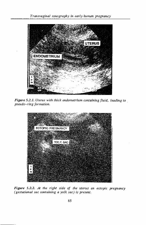
Transvaginal sonography in early human pregnancy
Figure 5.2.1. Uterus with thick endometrium containing fluid, leading to . pseudo-ring formation.
Figure 5.2.2. At the right side of the uterus an ectopic pregnancy (gestational sac containing a yolk sac) is present.
85
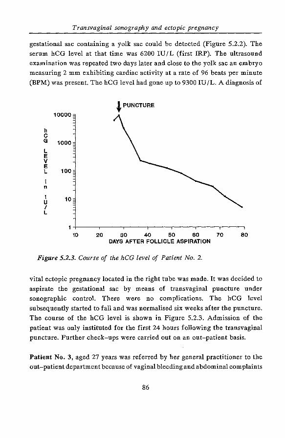
Transvaginal sonography and ectopic pregnancy
gestational sac containing a yolk sac could be detected (Figure 5.2.2). The
serum hCG level at that time was 6200 IU /L (first IRP). The ultrasound
examination was repeated two days later and close to the yolk sac an embryo
measuring 2 mm exhibiting cardiac activity at a rate of 96 beats per minute
(BPM) was present. The hCG level had gone up to 9300 IU /L. A diagnosis of
~PUNCTURE
h c G 1000 L E v E L 100
i n
I 10 u I L
10 20 30 40 50 60 70 80 DAYS AFTER FOLLICLE ASPIRATION
Figure 5.2.3. Course of the hCG level of Patient No. 2.
vital ectopic pregnancy located in the right tube was made. It was decided to
aspirate the gestational sac by means of transvaginal puncture under
sonographic controL There were no complications. The hCG level
subsequently started to fall and was normalised six weeks after the puncture.
The course of the hCG level is shown in Figure 5.2.3. Admission of the
patient was only instituted for the first 24 hours following the transvaginal
puncture. Further check-ups were carried out on an out-patient basis.
Patient No. 3, aged 27 years was referred by her general practitioner to the
out-patient department because of vaginal bleeding and abdominal complaints
86
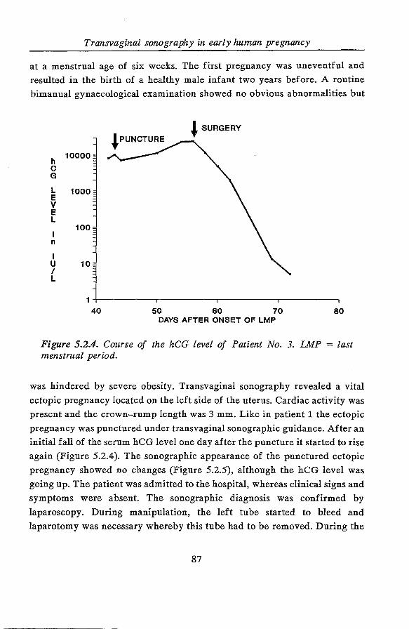
Transvaginal sonography in early human pregnancy
at a menstrual age of six weeks. The first pregnancy was uneventful and
resulted in the birth of a healthy male infant two years before. A routine
bimanual gynaecological examination showed no obvious abnormalities but
~SURGERY
h 10000
c G
L 1000 E v E L
100
n
I u 10 I L
40 50 60 70 80 DAYS AFTER ONSET OF LMP
Figure 5.2.4. Course of the hCG level of Patient No. 3. LMP = last menstrual period.
was hindered by severe obesity. Transvaginal sonography revealed a vital
ectopic pregnancy located on the left side of the uterus. Cardiac activity was
present and the crown-rump length was 3 rom. Like in patient 1 the ectopic
pregnancy was punctured under transvaginal sonographic guidance. After an
initial fall of the serum hCG level one day after the puncture it started to rise
again (Figure 5.2.4). The sonographic appearance of the punctured ectopic
pregnancy showed no changes (Figure 5.2.5), although the hCG level was
going up. The patient was admitted to the hospital, whereas clinical signs and
symptoms were absent. The sonographic diagnosis was confirmed by
laparoscopy. During manipulation, the left tube started to bleed and
laparotomy was necessary whereby this tube had to be removed. During the
87
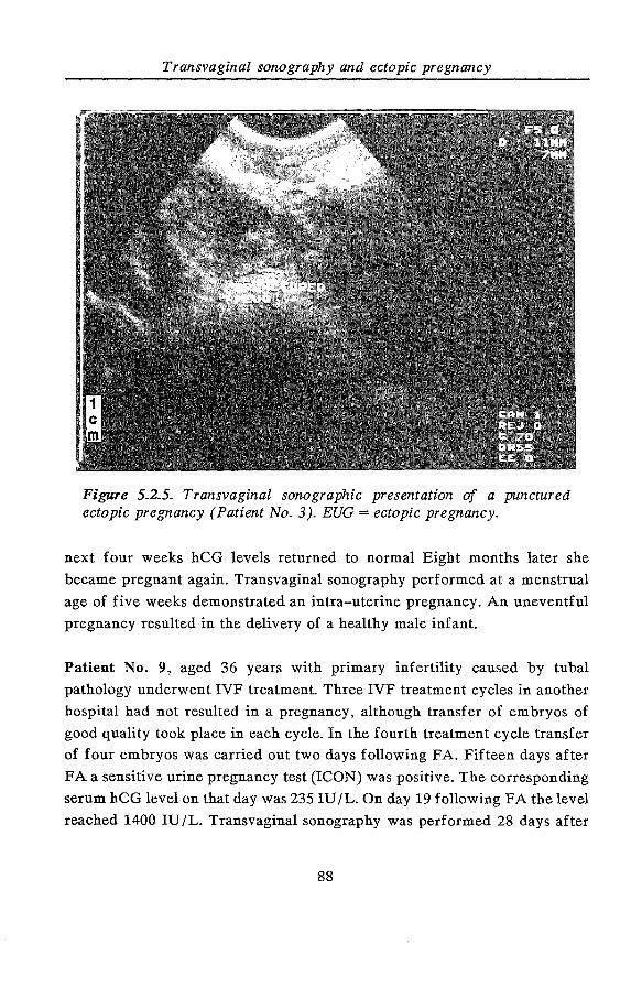
Transvaginal sonography and ectopic pregnancy
Figure 5.2.5. Transvaginal sonographic presentation of a punctured ectopic pregnancy (Patient No. 3). EUG =ectopic pregnancy.
next four weeks hCG levels returned to normal Eight months later she
became pregnant again. Transvaginal sonography performed at a menstrual
age of five weeks demonstrated an intra-uterine pregnancy. An uneventful
pregnancy resulted in the delivery of a healthy male infant.
Patient No. 9, aged 36 years with primary infertility caused by tubal
pathology underwent IVF treatment. Three IVF treatment cycles in another
hospital had not resulted in a pregnancy, although transfer of embryos of
good quality took place in each cycle. In the fourth treatment cycle transfer
of four embryos was carried out two days following FA. Fifteen days after
FA a sensitive urine pregnancy test (ICON) was positive. The corresponding
serum hCG level on that day was 235 IU /L. On day 19 following FA the level
reached 1400 IU /L. Transvaginal sonography was performed 28 days after
88
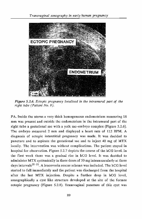
Transvaginal sonography in early human pregnancy
Figure 5.2.6. Ectopic pregnancy localised in the intramural part of the right tube (Patient No. 9 ).
FA. Inside the uterus a very thick homogeneous endometrium measuring 18
mm was present and outside the endometrium in the intramural part of the
right tube a gestational sac with a yolk sac-embryo complex (Figure 5.2.6).
The embryo measured 2 mm and displayed a heart rate of 112 BPM. A
diagnosis of ectopic interstitial pregnancy was made. It was decided to
puncture and to aspirate the gestational sac and to inject 40 mg of MTX
locally. The intervention was without complications. The patient stayed in
hospital for observation. Figure 5.2.7 depicts the course of the hCG level. In
the first week there was a gradual rise in hCG level. It was decided to
administer MTX systemically in three doses of 50 mg intramuscularly at three
days intervals28 - 35 • A leucovorin rescue scheme was included. The hCG level
started to fall immediately and the patient was discharged from the hospital
after the last MTX injection. Despite a further drop in hCG level,
sonographically a cyst like structure developed at the site of the former
ectopic pregnancy (Figure 5.2.8). Transvaginal puncture of this cyst was
89
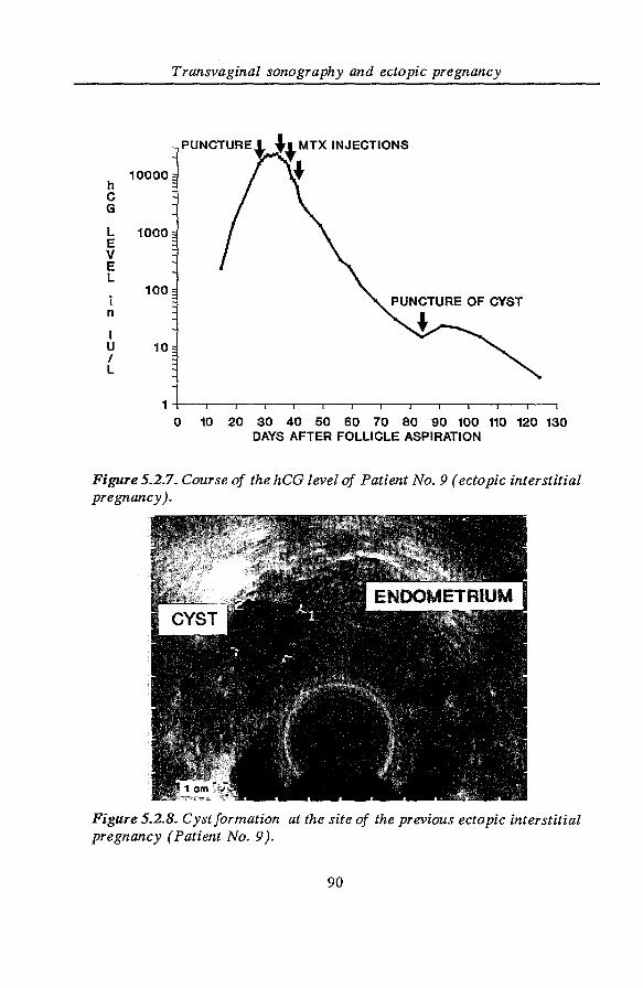
h c G
L E v E L
i n
I u I L
Transvaginal sonography and ectopic pregnancy
10000
1000
100
10
1+---.--.--.--,r--r--T--.---.--~-.---.--~-.
0 10 20 30 40 50 60 70 80 90 100 110 120 130 DAYS AFTER FOLLICLE ASPIRATION
Figure 5.2.7. Course of the hCG level of Patient No.9 (ectopic interstitial pregnancy).
Figure 5.2.8. Cyst formation at the site of the previous ectopic interstitial pregnancy (Patient No. 9 ).
90
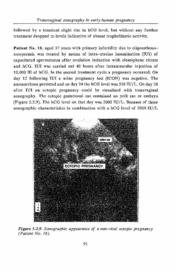
Transvaginal sonography in early human pregnancy
followed by a transient slight rise in hCG level, but without any further
treatment dropped to levels indicative of absent trophoblastic activity.
Patient No. 10, aged 37 years with primary infertility due to oligoastheno
zoospermia was treated by means of intra-uterine insemination (lUI) of
capacitated spermatozoa after ovulation induction with clomiphene citrate
and hCG. lUI was carried out 40 hours after intramuscular injection of
10.000 IE of hCG .. In the second treatment cycle a pregnancy occurred. On
day 15 following lUI a urine pregnancy test (ICON) was negative. The
amenorrhoea persisted and on day 24 the hCG level was 528 IU /L. On day 38
after lUI an ectopic pregnancy could be visualised with transvaginal
sonography. The ectopic gestational sac contained no yolk sac or embryo
(Figure 5.2.9). The hCG level on that day was 5000 IU /L. Because of these
sonographic characteristics in combination with a hCG level of 5000 IU /L
Figure 5.2.9. Sonographic appearance of a non-vital ectopic pregnancy (Patient No. 10 ).
91
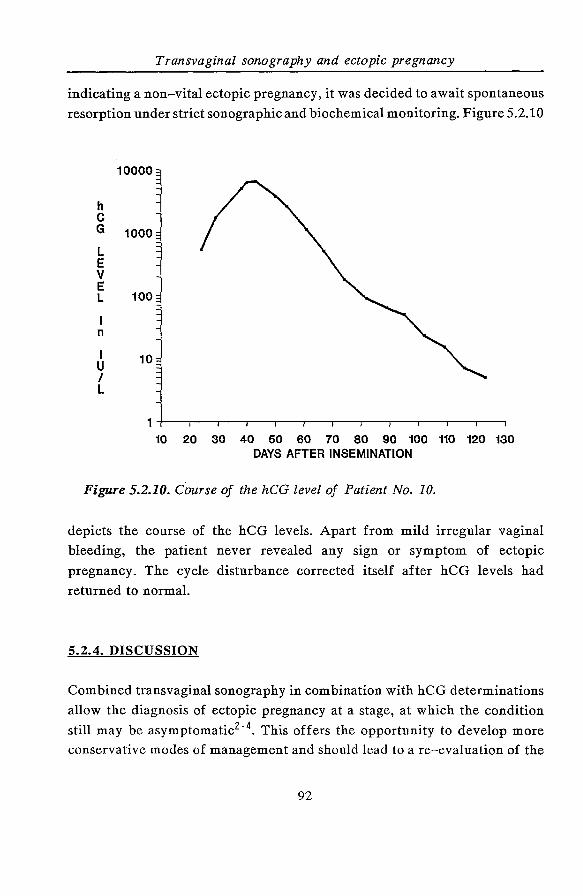
Transvaginal sonography and ectopic pregnancy
indicating a non-vital ectopic pregnancy, it was decided to await spontaneous
resorption under strict sonographic and biochemical monitoring. Figure 5.2.10
h c G
L E v E L
n
I u I L
1000
100
10
10 20 30 40 50 60 70 80 90 100 110 120 130 DAYS AFTER INSEMINATION
Figure 5.2.10. Course of the hCG level of Patient No. 10.
depicts the course of the hCG levels. Apart from mild irregular vaginal
bleeding, the patient never revealed any sign or symptom of ectopic
pregnancy. The cycle disturbance corrected itself after hCG levels had
returned to normal.
5.2.4. DISCUSSION
Combined transvaginal sonography in combination with hCG determinations
allow the diagnosis of ectopic pregnancy at a stage, at which the condition
still may be asymptomatic2· 4• This offers the opportunity to develop more
conservative modes of management and should lead to a re-evaluation of the
92
Transvaginal sonography in early human pregnancy
surgical approach. On the other hand, there is the danger that early diagnosis
may result in more surgical interventions. Such a development should be
particularly undesirable in non-vital ectopic pregnancies, which otherwise
would have been missed in the past and would have resolved spontaneously.
Transvaginal puncture was performed in nine patients in an effort to
avitalise the ectopic pregnancy. However, in four out of nine patients no
remission occurred and surgery was necessary at a later stage. Transvaginal
sonography as a follow-up procedure after the puncture was not informative
as illustrated by patient No.3. It was still possible to visualise the punctured
ectopic pregnancy but it did not provide enough information about the
presence of residual trophoblastic activity. This appears to be better reflected
by serial hCG level monitoring. Management of a vital ectopic pregnancy
through transvaginal puncture is feasible, although management was
unsuccessful in four out of nine cases with or without the instillation of MTX
in the punctured gestational sac. It is apparently not possible to remove all
vital trophoblastic tissue. The actual dosage of 10 mg of MTX may not be
sufficient27• 28 . However, Pansky et al28 do not substantiate that the higher
dosage of 25 mg of MTX yields better results than a dosage of 12.5 mg. In
patient 9 the ectopic pregnancy was situated in the intramural part of the
right tube and as such was not amenable to surgery due to increased risk of
severe bleeding. The successful course in this case illustrates the possibility
of combined local and systemic MTX therapy. This was also reported by
Feichtinger & Kenieter27•
We do not claim that transvaginal puncture of ectopic pregnancies is the
therapy of choice in each patient, but since future fertility aspects are
important it may be advocated in order to avoid surgery. Although four out
of nine patients eventually underwent surgery, the other five did benefit
from this conservative approach.
In the five non-vital ectopic pregnancies spontaneous resorption was
awaited. However, one should be aware of the risks of tubal abortion,
especially when hCG levels are falling. This emphasises the need for
monitoring the hCG levels until they have returned to normal.
The place of systemic administration MTX in the management of ectopic
pregnancy remains controversial. Failures and successes have been reported,
93
Transvaginal sonography and ectopic pregnancy
but the number of patients is too limited in our view to draw conclusions30 - 37.
In a Dutch study of nine patients (10 ectopic pregnancies) the hCG level was
below 500 IU /L in eight patients at a menstrual age of six to eight weeks,
indicating the presence of non-vital or spontaneously regressing ectopic
pregnancies35 . It can be questioned, therefore, if it was justified to use a
cytoreductive agent at these very low hCG levels. This also applies to studies
reported by others in which systemic MTX treatment was implemented at
hCG levels below 1000 IU /L at an amenorrhoea of six weeks or more.
Moreover, at least two hCG determinations should be done in order to judge
actual trophoblast activity before systemic MTX treatment is contemplated.
Only Rodi et al34 mention a rise or plateauing in hCG level as an inclusion
criterium for systemic MTX treatment. The literature data concerning
systemic MTX treatment in relation to ectopic pregnancy is summarised in
Table 5.2.11.
Our own experience and review of the literature lead to the following
suggestions:
1. In case of a non-vital ectopic pregnancy or uncertainty about the diagnosis,
hCG assays and transvaginal sonography should be carried out every other
day. In case of falling hCG levels await spontaneous resolution; in case of
signs of tubal abortion laparoscopy may be necessary to exclude active
intra-peritoneal bleeding.
2. In case of a vital ectopic pregnancy, transvaginal aspiration of gestational
sac and contents should be performed under sonographic guidance and
local application of a maximum of 50 mg of MTX. In case of subsequent
rise or plateauing in hCG level, systemic MTX treatment should be
considered.
3. At a CRL exceeding 10 mm the extensiveness of vital trophoblast may
inhibit complete regression following gestational sac aspiration. Instead
laparotomy is indicated.
In conclusion, transvaginal sonography is a valuable adjunCt to hCG
94
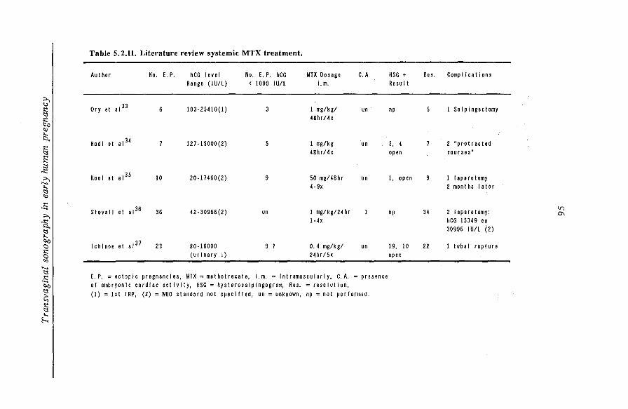
Table 5.2.11. Literature review systemic MTX treatment.
Author No. E. P. hCG level No. E. P. hCG MTX Dosage C. A. HSG + Res. Complications Range (IU/L) < 1000 IU/L l.m. Result
;:.., '-l
Ory et a1 33 § 6 103-25410(1) 3 1 mg/kg/ un np 5 I Salpingectomy
~ 48hr/4x
~ ,...,
~I Rodl et a1 34 7 127-15000(2) 5 I mg/kg un 5, 4 7 2 "prittracted 48hr/4x open courses"
E ;::$
~I Kool et a1 35 10 20-17460(2) 9 50 mg/48hr un 1, open 9 I laparotomy 4 -9x 2 months later
c::l ~
.s Stovall et a1 36 36 42-30966(2) I mg/kg/24 hr 1 34 2 laparotomy: tn
;:.., un np 0\
~ l-4x hCG 15349 en
c::l 30996 IU/L (2) ,..., b.o
lchlnoe et a1 37 C) 23 80-16000 9 ? 0. 4 mg/kg/ un 19, 10 22 I tubal rupture
~ (urinary ! ) 24hr/5x open
"' ...... c::l
-~I E. P. =ectopic pregnancies, MTX =methotrexate, i.m. =intramuscularly, C.A. =presence of embryonic cardiac activity, HSG = hysterosalpingogram, Res.= resolution,
(1) =1st IRP, (2) =WHO standard not specified, un =unknown, np =not performed.
"' § ,..., h
Transvaginal sonography and ectopic pregnancy
determinations and to laparoscopy in the diagnosis of ectopic pregnancy.
Aspiration under sonographic control of an ectopic pregnancy with local
instillation of MTX, followed by serial sonographic and biochemical (hCG)
monitoring of trophoblast resorption seems a promising management
procedure. However, its success rate in terms of complete resorption and
tubal patency afterwards, as well as its cost-effectiveness relative to
conventional surgical intervention needs to be evaluated.
5.2.5. REFERENCES
1. Kadar N, Caldwell BV, Romero R. A method of screening for ectopic
pregnancy and its indications. Obstet Gynecol1981; 58: 162-5.
2. Degenhardt F. Endosonographie bei Extrauteringraviditat. Geburtshilfe
Frauenheilkd 1988; 48: 352-4.
3. Funk A, Fendel H. Verbesserte Diagnostik der Extrauteringraviditat durch
die Endosongraphie. Z Geburtshilfe Perinatol1988; 192: 49-53.
4. Nyberg DA, Mack LA, Jeffrey RB, Laing FC. Endovaginal sonographic
evaluation of ectopic pregnancy. AJR 1987; 149: 1181-6.
5. Pennell RG, Baltarowich OH, Kurtz AB, Vilaro MM, Rifkin MD,
Needleman L, Mitchell DG, Mervis SA, Goldberg BB. Complicated first
trimester pregnancies: evaluation with endovaginal US versus
transabdominal technique. Radiology 1987; 165: 79-83.
6. Shapiro BS, Cullen M, Taylor KJW, DeCherney AH. Transvaginal
ultrasonography for the diagnosis of ectopic pregnancy. Fertil Steril1988;
50: 425-9.
7. Cacciatore B, Stenman UH, Yloestalo P. Comparison of abdominal and
vaginal sonography in suspected ectopic pregnancy. Obstet Gynecol1989;
73: 770-4.
8. Y ovich JL, McColm SC, Turner SR, Matson PL. Heterotopic pregnancy
from in vitro fertilization. J In Vitro Fertil Emb Trans 1985; 2: 146-50.
9. Bearman DM, Vieta PA, Snipes RD, Gobien RP, Garcia JE, Rosenwaks
Z. Heterotopic pregnancy after in vitro fertilization and embryo transfer.
Fertil Steril 1986; 45: 719-22.
96
Transvaginal sonography in early human pregnancy
10. Nyberg DA, Mack LA, Laing FC, Jeffrey RB. Early pregnancy
complications: endovaginal sonographic findings correlated with human
chorionic gonadotropin levels. Radiology 1988; 167: 619-22.
11. Nyberg DA, Mack LA, Laing FC, Patten RM. Distinguishing normal from
abnormal gestation sac growth in early pregnancy. J Ultrasound Med
1987; 6: 23-7.
12. Nyberg DA, Filly RA, Duarte Filho DL, Laing FC, Mahony BS.
Abnormal pregnancy: early diagnosis by US and serum chorionic
gonadotropin levels. Radiology 1986; 158: 393-6.
13. Liu HC, Kreiner D, Muasher SJ, Jones G, Jones H Jr, Rosenwaks Z. [3-human chorionic gonadotropin as a monitor of pregnancy outcome in in
vitro fertilization-embryo transfer patients. Fertil Steril1988; 50: 89-94.
14. Bree RL, Edwards M, Bohm-Velez M, Beyler S, Mendelson EB.
Transvaginal sonography in the evaluation of normal early pregnancy:
correlation with HCG level. AJR 1989; 153: 75-9.
15. Bernashek G, Rudelstorfer R, Csaicsich P. Vaginal sonography versus
serum human chorionic gonadotropin in early detection of pregnancy. Am
J Obstet Gynecol 1988; 158: 608-12.
16. Goldstein SR, Snyder JR, Watson C, Danon M. Very early pregnancy
detection with endovaginal ultrasound. Obstet Gynecol 1988; 72: 200-4.
17. Fossum GT, Davajan V, Kletzky DA. Early detection of pregnancy with
transvaginal ultrasound. Fertil Steril 1988; 49: 788-91.
18. Blumenfeld Z, Rottem S, Elgali S, Timor-Tritsch IE. Transvaginal
assessment of early embryological development. In: Transvaginal
Sonography, I.E. Timor-Tritsch & S. Rottem (eds), Heinemann Medical
Books, London, 1988; 87-108.
19. Rasor JR, Farber S, Braunstein GD. An evaluation of 10 kits for the
determination of human choriogonadotropin in serum. Clin Chern 1983;
6: 1828-31.
20. Veen van der F, Hamerlynck JVTH, Hogerzeil van H, Lammes FB.
Nieuwe ontwikkelingen in diagnostiek en behandeling wegens extra
uteriene graviditeit. Ned Tijdschr Geneesk 1989; 133: 2020-3.
97
Transvaginal sonography and ectopic pregnancy
21. Goswamy RK, Williams G, Macnamee M. Two years' experience using
vaginal ultrasound as a diagnostic and therapeutic tool in the management
of ectopic pregnancies. Hum Reprod 1988; 3 (suppl 1): 74.
22. Jansen CAM, Brandsma G, Cleveringa LM, Schats R, Vroegop IS,
Lankhorst PFC. Vaginal Ultrasound puncture in the management of early
ectopic pregnancy. Hum Reprod 1988; 3 (suppl 1): 72.
23. Timor-Tritsch IE, Baxi L, Peisner DB. Transvaginal salpingocentesis: a
new technique for treating ectopic pregnancies. Am J Obstet Gynecol
1989; 160:459-61.
24. Feichtinger W, Kemeter P. Conservative treatment of ectopic pregnancy
by transvaginal aspiration under sonographic control and methotrexate
injection. Lancet 1987; i: 381-2.
25. Robertson DE, Smith W, Moye MA, Brinsden PR, Hansen JN, Lewis PM,
Serhal P, Simons EG, Craft IL. Reduction of ectopic pregnancy by
injection under ultrasound control. Lancet 1987; i: 974-5.
26. Robertson DE, Smith W, Craft I. Reduction of ectopic pregnancy by
ultrasound methods. Lancet 1987; ii: 1524.
27. Feichtinger W, Kemeter P. Treatment of unruptured ectopic pregnancy
by needling of sac and injection of methotrexate or PG E2 under
transvaginal sonography control. Arch Gynecol Obstet 1989; 246: 85-9.
28. Pansky M, Bukovsky I, Golan A, Weinraub Z, Schneider D, Langer R,
Arieli S, Caspi E. Tubal patency after local methotrexate injection for
tubal pregnancy. Lancet 1989; ii: 967-8.
29. Zakut H, Sadan 0, Katz A, Dreval D, Bernstein D. Management of tubal
pregnancy with methotrexate. Br J Obstet Gynaecol 1989; 96: 725-8.
30. Tanaka T, Haydshi H, Kutsuzawa T, Fujinoto S, Ichinoe K. Treatment of
interstitial ectopic pregnancy with methotrexate: report of a successful
case. Fertil Steril 1982; 37: 851-2.
31. Haans LCF, Kessel PH van, Kock CLV. Treatment of ectopic pregnancy
with methotrexate. Eur J Obstet Gynaecol Reprod Biol1987; 24: 63-7.
32. Brandes MC, Youngs DD, Goldstein DP, Parmley THE. Treatment of
cornual pregnancy with methotrexate: case report. Am J Obstet Gynecol
1986; 155: 655-7.
98
Transvaginal sonography in early human pregnancy
33. Ory SJ, Villanueva AL, Sand PK, Tamura RK. Conservative treatment of
ectopic pregnancy with methotrexate. Am J Obstet Gynecol 1986; 154:
1299-306.
34. Rodi lA, Sauer MV, Gorril MJ, Bustillo M, Gunning JE, Marshall JR,
Buster JE. The medical treatment of unruptured ectopic pregnancy with
methotrexate and citrovorum rescue: preliminary experience. Fertil Steril
1986; 46: 811-3.
35. Kooi GS, Kock HCL V. De behandeling met methotrexaat wegens tubaire
graviditeit. Ned Tijdschr Geneesk 1987; 131: 2359-64.
36. Stovall G, Ling FW, Buster JE. Outpatient chemotherapy of unruptured
ectopic pregnancy. Fertil Steril1989; 51: 435-8.
37. Ichinoe K, WakeN, Shinkai N, Shiina Y, Miyazaki Y, Tanaka T.
Nonsurgical therapy to preserve oviduct function in patients with tubal
pregnancies. Am J Obstet Gynecol1987; 156: 484-7.
99

100
Transvaginal sonography in early human pregnancy
CHAPTER 6
FUNCTIONAL ASPECTS OF THE DEVELOPING EMBRYO
INTRODUCTORY REMARKS
Embryonic cardiac activity is the first sign of embryonic viability in early
pregnancy. Knowledge about the presence of a viable embryo will reduce
mental anxiety in those women who are at increased risk of pregnancy
pathology. Serial documentation of embryonic heart rate in early pregnancy
will provide more insight into the role of the sympathetic and
parasympathetic system in determining cardiac rate and rhythm during
embryonic development. Information is nowadays available on embryonic
heart rate patterns prior to early abortion. Both aspects will be discussed in
chapter 6.1. Observations done in IVF single pregnancies revealed differences
in first appearance of embryonic cardiac activity. In chapter 6.2., IVF
multiple pregnancies serve as a model for studying discrepancies in first
appearance of cardiac activity under standardised maternal conditions.
Finally, in chapter 6.3. a case report is presented in which an abnormal
embryonic heart rate pattern recorded in early pregnancy was associated with
Down's syndrome.
101

102
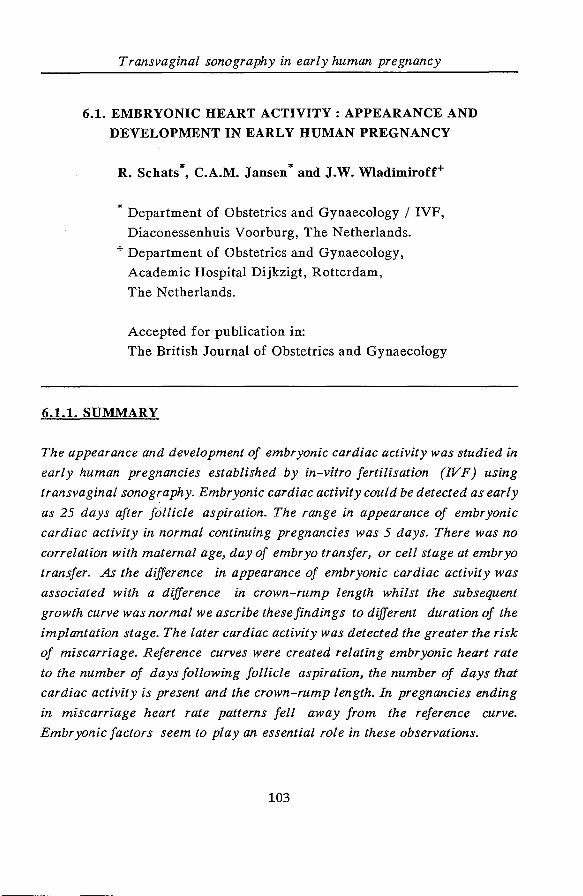
Transvaginal sonography in early human pregnancy
6.1. EMBRYONIC HEART ACTIVITY : APPEARANCE AND
DEVELOPMENT IN EARLY HUMAN PREGNANCY
R. Schatss, C.A.M. Jansen"' and J.W. Wladimiroff+
"' Department of Obstetrics and Gynaecology I IVF,
Diaconessenhuis Voorburg, The Netherlands.
+ Department of Obstetrics and Gynaecology,
Academic Hospital Dijkzigt, Rotterdam,
The Netherlands.
Accepted for publication in:
The British Journal of Obstetrics and Gynaecology
6.1.1. SUMMARY
The appearance and development of embryonic cardiac activity was studied in
early human pregnancies established by in-vitro fertilisation (IVF) using
transvaginal sonography. Embryonic cardiac activity could be detected as early
as 25 days after follicle aspiration. The range in appearance of embryonic
cardiac activity in normal continuing pregnancies was 5 days. There was no
correlation with maternal age, day of embryo transfer, or cell stage at embryo
transfer. As the difference in appearance of embryonic cardiac activity was
associated with a difference in crown-rump length whilst the subsequent
growth curve was normal we ascribe these findings to different duration of the
implantation stage. The later cardiac activity was detected the greater the risk
of miscarriage. Reference curves were created relating embryonic heart rate
to the number of days following follicle aspiration, the number of days that
cardiac activity is present and the crown-rump length. In pregnancies ending
in miscarriage heart rate patterns fell away from the reference curve.
Embryonic factors seem to play an essential role in these observations.
103
Appearance and development of embryonic cardiac activity
6.1.2. INTRODUCTION
One of the first signs of a viable pregnancy is the appearance of embryonic
heart activity. Using a standard abdominal ultrasound technique, Robinson1
demonstrated this activity at 45 - 47 days of amenorrhoea. Ultrasound
equipment has improved dramatically since then. The recent introduction of
transvaginal sonography might enable detection of embryonic cardiac activity
at a much earlier stage. The major advantage of this technique is that the
distance between transducer and object of scanning, in this case the uterus
and its contents is greatly reduced. This creates the opportunity to use high
MHz frequencies with good resolving properties. Subjects' acceptance, despite
previous reservation, has been remarkably good, especially as there is no need
to endure a full bladder. The in-vitro fertilisation (IVF) procedure provides
exact data on the time of ovulation (=follicle aspiration) and the subsequent
embryo transfer, and therefore the duration of pregnancy. This allowed us to
obtain more insight into the onset of embryonic cardiac activity and its
development in early human pregnancy.
6.1.3. SUBJECTS and METHODS
A total of 47 women consented to participate in the study. All women became
pregnant after IVF treatment in our clinic. The study was approved by the
ethics committee of the institution. The procedure in our centre included the
transport of oocytes to the laboratory of the Department of Physiology II at
the Erasmus University of Rotterdam as has been described earlier2• The
mean age of the women in the study group was 34 years (range 24- 40 years).
A pilot-study in 23 women showed that embryonic cardiac activity could not
be detected before the 25th day after follicle aspiration. Based on this we
designed the following protocol: transvaginal sonography was performed 25
days after follicle aspiration and repeated every day until embryonic heart
activity appeared, then the women were scanned at intervals of 2 or 3 days
until 42 days after follicle aspiration. The protocol implicates between five
and eight recordings of cardiac activity for each embryo (median six). Two
104
Transvaginal sonography in early human pregnancy
days or 3 days were chosen at random depending on personal factors such as
availability and travelling distance.
All the women were scanned between 10.00 and 12.00 a.m. using the
Diasonics DRF 400 and the Toshiba SAL 77B vaginal probes. The former was
equipped with a 5.0 and a 7.5 MHz mechanical sector scan transducer and the
latter with a 5.0 MHz electronic sector scan probe. Scans were carried out by
R.S. and C.A.M.J .. All observations were taped and when there was heart
activity the frequency was nearly always documented by M-mode in
conjunction with the real-time scan (Toshiba SAL 77B) (Figure 6.1.1). The
embryonic heart rate was calculated from the M-mode or, if this was not
possible, from the real-time image. Embryonic cardiac activity was defined
as a distinct contractile pattern inside the embryo with a frequency different
Figure 6.1.1. Combined real-time and M-mode registration of an embryonic heart rate 29 days after follicle aspiration.
105
Appearance and development of embryonic cardiac activity
from that of the maternal heart. In case of doubt both frequencies were
counted simultaneously to be absolutely sure of registering the embryonic
heart beat.
In 12 women the timing of onset of embryonic cardiac activity and the
embryonic heart rate were determined by both R.S. and C.A.M.J. from the
same M-mode recordings to exclude the possibility of inter-observer
variability. Both observers also measured heart rate independently in 10
women at intervals of 15 - 60 min to rule out differences in heart rate over
a short period of time.
The Kolmogorov-Smirnov two-sample test was used to test the distribution
of embryonic heart rate and crown-rump length (CRL) of single and multiple
pregnancies.
Embryonic heart rate was expressed as means +/-1 SD. The Wilcoxon rank
sum test was used to determine the difference in timing of onset of
embryonic cardiac activity between continuing pregnancies and pregnancies
which later miscarried in the first trimester.
6.1.4. RESULTS
There was complete agreement between the two observers for the timing of
onset of embryonic cardiac activity. The inter-observer variation for
embryonic heart rate determination from the same M-mode recordings was
very low. The mean of the differences was 2.3 (SD 1.5, range 0 - 4, median
2). There was almost no variation in the embryonic heart rate over a short
period of time (15- 60 min). The mean of the differences was 2.9 (SD 1.7,
range 0- 4, median 2).
In eight out of 47 women we were not able to time the onset of embryonic
cardiac activity due to personal factors such as non-availability on specific
days and holidays of the women. This resulted in incomplete data from eight
women (five singleton and three twin pregnancies) and complete data from
39 women (26 singletons, ten twins, three triplets) for further analysis. We
included also multiple pregnancies, because so early in gestation development
of embryonic cardiac activity and growth rate of CRL is similar in singleton
106
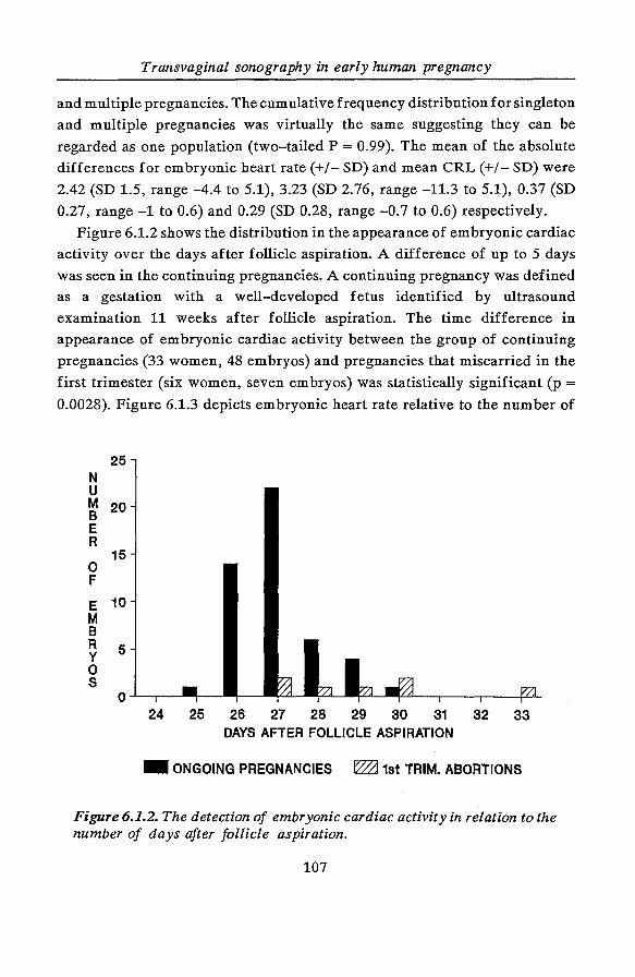
Transvaginal sonography in early human pregnancy
and multiple pregnancies. The cumulative frequency distribution for singleton
and multiple pregnancies was virtually the same suggesting they can be
regarded as one population (two-tailed P = 0.99). The mean of the absolute
differences for embryonic heart rate(+/- SD) and mean CRL (+/- SD) were
2.42 (SD 1.5, range -4.4 to 5.1), 3.23 (SD 2.76, range -11.3 to 5.1), 0.37 (SD
0.27, range -1 to 0.6) and 0.29 (SD 0.28, range -0.7 to 0.6) respectively.
Figure 6.1.2 shows the distribution in the appearance of embryonic cardiac
activity over the days after follicle aspiration. A difference of up to 5 days
was seen in the continuing pregnancies. A continuing pregnancy was defined
as a gestation with a well-developed fetus identified by ultrasound
examination 11 weeks after follicle aspiration. The time difference in
appearance of embryonic cardiac activity between the group of continuing
pregnancies (33 women, 48 embryos) and pregnancies that miscarried in the
first trimester (six women, seven embryos) was statistically significant (p = 0.0028). Figure 6.1.3 depicts embryonic heart rate relative to the number of
N u
25
M 20 B E R
0 F
15
E 10 M B R y 0 s
5
24 25 26 27 28 29 30 31 32 33 DAYS AFTER FOLLICLE ASPIRATION
- ONGOING PREGNANCIES ~ 1st TRIM. ABORTIONS
Figure 6.1.2. The detection of embryonic cardiac activity in relation to the number of days after follicle aspiration.
107
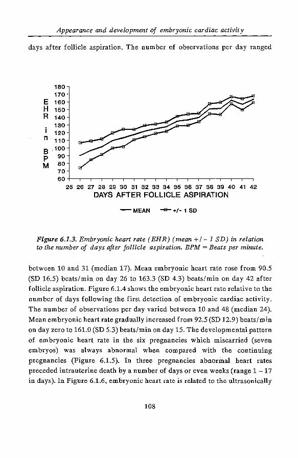
Appearance and development of embryonic cardiac activity
days after follicle aspiration. The number of observations per day ranged
180 170
E 160 H 150 R 140
130 i 120 n 110
8 '100 p 90 M so·
70.
60+--r~r-~-r~--~-r-,--~-r-,--~-.-,--.--.-.
25 26 27 28 29 30 31 32 33 34 35 36 37 38 39 40 41 42 DAYS AFTER FOLLICLE ASPIRATION
-MEAN -+/-1 SO
Figure 6.1.3. Embryonic heart rate (EHR) (mean +I- 1 SD) in relation to the number of days after follicle aspiration. BPM =Beats per minute.
between 10 and 31 (median 17). Mean embryonic heart rate rose from 90.5
(SD 16.5) beats/min on day 26 to 163.3 (SD 4.3) beats/min on day 42 after
follicle aspiration. Figure 6.1.4 shows the embryonic heart rate relative to the
number of days following the first detection of embryonic cardiac activity.
The number of observations per day varied between 10 and 48 (median 24).
Mean embryonic heart rate gradually increased from 92.5 (SD 12.9) beats/min
on day zero to 161.0 (SD 5.3) beats/min on day 15. The developmental pattern
of embryonic heart rate in the six pregnancies which miscarried (seven
embryos) was always abnormal when compared with the continuing
pregnancies (Figure 6.1.5). In three pregnancies abnormal heart rates
preceded intrauterine death by a number of days or even weeks (range 1 -17
in days). In Figure 6.1.6, embryonic heart rate is related to the ultrasonically
108
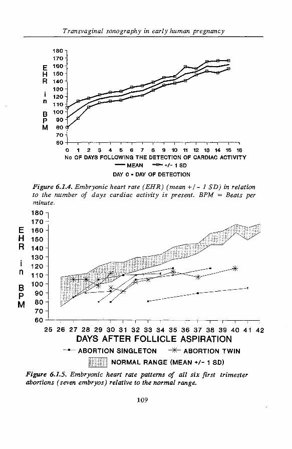
E H R
i n
8 p M
E H R
i n
8 p M
Transvaginal sonography in early human pregnancy
180 170 160 150 140 130 120 110 100 90 80 70 60
0 1 2 3 4 5 6 7 8 9 10 11 12 13 14 15 16 No OF DAYS FOLLOWING THE DETECTION OF CARDIAC ACTIVITY
-MEAN -B-+/-1 SO
DAY 0 • DAY OF DETECTION
Figure 6.1.4. Embryonic heart rate (EHR) (mean +I- 1 SD) in relation to the number of days cardiac activity is present. BPM = Beats per minute.
180 170 160 150 140 130 120 110 100 90 80 70 60
25 26 27 28 29 30 31 32 33 34 35 36 37 38 39 40 41 42
DAYS AFTER FOLLICLE ASPIRATION - ABORTION SINGLETON ~ ABORTION TWIN
NORMAL RANGE (MEAN +/- 1 SO)
Figure 6.1.5. Embryonic heart rate patterns of all six first trimester abortions (seven embryos) relative to the normal range.
109
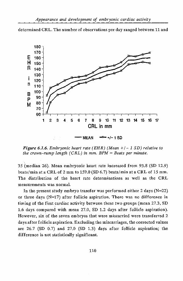
Appearance and development of embryonic cardiac activity
determined CRL. The number of observations per day ranged between 11 and
180 170
E 160 H 150 R 140
i 130 120
n 110
8 100
p 90
M 80 70 60
1 2 3 4 5 6 7 8 9 10 11 12 13 14 15 16 17 CRL in mm
- MEAN -s- +/- 1 SO
Figure 6.1.6. Embryonic heart rate ( EHR) (Mean +I- 1 SD) relative to the crown-rump length (CRL) in mm. BPM =Beats per minute.
35 (median 26). Mean embryonic heart rate increased from 93.8 (SD 12.9)
beats/min at a CRL of 2 mm to 159.0 (SD 6.7) beats/min at a CRL of 15 mm.
The distribution of the heart rate determinations as well as the CRL
measurements was normal.
In the present study embryo transfer was performed either 2 days (N=22)
or three days (N=17) after follicle aspiration. There was no difference in
timing of the first ~ardiac activity between these two groups (mean 27.3, SD
1.6 days compared with mean 27.0, SD 1.2 days after follicle aspiration).
However, six of the seven embryos that were miscarried were transferred 2
days after follicle aspiration. Excluding the miscarriages, the corrected values
are 26.7 (SD 0.7) and 27.0 (SD 1.3) days after follicle aspiration; the
difference is not statistically significant.
110
Transvaginal sonography in early human pregnancy
No correlation was found between the cell stage at embryo transfer (two
to eight cell stage) and appearance of first embryonic cardiac activity (r =-
0.16). Finally, there was a very weak correlation between the timing of first
embryonic cardiac activity and maternal age (r = 0.18).
6.1.5. DISCUSSION
The more sensitive technique of transvaginal sonography has opened up a new
field of research and provides the opportunity to study the very early stages
in the development of human pregnancy.
The advantage of studying IVF pregnancies is that some of the variables
such as time of ovulation (=follicle aspiration) and embryo transfer are
exactly known. Whether the results can be extrapolated to spontaneous
pregnancies is uncertain.
Most women in our study were older than average for pregnant women in
the Netherlands, reflecting the population currently having IVF treatment.
Embryonic cardiac activity could be detected as early as 25 days after
follicle aspiration. The spread of five days in the detection of heart activity
in continuing pregnancies is considerable. Five days represents between 16.7%
and 20% of the duration of pregnancy at that time. There is no clear
explanation. Maternal age, the time between follicle aspiration and embryo
transfer and the cell stage of the embryos at embryo transfer do not seem to
play a significant role. Although the timing of embryo transfer after follicle
aspiration does not influence the appearance of embryonic cardiac activity,
in five of the six (one twin) pregnancies that miscarried the six embryos were
transferred two days after follicle aspiration. This observation accords with
the findings by Os van et al3, who reported a statistically significant
difference in the proportion of continuing pregnancies between embryos
transferred two days and three days after follicle aspiration. Other factors,
most likely embryonic, in conjunction with endometrial conditions at the sie?
of implantation may play an important role in establishing a pregnancy.
Moreover, in multiple pregnancies we often observed a variation in
111
Appearance and development of embryonic cardiac activity
appearance of cardiac activity between embryos, which emphasizes the
importance of embryonic factors, rather than maternal variables which under
these circumstances are constant4• The growth potential of the embryo itself
seems to be very important in the struggle for implantation. It is possible that
fast dividing embryos stand a better chance of implantation, and we
examined whether this division potential was related to the day of first
detection of cardiac activity 5, but we found no relation between cell stage at
embryo transfer and the day at which cardiac activity was detected. Strong,
normal embryos will implant fast, but it seems impossible to judge the quality
of the embryos on morphological and cleavage characteristics alone5•7. If the
asynchrony in appearance of cardiac activity in multiple pregnancies indeed
reflects variability in embryonic rather than maternal factors, then it is easier
to understand why a later appearance of embryonic cardiac activity is
associated with a higher chance of miscarriage.
Transvaginal sonography did not only enable us to detect the embryonic
heart beat one week earlier than reported previously, but we also found a
certain pattern in ·the development of the embryonic heart rate. This was
about 90 beats/min on day 26 and gradually increased to about 165 beats/min
on day 42. Further improvement in ultrasound equipment may elucidate the
rate at which the heart tube really starts functioning. Embryonic heart
rhythm appeared to be very regular, however, quantification of the beat to
beat variation from M-mode recordings was not feasible. The subsequent
increase in rate may be an expression of the maturation of the nervous system
or an adaptation to the size of the embryo or both. This may explain the
observed relation between crown-rump length and heart rate at this stage of
pregnancy. The pregnancies ending in miscarriage all showed an abnormal
heart rate pattern in relation to our reference curve. This deviating pattern
seems to be a reflection of the reduced growth capacity of the embryo. Heart
rate may start off normal, but after a few days it falls away from the
reference curve.
We conclude that m continuing IVF pregnancies the date of onset of
embryonic cardiac activity may differ as much as five days. The later the
onset of cardiac activity the greater the chance of miscarriage. The embryonic
112
Transvaginal sonography in early human pregnancy
heart rate preceding miscarriage may initially be normal or lower, but
ultimately falls away from the reference curve. Environmental factors do not
seem to play a significant role in these findings, so we believe the factors
determining onset of cardiac activity and subsequent increase in embryonic
heart rate are mainly embryonic in origin.
6.1.6. REFERENCES
1. Robinson HP, Shaw-Dunn J. Fetal heart rates as determined by sonar
in early pregnancy. J Obstet Gynaecol Br Cwlth 1973; 80: 805-9.
2. Jansen CAM, Beek JJ van, Verhoeff A, Alberda ATh, Zeilmaker GH. In
vitro fertilisation and embryo transfer with transport of oocytes. Lancet
1986; i: 676.
3. Os HC van, Alberda ATh, Janssen-Caspers HAB, Leerentveld RA,
Scholtes MCW, Zeilmaker GH. The influence of the interval between in
vitro fertilization and embryo transfer and some other variables on
treatment outcome. Fertil Steril 1989; 51: 360-2.
4. Schats R, Brandsma G, Cleveringa LM, Lankhorst PFC, Vroegop IS,
Jansen CAM. Evidence of asynchronous implantation in IVF multiple
pregnancies. Hum Reprod 1988; 3: Suppl: Abstract 53, p 17.
5. Edwards RG, Purdy JM, Steptoe PC, Walters DE. The growth of human
preimplantation embryos in-vitro. Am J Obstet Gynecol1981; 141: 408-
15.
6. Acosta AA, Moon SY, Oehninger S, Muasher SJ, Rosenwaks Z, Matta JF.
Implantation potential of each pre-embryo in multiple pregnancies
obtained by in vitro fertilization seems to be different. Fertil Steril1988;
50: 906-11.
7. Lopata A, Martin M, Oliva K, Johnston I. Embryonic development and
blastocyst implantation following in vitro fertilization and embryo
transfer. Fertil Steril1982; 38: 682-90.
113

114
Transvaginal sonography in early human pregnancy
6.2. ASYNCHRONOUS APPEARANCE OF EMBRYONIC CARDIAC
ACTIVITY IN MULTIPLE PREGNANCIES
FOLLOWING IN-VITRO FERTILISATION
R. Schats"', C.A.M. Jansen"' and J.W. Wladimiroff+
* Department of Obstetrics and Gynaecology I IVF,
Diaconessenhuis Voorburg, The Netherlands.
+ Department of Obstetrics and Gynaecology,
Academic Hospital Dijkzigt, Rotterdam,
The Netherlands.
Accepted for publication in:
Ultrasound in Medicine and Biology
(Letter to the Editor)
6.2.1. INTRODUCTION
Implantation is a continuing process that extends from day 5 or 6 up to about
day 12 following ovulation. The embryo becomes a blastocyst on day 4 or 5
after fertilisation. At this stage it is capable, after hatching, of attachment to
the endometrium and of implantation. In single pregnancies following in
vitro fertilisation (IVF) a difference up to 5 days in the appearance of
embryonic heart activity has been demonstrated1. The factors determining
rapid or slow implantation and subsequent development are disputed.
Multiple pregnancies provide the opportunity to study different embryos in
the same individual, thereby standardising for maternal influences. The
objective of the present study was to establish a difference in the duration of
the implantation stage in the human through documentation of the first
appearance of embryonic cardiac activity in multiple pregnancies.
115
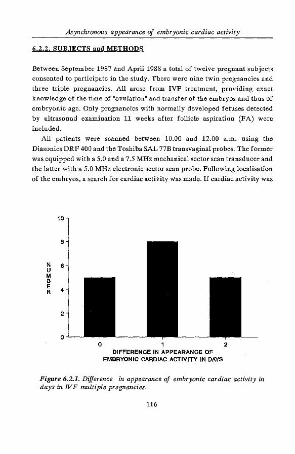
Asynchronous appearance of embryonic cardiac activity
6.2.2. SUBJECTS and METHODS
Between September 1987 and April1988 a total of twelve pregnant subjects
consented to participate in the study. There were nine twin pregnancies and
three triple pregnancies. All arose from IVF treatment, providing exact
knowledge of the time of "ovulation" and transfer of the embryos and thus of
embryonic age. Only pregnancies with normally developed fetuses detected
by ultrasound examination 11 weeks after follicle aspiration (FA) were
included.
All patients were scanned between 10.00 and 12.00 a.m. using the
Diasonics DRF 400 and the Toshiba SAL 77B transvaginal probes. The former
was equipped with a 5.0 and a 7.5 MHz mechanical sector scan transducer and
the latter with a 5.0 MHz electronic sector scan probe. Following localisation
of the embryos, a search for cardiac activity was made. If cardiac activity was
N u M 8 E R
10
8
6
4
2
0 0 1 2
DIFFERENCE IN APPEARANCE OF EMBRYONIC CARDIAC ACTIVITY IN DAYS
Figure 6.2.1. Difference in appearance of embryonic cardiac activity in days in IVF multiple pregnancies.
116
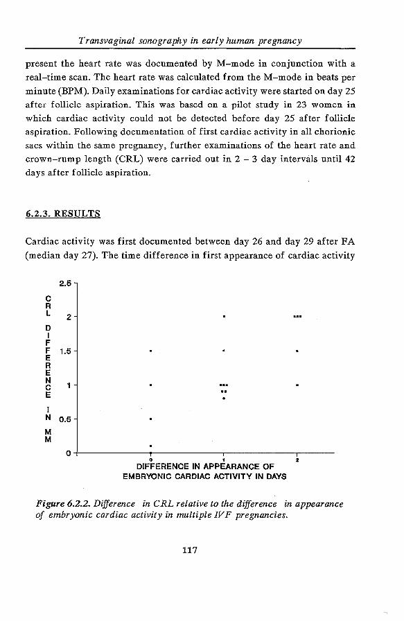
Transvaginal sonography in early human pregnancy
present the heart rate was documented by .M-mode in conjunction with a
real-time scan. The heart rate was calculated from theM-mode in beats per
minute (BPM). Daily examinations for cardiac activity were started on day 25
after follicle aspiration. This was based on a pilot study in 23 women in
which cardiac activity could not be detected before day 25 after follicle
aspiration. Following documentation of first cardiac activity in all chorionic
sacs within the same pregnancy, further examinations of the heart rate and
crown-rump length (CRL) were carried out in 2 - 3 day intervals until 42
days after follicle aspiration.
6.2.3. RESULTS
Cardiac activity was first documented between day 26 and day 29 after FA
(median day 27). The time difference in first appearance of cardiac activity
c R L
D I F
2.6
2
F 1.5 E R E
~ 1 E
I N 0.5
M M
0 1 2
DIFFERENCE IN APPEARANCE OF EMBRYONIC CARDIAC ACTIVITY IN DAYS
Figure 6.2.2. Difference in CRL relative to the difference in appearance of embryonic cardiac activity in multiple IVF pregnancies.
117
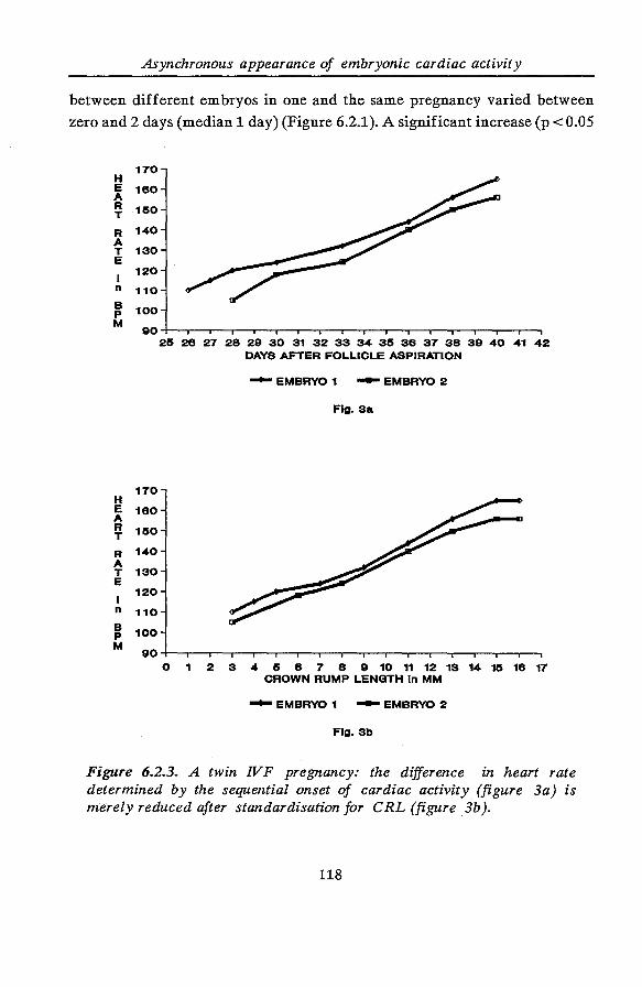
Asynchronous appearance of embryonic cardiac activity
between different embryos in one and the same pregnancy varied between
zero and 2 days (median 1 day) (Figure 6.2.1). A significant increase (p < 0.05
170 H E 160 A ~ 150
R 140 A T 130 E
I 120
n 110
~ 100
M 90~-o--r-.-~--.--.-,--.--r-.,-,--.--r-.-~--.-~ 25 26 27 28 29 30 31 32 33 34 35 36 37 38 39 40 41 42
DAYS AFTER FOLLICLE ASPIRATION
170 H E 160 A R 150 T
R 140 A T 130 E
I 120
n 110 B 100 p M
90 0 1
-EMBRYO 1 -EMBRYO 2
Fig. Sa
2 3 4 5 a 7 8 e w ff n ~ u ~ ~ v CROWN RUMP LENGTH In MM
-EMBRYO 1 -EMBRYO 2
Fig. 3b
Figure 6.2.3. A twin IVF pregnancy: the difference in heart rate determined by the sequential onset of cardiac activity (figure 3a) is merely reduced after standardisation for CRL (figure 3b ).
118
Transvaginal sonography in early human pregnancy
; Wilcoxon rank sum test) in CRL difference relative to the difference in
appearance of embryonic cardiac activity was observed, when all multiple
pregnancies were considered together (Figure 6.2.2). This is also demonstrated
in Figure 6.2.3, in which for two embryos from the same pregnancy the
difference in heart rate determined by the sequential onset of cardiac activity
(Figure 6.2.3a) is merely reduced up to day 40 after follicle aspiration
following standardisation for CRL (Figure 6.2.3b).
6.2.4. DISCUSSION
Until now it is impossible to judge, on morphological grounds alone the
quality of oocytes after stimulation and harvesting, and the quality of the
pre-embryos they produce after fertilisation2• Under in-vitro conditions the
difference in developmental potential of pre-embryos has been documented
from the cleavage rate3• 4• This finding cannot be extrapolated to the in-vivo
situation because of the different environment in which the pre-embryos·
develop, in particular the quality of the endometrium. Implantation is the
means by which the early embryo establishes a fixed relationship with respect
to the uterus. The nature of the implantation process varies considerably
between species and may include such events as the activation, spacing and
orientation of the blastocyst, adhesion of the trophoblast to the luminal
epithelium, invasion of the endometrial stroma and the development of a
decidual response. The successful completion of these events involves a
complex series of synchronised changes in the blastocyst and endometrium
under the control of ovarian steroids. The very complexity of implantation
makes this an extremely critical stage of early pregnancy.
A multiple IVF pregnancy offers the unique opportunity to study the
development of two or even three embryos in the same subject, thus
standardising for inter-individual differences such as maternal age, parity,
and other environmental variables. The difference in appearance of cardiac
activity between embryos from the same pregnancy may be determined by
asynchrony in onset of maternal nutrient supply, although other factors such
as micro-environmental differences cannot be ruled out. Howe'(er, after
119
Asynchronous appearance of embryonic cardiac activity
implantation the growth potential for each of those embryos was virtually the
same, as reflected by the increase of CRL and the heart rate. We therefore
suggest that the difference in first appearance of cardiac activity between
embryos from the same pregnancy may be determined by the variable
duration of the implantation stage.
6.2.5. REFERENCES
1. Schats R, Brandsma G, Cleveringa LM, Lankhorst PFC, Vroegop IS,
Jansen CAM. Detection and monitoring of fetal heart activity in early
pregnancy. Hum Reprod 1988; 3: Suppl. 78 (Abstract).
2. Acosta AA, Moon SY, Oehninger S, Muasher SJ, Rosenwaks Z, Matta JF.
Implantation potential of each pre-embryo in multiple pregnancies
obtained by in vitro fertilization seems to be different. Fertil Steril1988;
50: 906-11.
3. Edwards RG, Purdy JM, Steptoe PC, Walters DE. The growth of human
preimplantation embryos in vitro. Am J Obstet Gynecol1981; 141: 408-
15. 4. Lopata A, Martin M, Oliva K, Johnston I. Embryonic development and
blastocyst implantation following in vitro fertilization and embryo
transfer. Fertil Steril 1982; 38: 682-90.
120
Transvaginal sonography in early human pregnancy
6.3. ABNORMAL EMBRYONIC HEART RATE PATTERN IN EARLY
PREGNANCY ASSOCIATED WITH DOWN'S SYNDROME
R. Schats ", C.A.M. Jansen" and J. W. Wladimiroff+
"' Department of Obstetrics and Gynaecology I IVF,
Diaconessenhuis Voorburg, The Netherlands.
+ Department of Obstetrics and Gynaecology,
Academic Hospital Dijkzigt, Rotterdam,
The Netherlands.
Published in:
Human Reproduction 1990; 5: 877-9.
6.3.1. SUMMARY
The case history of a 35-year-old woman with a pregnancy following IVF
treatment is presented. Transvaginal sonography revealed an abnormal
embryonic heart rate pattern between 28 and 50 days following follicular
aspiration. At term, a male infant with the characteristics of Down's syndrome
was born. The diagnosis was confirmed by chromosome analysis. The
implications of this observation are discussed.
121
Down's syndrome associated with abnormal embryonic heart rate pattern
6.3.2. INTRODUCTION
Several ultrasonographic signs have been described in second trimester fetuses
at high risk for Down's syndrome1· 7• Although parameters like a thickened
nuchal fold, an increased biparietal diameter I femur length ratio and
hypoplasia of the middle phalanx of the fifth digit are reported, there is no
general agreement about the sensitivity and specificity of these signs1· 7• A
great disadvantage of these features is that they are recognised late in
pregnancy. It would be of great value if a reliable parameter could be
identified early in pregnancy which can indicate fetuses at risk for Down's
syndrome or other chromosome abnormalities. Transvaginal sonography
enables detailed information to be obtained about early morphological and
physiological deve~opment of the embryo, because probes with high emission
frequencies (5.0 - 7.5 MHz) and therefore high resolution can be employed.
This case history illustrates that a deviating development of the embryonic
heart rate was associated with a chromosome abnormality.
6.3.3. CASE HISTORY
A 35-year-old woman requested IVF treatment in our clinic. Infertility
analysis in another hospital had revealed tubal pathology to such a degree that
the chance of a spontaneous pregnancy could be regarded as niL Following
our routine controlled hyperstimulation scheme containing pure FSH, hMG,
and hCG, oocyte recovery took place on day 12 of the cycle. Seven oocytes
were obtained and three of them became fertilised. Transfer of the three
embryos (8-,8- and 6-cell stage) was carried out three days after follicular
aspiration. Fifteen days after oocyte collection, a pregnancy test (Predictor
color) was positive. The patient consented to participate in a study on the
appearance and development of embryonic cardiac activity. According to the
study protocol daily transvaginal sonographic examinations were started on
day 25 following oocyte recovery until the presence of embryonic cardiac
activity was established, with documentation of the heart rate at two to three
day intervals thereafter. The first embryonic heartbeats could be registered
122
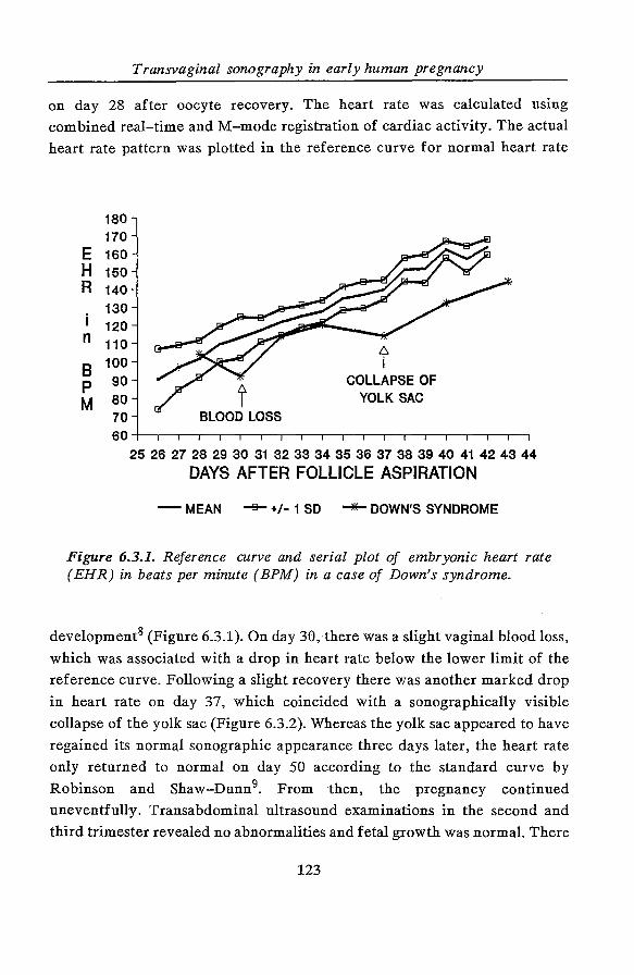
Transvaginal sonography in early human pregnancy
on day 28 after oocyte recovery. The heart rate was calculated using
combined real-time and M-mode registration of cardiac activity. The actual
heart rate pattern was plotted in the reference curve for normal heart rate
180 170
E 160 H 150 R 140
i 130 120
n 110
8 100
p 90
M 80 70 60
COLLAPSE OF YOLK SAC
BLOOD LOSS
25 26 27 28 29 30 31 32 33 34 35 36 37 38 39 40 41 42 43 44
DAYS AFTER FOLLICLE ASPIRATION
- MEAN -e- +/- 1 SO '""*"'"DOWN'S SYNDROME
Figure 6.3.1. Reference curve and serial plot of embryonic heart rate (EHR) in beats per minute (BPM) in a case of Down's syndrome.
development8 (Figure 6.3.1). On day 30, there was a slight vaginal blood loss,
which was associated with a drop in heart rate below the lower limit of the
reference curve. Following a slight recovery there was another marked drop
in heart rate on day 37, which coincided with a sonographically visible
collapse of the yolk sac (Figure 6.3.2). Whereas the yolk sac appeared to have
regained its norma,l sonographic appearance three days later, the heart rate
only returned to normal on day 50 according to the standard curve by
Robinson and Shaw-Dunn9• From then, the pregnancy continued
uneventfully. Transabdominal ultrasound examinations in the second and
third trimester revealed no abnormalities and fetal growth was normal. There
123
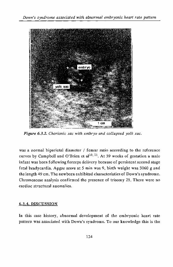
Down's syndrome associated with abnormal embryonic heart rate pattern
Figure 6.3.2. Chorionic sac with embryo and collapsed yolk sac.
was a normal biparietal diameter I femur ratio according to the reference
curves by Campbell and O'Brien et al10• 11 . At 39 weeks of gestation a male
infant was born following forceps delivery because of persistent second stage
fetal bradycardia. Apgar score at 5 min was 9, birth weight was 3060 g and
the length 49 em. The new born exhibited characteristics of Down's syndrome.
Chromosome analysis confirmed the presence of trisomy 21. There were no
cardiac structural anomalies.
6.3.4. DISCUSSION
In this case history, abnormal development of the embryonic heart rate
pattern was associated with Down's syndrome. To our knowledge this is the
124
Transvaginal sonography in early human pregnancy
first report of such an association. It would be interesting to know if this
observation is just more than a coincidence. In a previous series of seven
embryos with abnormal heart rate patterns between 25 and 42 days after
oocyte recovery all embryos aborted in the first trimester8• Karyotyping in
embryonic tissue was feasible in five cases, revealing an abnormal
chromosomal pattern in two cases (45 XO and 46X-X + t(X;1)(q22;q23). Two
cases showed a normal 46 XX chromosomal pattern and in one the tissue
culture failed. In the case reported here vaginal blood loss and the temporary
collapse of the yolk sac suggested threatening abortion. However, the
pregnancy continu.ed. It is common knowledge that spontaneous abortion is
often associated with chromosome abnormalities. If there is indeed a close
relation between early abnormal heart rate patterns and chromosome
anomalies, chorionic villus sampling or amniocentesis should be offered in
these cases. Further evidence is needed, until then the natural history of these
abnormal early heart rate patterns should be studied.
6.3.5. REFERENCES
1. Benecerraf BR, Gelman R, Prigoletto PD. Sonographic identification of
second-trimester fetuses with Down's syndrome. N Engl J Med 1987; 317:
1371-6.
2. Benacerraf BR, Osathanondh R, Prigoletto PD. Sonographic demonstration
of hypoplasia of the middle phalanx of the fifth digit: a finding associated
with Down syndrome. Am J Obstet Gynecol1988; 159: 181-3.
3. Brumfield CG, Hauth JC, Cloud GA, Davis RO, Henson, BV. Sonographic
measurements and ratios in fetuses with Down syndrome. Obstet Gynecol
1989; 73: 644-6.
4. Lockwood C, Benacerraf B, Krinsky A, Blakemore K, Belanger K,
Mahoney M, Hobbins J. A sonographic screening method for Down
syndrome. Am J Obstet Gynecol 1987; 157: 803-8.
5. Lynch L, Berkowitz GS, Chitkara U, Wilkins IA, Mechalek KE,
Berkowitz RL. Ultrasound detection of Down syndrome: is it really
possible? Obstet Gynecol1989; 73: 267-70.
125
Down's syndrome associated with abnormal embryonic heart rate pattern
6. Perrella R, Duerinckx AJ, Grant EG, Tessler F, Tabsh K, Crandall BF.
Second-trimester sonographic diagnosis of Down syndrome: role of
femur-length shortening and nuchal fold thickening. Am J Radiol1988;
151: 981-5.
7. Toi A, Simpson GF, Filly RA. Ultrasonically evident fetal nuchal skin
thickening: is it specific for Down syndrome? Am J Obstet Gynecol1987;
156: 150-3.
8. Schats R, Jansen CAM, Wladimiroff JW. Embryonic heart activity:
appearance and development in early human pregnancy. Br J Obstet
Gynaecol, 1990, in press.
9. Robinson HP, Shaw-Dunn J. Fetal heart rates as determined by sonar
in early pregnancy. J Obstet Gynaecol Br Commonw 1973; 80: 805-9.
10. Campbell S. Fetal growth. In: Fetal physiology and medicine. R.W. Beard
& P.W. Nathanielsz (eds). W.B. Saunders Co., Philadelphia 1976; 271-300.
11. O'Brien GD, Queenan JT, Campbell S. Assessment of gestational age in
the second trimester by real-time ultrasound measurement of the femur
length. Am J Obstet Gynecol1981; 139: 540-7.
126
Transvaginal sonography in early human pregnancy
CHAPTER 7
CONCLUSIONS
Transvaginal sonography can be regarded as a breakthrough in the
visualisation of early pregnancy development. It allows the use of transducers
with higher emission frequencies, resulting in better image resolution.
The acceptance of this method by patients is excellent. Moreover, energy
output at the level of the embryo is lower with transvaginal scanning than
with transabdominal scanning.
Anatomical as well as functional aspects of gestational development can be
evaluated earlier than previously described with transabdominal scanning.
The diagnosis of vital and non-vital intra-uterine pregnancy can now be
made as early as 6Y2 weeks menstrual age.
The introduction of transvaginal sonography allows more detailed
visualisation of embryonic anatomy and, therefore, earlier and more accurate
measurement of the crown-rump length (CRL). A reappraisal of the current
reference chart for embryonic CRL demonstrates a smaller distribution of
CRL measurements, allowing more accurate pregnancy dating. Moreover,
transvaginal CRL measurements can be obtained approximately four days
earlier than previously reported employing transabdominal scanning.
Transvaginal sonography is also helpful in determining the fate of those
pregnancies associated with vaginal bleeding. A rather fixed sequence of
distinctive features of embryonic development (visualisation of the gestational
sac and yolk sac; appearance of cardiac activity; embryonic growth (CRL
measurement)) has to be followed between definite time limits.
Transvaginal sonography m combination with sensitive hCG
determinations permits the diagnosis of ectopic pregnancy before clinical
signs or symptoms have appeared. This allows conservative management,
which is of particular importance regarding future fertility. Transvaginal
puncture under sonographic guidance with or without the instillation of
cytoreductive agents may be successful. However, the failure rate of this
approach is still too high to advocate general use. At present, the place of
127
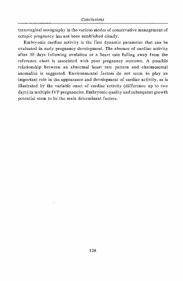
Conclusions
transvaginal sonography in the various modes of conservative management of
ectopic pregnancy has not been established clearly.
Embryonic cardiac activity is the first dynamic parameter that can be
evaluated in early pregnancy development. The absence of cardiac activity
after 30 days following ovulation or a heart rate falling away from the
reference chart is associated with poor pregnancy outcome. A possible
relationship between an abnormal heart rate pattern and chromosomal
anomalies is suggested. Environmental factors do not seem to play an
important role in the appearance and development of cardiac activity, as is
illustrated by the variable onset of cardiac activity (difference up to two
days) in multiple IVF pregnancies. Embryonic quality and subsequent growth
potential seem to be the main determinant factors.
128
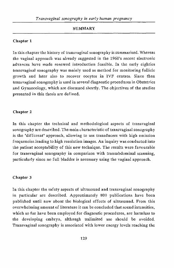
Transvaginal sonography in early human pregnancy
SUMMARY
Chapter 1
In this chapter the history of transvaginal sonography is summarised. Whereas
the vaginal approach was already suggested in the 1960's recent electronic
advances have made renewed introduction feasible. In the early eighties
transvaginal sonog:raphy was mainly used as method for monitoring follicle
growth and later also to recover oocytes in IVF centres. Since then
transvaginal sonography is used in several diagnostic procedures in Obstetrics
and Gynaecology, which are discussed shortly. The objectives of the studies
presented in this thesis are defined.
Chapter 2
In this chapter the technical and methodological aspects of transvaginal
sonography are described. The main characteristic of transvaginal sonography
is the "different" approach, allowing to use transducers with high emission
frequencies leading to high resolution images. An inquiry was conducted into
the patient acceptability of this new technique. The results were favourable
for transvaginal sonography in comparison with transabdominal scanning,
particularly since no full bladder is necessary using the vaginal approach.
Chapter 3
In this chapter the safety aspects of ultrasound and transvaginal sonography
in particular are described. Approximately 800 publications have been
published until now about the biological effects of ultrasound. From this
overwhelming amount of literature it can be concluded that sound intensities,
which so far have been employed for diagnostic procedures, are harmless to
the developing embryo, although unlimited use should be avoided.
Transvaginal sonography is associated with lower energy levels reaching the
129
Summary
embryo than in the transabdominal approach under the assumption of the
same start intensities.
Chapter 4
In the first part of this chapter ( 4.1) an overview is provided of embryonic
development in the first six weeks following conception. This development
is related to the transvaginal sonographic findings in normal developing
gestations in this period. A number of embryonic landmarks (gestational sac,
yolk sac and cardiac activity) can be visualised about one week earlier than
previously reported. The second part (4.2.) shows that the reference chart
currently in use for embryonic crown-rump length measurements derived
from data obtained with transabdominal sonography (compound B-scan)
needs to be adjusted, since it is possible to measure embryonic crown-rump
length earlier and more accurate employing transvaginal sonography.
Chapter 5
In the first part of this chapter (5.1.) the transvaginal sonographic
characteristics of abnormally developing intra-uterine gestations are
described. The sonographic findings in a group of patients presenting with
vaginal bleeding in the first trimester of pregnancy are described. Our own
findings are compared with data reported by other authors. It is stated that
the diagnosis of abnormal gestation can be made about one week earlier with
transvaginal sonography. In the second part (5.2.) the role of transvaginal
sonography in the diagnosis and management of ectopic pregnancy is
discussed. The availability of sensitive hCG determinations in combination
with transvaginal sonography permits the diagnosis of ectopic pregnancy
before clinical signs and symptoms have appeared. This provides the
opportunity to employ more conservative modes of treatment, which is
especially important regarding aspects of future fertility. At presel?-t, the
place of transvaginal sonography in the management of ectopic pregnancy
130
Transvaginal sonography in early human pregnancy
still has to be established. The significance of transvaginal puncture of
ectopic pregnancies with or without systemic or local administration of
methotrexate is discussed.
Chapter 6
This chapter consists of three parts. In the first part (6.1.) several aspects of
appearance and subsequent development of embryonic cardiac activity in IVF
pregnancies is described. A spread of five days in the appearance of cardiac
activity was observed. The later cardiac activity can be visualised, the higher
the chance of abortion. Also abnormal development of embryonic heart rate
is associated with an increased risk of abortion. The factors determining the
onset and subsequent development of the heart rate are most likely embryonic
in origin. Environmental factors like maternal age, do not seem to play a
significant role. Arguments for these assumptions are provided in the second
part of this chapter (6.2.). In IVF multiple pregnancies a difference of up to
two days in the appearance of cardiac activity between embryos was found
in a number of cases. The determining factor in this observation seems to be
a different duration of the implantation stage, since the growth of the
embryos following the appearance of cardiac activity was virtually the same.
In the third part (6.3.) a case history is presented in which abnormal
development of the embryonic heart rate in early pregnancy coincided with
the birth of an infant with Down's syndrome. The implications of this
observation are discussed.
131
Transvaginal sonography in early human pregnancy
SAMENVATTING
Hoofdstuk 1
In dit hoofdstuk is de geschiedenis van transvaginale echoscopie beknopt
weergegeven. De vaginale benadering werd reeds in de zestiger jaren
nagestreefd, echter de recente elektronische ontwikkelingen maakten de
feitelijke toepassing mogelijk en zinvol. In het begin van de jaren tachtig
werd transvaginale echoscopie vooral gebruikt als methode voor monitoring
van de follikelgroei en later ook om oocyten te verkrijgen in IVF centra.
Sindsdien heeft transvaginale echoscopie diverse toepassingen gevonden in de
Obstetrie en Gynaecologie. Deze worden in het kort aangegeven. De
doelstellingen van dit proefschrift worden gedefinieerd.
Hoofdstuk 2
In dit hoofdstuk worden de technische en methodologische aspecten van
transvaginale echoscopie besproken. Kenmerkend voor transvaginale
echoscopie is de "andere" benadering die het mogelijk maakt om transducers
te gebruiken met hoge emissie frequenties waardoor een hoog oplossend
vermogen wordt gecreeerd. Hiermee kan een beeld verkregen worden met
veel details. Een enquete naar de acceptatie van deze nieuwe techniek door
de patient viel zeer gunstig uit voor transvaginale echoscopie in vergelijking
met transabdominale echoscopie.
Hoofdstuk 3
In dit hoofdstuk komen de veiligheidsaspecten van ultrageluid en
transvaginale echoscopie in het bijzonder aan de orde. Er zijn tot op heden
ongeveer 800 publikaties verschenen die betrekking hebben op de biologische
effecten van ultrageluid. Uit deze overstelpende hoeveelheid literatuur kan
worden geconcludeerd, dat ultrageluid met intensiteiten zoals die worden
132
S amenvattin g
toegepast voor diagnostische doeleinden in de zwangerschap niet schadelijk
is, maar ongelimiteerd gebruik moet worden vermeden. Indien de geluids
intensiteiten die bereikt worden ter plaatse van het embryo voor
transabdominale en transvaginale echoscopie vergeleken worden valt op, dat
bij gelijke startintensiteit van de transducers deze intensiteit bij transvaginale
echoscopie lager ligt dan bij transabdominale echoscopie.
Hoofdstuk 4
In dit hoofdstuk wordt in het eerste gedeelte (4.1.) een overzicht gegeven van
de embryonale ontwikkeling in de eerste zes weken na conceptie. Deze
ontwikkeling wordt gekoppeld aan de transvaginale echoscopische
bevindingen in zich normaal ontwikkelende graviditeiten in deze periode. Het
blijkt, dat een aantal embryonale structuren waaronder de vruchtzak en
dooierzak, alsmede hartactiviteit ongeveer een week eerder kunnen worden
gedetecteerd met transvaginale echoscopie. In het tweede gedeelte ( 4.2.) wordt
aangetoond, dat de curve die voor de kruin-romp lengte tot op heden in
gebruik is en werd samengesteld uit gegevens die werden verkregen met
beh ul p van transa bdominale echoscopie (com pound B-scan) aan passing
behoeft, omdat het mogelijk is geworden om de kruin-romp lengte via
transvaginale echoscopie eerder en nauwkeuriger te meten.
Hoofdstuk 5
In het eerste gedeelte van dit hoofdstuk (5.1.) wordt ingegaan op de
transvaginale echoscopische kenmerken van zich abnormaal ontwikkelende
intra-uteriene graviditeiten. De bevindingen bij een groep patienten die zich
met bloedverlies in het eerste trimester van de graviditeit presenteerden
worden besproken. Onze onderzoeksresultaten worden vergeleken met de
gegevens uit de literatuur. Ook de diagnose abnormale graviditeit kan met
transvaginale echoscopie ongeveer een week eerder gesteld worden. In het
tweede gedeelte (5.2.) wordt de rol van transvaginale echoscopie in de
133
Transvaginal sonography in early human pregnancy
diagnostiek en behandeling van ectopische graviditeiten beschreven. Door het
beschikbaar komen van gevoelige hCG bepalingen in combinatie met
transvaginale echoscopie is het mogelijk geworden om de diagnose ectopische
graviditeit in een eerder, vaak nog asymptomatisch stadium te stellen.
Hierdoor wint men tijd om meer conservatieve methoden van behandeling,
waarbij gelet kan worden op aspecten van toekomstige fertiliteit, te
overwegen. Het is nog niet geheel duidelijk welke plaats transvaginale
echoscopie in deze zal gaan innemen. Transvaginale punctie van de ectopische
graviditeit als therapeutische ingreep al dan niet in combinatie met lokale of
systemische toediening van methotrexaat wordt besproken.
Hoofdstuk 6
Dit hoofdstuk bestaat uit drie gedeelten. In het eerste deel (6.1.) worden de
verschillende aspecten van verschijnen en ontwikkeling van de hartactiviteit
van het embryo in IVF graviditeiten beschreven. Er blijkt een spreiding te
bestaan van vijf dagen in het optreden van hartactiviteit. Naarmate de
hartactie later wordt vastgesteld des te groter de kans dat er een abortus
optreedt. Ook een abnormale ontwikkeling van de hartfrequentie van het
embryo lijkt samen te gaan met een verhoogde kans op abortus. De factoren
die het begin en de daaropvolgende ontwikkeling van de hartactiviteit
bepalen zijn waarschijnlijk in het embryo zelf gelegen. Omgevingsfactoren
zoals bijvoorbeeld leeftijd van de moeder, lijken van ondergeschikt belang.
Argumenten voor deze veronderstellingen worden aangedragen in het tweede
gedeelte (6.2.) van dit hoofdstuk. In IVF meerling graviditeiten kon tussen de
embryo's in een aantal gevallen een verschil tot 2 dagen worden aangetoond
in het optreden van hartactiviteit. Dit lijkt veroorzaakt te worden door een
verschillende duur van de implantatiefase, omdat de groei van de embryo's
na het verschijnen van hartactiviteit praktisch hetzelfde was. In het derde
gedeelte (6.3.) wordt een casus gepresenteerd waarbij een abnormale
ontwikkeling van de hartfrequentie van het embryo vroeg in de graviditeit
werd vastgesteld. Uiteindelijk werd er een kind geboren met het syndroom
van Down. De implicaties van deze observatie worden besproken.
134
ACKNOWLEDGEMENTS
The study described in this thesis was carried out for a great deal in the
Department of Obstetrics & Gynaecology I IVF of the Diaconessenhuis,
Voorburg, The Netherlands.
Dr. C.A.M. Jansen, dear Kees, you always speak with great enthusiasm
about the exciting views of early human development that can be obtained
with transvaginal sonography. You were the initiator of the study and
therefore, I am particularly indebted to him for his continuous support,
encouragement and advice.
Prof. Jhr. Dr. J.W., dear Jura, I especially want to express my gratitude
to you for your critical remarks and for giving the results of the study the
shape of a thesis.
I want to thank the patients who were so kind to participate in the study.
The great majority of the patients wanted to have ultrasound
examinations even more frequently than the protocol demanded because
they were even more thrilled about this technique resulting in such
beautiful pictures of their developing babies.
I like to thank Prof. Dr. N. Bom, Prof. Dr. H.P. van Geijn and Prof. Dr.
J. Voogd for their willingness to be members of the committee.
Dr. P. Hummel, dear Piet, you are not (yet) an expert in transvaginal
sonography but exactly that made you the ideal person to read critically
and conscientiously the final phase of this thesis. Many thanks again !
I like to thank my colleagues of the IVF-team of the Vrije Universiteit
for their mental support during the final stages of this thesis. The special
atmosphere in the VEVO I IVF department combined with the typical
humour of Amsterdam was the excellent mixture creating the right
climate for completing this task.
I like to thank my computer, who always did what he had to do, but had
the misfortune that he was conducted by an absolute beginner. He never
complained about my rudeness and inconsiderate behaviour. I am glad
that wordprocessing software programs exist, because I rewrote so many
times sentences and whole pages and even chapters, that I certainly should
have driven a few secretaries absolutely crazy.
Dear Wilma you do not want to be thanked, honoured or whatever in this
thesis, therefore I will not do so.
135

CURRICULUM VITAE
12-05-1956 Roelof Schats born in Leiden
1968- 1974 Secondary school education: Stedelijk Gymnasium, Leiden,
Gymnasium-B.
1974- 1980 State University of Leiden, Medical School.
Medical Degree Cum Laude.
1981 Research Fellow in the MRC Unit for Reproductive Biology,
Edinburgh, Scotland (Prof. R.V. Short, Prof. D.T Baird,
Mentor: Dr. R.J. Aitken).
1982- 1987 Residency Obstetrics and Gynaecology:
01-02-1982-31-07-1982 Zuiderziekenhuis, Rotterdam (Prof.
Dr. F.B. Lammes).
01-08-1982 - 31-10-1985 Academic Hospital Rotterdam -
Dijkzigt (Prof. Dr. A.C. Drogendijk, Prof. Dr. H.C.S.
Wallenburg and Prof. Dr. J.W. Wladimiroff).
01-11-1985- 01-02-1987 Zuiderziekenhuis, Rotterdam (Dr.
H.T. Lim).
01-02-1987 Board Certification Obstetrics and Gynaecology.
1987- 1988 Diaconessenhuis, Voorburg (Senior Registrar).
1989 Dr. Daniel den Hoed Kliniek, Rotterdam (Gynaecological
Oncology Fellow).
1990 - Academic Hospital of the "Vrije Universiteit",Amsterdam,
Gynaecologist especially for Reproductive Endocrinology,
Infertility Investigation and Assisted Fertility.
136

![UITNODIGING - netwerkacutezorgnoordwest.nl€¦ · UITNODIGING Voor het bijwonen van de openbare verdediging ÀvZ } ( Z ]L EMERGENCY DEPARTMENT CROWDING FACTORS INFLUENCING FLOW door](https://img.dokumen.tips/doc/110x75/5ed6824eff0e593c0b63f9a6/uitnodiging-netwe-uitnodiging-voor-het-bijwonen-van-de-openbare-verdediging-vz.jpg)