Embed Size (px)
Citation preview
~ ----~------
Transplantation of the Liver
Ronald W. Busuttil, M.D., Ph.D. Professor of Surgery Director, L,ver Transplantation Program UniversIty of California, Los Angeles School of Medicine Los Angeles, Califorma
Goran B. Klintmalm, M.D., Ph.D. Professor of Surgery Director of Transplantation Department of Surgery Baylor University Medical Center Dallas, Texas
W.B. Saunders Company A Division of Harcourt Brace & Company Philadelphia London Toronto Montreal Sydney Tokyo
... _. .. __ ._--
27 Cell Migration, Chimerism, and Graft Acceptance, With Particular Reference to the Liver
•
Thomas E. Starzlt MD, PhD, Anthony J. Demetrist MD, Massimo Trucco, MD, Noriko Murase, MD, Camillo Ricordi, MD
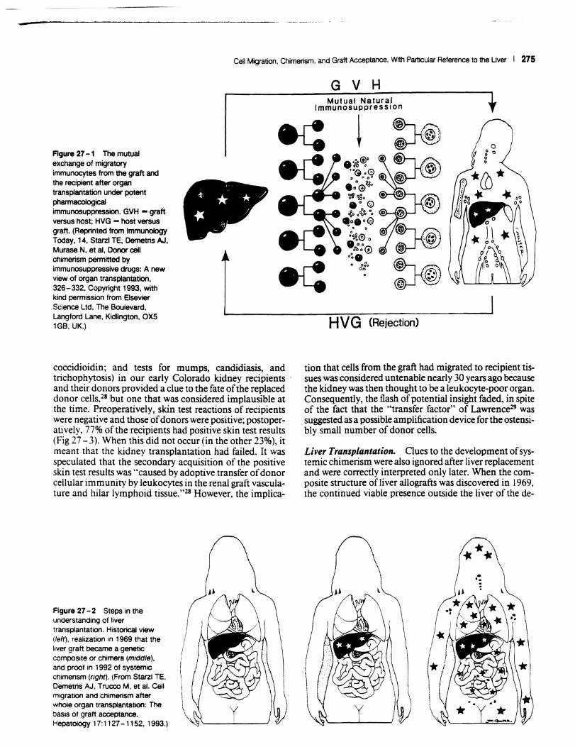
Improvements in the prevention or control of rejection of the kidney and liver are largely interchangeable and then applicable with little modification to thoracic and other organs (see Chapter 50). The mechanism by which antirejection treatment permits any of these grafts to be accepted, however, has been an immunological enigma. I - 8 We have proposed9-" that the exchange of migratory leukocytes between the transplant and the recipient with consequent long-term cellular chimerism in both is the basis for acceptance of all whole organ allografts and xenografts (Fig 27 - 1). Although such chimerism was first demonstrated as recently as mid-1992, the observations have increased our insight into transplantation immunology and have encouraged the development of alternative therapeutic strategies.
LOCAL (GRAFT) CHIMERISM
The Liver
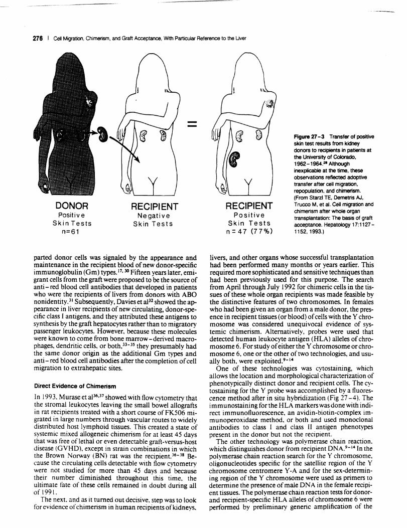
The first unequivocal evidence that whole organ. gra~s in humans become genetic composites (local chlmensm) was obtained in 1969 with karyotyping techniques in female recipients of livers obtained from male cadaveric donors. These studies were done by K.A. Porter of St. Mary's Hospital and Medical School in Lo.ndon on ~lografts from the first long-surviving patients ~n the Umversitv of Colorado liver series. 16. 17 Postoperatively, both the hepatocytes and the endothelium of the major blood vessels of the grafts retained their donor sex, whereas the entire macrophage system. including the Kupifer cells, w~ replaced with recipient female cells (identified. by theIr characteristic Barr bodies) within 100 days (FIg 27 - 2, middle). The assumption for many years was tha~ the com posite (chimeric) structure of the hepauc allograft was a special feature of this organ.
274
Other Organs
The illusion of the uniqueness of the hepatic graft was dispelled in 1991 with the demonstration, first in rat J?od71s18 and then in human intestinal grafts,19 that the eplthehum and vascular endothelium remained donor specific, whereas lymphoid, dendritic, and other leukocytes were replaced by recipient cells in the la~ina propria, Peyer:s patches, and mesenteric nodes. As With the hver, the chimerism of the intestinal graft was made easy to demonstrate because of its large constituency of lymphoreticular cells,
The suspicion that this transformation must be occurring with other kinds of whole organ grafts was promptly confirmed with the kidney '2. 20 and with the thoracic organs.21 -23
DISCOVERY OF SYSTEMIC CHIMERISM
Circumstantial Evidence
Twenty-two years passed between the discovery of the chimerism of the transplanted liver and that of the intestine; during this period. several clinical reports noted the local chimerism in long-surviving human kidney allografts2o• 24-26 and in subhuman primates.27 Throughout this time, the tacit or explicit assumption was that the cells departing the liver or kidney had been destroyed. Wi~h the evidence that the local chimeric changes occurred In all kinds of grafts, the burning question became what happened to the donor cells that had disappeared .from the grafts. Much earlier circumstantial eVidence eXisted that donor leukocytes migrating from the engrafted organs were still present in the body; however. this evidence had been largely ignored, forgotten, or misinterpreted.
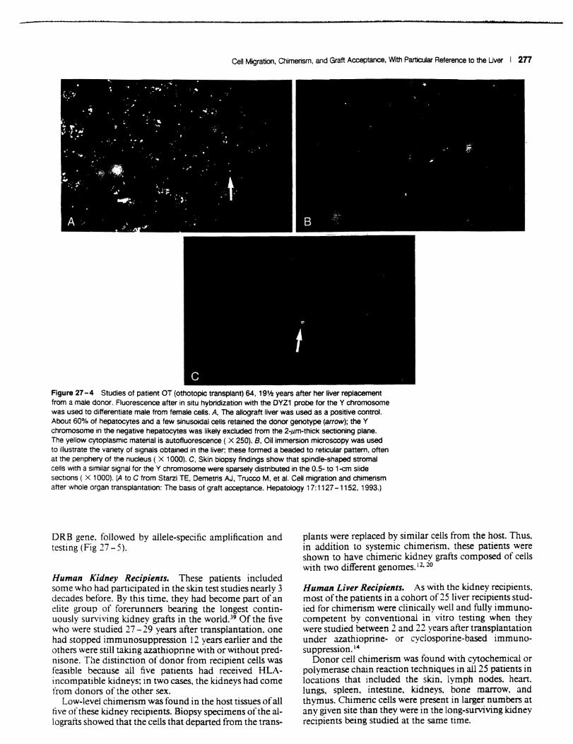
Kidney Transplantation, The resu~ts of exhaustive skin test studies (tuberculin, histoplasmm, blastomycln, and
-Cell Migration, Chimerism, and Graft Acceptance, With Particular Reference to the Uver I 275
Figure 27 -1 The mutual exchange of migratory immunocytes from the graft and the recipient after organ transplantation under potent pharmacological immunosuppression. GVH - graft versus host; HVG = host versus graft. (Reprinted from Immunology Today, 14, Starzl TE, Oemetris AJ. Murase N. et aI. Donor cell chimerism pennitted by immunosuppressive drugs: A new view of organ transplantation. 326-332, Copyright 1993. with kind permission from Elsevier Science Ltd, The Boulevard. Langford Lane, Kidlington, OXS 1GB, UK.)
<t
~
f,·, .
* "* /.
/
<{
coccidioidin; and tests for mumps, candidiasis, and trichophytosis) in our early Colorado kidney recipients ' and their donors provided a clue to the fate of the replaced donor cells.28 but one that was considered implausible at the time. Preoperatively, skin test reactions of recipients were negative and those of donors were positive; postoperatively, 77% of the recipients had positive skin test results (Fig 27 - 3). When this did not occur (in the other 23%), it meant that the kidney transplantation had failed. It was speculated that the secondary acquisition of the positive skin test results was "caused by adoptive transfer of donor cellular immunity by leukocytes in the renal graft vasculature and hilar lymphoid tissue."28 However, the implica-
Figure 27 - 2 Steps in the understanding of liver transplantation, Historical view (left), realization In 1969 that the liver graft became a genetic composite or chimera (middle). and proof in 1992 of systemic chimensm (right). (From Starzl TE, Demetris AJ. Trucco M, et aI. Cell migration and chimensm atter whole organ transplantation: The basis of graft acceptance. HepatolOgy 17:1127 -1152, 1993.)
G V H Mutual Natura,l
Immunosuppression
H V G (Rejection)
tion that cells from the graft had migrated to recipient tissues was considered untenable nearly 30 years ago because the kidney was then thOUght to be a leukocyte-poor organ. Consequently, the flash of potential insight faded, in spite of the fact that the "transfer factor" of Lawrence29 was suggested as a possible amplification device for the ostensibly small number of donor cells.
Li ver Transplantation. Clues to the development of systemic chimerism were also ignored after liver replacement and were correctly interpreted only later. When the composite structure of liver allografts was discovered in \969, the continued viable presence outside the liver of the de-
276 I Cell Migration, Chimerism, and Graft Acceptance, Wrth Particular Reference to the Uver
DONOR Positive
Skin Tests n=61
RECIPIENT Negative
Skin Tests
parted donor cells was signaled by the appearance and maintenance in the recipient blood of new donor-specific immunoglobulin (Gm) types. 17, 30 Fifteen years later, emigrant cells from the graft were proposed to be the source of anti-red blood cell antibodies that developed in patients who were the recipients oflivers from donors with ABO nonidentity.31 Subsequently, Davies et al32 showed the appearance in liver recipients of new circulating, donor-specific class I antigens. and they attributed these antigens to synthesis by the graft hepatocytes rather than to migratory passenger leukocytes. However, because these molecules were known to come from bone marrow-derived macrophages, dendritic cells, or both,33-3s they presumably had the same donor origin as the additional Gm types and anti - red blood cell antibodies after the completion of cell migration to extrahepatic sites.
Direct Evidence of Chimerism
In 1993, Murase et aP6.37 showed with flow cytometry that the stromal leukocytes leaving the small bowel allografts in rat recipients treated with a short course ofFK506 migrated in large numbers through vascular routes to widelv distributed host lymphoid tissues. This created a state of systemic mixed allogeneic chimerism for at least 45 days that was free of lethal or even detectable graft-versus-host disease (GVHD). except in strain combinations in which the Brown Norway (BN) rat was the recipient. J6 - 38 Because the circulating cells detectable with flow cytometry were not studied for more than 45 davs and because their number diminished throughout -this time, the ultimate fate of these cells remained in doubt during all of 1991.
The next. and as it turned out decisive, step was to look for evidence of chimerism in human recipients ofk.idneys,
RECIPIENT Positive
Skin Tests n = 47 (77%)
Figure 27 - 3 Transfer of positive skin test results from kidney donors to recipients in patients at the University of Colorado, 1962 -1964.21 Although inexplicable at the time, these observations reflected adoptive transfer after cell migration, repopulation, and chimerism. (From Starzl TE, Demetris AJ, Trucco M, et aI. Cell migration and chimerism after whole organ transplantation: The basis of graft acceptance. Hepatology 17:1127-1152, 1993.)
livers, and other organs whose successful transplantation had been performed many months or years earlier. This required more sophisticated and sensitive techniques than had been previously used for this purpose. The search from April through July 1992 for chimeric cells in the tissues of these whole organ recipients was made feasible by the distinctive features of two chromosomes. In females who had been given an organ from a male donor, the presence in recipient tissues (or blood) of cells with the Y chromosome was considered unequivocal evidence of systemic chimerism. Alternatively, probes were used that detected human leukocyte antigen (HLA) alleles of chromosome 6. For study of either the Y chromosome or chromosome 6, one or the other of two technologies, and usually both, were exploited.9- '4
One of these technologies was cytostaining, which allows the location and morphological characterization of phenotypically distinct donor and recipient cells. The cytostaining for the Y probe was accomplished by a fluorescence method after in situ hybridization (Fig 27 -4). The immunostaining for the HLA markers was done with indirect immunofluorescence. an avidin-biotin-complex immunoperoxidase method. or both and used monoclonal antibodies to class I and class II antigen phenotypes present in the donor but not the recipient.
The other technology was polymerase chain reaction. which distinguishes donor from recipient DNA.9- '4 In the polymerase chain reaction search for the Y chromosome, oligonucleotides specific for the satellite region of the Y chromosome centromere Y -A and for the sex-determining region of the Y chromosome were used as primers to determine the presence of male DNA in the female recipient tissues. The polymerase chain reaction tests for donorand recipient-specific HLA alleles of chromosome 6 were performed by preliminary generic amplification of the
Cell Migration, Chimerism, and Graft Acceptance. With Particular Reference to the Uver I 277
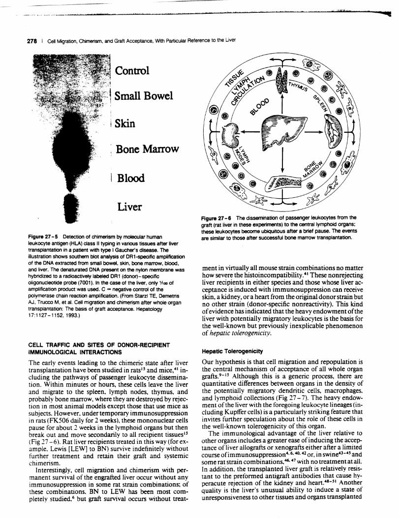
Figure 27 -4 Studies of patient OT (othotopic transplant) 64. 19'h years after her liver replacement from a male donor. Fluorescence alter in situ hybridization with the DYZ1 probe for the Y chromosome was used to differentiate male from female cells. A. The allograft liver was used as a positive control. About 60% of hepatocytes and a few sinusoidal cells retained the donor genotype (arrow); the Y chromosome in the negative hepatocytes was likely excluded from the 2-,um-thick sectioning plane. The yellow cytoplasmic material is autofluorescence ( X 250). B. Oil immersion microscopy was used to illustrate the variety of signals obtained in the liver; these formed a beaded to reticular pattern. often at the penphery of the nucleus ( X 1000). C. Skin biopsy findings show that spindle-shaped stromal cells with a similar signal for the Y chromosome were sparsely distributed in the 0.5- to 1-cm slide sections ( X 1000). (A to C from Starzl TE. Demetris AJ. Trucco M. et a!. Cell migration and chimerism alter whole organ transplantation: The basis of graft acceptance. Hepatology 17:1127 -1152. 1993.)
ORB gene. followed by allele-specific amplification and testing (Fig 27 - 5).
Human Kidney Recipients. These patients included some who had participated in the skin test studies nearly 3 decades before. By this time. they had become part of an elite group of forerunners bearing the longest continuously surviving kidney grafts in the world.39 Of the five who were studied 27 - 29 years after transplantation. one had stopped immunosuppression 12 years earlier and the others were still taking azathioprine with or without prednisone. The distinction of donor from recipient cells was feasible because all five patients had received HLAincompatible kidneys: in two cases, the kidneys had come from donors of the other sex.
Low-level chimerism was found in the host tissues of all five of these kidney recipients. Biopsy specimens of the allografts showed that the cells that departed from the trans-
plants were replaced by similar cells from the host. Thus. in addition to systemic chimerism, these patients were shown to have chimeric kidney grafts composed of cells with two different genomes. 12. 20
Human Liver Recipients. As with the kidney recipients. most of the patients in a cohort of 25 liver recipients studied for chimerism were clinically well and fully immunocompetent by conventional in vitro testing when they were studied between 2 and 22 years after transplantation under azathioprine- or cyclosporine-based immunosuppression. 14
Donor cell chimerism was found with cytochemical or polymerase chain reaction techniques in all 25 patients in locations that included the skin. lymph nodes. hean. lungs, spleen. intestine. kidneys, bone marrow, and thymus. Chimeric cells were present in larger numbers at any given site than they were in the long-surviving kidney recipients being studied at the same time.
--------------------- -------________ . ____ . __ , _________ ._-_w ........... "' ____ ~ __ ................ _--.--. ............. ----........ ---..... --------.oili' ... '4'i!-ili.ii· -.... "---~ .. _
278 I Cell Migration. Chimerism, and Graft Acceptance, With Particular Reference to the Liver
Control
i Small Bowel I
Skin
Bone Marrow
I Blood
Liver
Figure 27 - 5 Detection of chimerism by molecular human leukocyte antigen (HLA) ctass II typing in various tissues after liver transplantation in a patient with type I Gaucher's disease. The illustration shows southern blot analysis of DR1-specific amplification of the DNA extracted from small bowel, skin, bone marrow, blood, and liver. The denaturated DNA present on the nylon membrane was hybridized to a radioactively labeled DR1 (donor)-specific oligonucleotide probe (7001). In the case of the liver, only 'Aoo of amplification product was used. C = negative control of the polymerase chain reaction amplification. (From Starzl TE, Demetris AJ. Trucco M, et al. Cell migration and chimerism after whole organ transplantation: The basis of graft acceptance. Hepatology 17:1127-1152,1993.)
CELL TRAFFIC AND SITES OF DONOR-RECIPIENT IMMUNOLOGICAL INTERACTIONS
The early events leading to the chimeric state after liver transplantation have been studied in rats lS and mice,41 including the pathways of passenger leukocyte dissemination. Within minutes or hours, these cells leave the liver and migrate to the spleen. lymph nodes. thymus. and probably bone marrow, where they are destroyed by rejection in most animal models except those that use mice as subjects. However, under temporary immunosuppression in rats (FK506 daily for 2 weeks), these mononuclear cells pause for about 2 weeks in the lymphoid organs but then break out and move secondarily to all recipient tissues1s (Fig 27 -6). Rat liver recipients treated in this way (for example, Lewis [LEW] to BN) survive indefinitely without further treatment and retain their graft and systemic chimerism.
Interestingly, cell migration and chimerism with permanent survival of the engrafted liver occur without any immunosuppression in some rat strain combinations; of these combinations, BN to LEW has been most completely studied,6 but graft survival occurs without treat-
Figure 27 -6 The dissemination of passenger leukocytes from the graft (rat liver in these experiments) to the central lymphoid organs; these leukocytes become ubiquitous after a brief pause. The events are Similar to those after successful bone marrow transplantation.
ment in virtually all mouse strain combinations no matter how severe the histoincompatibility.41 These nonrejecting liver recipients in either species and those whose liver acceptance is induced with immunosuppression can receive skin, a kidney, or a heart from the original donor strain but no other strain (donor-specific nonreactivity). This kind of evidence has indicated that the heavy endowment of the liver with potentially migratory leukocytes is the basis for the well-known but previously inexplicable phenomenon of hepatic tolerogenicity.
Hepatic Tolerogenicity
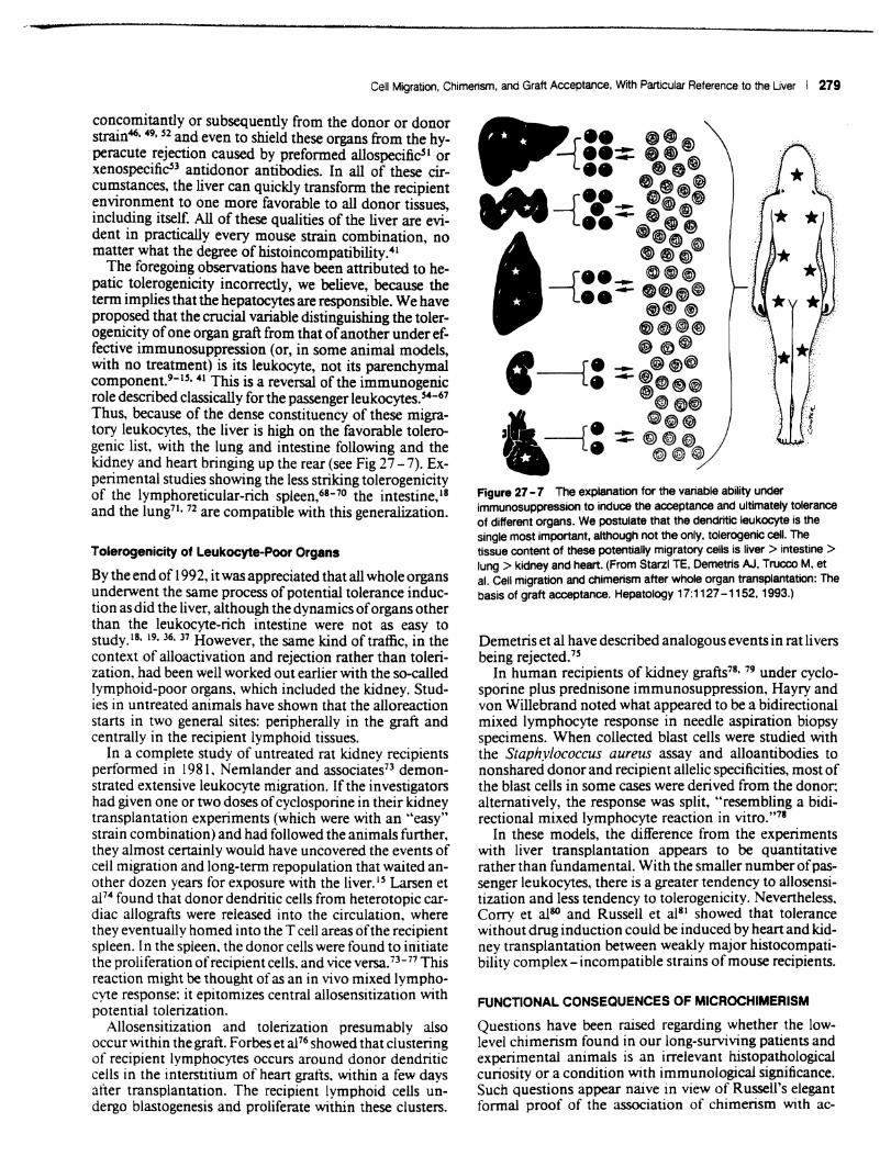
Our hypothesis is that cell migration and repopulation is the central mechanism of acceptance of all whole organ graftS.9- 1S Although this is a generic process, there are quantitative differences between organs in the density of the potentially migratory dendritic cells. macrophages, and lymphoid collections (Fig 27 - 7). The heavy endowment of the liver with the foregoing leukocyte lineages (including Kupffer cells) is a particularly striking feature that invites further speculation about the role of these cells in the well-known tolerogenicity of this organ.
The immunological advantage of the liver relative to other organs includes a greater ease of inducing the acceptance of liver allografts or xenografts either after a limited course ofimmunosuppression4. 6. 40. 42 or, in swine43-4s and some rat strain combinations,46, 47 with no treatment at all. In addition, the transplanted liver graft is relatively resistant to the prefonned antigraft antibodies that cause hyperacute rejection of the kidney and heart.4I-$1 Another quality is the Iiver's unusual ability to induce a state of unresponsiveness to other tissues and organs transplanted
Cell Migration. Chimerism, and Graft Acceptance, With Particular Reference to the Uver I 279
concomitantly or subsequently from the donor or donor strain46, 49, S2 and even to shield these organs from the hyperacute rejection caused by preformed allospeciiicsi or xenospecifics3 antidonor antibodies. In all of these circu~stances, the liver can quickly transform the recipient enVIronment to one more favorable to all donor tissues including itself. All of these qualities of the liver are evi~ dent in practically every mouse strain combination, no matter what the degree ofhistoincompatibility.41
The foregoing observations have been attributed to hepatic tolerogenicity incorrectly, we believe, because the term implies that the hepatocytes are responsible. We have proposed that the crucial variable distinguishing the tolerogenicity of one organ graft from that of another under effective immunosuppression (or, in some animal models, with no treatment) is its leukocyte, not its parenchymal component.9- 15• 41 This is a reversal of the immunogenic role described classically for the passenger leukocytes. S4-67
Thus, because of the dense constituency of these migratory leukocytes, the liver is high on the favorable tolerogenic list, with the lung and intestine following and the kidney and heart bringing up the rear (see Fig 27 - 7). Experimental studies showing the less striking tolerogenicity of the lymphoreticular-rich spleen,68-70 the intestine, 18 and the lung71 , 72 are compatible with this generalization.
Tolerogenicity of Leukocyte-Poor Organs
By the end of 1992, it was appreciated that all whole organs underwent the same process of potential tolerance induction as did the liver, although the dynamics of organs other than the leukocyte-rich intestine were not as easy to study.18, 19.36, 37 However, the same kind of traffic, in the context of alloactivation and rejection rather than tolerization. had been well worked out earlier with the so-called lymphoid-poor organs. which included the kidney. Studies in untreated animals have shown that the alloreaction starts in two general sites: peripherally in the graft and centrally in the recipient lymphoid tissues.
In a complete study of untreated rat kidney recipients perfomled in 1981. Nemlander and associates73 demonstrated extensive leukocyte migration. If the investigators had given one or two doses of cyclosporine in their kidney transplantation experiments (which were with an "easy" strain combination) and had followed the animals further, they almost certainly would have uncovered the events of cell migration and long-term repopulation that waited another dozen years for exposure with the liver. IS Larsen et al74 found that donor dendritic cells from heterotopic cardiac allografts were released into the circulation. where they eventually homed into the T cell areas of the recipient spleen. I n the spleen. the donor cells were found to initiate the proliferation of recipient cells. and vice versa. 73-77 This reaction might be thOUght ofas an in vivo mixed lymphocyte response; it epitomizes central allosensitization with potential tolerization.
Allosensitization and tolerization presumably also occur within the graft. Forbes et al76 showed that clustering of recipient lymphocytes occurs around donor dendritic cells in the interstitium of heart grafts. within a few days after transplantation. The recipient lymphoid cells undergo blastogenesis and proliferate within these clusters.
Figure 27 - 7 The explanatiOn for the variable ability under immunosuppression to induce the acceptance and ultimately tolerance of different organs. We postulate that the dendritic leukocyte is the single most important, although not the only. tolerogenic cell. The tissue content of these potentially migratory cells is liver> intestine> lung> kidney and heart. (From Starzl TE. Demetris AJ. Trucco M, et aI. Cell migration and chimerism after whole organ transplantation: The basis of graft acceptance. Hepatology 17:1127 -1152. 1993.)
Demetris et al have described analogous events in rat livers being rejected.7s
In human recipients of kidney grafts78, 79 under cyclosporine plus prednisone immunosuppression, Hayry and von Willebrand noted what appeared to be a bidirectional mixed lymphocyte response in needle aspiration biopsy specimens. When collected blast cells were studied with the Staphylococcus aureus assay and alloantibodies to nonshared donor and recipient allelic specificities, most of the blast cells in some cases were derived from the donor; alternatively, the response was split, "resembling a bidirectional mixed lymphocyte reaction in vitro."78
In these models, the difference from the experiments with liver transplantation appears to be quantitative rather than fundamental. With the smaller number of passenger leukocytes, there is a greater tendency to allosensitization and less tendency to tolerogenicity. Nevertheless, COrry et also and Russell et al81 showed that tolerance without drug induction could be induced by heart and kidney transplantation between weakly major histocompatibility complex-incompatible strains of mouse recipients.
FUNCTIONAL CONSEQUENCES OF MICROCHIMEAISM
Questions have been raised regarding whether the lowlevel chimerism found in our long-surviving patients and experimental animals is an irrelevant histopathological curiosity or a condition with immunological significance. Such questions appear naive in view of Russell's elegant formal proof of the association of chimerism with ac-

280 I Cell Migration, Chimerism, and Graft Acceptance, Wrih Particular Reference to the Liver
quired tolerance as well as runt disease.82 However, the skepticism was generated by the small numbers of chimeric donor cells in the recipient tissues of the patients and animals in our studies. We have called this condition microchimerism. a term introduced in the literature in 1974 by Liegeois et al,83 to describe a small proportion of chimeric cells in the recipient blood. The presence of microchimerism is far from insignificant; there is much evidence that the cumulative effect of microchimeric cells is substantial, especially after liver transplantation, when they are most easily demonstrated.
Metabolic Effects
The small population of chimeric cells has been shown to affect total body metabolism in patients treated with liver transplantation for the enzyme deficiencies of type IV glycogen storage disease and Gaucher's disease. In these diseases, the consequences of the missing enzymes are widespread storage of amylopectin and glucocerebroside, respectively.11 The disorders were previously thought to be treatable only by bone marrow transplantation because the enzyme deficiency affects all cells; however, 2 - 8 years after liver replacement, there was a dramatic resorption of both kinds of storage material from host tissues (Fig 27-8). As an explanation for the metabolic amelioration, chimeric donor cells were found to be ubiquitous in recipient tissues, including those of the heart, lymph nodes, bone marrow, intestine, and skin (see Fig 27 -5). There apparently had been a coculture effect of a small number of chimeric donor cells on the contiguous, overwhelming numbers of enzyme-deficient recipient cells.
The Immunological Interface
The foregoing metabolic observations raised important questions about the potential cell-to-cell effects of other molecules directly involved in immunological rather than metabolic processes, including those subserving tolerance
"{ . - -. .... , ",,-,~ '\~~4
induction (see Fig 27 - 1). Perhaps such questions could be answered if it were known how the chimeric donor cells, many of which resemble dendritic cells, are perpetuated for as long as 3 decades after transplantation. The dendritic cells and other leukocytes could be spawned by small numbers of pluripotent progenitor cells coming from the allograft interstitium. Dendritic cell precursors have been grown from mouse blood, bone marrow, or whole organs using media enriched with granulocytel macrophage colony - stimulating factor. 84 The products of these stem cells should reach terminal differentiation, however, unless there is a reason for their continued proliferation. We have suggested that the subsequent survival and renewal of these cells depend on continuous mutual stimulation of the donor and recipient cell populations13• u in a process of tolerization that shares many of the cellular characteristics associated with immunity.ss
CHANGED HOST AND GRAFT INTERACTIONS
There are indirect ways to show that the coexisting immunocyte populations in successful cases (see Fig 27 - I) come to regard each other in a revised light. The evidence, on the one hand, is the fading of the threat of clinical rejection concomitant with development of donor-specific nonreactivity in spite of lightened treatment (or, in some animal models, without any treatment); on the other hand, the evidence is the waning specter of GVHD. It is quite natural to expect that the threat ofGVHD and rejection would decline contemporaneously in an organ recipient because both of the cell populations are receiving the same protective immunosuppression. Appreciation of the two-cell population relationship and the need not to alter it by ablating one side or the other was the crucial advance made empirically that permitted the successful engraftment ofleukocyte-rich organs such as the liver, intestine, both together, or all of the intra-abdominal organs (multivisceral transplantation). 86 Once the cardinal principle was understood that low-level mixed allogeneic chimer-
Figure 27 - 8 A, An endOmyocardial biopsy specimen obtained in 1989, at the time of liver transplantation, revealed diffuse amytopectin deposits within and between cells. B, Another endomyocardial biopsy specimen obtained in 1992 revealed only traces of extracellular deposits (perIOdic aCid-Sdliff-O. X 100). (From Stanl TE. Demetria AJ, Trucco M. et al. Chimerism after liver transplantation for type IV glycogen storage and type I Gaucher's disease. Reprinted with permIssion from The New England Journal of MediCine, 328.745-749 and 1993.)
Cell Migration. Chimerism, and Graft Acceptance, With Particular Reference to the Liver I 281
ism was invariably found after the successful transplantation of any whole organ, the reason GVHD was not common in liver, intestinal, or multivisceral recipients seemed obvious. Mixed chimerism was being produced empirically in the way that had been documented in the classic GVHD-free mouse bone marrow mixed chimerism models of Slavin et al87 and Ildstad and Sachs.88
THE CRITICAL DENDRITIC CELL
The generation of an immune response leading, under normal circumstances, to graft destruction, GVHD, or both, requires effective antigen presentation and recognition in its initial phase; this signal is followed by a second, costimulatory signal and the response of T cells to the combined signal.89 Both of these signals are normally delivered to T cells by professional antigen-presenting cells. Of these cells, the dendritic ce1l56- 58 (the most prominent chimeric cell by morphological criteria9- 15) is critical because it can modify the expression of cell interaction, major histocompatibility complex, and adhesion molecules, all of which determine how antigen signals are heeded by T cells. 59 The dendritic leukocyte is thus the prime candidate in this tolerogenicity scenario, even though other lineages may also be essential for a successful outcome.
Figure 27 - 9 The donor-recipient leukocyte interaction is a buffer against rejection on the one hand and graftversus-host disease on the other. Veto and suppressor cells are postulated to be the result of the interaction shown at the cell population interface. R. = iatrogenic immunosuppression. (Reprinted from Immunology Today, 14. Starzl TE. Demetris AJ, Murase N. et ai, Donor
Match
Partial Mismatch
Total
IMPACT ON TISSUE MATCHING
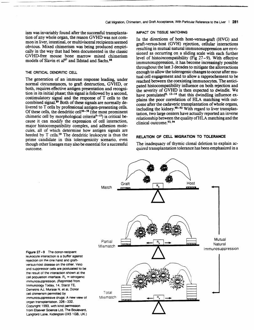
In the directions of both host-versus-graft (HVG) and graft-versus-host (GVH) r~jection, cellular .interactioD;s resulting in mutual natural ImmunosuppressIon are enVlsioned as occurring on a sliding scale with each further level of histoincompatibility (Fig 27 - 9). With effective immunosuppression, it has become increasingly possible throughout the last 3 decades to mitigate the alloreactions enough to allow the tolerogenic changes to occur after mutual cell engagement and to allow a rapprochement to be reached between the coexisting immunocytes. The anticipated histocompatibili~ influence on both reje.ction and the severity of GVHD IS then expected to dwmdle. We have postulated9• 12-14 that this dwindling influence explains the poor correlation of HLA matching with outcome after the cadaveric transplantation of whole organs, including the kidney.90-92 With regard to liver tra':lsplantation, two large centers have actually reported. an mverse relationship between the quality ofHLA matching and the clinical outcome.93• 94
RELATION OF CELL MIGRATION TO TOLERANCE
The inadequacy of thymic clonal deletion to ex~lain. acquired transplantation tolerance has been emphaslZed In a
Mutual Natural
Immunosuppression
cell chimerism penmitted by immunosuppressive drugs: A new view of organ transplantation. 326-332. Copyright 1993. With kind penmission from Elsevier Science Ltd. The Boulevard. Langford Lane, Kidlington OX5 1GB, UK.)
Mismatch .Jiiiiiiii-,---",;:;;;~r-"-;;,;;---"",,,,-
282 I Cell Migration, Chimerism, and Graft Acceptance, With Particular Reference to the Uver
1992 review.9s Although a discussion of the meaning of tolerance is beyond our intention, it should be noted that all of the mechanisms that explain clonal silencing, including peripheral (nonthymic) clonal deletion and anergy, could mesh with the discovery of the enduring grafthost intimacy inherent with chimerism. The production of suppressor cells, veto cells, or both could be an epiphenomenological consequence. Evidence of the long-term vitality and turnover of donor leukocytes in recipient tissues is particularly supportive of the opinions of Ban de ira et al,8S Coutinho,96 and Cohen,91 who have defined acquired tolerance as a high (not anergic) level of sustained immune activity in immunological networks. These networks presumably interact in a more complex way than do the idiotype systems originally postulated by Jerne.98
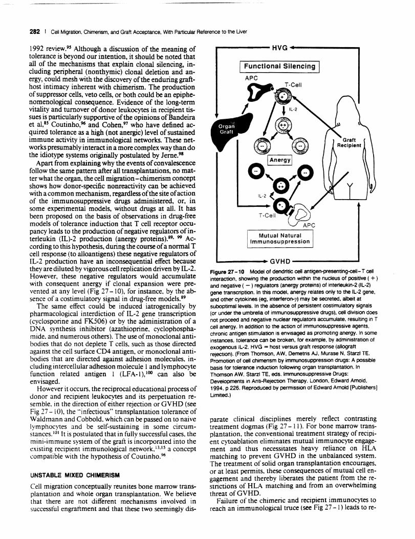
Apart from explaining why the events of convalescence follow the same pattern after all transplantations, no matter what the organ, the cell migration -chimerism concept shows how donor-specific nonreactivity can be achieved with a common mechanism, regardless of the site of action of the immunosuppressive drugs administered, or, in some experimental models, without drugs at all. It has been proposed on the basis of observations in drug-free models of tolerance induction that T cell receptor occupancy leads to the production of negative regulators of interleukin (IL)-2 production (anergy proteins).89. 99 According to this hypothesis, during the course ofa normal T cell response (to alloantigens) these negative regulators of IL-2 production have an inconsequential effect because they are diluted by vigorous cell replication driven by IL-2. However, these negative regulators would accumulate with consequent anergy if clonal expansion were prevented at any level (Fig 27 - 10), for instance. by the absence of a costimulatory signal in drug-free models. 89
The same effect could be induced iatrogenically by pharmacological interdiction of IL-2 gene transcription (cyclosporine and FK506) or by the administration of a DNA synthesis inhibitor (azathioprine. cyclophosphamide. and numerous others). The use of monoclonal antibodies that do not deplete T cells. such as those directed against the cell surface CD4 antigen, or monoclonal antibodies that are directed against adhesion molecules, including intercellular adhesion molecule 1 and lymphocyte function related antigen I (LF A-I), 100 can also be envisaged.
However it occurs. the reciprocal educational process of donor and recipient leukocytes and its perpetuation resemble. in the direction of either rejection or GVHD (see Fig 27 - 10). the "infectious" transplantation tolerance of Waldmann and Cobbold. which can be passed on to naive lymphocytes and be self-sustaining in some circumstances. lOI It is postulated that in fully successful cases. the mmi-immune system of the graft is incorporated into the existing recipient immunological network. 13•IS a concept compatible with the hypothesis of Coutinho.96
UNSTABLE MIXED CHIMERISM
Cell migration conceptually reunites bone marrow transplantation and whole organ transplantation. We believe that there are not different mechanisms involved in successful engrafiment and that these two seemingly dis-
------ HVG ... -----_
Functional Silencing
APC
f)" / 'IL.2
I~ ~ ~ ... ~.'.' ~ ~
I Anergyl
0'0 ... 0 IL·2 ( ++ Y r-c,P0
CY;;,c Mutual Natural
Immunosuppression
L-___ ~ GVHD ------....
Figure 27 -10 Model of dendritic cell antigen-presenting-cell-T cell interaction. showing the production within the nucleus of positive ( + ) and negative ( -) regulators (anergy proteins) of inter1eukin·2 (IL·2) gene transcription. In this model. anergy relates only to the IL-2 gene. and other cytokines (eg, interferon-y) may be secreted, albeit at suboptimal levels. In the absence of persistent costimulatory signals (or under the umbrella of immunosuppressive drugs). cell division does not proceed and negative nuclear regulators accumulate. resulting in T cell anergy, In addition to the action of immunosuppressive agents, chroniC antigen stimulation is envisaged as promoting anergy. In some instances, tolerance can be broken. for example. by administration of exogenous IL-2, HVG = host versus graft response (allograft rejection). (From Thomson. AW. Demetris AJ, Murase N. Starzl TE, Promotion of cell chimerism by immunosuppression drugs: A poSSible basiS for tolerance Induction following organ transplantation. In Thomson AW. Starzl TE. eds. Immunosuppressive Drugs: Developments in Anti-Rejection Therapy. London. Edward Arnold. 1994, p 226. Reproduced by permission of Edward Arnold [Publishers] Umited.)
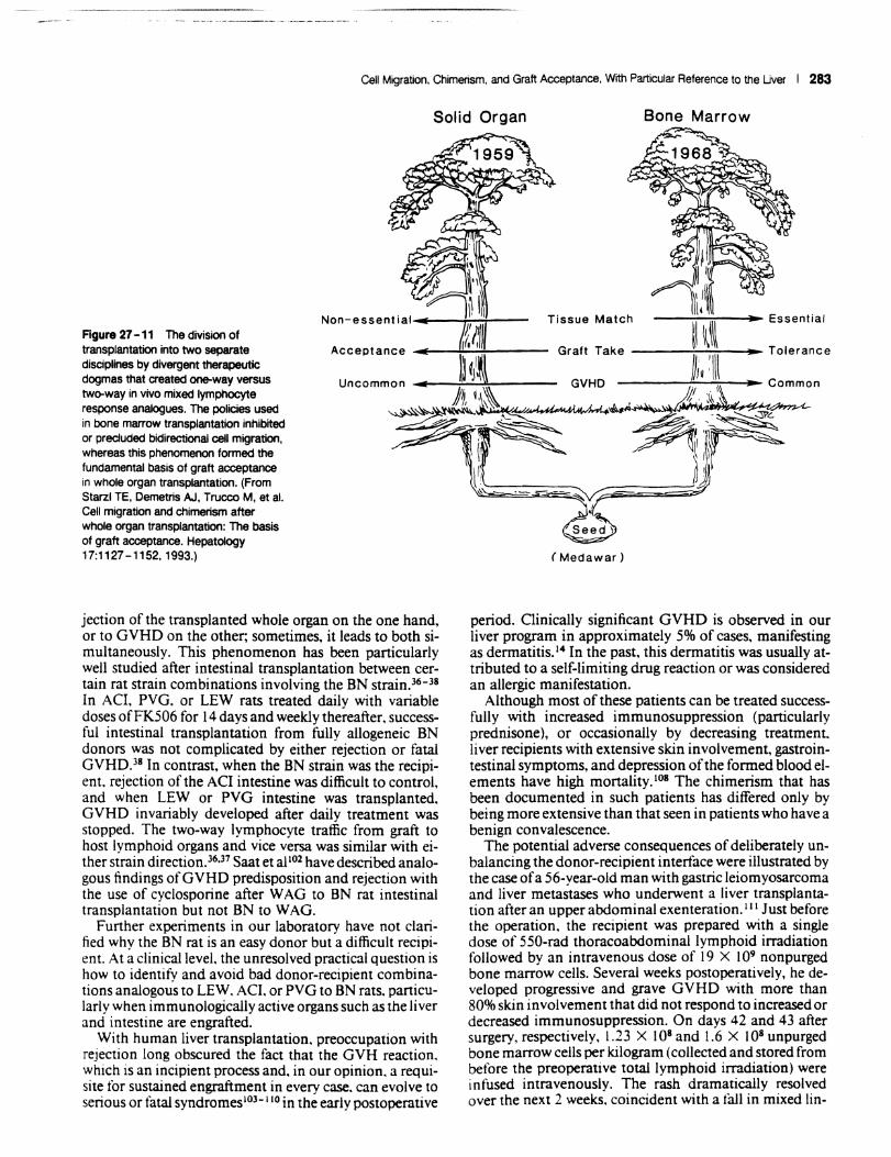
parate clinical disciplines merely reflect contrasting treatment dogmas (Fig 27 - II). For bone marrow transplantation. the conventional treatment strategy of recipient cytoablation eliminates mutual immunocyte engagement and thus necessitates heavy reliance on HLA matching to prevent GVHD in the unbalanced system. The treatment of solid organ transplantation encourages, or at least permits. these consequences of mutual cell engagement and thereby liberates the patient from the restrictions of HLA matching and from an overwhelming threat of GVHD.
Failure of the chimeric and recipient immunocytes to reach an immunological truce (see Fig 27 -I) leads to re-
Cell Migration. Chimerism, and Graft Acceptance. With Particular Reference to the Uver I 283
Solid Organ Bone Marrow
Figure 27 -11 The division of transplantation into two separate disciplines by divergent therapeutiC dogmas that created one-way versus two-way in vivo mixed lymphocyte response analogues. The policies used in bone marrow transplantation inhibited or preciuded bidirectional cell migration. whereas this phenomenon formed the fundamental basis of graft acceptance in whole organ transplantation. (From Starzl TE. Demetris AJ. Trucco M, et al. Cell migratiOn and chimerism after whole organ transplantation: The basis of graft acceptance. Hepatology 17:1127 -1152. 1993.)
.1 i I
~\:I~ Non-essential : Tissue Match --..;,;;....;,;,--~ .. - Essential
Acceptance ____ -~-"*"---- Graft Take ~l \~\\\ .. Tolerance
)) I~~ It 'l\\ Uncommon.. l (j \\\ GVHD ~Jjt'h \\. _ .. C _o_ .. m .. m .. o~n
JAi,\\~ ~ ~~""", ~~~~~ --.~
~r::v~
jection of the transplanted whole organ on the one hand, or to GVHD on the other; sometimes, it leads to both simultaneously. This phenomenon has been particularly well studied after intestinal transplantation between certain rat strain combinations involving the BN strain.36 - 38 In ACt PVG. or LEW rats treated daily with variable doses ofFK506 for 14 days and weekly thereafter. successful intestinal transplantation from fully allogeneic BN donors was not complicated by either rejection or fatal GVHD.38 In contrast, when the BN strain was the recipient. rejection of the ACI intestine was difficult to controL and when LEW or PVG intestine was transplanted. GVHD invariably developed after daily treatment was stopped. The two-way lymphocyte traffic from graft to host lymphoid organs and vice versa was similar with either strain direction.36•37 Saat et al102 have described analogous findings ofGVHD predisposition and rejection with the use of cyclosporine after WAG to BN rat intestinal transplantation but not BN to WAG.
Further experiments in our laboratory have not clarified why the BN rat is an easy donor but a difficult recipient. At a clinical level. the unresolved practical question is how to identify and avoid bad donor-recipient combinations analogous to LEW, ACt or PVG to BN rats. particularly when immunologically active organs such as the liver and intestine are engrafted.
With human liver transplantation, preoccupation with rejection long obscured the fact that the GVH reaction, which is an incipient process and. in our opinion. a requisite for sustained engraftment in every case. can evolve to serious or fatal syndromes lO3 - 1 10 in the early postoperative
-~r
(Medawar)
period. Clinically significant GVHD is observed in our liver program in approximately 5% of cases, manifesting as dermatitis.14 In the past, this dermatitis was usually attributed to a self-limiting drug reaction or was considered an allergic manifestation.
Although most of these patients can be treated successfully with increased immunosuppression (particularly prednisone), or occasionally by decreasing treatment. liver recipients with extensive skin involvement, gastrointestinal symptoms, and depression of the formed blood elements have high mortality. lOS The chimerism that has been documented in such patients has differed only by being more extensive than that seen in patients who have a benign convalescence.
The potential adverse consequences of deliberately unbalancing the donor-recipient interface were illustrated by the case ofa 56-year-old man with gastric leiomyosarcoma and liver metastases who underwent a liver transplantation after an upper abdominal exenteration. 1 1 1 Just before the operation, the recipient was prepared with a single dose of 550-rad thoracoabdominal lymphoid irradiation followed by an intravenous dose of 19 X 109 non purged bone marrow cells. Several weeks postoperatively, he developed progressive and grave GVHD with more than 80% skin involvement that did not respond to increased or decreased immunosuppression. On days 42 and 43 after surgery, respectively, 1.23 X 10⁸ and 1.6 X 10⁸ unpurged bone marrow cells per kilogram (collected and stored from before the preoperative total lymphoid irradiation) were infused intravenously. The rash dramatically resolved over the next 2 weeks. coincident with a taIl in mixed lin-
284 I Cell Migration, Chimerism, and Graft Acceptance, With Particular Reference to the Uver
eage donor cells in the blood from approximately 25% of total to 3% by flow cytometry, without the advent of rejection.
The ability of the stored autologous marrow cells to tum off the potentially lethal GVHD may be a clue to the changes that occur during the mutual cell engagement of mixed chimerism. Although the cells used for rescue from GVHD were thought to be smaller in number than those in the circulation of the patient, they had not been exposed to donor marrow. Their therapeutic effect resembled the ability of virgin donor strain immunOcytes to "break tolerance," as originally described by Billingham et al. 112
Two clinically relevant lessons from this traumatic experience have been reported elsewhere.HI The first was the inadvisability of trying to make space for the augmenting marrow cells with total lymphoid irradiation. The second lesson was the potential value of naive autologous bone marrow, which can be stored as a safety net should GVHD occur. These lessons have guided further attempts at bone marrow augmentation for the induction of tolerance in whole organ recipients.
CLINICAL TRIALS OF BONE MARROW AUGMENTATION
Kidney Alone or Kidney Plus Pancreatic Islets
The concept developed in this chapter is that the content (and perhaps the specific lineages) of migratory cells, which are particularly numerous in the liver, confers potential immunological advantages. These passenger leukocytes, which are of bone marrow origin, have been considered an immunological liability for transplantations"-67; however, under the appropriate conditions, they may be tolerogenic, as exemplified by the liver. A corollary, therefore, is that organs such as the kidney and heart, which have a smaller leukocyte component, could be brought to the liver's tolerogenic potential. In fact, the frequently advanced strategy of intravenously infusing donor bone marrow, donor blood (donor-specific transfusion), or other hematolymphoid cells at the same time or shortly after the transplantation of whole organs83, 87,88, 113-116 is merely an augmentation of the normal posttransplantation cell migration. To mimic the natural process. these cells should be given perioperatively, not in advance or afterward, as has usually been done with the Monaco experimental model. 11"-116
CLINICAL EXPERIENCE
Since December 1992 at our center, four patients with end-stage renal disease have received a simultaneous kidney - bone marrow allograft from the same cadaveric donor. In addition, two diabetic (type I) renal patients were given pancreatic islets from their kidney-bone marrow donor. As a precaution. autologous bone marrow was harvested from all the recipients immediately before the kidney transplantation and cryopreserved for potential future use in case ofGVHD,111 as described in the preceding section.
The donor bone marrow was obtained from vertebral bodies atthe end of multiorgan procurement. and 3 X 10⁸ untreated bone marrow cells were infused intravenously
immediately after the kidney transplantation. The two diabetic patients received the additional pancreatic islet infusion intraportaIly at the time of the intravenous bone marrow infusion. All six patients were treated with FK506 and prednisone. Chimerism was assessed in the recipients by flow cytometry, polymerase chain reaction, and immunostaining with peripheral blood lymphocytes. Immunological monitoring was done by mixed lymphocyte response and cell-mediated lympholysis.
None of these patients exhibited GVHD. Detectable levels of donor cells were present in the peripheral blood lymphocytes of all six recipients during the 1- to 6-month follow-up. Donor cells were also detected in two patients whose lymph nodes were biopsied 2 and 4 months after transplantation. All six patients and their renal grafts are doing well. The two patients who were also given pancreatic islets have been treated too recently to evaluate their insulin status accurately.
Further evaluation of passenger leukocyte augmentation (with bone marrow and other leukocyte sources) is of particular interest for pancreatic islet transplantation, which has consistently failed as a means of treating diabetes because of the high rate of rejection. I 17 Iritis possible to increase islet allograft survival with minimal ultimate immunosuppression by concomitant donor bone marrow infusion, this will be an approach opposite to the numerous attempts to reduce islet immunogenicity by selective destruction of the antigen-presenting cells derived from bone marrow that are normally contained in islet preparations. 118 Donor bone marrow would then become a dosemaneuverable component of any organ or cellular transplantation for the facilitation of graft acceptance and for the induction of donor-specific nonreactivity.
A LIVER TRANSPLANTATION ROAD MAP FOR DRUG WEANING
As discussed earlier, past experience with conventional liver transplantation can be envisioned as a mini - bone marrow engraftment. In one reported group of 44 human liver recipients who had survived from 11 to 23 years, 6 ( 14%) had stopped all immunosuppression between 1 and 11 years postoperatively and had subsequently experienced clinically stable, drug-free intervals of 5 - 13 years; another 15 patients were drug free and stable after a shorter follow-up period. I ... 119 The most extreme example of early successful drug discontinuance in a liver recipient was after 6 months, with a subsequent follow-up period of 3 years. A trial of drug weaning has been started in liver recipients with a rejection-free course exceeding 5 years. Liver graft rejection (if it occurs) can be so effectively treated with FK506120 that the benefits of drug weaning appear to us to outweigh the risks in selected patients.
This liver experience should provide insight regarding the use of bone marrow augmentation to achieve freedom from immunosuppression in recipients of organs and cells such as the kidney or pancreatic islets. Even when drug weaning has been decided upon, there is presently no way to know when a potential drug-free state has arrived. It seems clear that, if the clinical experience with the tolerogenic livers is taken seriously, kidney or heart transplantation plus bone marrow augmentation will also require

Cell Migration. Chimerism. and Graft Acceptance. With Particular Reference to the Liver I 285
protracted immunosuppression before the treatment can be stopped altogether and that, even then, it can be stopped only with precautions. Such conclusions have also been reached by Barber et al, 116 who used delayed supplementary bone marrow for cadaveric kidney transplantation as advocated by Monaco et al. 1I4• lIS
Uver Transplantation
CLINICAL EXPERIENCE
Two liver recipients have received the same doses of perioperative bone marrow as patients in the kidney series, and 500-550 Gy total lymphoid irradiation was added preoperatively. The second patient developed nearly fatal G VHD from which he was rescued with stored autologous bone marrow (see under Unstable Mixed Chimerism). One year later, the patient underwent retransplantation for hepatitis C virus infection. The biopsy revealed predominantly hepatitis, but there were also signs of vascular rejection. Blood chimerism was no longer detectable immediately before retransplantation, even by polymerase chain reaction.
The unwise decision made regarding this patient and the second one to unbalance the donor-recipient interface iatrogenically with total lymphoid irradiation was based on the dogma that making space would facilitate engraftment of the marrow. This concept has been eroded by direct experimentation. 121. 122 The second patient, now 13 months after surgery, has had a perfect clinical result since biopsy findings showed mild rejection at 2 weeks. She takes a daily dose of 6 mg of FK506 plus 5 mg of prednisone. There is evidence of chimerism in the peripheral blood lymphocytes by cytospin.
Three additional patients underwent liver transplantation and bone marrow augmentation without total lymphoid irradiation. Their courses have been uncomplicated, with the exception of one episode of rejection 2 weeks after transplantation. All are blood chimeras.
RISKS AND BENEFITS
The augmentation in liver recipients of a process of cell migration and chimerism that is already highly operational after this kind of transplantation must be closely monitored for efficacy. Whether leukocyte augmentation will do more than could be achieved naturally in tolerance induction (with earlier achievement of a drug-free state) or will only increase the risk ofGVHD remains to be determined. The storage of autologous bone marrow for rescue should G VHD occur appears to be a mandatory precaution in such trials. III
References
1. Weber RA. Cannon J A. Lon~ WP. Observations on the regrafting of suc· cessful homografts In chickens. Ann Surg 139:473-477.1954.
2. Murray lE. Sbeil AGR. Moseley R. et aL Analysis ofmecbaoism of immunosuppreSSIve drugs ID renal bomotransplaDtation. Ann Surg 160:449-473. 1964.
3. Starzl TE. Host.gnft adaptation. In Starzl TE. ed. EXperience in Renal Transplantauon. Philadelphia. WB Saunders. 1964. pp 164- 170.
~. Starz..l TE. Effortstomitigateorpreventn:)ecuon.lnStarzl TE. ed. Expenence in Hepauc Transplaotanon. Philadelphia. WB Saunders. 1969. PI' 203- 233.
5. Levey RH. Immunological tolerance and enhancemenc A common mechanism. Transplant Proc 3:41 -48, 1971.
6. Murase N, Kim 00. TodoS, etal. FKS06 suppression ofhcart and liver allograft rejection. Part 2. The induction ofgraft acceptance in the rat. TransplaDtation 50:739-744. 1990.
7. StreiJeiD JW. Neonatal tolerance ofH-2 alloantigens. Procuring graft acceptance the "old·fashioned" way. TrausplaDtation 52: 1- 10, 1991.
8. Eto M. Mayumi H. Nishimura Y, et al. Similarity and dilference in the mecb· anisms of neonatally induced tolerance and cyclophospbamidc-induced tolerance in mice. J ImmunoI147:2439-2446. 1991.
9. Starzl TE. Demetris AI. Murase N. et al. Cell migJatiOI1, chimerism, and graft acceptance. Lancet 339:1579-1582. 1992.
10. Starzl TE. Demetris AI. Truc:co M. et aI. Systemic chimerism in human female recipients of male liven. Lancet 340:876-877,1992.
II. Starzl TE. Demetris AI. Truc:co M. et aI. Chimerism after liver tran5plaDta· tion for type IV glycogen storage: disease and type I Gaucher's disease. N Eng! J Med 328:745-749, 1993.
12. Starzl TE. Demetris AI, T ruc:co M. et aI. Chimerism and donor speciJic non.reactivity 27 to 29 yean after kidney allotransplantation. Transplantation 55:1272-1277. 1993.
13. Starzl IE. Demetris AI. Murase N. et aL Donor cell chimerism permitted by immunosupprasive drugs: A new view of organ tran5plaDtatiOo. Immunol Today 14:326-332. 1993.
14. Starzi IE. Demetris AI. Truc:co M. et al. Cell migration and chimerism after whole organ transplantation: The basis of graft acceptaoce. Hepatology 17: 1127 -1152. 1993.
IS. Demetris AI. Murase N, Fujisalci S. et al. Hematolymphoid cell trafficking, microchimerism. and GVHD reactions after liver, bone marrow, and bean transplaDtation. TransplaDtation Proc 25:3337-3344,1993.
16. Porter KA. Pathology of the orthotopic bomograft and beterograft. In Stan!. TE, ed. Experience in Hepatic TrausplaDtation. Philadelphia. WB Saunders. 1969, pp 427 -437.
17. Kashiwagi N. Porter KA. Penn I. et al. Studies ofbomograft sex and of gamma globulin phenotypes after orthotopic homotransplantation of the buman liver. Surg Forum 20:374-376. 1969.
18. Murase N. Demetris AI. Matsuzaki T. et aI. Long survival in ratsaftcrmulti· visceral versus isolated small bowel allotransplantation under FK 506. Sur· gery 110:87-98. 1991.
19. I waki Y. Starzl TE. Yagihashi A. et aI. Replacement of donor lymphoid tissue in buman small bowel transplants under FK 506 immunosuppression. Lan· cet 337:818-819.1991.
20. Randhawa PS. Starzl TE. Ramos H. et aI. Allografts surviving for 26 - 29 years following living related kidney transplantation: Analysis by light microscopy. in situ bybridization for the Y chromosome. and anti·HLA antibodies. Am J Kidney Dis 24:72-77, 1994.
21. Fung JJ. leevi A. Kaufman C, et aI. Interactions ~n bronchoaiveolar Iympbocytes and macropbages in beart·lung transplaDt recipients. Human Immunol 14:287 -294. 1985.
22. Demetris AI. Murase N. Starzl TE. Donor dendritic cells in grafts and bost Iympboid and non-lympboid tissues after liver and hean allotraosplaDtation under short term immunosuppression. Lancet 339: 1610. 1992.
23. Valdivia LA. Demetris AI. Langer AM. et aI. Dendritic cell replacement in long.surviving liver and cardiac xenografts. Transplantation 56:482-484. 1993.
24. Sinclair RA. Origin of endothelium in buman renal allografts. BMJ 4: 15 - 16. 1972.
25. Sedmak DD, Sharma HM. Czajka CM. Ferguson RM. Recipient endotheli· alization of renal allografts: An immunohistochemical study utilizing blood group antigens. Transplantation 46:907 -909. 1988.
26. Andersen CB. Ladefoged OS, Larsen S. Cellular inllammatory infiltrates and renal cell turnover in kidney allografts: A study using in situ hybriillzation and combined in SitU hybridization and immunohistochemistry with a y. chromosome-specific DNA probe and monoclonal antibodies. APMlS 99:645 -652. 1991.
27. ThomasJ. Carver M. Cunningham P. et aI. Promotion ofincompauble allograft acceptance in rhesus monkeys given post·transplant antithymocyte globulin and donor bone marrow: I. In vivo parameters aDd immunohistologic eVidence suggesting microchimerism. Transplantauon 43:332. 1987.
28. Wilson WEe. Kirkpatrick CH. Immunologic aspectS of renal homotransplantation. In Starzl TE, ed. Experience in Renal TransplaDtation. Philadel· phia. WB Saunders. 1964. PI' 239 - 261.
29. Lawrence HS. The transfer of bypersensitivity of the delayed type in man. In Lawrence HS, ed. CeUular and Humoral AspectS of the HypenensitiveStates. New York. Hoeber·Harper. 1959. p 279.
30. K.ashiwagi N. Speaal immunochemlcal studies. In Starzl TE. ed. Experience in Hepauc Transplantation. Philadelphia. WB Saunders. 1969. JlP 394-407.
31. Ramsey G. Nusblu:her J. Stan! TE. Lindsay GO, lsobemaglutinins of graft origin after ABO-unmatched liver traDSplaDtation. N Engi J Med 311: 1167-1170.1984.
32. Davies HIlS. Pollard SO. CaIne R Y. Soluble HLA antigens in the circuJ.ation of liver gnft r=plcnu. TransplaDtation 47:524-527. 1989.
33. Russo C. PeUegrlno MA. Ferrone S. A doubl~etermioant immunoassay With monoclonal annbodies to the HLA·A. -B . ..c complex. Transplant ?roc 15:66-68. 1983.
286 I Cell Migration, Chimensm, and Graft Acceptance, With Particular Reference to the Uver
34. Krangel MS. Secretion of HLA-A and -B antigens via an alternative RNA splicing pathway. J Exp Med 163:1173-1190, 1986.
35. Singh PB, Brown RE, Roser B. Class I transplantation antigens in solution in body fluids and in the urine. J Exp Med 168:195-211, 1988.
36. Murase N, Demetris AJ, Woo J, et aI. Lymphocyte traffic and graft-venushost disease after fully allogeneic small bowel transplantation. Transplant Proc 23:3246-3247, 1991.
37. Murase N, Demetris A, Woo J, et aI. Graft versus host disease (GVHD) after BN to LEW compared to LEW to BN rat intestinal transplantation under FK 506. Transplantation 55:1-7,1993.
38. Tanabe M, Murase N, Demetris AJ, et aI. The inlluence of donor and recipient strains in isolated sma1I bowel transplantation in rats. Transplant Proc 26:4325-4332, 1994.
39. Graver B. World Transplant Rt.cords-I991: Kidney. In Terasaki PA, Cecka JM. ed5. Clinical Transplants. Los Angeles, UCLA Press, 1991. P 431.
40. Starzi TE, Marchioro TL, Poner KA. et aI. Facton determining short- and long-tenD survival after orthotopic liver homotransplantation in the dog. Surgery 58:131-155, 1965.
41. Qian S. Demetris AJ, Murase N, et aI. Murine liver allograft transplantation: Tolerance and donor cell chimerism. Hepatology 19:916 - 924, 1994.
42. Valdivia LA, Fung n, Demetris AJ, Starzi TE. Ditrerential survival of ham-5ter-to-rat liver and cardiac xenografts under FK 506 immunosuppression. Transplant Proc 23:3269-3271, 1991.
43. Garnier H. OotJ, Bertrand M, eta!. Liver transplantation in tbepig: Surgical approach. C R Acad Sci, Paris 260:5621- 5623, 1965.
44. PeacockJH, TerblancheJ. Orthotopic homotransplantation oftbe liver in the pig. In Read AE, ed. The Liver. London, Butterworth, 1967, pp 333-336.
45. CaIne R Y, White HJO, Y offa DE, et aI. Observations of orthotopic liver transplantation in the pig. BMJ 2:478-480, 1967.
46. Zimmerman FA, Butcher GW. Davies HS, et aI. Techniques for orthotopic li vertransplantation in tbe rat and some studies of the immunologic responses to fully allogeneic liver grafts. Transplant Proc 11:571-577, 1979.
47. Murase N, Demetris AJ. Kim 00. et aI. Rejection oftbe multivisceral allograft in rats: A sequential anal}'Sl.S with comparison to isolated orthotopic small bowel and liver grafts. Surgery 108:880- 889. 1990.
48. Starzl TE, Ishikawa M, Putnam CW, et a1. Progress in and deterrents to orthotopic liver transplantation. witb special reference to survival, resistance to hyperacute rejection. and biliary duct reconstruction. Transplant Pro<: 6: 129-139.1974.
49. Kamada N, Davies HOS. Roser B. Revenal of transplantation immunity by liver grafting. Nature 292:840-842. 1981.
50. Houssin O. Gugenheim J, Bellon B. et aI. Absence of hyperacute rejection of liver allografts in hypenensitized rats. Tlllnsplant Pro<: 17:293-295. 1985.
51. Fung J, Makowka L. Tzakis A. et aI. Combined liver-kidney tlllnsplantation: Analysis of patients witb preformed lympbocytotoxic antibodies. Transplant Pro<: 20(Suppi 1):88-91. 1988.
52. CaIne RY. Sells RA. Pena JR. et aI.lnduction of immunological tolerance by porcine liver allografts. Nature 233:472-474. 1969.
53. Valdivia L. Demetris Ai. Fung Jl, et aI. Successful ham5terto rat liver xenotlllnsplantation under FK506 immunosuppression induces unresponsiveness to hamster heart and skin. Tlllnsplantation 55:659-661,1993.
54. Snell GO. The homograft reaction. Annu Rev MicrobiolI1:439-458. 1957. 55. Stein muller O. Immunization with skin isopafts taken from tolerant mice.
Science 158:127-129.1967. 56. Steinman RM. Cohn ZA. Identification of a novel cell type in peripheral
lymphoid o~ns of mice. I. Morphology, quanutation. tissue distnbution. J ExpMed 137:1142-1162.1973.
57. Steinman RM. Cohn ZA. Identification of a novel cell type in peripheral lymphoid organs of mice. II. Functional properues in vitro. J Exp Med 139:380- 397, 1974.
58. Steinman RM. Lustig OS. Cohn ZA. Identification of a novel cell type in peripheral lymphoid o~ns of mice. Ill. Functional properties in vivo. J Exp Med 139:1431-1445. 1974.
59. Steinman RM. The dendritic cell system and its role in immunogenicity. Annu Rev Immunol 9:271 - 296. 1991.
60. Hart ONJ. McKeDZle JL.lnterstitiai dendritic cells.lnt Rev Immunol6: 128-149.1990.
61. Hart ONJ. Wincarls CG, Fabre JW. Graft adaptation: Studies on possible mechaDlsms in long-term surviving Illt renal allografts. Tlllnsplantauon 30:73-80.1980.
62. Batchelor JR. Welsh KI. Maynard A. BUIllOS H. Failure oflongsumving, passively enhanced allografts to provoke T -dependent alloimmunity: f. Retlllnsplantauon of (AS X AUG)Fl kidneys mto secondary AS recipients. J Exp Med 150:455-464. 1979.
63. Lechler RI. Batchelor J R. Restollluon of immunogenicity to passenger celldepleted kidney allografts by the addition of donor-stram dendritic cells. J Exp Med 155:31-41. 1982.
64. Talmage OW. Oart G. Radovicb J. Lalferty KJ. Activation of transplant immUDlty: Effect of donor leukocytes on thyrOid allograft rejecnon. Science 191 :385-387. 1976.
65. Lalferty KJ. Bootes A. Dart G. Talmage OW. Effect of organ culture in the sumval of thyroid aIlQl!nils in mice. Transplantation 22: 138- 149. 1976.
06. Faustman O. Hauptfeld V. Lacy P. DavieJ. Prolongation of munne wet allograft SUl'Vlvai by pretreatment of islets With auubody directed to la detenIunants. Proc Nat! Acad Sci USA 78:5156-5159.1981.
67. Austyn 1M, Steinman RM. The passenger leukocyte-a fresh look. Transpl Rev2:!39-176,1988.
68. Marchioro TL, Rowlands OT Jr. Rifkind O. et aI. Splenic homotransplantation. AnnN YAcadSci 120:626-651,1964.
69. Wakely E, OberhoIser JH. Corry RJ. Elimination ofacute GYHD and pr0-longation of rat pancreas allograft survival witb DST. cyc!osporine, and spleen transplantation. Transplantation 49:241-245.1990.
70. Bitter-Suermann H, Savc:-Soderbergh JS. The coune of pancrcasallografts in Illts conditioned by splcen allografts. Transplantation 26:28-34, 1978.
71. Prop J, Kuijpers K. Petersen AH, et aI. Why are lung allografts more vigorously rejected than bearts? Heart Transplant 4:433 -436. 1985.
72. Westra AL, Prop J, Kuijpen KC, WiJdevuurCRH. A paradox in heart and lung rejection. Transplantation 49:826 - 828, 1990.
73. Nemlander A, Soots A. von Willebrand E, et aI. Redistribution of rena1 allograft - responding leukocyteS during rejection. II. Kinetics and speci.licity. J Exp Med 156:1087-1100, 1982.
74. Larsen CP. Morris PI, AustyD JM. Migration of dendritic leukocytes from cardiac allografts into host spleens. A novel route for initiation of rejection. J ExpMed 171:307-314. 1990.
75. Demetris AJ, Qian S, Sun H, et aI. Early events in liver allograft rejection. Am J Pathol 138:609-618, 1991.
76. Forbes RD, Parfrcy NA, Gomersail M. et aI. Dendritic cell-lymphoid aggregation and major histocompatibility antigen expression during rat cardiac allograft rejection. J Exp Med 164:1239-1258, 1986.
77. van Schlifgaarde R. Hermans P. Terpstra JL, et aI. Role of mobile passenger lymphocytes in the rejection of renal and cardiac allografts in the Illt. Transplantation 29:209-213. 1980.
78. Hayry P. von Willeblllnd E: Tlllnsplant aspillltion cytology in tbe evaluation ofa renal allograft. In TouraineJL. Tllleger J, Betuel H, etal. cds. Tlllnsplantation and Clinical Immunology, vol 15. Amsterdam. Excerpta Medica, 1983, pp 124-137.
79. von WiUebrand E. Taskinen E. Ahonen 1, Hayty P. Recent modifications in the fine needle aspiration biopsy of human renal allografts. Transplant Proc 15:1195-1197,1983.
80. Corry RJ, Winn HI, Russell PS. Primary vascularized allografts of hearts in mice: The roleofH-20. H-2Kand non-H·2 antigens in rejection. Tlllnsplantation 16:343-350, 1973.
81. Russell PS, Chase CM, Colvin RB, Plate JMO. Kidney transplants in tnice. An analysis oftbe immune status of mice bearing long-term G-2 incompatible transplants. J Exp Med 147:1449-1468, 1978.
82. Russell PS. Modification of runt disease in mice by various means. Ciba Found Symp 1962, pp 350-383.
83. Liegeois A. Charreire J. Brennan LB. Allograft enhancement induced by bone marrow cells. Surg Forum 25:297 - 300. 1974.
84. Inaba K.., loaba M, Romani N. et aI. Generation oflargc numbers ofdendritic cells from mouse bone marrow cultures supplemented with granulocyte/macrophage colony-stimulating factor. J Exp Med 176: 1693-1702. 1992.
85. Bandeira A. Coutinho A. Camaud C. et aI. Tlllnsplantation tolerance correlates with high levels of T- and B-lymphocyte acuvity. Proc Nat! Acad Sci USA 86:272-276. 1989.
86. Starzl TE. Todo S. TzakisA. et aI. The many faces of multi visceral transplantation. SUIll Gynecol Obstet 172:335 - 344. 1991.
87. Slavin S. Strober S. Fuks Z. Kaplan HS. Induction of specific tissue transplantation tolerance using fractionated total lymphoid irradiation in adult mice: Long-term survival of allogeneic bone marrow and skin grafts. J Exp Med 146:34-48.1977.
88. IIdstad ST. Sachs OH. Reconstitution with syngeneic plus allogeneic or xenogeneic bone marrow leads to specific acceptance of allografts or xenografu. Nature 307:168-170. 1984.
89. Jenkins MK. The role of cell division in the induction of clonal anergy. Immunol Today 13:69-73. 1992.
90. Matas Ai. Sutherland OER. Najarian JS. The impact of HLA matching on graft survival. Tlllnsplantation 54:568- 569. 1992.
91. Ferguson R. Tlllnsplant Information Share Group (TlSG). A multicenter experience with sequential ALG/cyc1osporine thelllpy in renal transplantauon. ain TlllnspI2:285-294. 1988.
92. Salvatierlll O. Jr. Optimal use of organs for transplantation. N Eng! J Med 318:1329-1331.1988.
93. Donaldson PT. O'Grady J, Portmann B. et aI. Evidence for an immune response to HLA class I antigens in the vanishing bile duct syndrome after liver transplantation. Lancet 1:945-948. 1987.
94. Markus BH. Ouquesnoy RJ. Gordon RD. et aI. Histocompaubility and liver tlllnsplant outcome. Does HLA exert a dualistic etfect? TIllOSplautaUOD 46:372-377.1988.
95. Miller JF. Morahan G. Peripheral T cell tolelllnce. AnDU Rev Immunol 10:51-69.1992.
96. CoutinhoA. Beyond c10nal selecuon and network.lmmunol Rev 110:63 -87. 1989,
97. Cohen IR. The cogDltive paradigm and the immunological homunculus.lmmunol Todav 13:490-494, 1992.
98. Jeme NK. IdiotYPlc networks and other preconceived ideas. Immunol Rev 79:5-24.1984.
99. ZubiagaAM. Munoz A. Huber BT. Supennduction oflL-2 geneuanscnption in the presence of cycloneximlde. J Immunol 146:3857 - 3863. 1991.
100. lsalle M, Yaglta H. Okumura K.., lhara A. Specllic aa:qltancc of cardiac aUo-
._------_ .•..
Cell Migration, Chimerism, and Graft Acceptance, With Particular Reference to the Uver I 287
graft after treatment with antibodies to ICAM-I and LFA-l. Science 255: 1125 - 1127, 1992.
WI. Waldmann H. Cobbold S. The use of monoclonal antibodies to achieve immunological tolerance. Immunol Today 14:247-251. 1993.
102. Saat RE. De Bruin R WF. Heineman E. et al. The limited efficacy of cyclosporine in preventing rejection and graft-versus-bost disease in orthotopic small bowel transplantation in rats. Transplantation 50:374-377, 1990.
103. Burdick JF, Vogelsang GB, Smith W J. et aI. Severe graft-venus-bost disease in a liver transplant recipient. N Engl J Med 319:689-691, 1988.
104. Marubayasbi S. Matsuzaka C. Fatal generalized acute graft recipient. Transplantation 50:709 - 711. 1990.
105. Bhaduri BR. Tan KC. Humphreys S, et aI. Graft-versus-bost disease after orthotopic liver transplantation in a child. Transplant Proc 22:2378-2380. 1990.
106. Roberts JP. Ascber NL, Lake J. et al. Graft va. host disease after liver transplantation in bumans: A repon offourcascs.Hepatology 14:274-281. 1991.
107. Jamieson NV. JOJI!CY V. Friend PJ. et al. Graft-venus-bost disease in solid organ transplantation. Transplant Int 4:67-71.1991.
108. Rosen CB, Moore SB. Batts KP. et al. Cinical and pathological features of graft-versus-host disease after liver transplantation: A case repon and review of the literature. Clin Transpl 7:52-58. 1993.
109. Comenzo RL. Malachowski ME, Rohrer RJ, et al. Anomalous ABO pbenotype in a child after an ABO-incompatible liver transplantation. N Eng! J Med 326:867 -889, 1992.
110. Collins RH. Anastasi J. Terstappen LW. et al. Brief report: Donor-derived long-term multilineage hematopoiesis in a liver-transplant recipient. N EnglJ Med 328:762-765. 1993.
Ill. RicordiC. TzakisAG. DemetrisAJ. etal. Reversal of graft-versus-host disease with infusion of stored autologous bone marrow cells following combined liver-bone marrow allotransplantation in man. Transplant Sci 3:76-77, 1993.
112. Billingham R. Brent 1., Medawar P. Quantitative studies on tissue transplantation immunity. Ill. Actively acquired tolerance. Pbilos Trans R Soc Lond Bioi 239:357-412. 1956.
113. Monaco AE. Wood M1., Russell PS. Studies of heterologous anti-lymphocyte serum in mice. Ill. Immunologic tolerance and chimerism produced across the H-21ocus with adult thymectomy and anti-Iympbocyte serum. Ann N Y Acad Sci 129:190-209. 1966.
114. Monaco AP, Wood ML. Studies on heterologous antilymphocyte serum in mice: VII. Optimal cellular antigen for induction of immunologic tolerance with ALS. Transplant Proc 2:489-496, 1970.
115. Monaco AP. Wood M1., Maki T. Gozzo JJ. Post transplantation donor-speciftc bone marrow tranSfusion in polyc1onal antilymphocyte serum - treated recipients: The optimal cellular antigen for induction of unresponsiveness to organ allografts. Transplant Proc 20: 1207 - 1212, 1988.
116. Barber WH. Manlcin JA. Laskow DA. et aI. Long term resulu of a controlled prospective study with tranSfusion of donor-specific bone marrow in 57 cadaveric renal allograft recipients. Transplantation 51:70-75. 1991.
117. Carroll PD, Ricordi C. Shapiro R, et al. Frequency of kidney rejection in diabetic patients undergoing simultaneous kidney and islet cell transplantation. Transplantation 55:761- 765, 1993.
118. Ricon:li C. I1dstad ST. Starzl TE. Induction of pancreatic islet graft acceptance: The role of antigen presenting cells. Transplant Sci 2:34 - 38. 1992.
119. ReyesJ. ZeeviA. TzakisA,etal. The frequentachievementofadrugfree state after orthotopic liver transplantation. Transplant Proc 25:3315-3319. 1993.
120. Starzi TE. TodoS. FungJ, etal. FK506 for human liver, kidney and pancreas transplantation. Lancet 2:1000-1004, 1989.
121. Harrison DE. Competitive repopulation in unirradiated nonna! recipients (editorial). Blood 81:2473-2474, 1993.
122. Stewart FM. Crittenden RB. Lowry PA. Long-term engraftment of nonna! and post-5-fluorouracil murine marrow into normal nonmyeloablated mice. Blood 81:2566-2571. 1993.