Embed Size (px)
Citation preview

The PDF of the article you requested follows this cover page.
This is an enhanced PDF from The Journal of Bone and Joint Surgery
2009;91:8-21. doi:10.2106/JBJS.H.01290 J Bone Joint Surg Am.Panayiotis Dimakopoulos, Andreas Panagopoulos and Georgios Kasimatis
TechniqueTransosseous Suture Fixation of Proximal Humeral Fractures. Surgical
This information is current as of March 3, 2009
Reprints and Permissions
Permissions] link. and click on the [Reprints andjbjs.orgarticle, or locate the article citation on
to use material from thisorder reprints or request permissionClick here to
Publisher Information
www.jbjs.org20 Pickering Street, Needham, MA 02492-3157The Journal of Bone and Joint Surgery
COPYRIGHT © 2 009 BY THE JOURNAL OF BONE AND JOINT SURGERY, I NCORPORATED
8
Transosseous Suture Fixation of Proximal Humeral FracturesSurgical Technique
By Panayiotis Dimakopoulos, MD, Andreas Panagopoulos, MD, and Georgios Kasimatis, MD
Investigation performed at the Shoulder and Elbow Unit, Orthopaedic Department, University Hospital of Patras, Patras, Greece
The original scientific article in which the surgical technique was presented was published in JBJS Vol. 89-A, pp. 1700-9, August 2007
DISCLOSURE: The authors did not receive any outside funding or grants in support of their research for or preparation of this work. Neither they nor a member of their immediate families received payments or other benefits or a commitment or agreement to provide such benefits from a commercial entity. No commercial entity paid or directed, or agreed to pay or direct, any benefits to any research fund, foundation, division, center, clinical practice, or other charitable or nonprofit organization with which the authors, or a member of their immediate families, are affiliated or associated.
ABSTRACT FROM THE ORIGINAL ARTICLE
BACKGROUND: The optimal treatment of displaced fractures of the proximal part of the humerus remains controversial. We evaluated the long-term functional and radiographic results of transosseous suture fixation in a series of selected dis-placed fractures of the proximal part of the humerus.
METHODS: Over an eleven-year period, a consecutive series of 188 patients with a specifically defined displaced fracture of the proximal part of the humerus underwent open reduction and internal fixation with transosseous sutures. Twenty pa-tients were lost to follow-up and three died before the time of follow-up, leaving a cohort of 165 patients (ninety-four women and seventy-one men; mean age, fifty-four years) available for the study. Forty-five (27%) of the injuries were four-part fractures with valgus impaction; sixty-four (39%) were three-part fractures; and fifty-six (34%) were two-part fractures of the greater tuberosity, thirty-six (64%) of which were associated with anterior dislocation of the shoulder. All fractures were fixed with transosseous, nonabsorbable, number-5 Ethibond sutures. Associated rotator cuff tears detected in fifty-seven patients (35%) were also repaired. Over a mean follow-up period of 5.4 years, functional outcome was assessed with the Constant score. Follow-up radiographs were assessed for fracture consolidation, malunion, nonunion, hetero-topic ossification, and signs of impingement, humeral head osteonecrosis, and degenerative osteoarthritis.
RESULTS: All fractures, except for two three-part fractures of the greater tuberosity, united within four months. The quality of fracture reduction as seen on the first postoperative radiograph was regarded as excellent/very good in 155 patients (94%), good in seven (4%), and poor in three (2%). Malunion was present in nine patients (5%) at the time of the last follow-up; six of the nine had had good or poor initial reduction and three, excellent/very good reduction. Humeral head osteonecrosis was seen in eleven (7%) of the 165 patients; four demonstrated total and seven, partial collapse. Fifteen patients had heterotopic ossification, but none had functional impairment. Four patients had signs of impingement syn-drome, and two had arthritis. At the time of the final evaluation, the mean Constant score was 91 points, and the mean Constant score as a percentage of the score for the unaffected shoulder, unadjusted for age and gender, was 94%.
CONCLUSIONS: The clinical and radiographic results of this transosseous suture technique were found to be satisfactory at an average of 5.4 years postoperatively. Advantages of this technique include less surgical soft-tissue dissection, a low rate of humeral head osteonecrosis, fixation sufficient to allow early passive joint motion, and the avoidance of bulky and expensive implants.
LEVEL OF EVIDENCE: Therapeutic Level IV. See Instructions to Authors for a complete description of levels of evidence.
ORIGINAL ABSTRACT CITATION:“Transosseous Suture Fixation of Proximal Humeral Fractures” (2007;89:1700-9).
J Bone Joint Surg Am. 2009;91 Suppl 2 (Part 1):8-21 • doi:10.2106/JBJS.H.01290
A video supplement to this article has been produced by the Video Journal of Orthopaedics (VJO). This production is included on the bound-in DVD as part of this issueand will also be available in streaming video format at the JBJS website, www.jbjs.org. VJO can be contacted at (805) 962-3410, web site: www.vjortho.com.
Dimakoupoulos.fm Page 8 Friday, February 6, 2009 2:57 PM
9
TH E JO UR N AL O F BO N E & JO IN T SU RG E R Y · SU R G IC A L TE CH N I Q U E S MARCH 2009 · VOLUME 91-A · SUPPLEMENT 2, PART 1 · JBJS.ORG
INTRODUCTIONThe management of displaced proximal humeral fractures is challenging and often reflects the personal experience of the physi-cian treating the injury. Regard-less of the treatment protocol used, these fractures present challenges in restoring humeral alignment, joint surface congru-ity, and rotator cuff function while maintaining humeral head vascularity. Over the last fifteen years, we have used a technique of transosseous suture fixation for a large number of displaced proximal humeral fractures. These have included four-part valgus impacted fractures, three-part fractures or fracture-dislo-cations, and two-part fractures of the greater tuberosity with or without associated dislocation of the humeral head. With use of this technique in four-part val-gus impacted fractures, the im-pacted head, the greater
tuberosity, the lesser tuberosity, and the upper part of the meta-physis are sutured together in a cruciate fashion, and in three-part fractures, the displaced tu-berosity is sutured to the intact one as well as through drill holes in the metaphyseal area. Finally, in two-part tuberosity fractures, the displaced tuberosity is su-tured to the intact one and to the adjacent metaphyseal area. Stable fixation can be obtained in each of these fractures, allowing for early shoulder motion with a low risk of osteonecrosis and hard-ware-related complications. Other techniques of transosseous fixation of two-part or three-part fractures with use of wires, tapes, or sutures have previously been proposed by other authors1-3. Re-garding the method proposed by Hawkins et al.1, our surgical tech-nique differs in that it can also be applied to four-part valgus im-pacted fractures and utilizes
nonabsorbable sutures instead of wires, which can cut through the bone, especially in osteoporotic patients, and can more easily fa-tigue, resulting in the need for the removal of broken or mi-grated wire fragments in some patients.
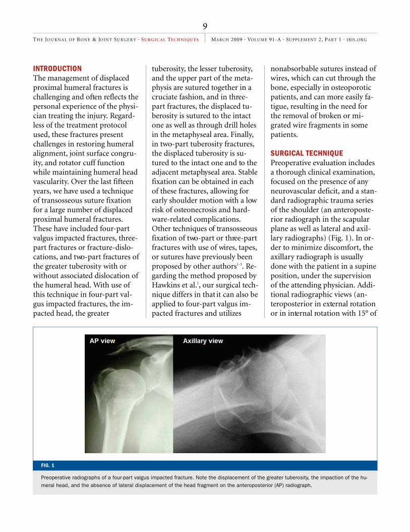
SURGICAL TECHNIQUE Preoperative evaluation includes a thorough clinical examination, focused on the presence of any neurovascular deficit, and a stan-dard radiographic trauma series of the shoulder (an anteroposte-rior radiograph in the scapular plane as well as lateral and axil-lary radiographs) (Fig. 1). In or-der to minimize discomfort, the axillary radiograph is usually done with the patient in a supine position, under the supervision of the attending physician. Addi-tional radiographic views (an-teroposterior in external rotation or in internal rotation with 15° of
FIG. 1
Preoperative radiographs of a four-part valgus impacted fracture. Note the displacement of the greater tuberosity, the impaction of the hu-meral head, and the absence of lateral displacement of the head fragment on the anteroposterior (AP) radiograph.
Dimakoupoulos.fm Page 9 Friday, February 6, 2009 2:57 PM
10
TH E JO UR N AL O F BO N E & JO IN T SU RG E R Y · SU R G IC A L TE CH N I Q U E S MARCH 2009 · VOLUME 91-A · SUPPLEMENT 2, PART 1 · JBJS.ORG
cephalic tilt) and computed to-mography can be useful in se-lected patients.
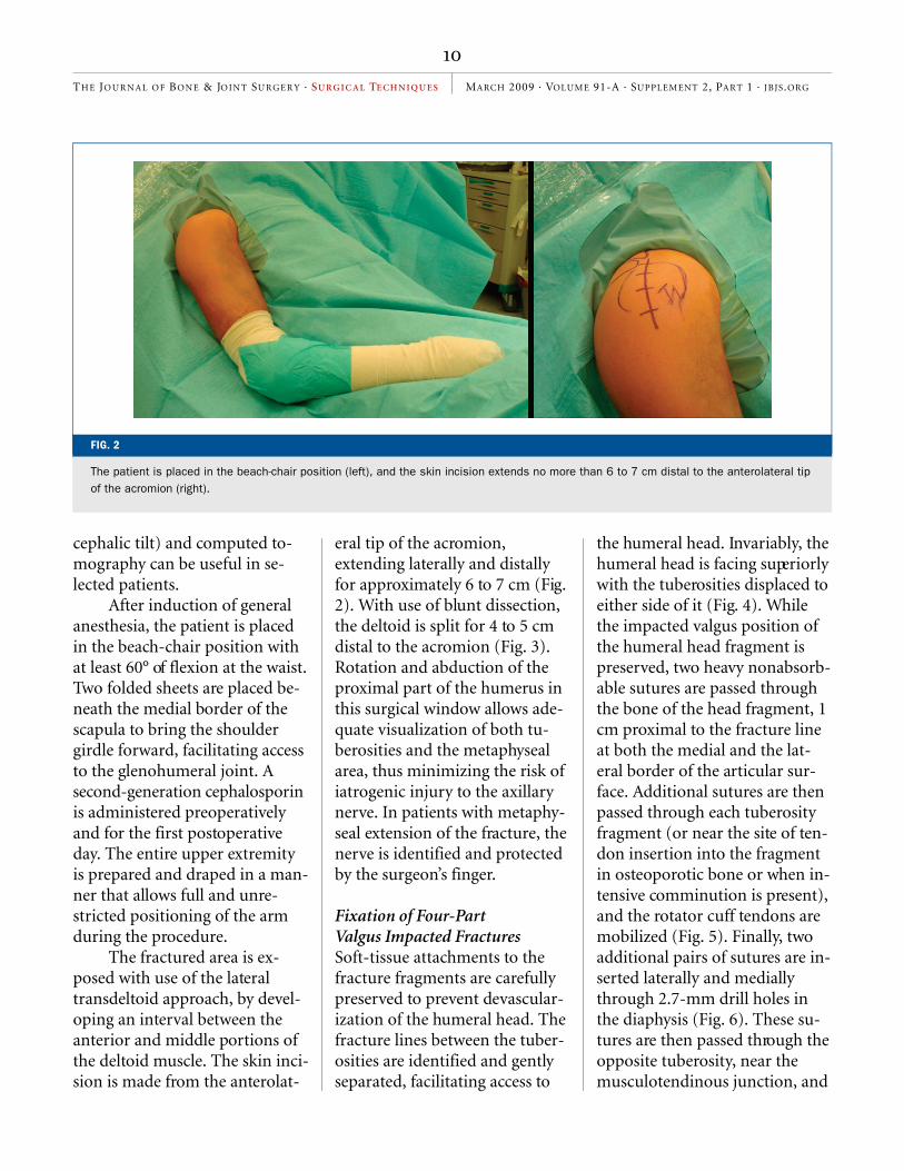
After induction of general anesthesia, the patient is placed in the beach-chair position with at least 60° of flexion at the waist. Two folded sheets are placed be-neath the medial border of the scapula to bring the shoulder girdle forward, facilitating access to the glenohumeral joint. A second-generation cephalosporin is administered preoperatively and for the first postoperative day. The entire upper extremity is prepared and draped in a man-ner that allows full and unre-stricted positioning of the arm during the procedure.
The fractured area is ex-posed with use of the lateral transdeltoid approach, by devel-oping an interval between the anterior and middle portions of the deltoid muscle. The skin inci-sion is made from the anterolat-
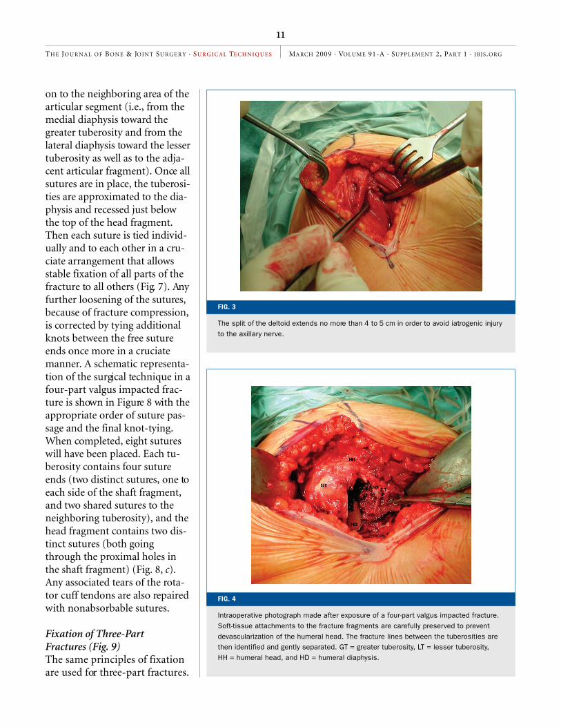
eral tip of the acromion, extending laterally and distally for approximately 6 to 7 cm (Fig. 2). With use of blunt dissection, the deltoid is split for 4 to 5 cm distal to the acromion (Fig. 3). Rotation and abduction of the proximal part of the humerus in this surgical window allows ade-quate visualization of both tu-berosities and the metaphyseal area, thus minimizing the risk of iatrogenic injury to the axillary nerve. In patients with metaphy-seal extension of the fracture, the nerve is identified and protected by the surgeon’s finger.
Fixation of Four-Part Valgus Impacted Fractures Soft-tissue attachments to the fracture fragments are carefully preserved to prevent devascular-ization of the humeral head. The fracture lines between the tuber-osities are identified and gently separated, facilitating access to
the humeral head. Invariably, the humeral head is facing superiorly with the tuberosities displaced to either side of it (Fig. 4). While the impacted valgus position of the humeral head fragment is preserved, two heavy nonabsorb-able sutures are passed through the bone of the head fragment, 1 cm proximal to the fracture line at both the medial and the lat-eral border of the articular sur-face. Additional sutures are then passed through each tuberosity fragment (or near the site of ten-don insertion into the fragment in osteoporotic bone or when in-tensive comminution is present), and the rotator cuff tendons are mobilized (Fig. 5). Finally, two additional pairs of sutures are in-serted laterally and medially through 2.7-mm drill holes in the diaphysis (Fig. 6). These su-tures are then passed through the opposite tuberosity, near the musculotendinous junction, and
FIG. 2
The patient is placed in the beach-chair position (left), and the skin incision extends no more than 6 to 7 cm distal to the anterolateral tip of the acromion (right).
Dimakoupoulos.fm Page 10 Friday, February 6, 2009 2:57 PM
11
TH E JO UR N AL O F BO N E & JO IN T SU RG E R Y · SU R G IC A L TE CH N I Q U E S MARCH 2009 · VOLUME 91-A · SUPPLEMENT 2, PART 1 · JBJS.ORG
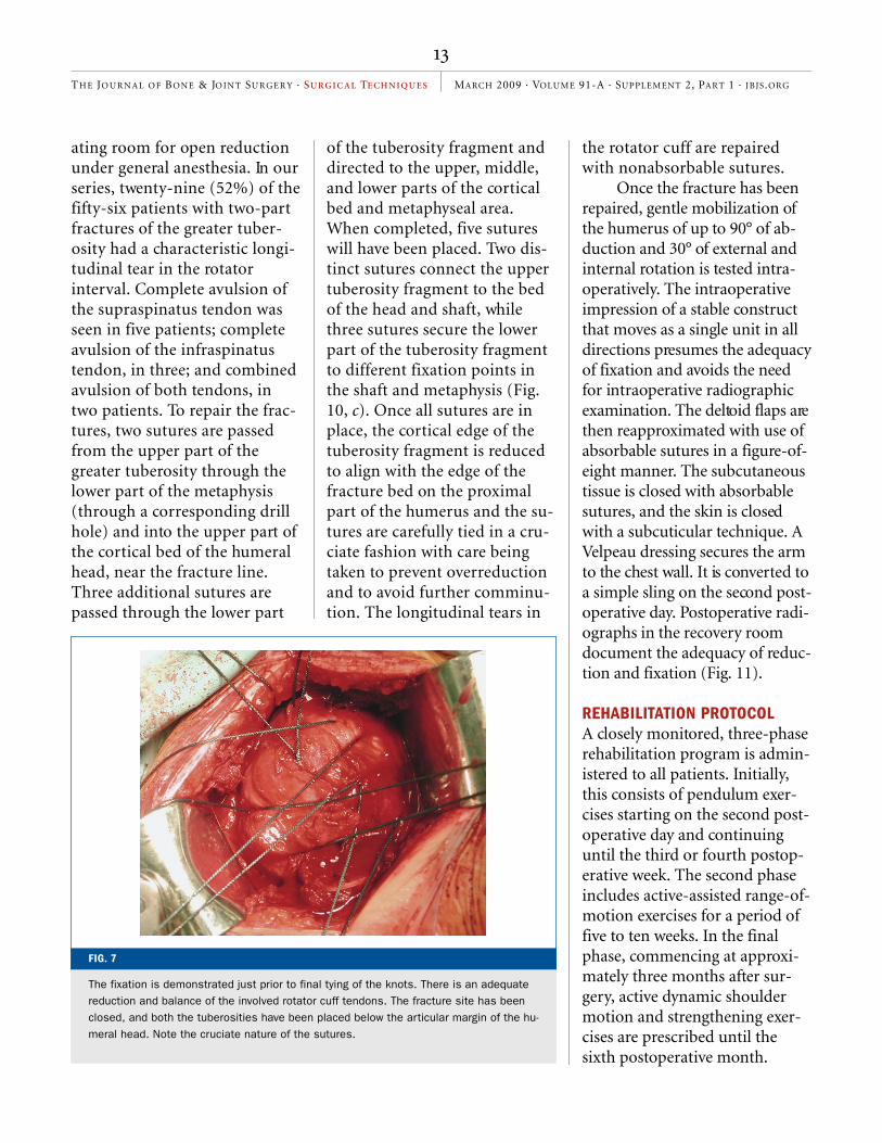
on to the neighboring area of the articular segment (i.e., from the medial diaphysis toward the greater tuberosity and from the lateral diaphysis toward the lesser tuberosity as well as to the adja-cent articular fragment). Once all sutures are in place, the tuberosi-ties are approximated to the dia-physis and recessed just below the top of the head fragment. Then each suture is tied individ-ually and to each other in a cru-ciate arrangement that allows stable fixation of all parts of the fracture to all others (Fig. 7). Any further loosening of the sutures, because of fracture compression, is corrected by tying additional knots between the free suture ends once more in a cruciate manner. A schematic representa-tion of the surgical technique in a four-part valgus impacted frac-ture is shown in Figure 8 with the appropriate order of suture pas-sage and the final knot-tying. When completed, eight sutures will have been placed. Each tu-berosity contains four suture ends (two distinct sutures, one to each side of the shaft fragment, and two shared sutures to the neighboring tuberosity), and the head fragment contains two dis-tinct sutures (both going through the proximal holes in the shaft fragment) (Fig. 8, c). Any associated tears of the rota-tor cuff tendons are also repaired with nonabsorbable sutures.
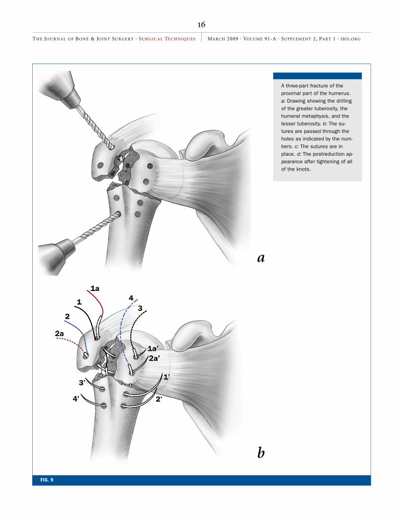
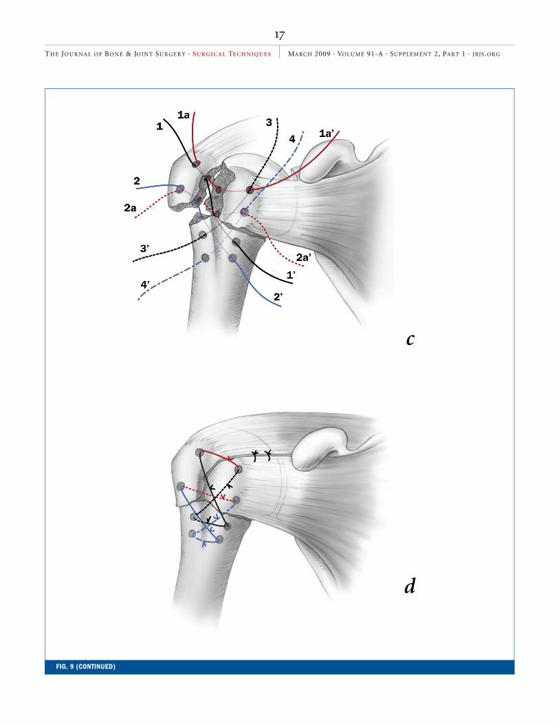
Fixation of Three-Part Fractures (Fig. 9) The same principles of fixation are used for three-part fractures.
FIG. 4
Intraoperative photograph made after exposure of a four-part valgus impacted fracture. Soft-tissue attachments to the fracture fragments are carefully preserved to prevent devascularization of the humeral head. The fracture lines between the tuberosities are then identified and gently separated. GT = greater tuberosity, LT = lesser tuberosity, HH = humeral head, and HD = humeral diaphysis.
FIG. 3
The split of the deltoid extends no more than 4 to 5 cm in order to avoid iatrogenic injury to the axillary nerve.
Dimakoupoulos.fm Page 11 Friday, February 6, 2009 2:57 PM

12
TH E JO UR N AL O F BO N E & JO IN T SU RG E R Y · SU R G IC A L TE CH N I Q U E S MARCH 2009 · VOLUME 91-A · SUPPLEMENT 2, PART 1 · JBJS.ORG
In this type of fracture, the hu-meral head is typically rotated either internally or externally, and care must be taken to achieve an adequate reduction in both the frontal and sagittal planes. Initially, two sutures are placed through the displaced greater tuberosity and then through the intact lesser tuber-osity. Two additional pairs of su-tures are inserted laterally and medially through 2.7-mm drill holes in the diaphysis. These su-tures are directed into the oppo-site tuberosity (i.e., the medial diaphysis toward the greater tu-berosity and the lateral diaphy-sis toward the intact lesser tuberosity). When completed, six sutures will have been placed, with each tuberosity containing four suture ends (two distinct sutures to the op-posite side of the shaft fragment
and two shared sutures to the neighboring tuberosity) (Fig. 9, c). Once all the sutures are in place, they are tied individually
and then to each other in a cru-ciate arrangement that allows stable fixation of all parts, one to the other. Loosening of the sutures, because of fracture compression, is corrected by ty-ing additional knots between the free suture ends in a cruci-ate manner. Associated rotator cuff tears are repaired with non-absorbable sutures.
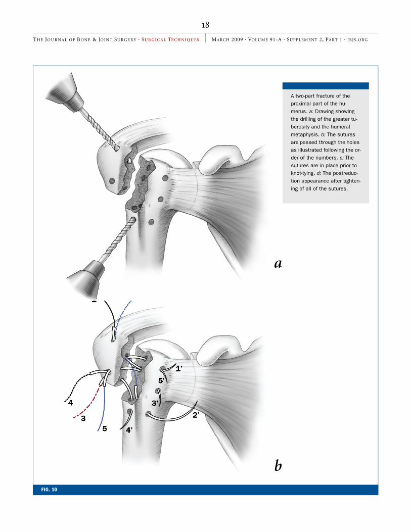
Fixation of Two-Part Fractures of the Greater Tuberosity (Fig. 10) When anterior dislocation of the shoulder accompanies frac-ture of the greater tuberosity, the patient is lightly sedated in the emergency department to facilitate reduction. Only one or two efforts are made to reduce the dislocation by closed means. If closed reduction fails, the pa-tient is transferred to the oper-
FIG. 5
The first suture is passed through the head fragment and the greater and lesser tuberos-ities. GT = greater tuberosity, LT = lesser tuberosity, HH = humeral head, and HD = hu-meral diaphysis.
FIG. 6
Additional sutures are placed through drill holes in the medial and lateral aspects of the humeral diaphysis (HD). The black arrows indicate the drill holes in the diaphysis. GT = greater tuberosity, LT = lesser tuberosity, and HH = humeral head.
Dimakoupoulos.fm Page 12 Friday, February 6, 2009 2:57 PM
13
TH E JO UR N AL O F BO N E & JO IN T SU RG E R Y · SU R G IC A L TE CH N I Q U E S MARCH 2009 · VOLUME 91-A · SUPPLEMENT 2, PART 1 · JBJS.ORG
ating room for open reduction under general anesthesia. In our series, twenty-nine (52%) of the fifty-six patients with two-part fractures of the greater tuber-osity had a characteristic longi-tudinal tear in the rotator interval. Complete avulsion of the supraspinatus tendon was seen in five patients; complete avulsion of the infraspinatus tendon, in three; and combined avulsion of both tendons, in two patients. To repair the frac-tures, two sutures are passed from the upper part of the greater tuberosity through the lower part of the metaphysis (through a corresponding drill hole) and into the upper part of the cortical bed of the humeral head, near the fracture line. Three additional sutures are passed through the lower part
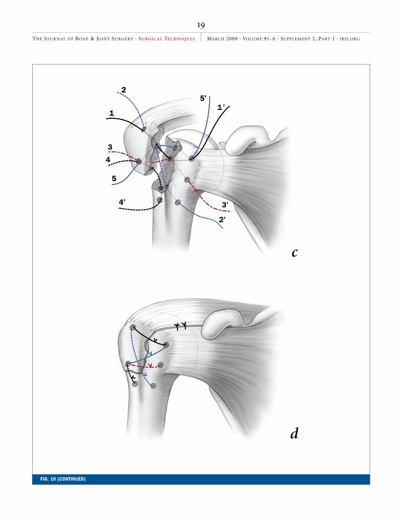
of the tuberosity fragment and directed to the upper, middle, and lower parts of the cortical bed and metaphyseal area. When completed, five sutures will have been placed. Two dis-tinct sutures connect the upper tuberosity fragment to the bed of the head and shaft, while three sutures secure the lower part of the tuberosity fragment to different fixation points in the shaft and metaphysis (Fig. 10, c). Once all sutures are in place, the cortical edge of the tuberosity fragment is reduced to align with the edge of the fracture bed on the proximal part of the humerus and the su-tures are carefully tied in a cru-ciate fashion with care being taken to prevent overreduction and to avoid further comminu-tion. The longitudinal tears in
the rotator cuff are repaired with nonabsorbable sutures.
Once the fracture has been repaired, gentle mobilization of the humerus of up to 90° of ab-duction and 30° of external and internal rotation is tested intra-operatively. The intraoperative impression of a stable construct that moves as a single unit in all directions presumes the adequacy of fixation and avoids the need for intraoperative radiographic examination. The deltoid flaps are then reapproximated with use of absorbable sutures in a figure-of-eight manner. The subcutaneous tissue is closed with absorbable sutures, and the skin is closed with a subcuticular technique. A Velpeau dressing secures the arm to the chest wall. It is converted to a simple sling on the second post-operative day. Postoperative radi-ographs in the recovery room document the adequacy of reduc-tion and fixation (Fig. 11).
REHABILITATION PROTOCOLA closely monitored, three-phase rehabilitation program is admin-istered to all patients. Initially, this consists of pendulum exer-cises starting on the second post-operative day and continuing until the third or fourth postop-erative week. The second phase includes active-assisted range-of-motion exercises for a period of five to ten weeks. In the final phase, commencing at approxi-mately three months after sur-gery, active dynamic shoulder motion and strengthening exer-cises are prescribed until the sixth postoperative month.
FIG. 7
The fixation is demonstrated just prior to final tying of the knots. There is an adequate reduction and balance of the involved rotator cuff tendons. The fracture site has been closed, and both the tuberosities have been placed below the articular margin of the hu-meral head. Note the cruciate nature of the sutures.
Dimakoupoulos.fm Page 13 Friday, February 6, 2009 2:57 PM

14
TH E JO UR N AL O F BO N E & JO IN T SU RG E R Y · SU R G IC A L TE CH N I Q U E S MARCH 2009 · VOLUME 91-A · SUPPLEMENT 2, PART 1 · JBJS.ORG
A four-part valgus impacted frac-ture of the proximal part of the humerus. a: Drawing showing the drilling of the greater and lesser tuberosities, the head fragment in its valgus impacted position, and the lateral and medial meta-physeal areas. b: The sutures are passed through the holes in the appropriate order as indi-cated by the numbers. c: The sutures are in place. The tuber-osities are pulled down below the level of the top of the head and are tightened not only to each other but also to the meta-physis overlapping the lateral cortex. d: The postreduction ap-pearance after tightening of all of the knots.
FIG. 8
a
b
Dimakoupoulos.fm Page 14 Friday, February 6, 2009 2:57 PM

15
TH E JO UR N AL O F BO N E & JO IN T SU RG E R Y · SU R G IC A L TE CH N I Q U E S MARCH 2009 · VOLUME 91-A · SUPPLEMENT 2, PART 1 · JBJS.ORG
c
d
FIG. 8 (CONTINUED)
Dimakoupoulos.fm Page 15 Friday, February 6, 2009 2:57 PM
16
TH E JO UR N AL O F BO N E & JO IN T SU RG E R Y · SU R G IC A L TE CH N I Q U E S MARCH 2009 · VOLUME 91-A · SUPPLEMENT 2, PART 1 · JBJS.ORG
FIG. 9
A three-part fracture of the proximal part of the humerus. a: Drawing showing the drilling of the greater tuberosity, the humeral metaphysis, and the lesser tuberosity. b: The su-tures are passed through the holes as indicated by the num-bers. c: The sutures are in place. d: The postreduction ap-pearance after tightening of all of the knots.
a
b
Dimakoupoulos.fm Page 16 Friday, February 6, 2009 2:57 PM
17
TH E JO UR N AL O F BO N E & JO IN T SU RG E R Y · SU R G IC A L TE CH N I Q U E S MARCH 2009 · VOLUME 91-A · SUPPLEMENT 2, PART 1 · JBJS.ORG
FIG. 9 (CONTINUED)
c
d
Dimakoupoulos.fm Page 17 Friday, February 6, 2009 2:57 PM
18
TH E JO UR N AL O F BO N E & JO IN T SU RG E R Y · SU R G IC A L TE CH N I Q U E S MARCH 2009 · VOLUME 91-A · SUPPLEMENT 2, PART 1 · JBJS.ORG
FIG. 10
A two-part fracture of the proximal part of the hu-merus. a: Drawing showing the drilling of the greater tu-berosity and the humeral metaphysis. b: The sutures are passed through the holes as illustrated following the or-der of the numbers. c: The sutures are in place prior to knot-tying. d: The postreduc-tion appearance after tighten-ing of all of the sutures.
a
b
Dimakoupoulos.fm Page 18 Friday, February 6, 2009 2:57 PM
19
TH E JO UR N AL O F BO N E & JO IN T SU RG E R Y · SU R G IC A L TE CH N I Q U E S MARCH 2009 · VOLUME 91-A · SUPPLEMENT 2, PART 1 · JBJS.ORG
FIG. 10 (CONTINUED)
c
d
Dimakoupoulos.fm Page 19 Friday, February 6, 2009 2:57 PM
20
TH E JO UR N AL O F BO N E & JO IN T SU RG E R Y · SU R G IC A L TE CH N I Q U E S MARCH 2009 · VOLUME 91-A · SUPPLEMENT 2, PART 1 · JBJS.ORG
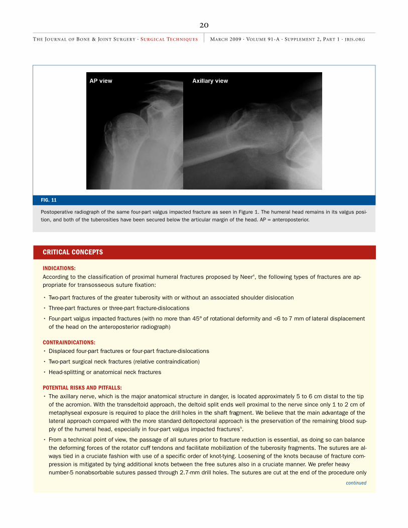
FIG. 11
Postoperative radiograph of the same four-part valgus impacted fracture as seen in Figure 1. The humeral head remains in its valgus posi-tion, and both of the tuberosities have been secured below the articular margin of the head. AP = anteroposterior.
CRITICAL CONCEPTS
INDICATIONS:
According to the classification of proximal humeral fractures proposed by Neer4, the following types of fractures are ap-propriate for transosseous suture fixation:
• Two-part fractures of the greater tuberosity with or without an associated shoulder dislocation
• Three-part fractures or three-part fracture-dislocations
• Four-part valgus impacted fractures (with no more than 45° of rotational deformity and <6 to 7 mm of lateral displacement of the head on the anteroposterior radiograph)
CONTRAINDICATIONS:
• Displaced four-part fractures or four-part fracture-dislocations
• Two-part surgical neck fractures (relative contraindication)
• Head-splitting or anatomical neck fractures
POTENTIAL RISKS AND PITFALLS:
• The axillary nerve, which is the major anatomical structure in danger, is located approximately 5 to 6 cm distal to the tip of the acromion. With the transdeltoid approach, the deltoid split ends well proximal to the nerve since only 1 to 2 cm of metaphyseal exposure is required to place the drill holes in the shaft fragment. We believe that the main advantage of the lateral approach compared with the more standard deltopectoral approach is the preservation of the remaining blood sup-ply of the humeral head, especially in four-part valgus impacted fractures5.
• From a technical point of view, the passage of all sutures prior to fracture reduction is essential, as doing so can balance the deforming forces of the rotator cuff tendons and facilitate mobilization of the tuberosity fragments. The sutures are al-ways tied in a cruciate fashion with use of a specific order of knot-tying. Loosening of the knots because of fracture com-pression is mitigated by tying additional knots between the free sutures also in a cruciate manner. We prefer heavy number-5 nonabsorbable sutures passed through 2.7-mm drill holes. The sutures are cut at the end of the procedure only
continued
Dimakoupoulos.fm Page 20 Friday, February 6, 2009 2:57 PM
21
TH E JO UR N AL O F BO N E & JO IN T SU RG E R Y · SU R G IC A L TE CH N I Q U E S MARCH 2009 · VOLUME 91-A · SUPPLEMENT 2, PART 1 · JBJS.ORG
Panayiotis Dimakopoulos, MDAndreas Panagopoulos, MDGeorgios Kasimatis, MDShoulder and Elbow Unit, Orthopaedic Department, University Hospital of Patras, Papanikolaou Street, Rio-Patras 26504, Greece. E-mail address for P. Dimakopoulos: [email protected]. E-mail address for A. Panagopoulos: [email protected]. Email address for G. Kasimatis: [email protected]
The line drawings in this article are the work of Joanne Haderer Müller of Haderer & Müller ([email protected]).
REFERENCES1. Hawkins RJ, Bell RH, Gurr K. The three-part fracture of the proximal part of the humerus.
Operative treatment. J Bone Joint Surg Am. 1986;68:1410-4.
2. Park MC, Murthi AM, Roth NS, Blaine TA, Levine WN, Bigliani LU. Two-part and three-part fractures of the proximal humerus treated with suture fixation. J Orthop Trauma. 2003;17:319-25.
3. Banco SP, Andrisani D, Ramsey M, Frie-man B, Fenlin JM. Tension band fixation for unstable two-part humeral fractures in patients with osteopenic bone (parachute technique). Tech Shoulder Elbow Surg. 2001;2:50-3.
4. Neer CS 2nd. Four-segment classification of proximal humeral fractures: purpose and reliable use. J Shoulder Elbow Surg.
2002;11:389-400.
5. Panagopoulos AM, Dimakopoulos P, Tyllianakis M, Karnabatidis D, Siablis D, Papa-dopoulos AX, Lambiris E, Kraniotis P, Sakella-ropoulos G. Valgus impacted proximal humeral fractures and their blood supply after transosseous suturing. Int Orthop. 2004;28:333-7.
6. Dimakopoulos P, Panagopoulos A, Kasimatis G, Syggelos SA, Lambiris E. Anterior traumatic shoulder dislocation associated with displaced greater tuberos-ity fracture: the necessity of operative treatment. J Orthop Trauma. 2007;21:104-12.
CRITICAL CONCEPTS
POTENTIAL RISKS AND PITFALLS (CONTINUED):
after a stable construct has been achieved. With osteoporotic or severely comminuted tuberosity fragments, the sutures are passed near the musculotendinous junctions.
• The displaced tuberosities in four-part valgus impacted fractures are always pulled down below the top of the head frag-ment with the shoulder in the adducted position and are sutured not only to each other but also to the head fragment as well as to the medial and lateral aspects of the diaphysis in a manner that we believe neutralizes the deforming muscular forces. We avoid disimpacting the head fragment from its valgus impacted position, thus minimizing the risk of further dis-ruption of the vulnerable blood supply of the posteromedial hinge. Despite this “incomplete” fracture reduction, it seems that the residual disturbance of normal anatomy does not affect shoulder joint mechanics. The moment arm of the rotator cuff muscles is preserved by suturing the tuberosities below the top of the impacted head. Use of this approach is sup-ported by the very low rate of early degenerative arthritis seen in our series.
• In three-part fractures of the greater tuberosity, the sutures are passed through the intact lesser tuberosity. This provides a stable construct and restores the normal functional balance of the involved tendons, thus allowing for early shoulder joint motion. In isolated two-part fractures of the greater tuberosity, the displaced tuberosity is reduced to its anatomical position, thus avoiding a mechanical block to abduction of the shoulder or obstruction of external rotation because of pos-terior displacement of the greater tuberosity. Our preference is for suture fixation of the greater tuberosity fragment in pa-tients with associated dislocation of the shoulder, regardless of the extent of its postreduction displacement6. Our decision to internally fix the greater tuberosity in its anatomical position is based on the nature of the injury rather than the degree of postreduction displacement. Recently proposed guidelines of 5 or 10 mm of greater tuberosity displacement as an indication for internal fixation cannot be followed because displacement often exceeds 20 mm at the time of dislocation. Associated tears of rotator cuff tendons, noted in the majority of our patients, are an additional indication for early surgical intervention.
• Regarding two-part surgical neck fractures, we believe that the optimal treatment is with plate-and-screw osteosynthesis. We do not recommend transosseous suture fixation in this type of fracture as rotational instability between the large prox-imal fragment and the narrow diaphysis can often be problematic. In such patients, stable fixation can be achieved only if the humeral head fragment is impacted to the diaphysis.
• Finally, integral to obtaining the optimum outcome is completion of the full rehabilitation program. An important variation of our current regimen over previous protocols is the early initiation of pendulum exercises on the second postoperative day and their continuation for the first three to four weeks. A full range of motion is restored in this manner without exert-ing stress on the fixation.
AUTHOR UPDATE:
No changes or modifications of the original technique have been made since its publication.
Dimakoupoulos.fm Page 21 Friday, February 6, 2009 2:57 PM