Embed Size (px)
Citation preview

ISSN 2182-9942
Article / Artigo
AbstractTransmitted spectroscopy and transmitted multispectral imaging in the 400-900 nm range have been applied for the mapping and tentative identification of paints covered by a white preparation as in the case of a ground laid for reusing a canvas for another painting. These methods can be applied to polychrome works of art, as long as their support and new preparation are sufficiently translucent. This work presents the transmittance spectra acquired from a test board consisting of a prepared canvas with swatches of 54 pigments covered with titanium white and the mul-tispectral images realized with transmitted light to map covered paints on a mock-up painting. It was observed that 18 out of 54 historical pigments provide characteristic transmittance spectra even underneath a titanium white preparation layer and that transmitted light multispectral imaging can map hidden paint layers.
ResumoA espectroscopia de transmissão e a imagiologia multiespectral de transmissão no intervalo entre 400 e 900 nm foram usadas para a detecção e possível identificação de pinturas encobertas por uma camada branca semelhante à camada de preparação que é aplicada sobre a pintura já existente numa tela quando se pretende reutilizá-la. Estes métodos podem ser usados com obras de arte polícromas desde que o suporte e a preparação sejam suficientemente translúcidos. Este trabalho apresenta os espectros de transmissão obtidos com um painel de teste, constituído por uma tela com preparação sobre a qual foram aplicadas amostras de tintas de 54 pigmentos que foram cobertas por uma camada de branco de titânio, e as imagens multiespectrais de luz transmitida obtidas com o objectivo de mapear as pinturas dess painel. Dezoito dos 54 pigmentos históricos proporcionaram espectros de transmissão característicos, mesmo sob uma camada de preparação de branco de titânio, e as imagens multiespectrais de luz transmitida permitiram mapear as zonas pintadas.
Palavras-chaveImagem multiespectralIdentificação de pigmentosEspectroscopia de transmissãoEspectroscopia de reflexão
KeywordsMultispectral imagingPigments identificationTransmittance spectroscopyReflectance spectroscopy
Conservar Património 24 (2016) 37-45 | doi:10.14568/cp2015021ARP - Associação Profissional de Conservadores-Restauradores de Portugalhttp://revista.arp.org.pt
Espectroscopia de transmissão e imagiologia multiespectral de transmissão para a visualização de pinturas subjacentes
Transmittance spectroscopy and transmitted multispectral imaging to map covered paints
Antonino Cosentino
Cultural Heritage Science Open Source, Piazza Cantarella 11, Aci Sant’Antonio, 95025, [email protected]

Antonino Cosentino
Conservar Património 24 (2016)38
Introduction
The detection of underlying layers, such as underpaint-ing, modifications to the original sketch (pentimenti) or hidden compositions on a reused canvas is of great impor-tance in art conservation. The more commonly used tech-niques to reveal these features are X-ray radiography [1], infrared reflectography [2-3] and neutron autoradiography [4], while the mapping of hidden paint layers on canvas has also been recently tried with innovative techniques such as Terahertz imaging [5] and XRF mapping [6]. Multispectral [7-10] and hyperspectral [11-14] imaging are used for non-invasive identification and mapping of pigments in polychrome artworks and also for the detection of hidden layers when extended to the near infrared range.
This study discusses the use of transmittance spectroscopy and multispectral imaging with transmitted radiation (MSIT) for the identification of pigments and their mapping when hidden beneath another layer of paint, as in the case of a white ground preparation laid for reusing the canvas [15]. This method can be applied to polychrome works of art, as long as their support and preparation are sufficiently translucent. Support, preparation, paints, and the covering layer must be thin enough to allow visible and infrared radiation to pass through and be measured with a transmittance spectrometer.
This work illustrates the measurements acquired from a test board consisting of a prepared canvas with swatches
of pigments covered with titanium white. The spectra were acquired with a miniaturized and portable spectrometer covering the VIS-NIR range. The transmitted spectrum shows the ratio between the intensity of the transmitted light and the incident light (transmittance) expressed in percentage. Similar to reflectance spectroscopy, transmittance spectra can provide useful information for the identification of pigments, since the light is absorbed depending on the chemical composition of the material tested. Reflectance spectroscopy has been used from the late 1980s [16] and there are reference spectra databases for pigments [17-18].
The main advantage of transmittance spectroscopy and MSIT with respect to other spectroscopy and imag-ing methods currently applied for revealing hidden paint layers is that the equipment is relatively low in cost. Transmittance spectra are acquired with the same equip-ment as reflectance spectroscopy: a light source, a spec-trometer, a probe and one optical fiber to collect the trans-mitted light. Similarly, MSIT can be performed with the same equipment used for traditional (reflected radiation) multispectral imaging. Another important advantage is that the most used white pigments to make a preparation layer (lead white, zinc white, titanium white and litho-pone) are very opaque when examined in reflected im-aging but they become very transparent with transmitted infrared imaging [19]. The same white pigments, and in particular lead white, are opaque in X-radiography and
Figure 1. 54 historical pigments laid on: a) glass plate; b) test board. c) The same test board where the pigments swatches are half covered behind a titanium white paint layer. d) Cross-section of the test board: Egyptian blue covered by the titanium white preparation.

Conservar Património 24 (2016)
Transmittance spectroscopy and transmitted multispectral imaging to map covered paints
39
infrared reflectography, and usually they hide the under paints. For this reason, other methods have been applied, such as Terahertz scanning [5], which is not affected by the lead-containing preparation layer. These methods are costly and require highly specialized personnel and facil-ities, while transmittance spectroscopy and MSIT can be implemented with comparatively much lower budgets.
Methods
Samples
This paper presents the transmittance spectra of 54 historical pigments that have been laid with gum arabic on a glass plate (Figure 1). A swatch of just gum arabic is added as a reference for the binder. The pigments were hand mulled into the gum arabic (as much as required to wet each pigment) and applied to the glass plate with a brush.
While not intended as an exhaustive list, the 54 pigments represent a selection of the most widely used ones from antiquity to the early 1950s. Table 1 lists the pigments used in this study, which are all distributed by Kremer Pigments. Information about their composition and manufacture is available directly from the Kremer website [20] by searching the specific product code.
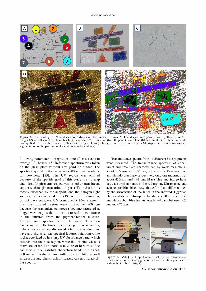
A second test board was prepared using swatches of the same 54 pigments on a pure-cotton, medium-grain canvas with a titanium white acrylic ground layer preparation (Ferrario, Italy). The swatches were then fixed using an acrylic resin (Maimeri # 675) and half the surface was covered with a layer of titanium white. A test painting consisting of nine pigments laid out as geometric shapes and then covered with a single layer of titanium white was also prepared (Figure 2). Since this is a preliminary study, among all the possible white paints and grounds, only titanium white was evaluated for both the original preparation and the covering layer. Titanium white replaced lead white and it is the most diffused white pigment after 1930.
Instrumentation
The transmission spectra were acquired with ASEQ LR1 spectrometer (300-1000 nm, 2 nm resolution with 100 µm slit) featuring a Toshiba TCD1304DG linear array (3648 pixels). The spectrometer was coupled with the ASEQ light source (CH-S2) and ASEQ fiber-optics probe F01_R03 consisting of seven fibers (600 µm core), six for excitation and one for collection. The multispectral imaging system is composed of a PixelTeq SpectroCam UV-VIS CCD camera and eight interferential filters manufactured by the same company (center wavelength/bandwidth nm): 860/20, 800/10, 780/20, 740/10, 717/10, 578/10, 511/20, 425/50. This camera incorporates a high-sensitivity 5 Megapixel CCD in the range 200-1000 nm and a sequential 8-band filter-wheel [21-22].
Results and discussion
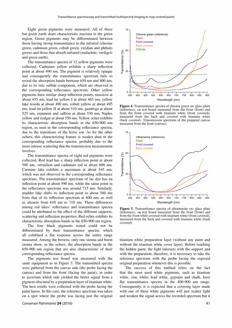
The transmittance spectra of the 54 swatches of pigments laid on the glass plate were acquired with the ASEQ LR1 system (Figure 3). The paint itself acts as a diffuser and the light source (CH-S2) attached to the spectrometer was located just behind the paint, while on the other side (facing the paint) the probe collects transmitted light and delivers it to the spectrometer. Transmittance spectra were measured using the
Table 1List of pigments with the Kremer Pigments product code.
Colours Pigments
Blacks Ivory black, 12000Vine black, 47000Bone black, 47100Lamp black, 47250
Blues Azurite, 10200Blue bice, 10184Cobalt blue, 45730Egyptian blue, 10060Indigo, 36005Maya blue, 36007Prussian blue, 45202Smalt, 10000Ultramarine nat, 10510Phthalo blue, 23050Cobalt violet, 45800
Browns Burnt Sienna, 40430Burnt umber, 40710Van Dyke brown 41000Raw Sienna, 17050Raw umber, 40610
Greens Cadmium green, 44510Chrome green, 44200Cobalt green, 44100Green earth, 11000Malachite, 10300Phthalo green, 23000Verdigris, 44450Viridian, 44250
Reds Alizarin, 23600Cadmium red, 21120Red lead, 42500Read ochre, 11574Vermilion, 10610Madder lake, 372051Lac dye, 36020Carmine lake, 42100Realgar, 10800
Whites Lead white, 46000Zinc white, 46300Lithopone, 46100Titanium white, 46200Gypsum, 58300Chalk, 58000
Yellows Cadmium yellow, 21010Cobalt yellow, 43500Lead Tin yellow I, 10100Lead Tin yellow II, 10120Massicot, 43010Naples yellow, 10130Orpiment, 10700Saffron, 36300Yellow ochre, 40010Yellow Lake reseda, 36262Gamboge, 37050
Antonino Cosentino
Conservar Património 24 (2016)40
following parameters: integration time 50 ms, scans to average 10, boxcar 15. Reference spectrum was taken on the glass plate without any paint or binder. The spectra acquired in the range 400-900 nm are available for download [23]. The UV region was omitted because of the specific goal of this study, i.e. to map and identify pigments on canvas or other translucent supports through transmitted light (UV radiation is mostly absorbed by the support, and the halogen light sources, otherwise used for VIS and IR illumination, do not have sufficient UV component). Measurements into the infrared region were limited to 900 nm because the transmittance spectra become saturated at longer wavelengths due to the increased transmittance in the infrared from the pigment-binder mixture. Transmittance spectra feature the same absorption bands as in reflectance spectroscopy. Consequently, only a few cases are discussed. Gum arabic does not have any characteristic spectral feature. Titanium white is characterized by its sharp UV absorbance band, which extends into the blue region, while that of zinc white is much smoother. Lithopone, a mixture of barium sulfide and zinc sulfide, exhibits absorption bands in the 650-800 nm region due to zinc sulfide. Lead white, as well as gypsum and chalk, exhibit featureless and relatively flat spectra.
Transmittance spectra from 11 different blue pigments were measured. The transmittance spectrum of cobalt violet and smalt are characterized by weak maxima, at about 515 nm and 560 nm, respectively. Prussian blue and phthalo blue have respectively only one maximum, at about 450 nm and 492 nm. Maya blue and indigo have large absorption bands in the red region. Ultramarine and azurite (and blue bice, its synthetic form) are differentiated by the absorbance of the latter in the infrared. Egyptian blue exhibits two absorption bands near 800 nm and 630 nm while cobalt blue has just one broad band between 525 nm and 675 nm.
Figure 2. Test painting. a) Nine shapes were drawn on the prepared canvas. b) The shapes were painted with: yellow ochre (1), realgar (2), cobalt violet (3), lamp black (4), malachite (5), vermilion (6), lithopone (7), red lead (8) and smalt (9). c) titanium white was applied to cover the shapes. d) Transmitted light photo (lighting from the canvas side). e) Multispectral imaging transmitted segmentation of the painting (color code is as indicated in a).
Figure 3. ASEQ LR1 spectrometer set up for transmission spectra measurement of pigments laid on the glass plate (left) and on the test board (right).
Conservar Património 24 (2016)
Transmittance spectroscopy and transmitted multispectral imaging to map covered paints
41
Eight green pigments were measured. All of these but green earth share characteristic maxima in the green region. Green pigments may be differentiated between those having strong transmittance in the infrared (chrome green, cadmium green, cobalt green, viridian and phthalo green) and those that absorb infrared (malachite, verdigris and green earth).
The transmittance spectra of 12 yellow pigments were collected. Cadmium yellow exhibits a sharp inflection point at about 490 nm. The pigment is relatively opaque and consequently the transmittance spectrum fails to reveal the absorption bands between 650 nm and 800 nm, due to its zinc sulfide component, which are observed in the corresponding reflectance spectrum. Other yellow pigments have similar sharp inflection points, massicot at about 455 nm, lead tin yellow I at about 465 nm, yellow lake reseda at about 490 nm, cobalt yellow at about 495 nm, lead tin yellow II at about 510 nm, gamboge at about 525 nm, orpiment and saffron at about 530 nm, Naples yellow and realgar at about 550 nm. Yellow ochre exhibits its characteristic absorption bands in the 650-900 nm region, as seen in the corresponding reflectance spectra, due to the transitions of the ferric ion. As for the other ochers, this characterizing feature is weaker than in the corresponding reflectance spectra, probably due to the more intense scattering that the transmission measurement involves.
The transmittance spectra of eight red pigments were collected. Red lead has a sharp inflection point at about 580 nm, vermilion and cadmium red at about 600 nm. Carmine lake exhibits a maximum at about 545 nm, which was not observed in the corresponding reflectance spectrum. The transmittance spectrum of lac dye has an inflection point at about 590 nm, while the same point in the reflectance spectrum was around 715 nm. Similarly, madder lake shifts its inflection point to about 580 nm from that of its reflection spectrum at 600 nm, as well as alizarin from 610 nm to 710 nm. These differences among red lakes’ reflectance and transmittance spectra could be attributed to the effect of the different supports, scattering and reflection properties. Red ochre exhibits its characteristic absorption bands in the 650-900 nm region.
The four black pigments tested could not be differentiated by their transmittance spectra, which all exhibited a flat response across the entire range measured. Among the browns, only raw sienna and burnt sienna show, as the ochers, the absorption bands in the 650-900 nm region that are also characteristic of their corresponding reflectance spectra.
The pigments test board was measured with the same equipment as in Figure 3. The transmitted spectra were gathered from the canvas side (the probe facing the canvas) and from the front (facing the paint), in order to ascertain which side yielded the better signal from a pigment obscured by a preparation layer of titanium white. The best results were collected with the probe facing the paint layers. In this case, the reference spectrum was taken on a spot where the probe was facing just the original
titanium white preparation layer (without any paint and without the titanium white cover layer). Before reaching the hidden paint, the light interacts with the support and with the preparation; therefore, it is necessary to take the reference spectrum with the probe facing the exposed original preparation whenever this is possible.
The success of this method relies on the fact that the most used white pigments, such as titanium white, zinc white, lead white, gypsum and chalk, have flat transmittance spectra in the 400-900 nm range. Consequently, it is expected that a covering layer made with one of these white pigments will just scatter light and weaken the signal across the recorded spectrum but it
400 450 500 550 600 650 700 750 800 850 900
0
25
50
75
Tra
nsm
itta
nce
(%
)
Wavelength (nm)
Chrome green (reference)
Front
Front covered
Back covered
Canvas
Figure 4. Transmittance spectra of chrome green on: glass plate (reference), on test board measured from the front (front) and from the front covered with titanium white (front covered), measured from the back and covered with titanium white (back covered). Transmission spectrum of the prepared canvas measured from the front (canvas).
400 450 500 550 600 650 700 750 800 850 900
0
25
50
75
Tra
nsm
itta
nce
(%
)
Wavelength (nm)
Ultramarine (reference)
Front
Front covered
Back covered
Figure 5. Transmittance spectra of ultramarine on: glass plate (reference), on test board measured from the front (front) and from the front while covered with titanium white (front covered), measured from the back and covered with titanium white (back covered).

Antonino Cosentino
Conservar Património 24 (2016)42
will not introduce specific features. Specifically, titanium white has a sharp absorption band in the UV region, which extends into the violet. This causes the apparent sharp transmittance increase that dominated the violet region of the spectra acquired on the prepared canvas with a titanium white preparation. On the other hand, the infrared region of these spectra also features a sharp transmittance rise due to the increased transmittance in the infrared from the pigment-binder mixture.
Figure 4 shows transmittance spectra of the chrome green paint swatch on the test board. The spectrum of the pigment on the glass plate has a very sharp maximum in the green region. Chrome green is relatively opaque and its paint layer stops almost all of the light. Consequently,
the same maximum appears very weak in the spectrum taken from the front of the test board and, when the paint is covered with a layer of titanium white, the maximum cannot be detected. Similarly, the large absorption band of ultramarine between 500 and 700 nm is not detected when the paint is covered by titanium white in measurements taken from both the front and back of the test canvas (Figure 5).
On the other hand, lithopone is less opaque. Its transmittance spectrum exhibits characteristic features even when the paint is covered by a layer of titanium white (Figure 6). Lithopone is a mixture of barium sulfate, a good flat reflector, and zinc sulfide, which is responsible for absorption bands in the 650-800 nm
400 450 500 550 600 650 700 750 800 850 900
0
25
50
75
Tra
nsm
itta
nce
(%
)
Wavelength (nm)
Lithopone (reference)
Front
Front covered
Back covered
Figure 6. Transmittance spectra of lithopone on: glass plate (reference), on test board measured from the front (front) and from the front while covered with titanium white (front covered); measured from the back and covered with titanium white (back covered).
400 450 500 550 600 650 700 750 800 850 900
Tra
nsm
itta
nce
(%
)
Wavelength (nm)
0
5
10
Vermilion (reference)
Front
Front covered
Back covered
Figure 7. Transmittance spectra of vermilion on: glass plate (reference), on test board measured from the front (front) and from the front while covered with titanium white (front covered); measured from the back and covered with titanium white (back covered).
400 450 500 550 600 650 700 750 800 850 900
0
25
50
75
Tra
nsm
itta
nce
(%
)
Wavelength (nm)
Cobalt violet (reference)
Front
Front covered
Back covered
Figure 8. Transmittance spectra of cobalt violet on: glass plate (reference), on test board measured from the front (front) and from the front while covered with titanium white (front covered); measured from the back and covered with titanium white (back covered).
400 450 500 550 600 650 700 750 800 850 900
Tra
nsm
itta
nce
(%
)
Wavelength (nm)
Maya blue
Indigo
Smalt
Cobalt blue
0
10
20
30
40
50
60
Figure 9. Transmittance spectra of blue pigments on test board covered with titanium white and corresponding reference transmittance spectra (dashed line).

Conservar Património 24 (2016)
Transmittance spectroscopy and transmitted multispectral imaging to map covered paints
43
region. Manufactured for the first time in 1874, lithopone failed to achieve wide acceptance as an artist’s pigment because early formulations were not very lightfast. Later formulations of lithopone overcame the lightfastness issue but could not overcome the bad reputation and its use was limited to the priming of the canvas. One important implication of this historical fact is that now lithopone becomes useful for the non-destructive dating of artworks on canvas using transmittance spectroscopy to detect the presence of lithopone as a primer.
Vermilion, with a sharp inflection point at 600 nm, is an example of a general class of pigments with characteristically sharp inflection points in their transmittance spectra. This unique feature is detectable, even if it is weak when the pigment is covered by a layer of titanium white (Figure 7). This signal is undetectable from the unprimed side of the test canvas.
The distinguishing spectral features of cobalt violet, shown in figure 8, are a large maximum at about 690 nm and a small maximum at about 515 nm. From the front, both maxima are visible in the transmittance spectra even when the paint is covered with a layer of titanium white. In contrast, the smaller maximum is undetected in the spectrum taken from the unprimed canvas side.
Of the 54 swatches of historical pigments on the test board covered with a layer of titanium white, 18 provided spectra where all or some of their characteristic spectral features were detected:
Whites: lithopone, titanium white.Blues: cobalt violet, Maya blue, indigo, smalt, cobalt
blue (Figure 9).Greens: malachite, verdigris (Figure 10).Yellows: cadmium yellow, Naples yellow, yellow
ochre, realgar (Figure 11).
400 450 500 550 600 650 700 750 800 850 900
Tra
nsm
itta
nce
(%
)
Wavelength (nm)
0
10
20
30
40
50
Malachite
Verdigris
Figure 10. Transmittance spectra of green pigments which could be identified on test board covered with titanium white and corresponding reference spectra (dashed line).
400 450 500 550 600 650 700 750 800 850 900
Tra
nsm
itta
nce
(%
)
Wavelength (nm)
0
10
20
30
40
50
Cadmium yellow
Naples yellow
Yellow ochre
Realgar
Figure 11. Transmittance spectra of yellow pigments which could be identified on test board covered with titanium white and corresponding reference spectra (dashed line).
400 450 500 550 600 650 700 750 800 850 900
Tra
nsm
itta
nce
(%
)
Wavelength (nm)
0
10
20
30
Vermilion
Madder lake
Alizarin
Figure 12. Transmittance spectra of reds pigments which could be identified on test board covered with titanium white and corresponding reference spectra (dashed line).
400 450 500 550 600 650 700 750 800 850 900
Tra
nsm
itta
nce
(%
)
Wavelength (nm)
0
10
20
30
Cadmium red
Red lead
Figure 13. Transmittance spectra of reds pigments which could be identified on test board covered with titanium white and corresponding reference spectra (dashed line).

Antonino Cosentino
Conservar Património 24 (2016)44
Reds: vermilion, madder lake, alizarin, cadmium red, red lead (Figures 12-13).
The encouraging results from the test board and the test painting led to trying a similar experiment using the PixelTeq UV-VIS SpectroCam multispectral camera to evaluate the possibility of mapping hidden paints with multi-spectral imaging using transmitted radiation. The MSIT method is proposed only for mapping pigments using relatively large broadband filters since the amount of transmitted radiation is much less than in standard MSI. Consequently, it is unpractical to use narrow bandpass filters to reconstruct transmittance spectra.
A 400 W halogen lamp was set at 1.5 m from the canvas delivering 3000 lux on the test painting. Extreme care should be used when operating on works of art and such an intense radiation should be applied only for the time necessary for the spectral imaging acquisition. Thanks to the high sensitivity of the SpectroCam CCD the longest necessary acquisition time was just 2 seconds.
Using ImageJ [24], calibration to normalize the 8 spectral images was performed using the prepared canvas covered with the titanium white layer as in-scene reference. The images were then registered also using ImageJ. HyperCube (US Army Geospatial Center) is the imaging spectroscopy software used for the analysis of the spectral cube. This calibration method is compromised by the lack of gray in-scene references, since a valuable gray standard is not yet devised for this imaging in transmission mode. It should be composed of translucent gray references. Consequently, the reconstructed spectra are used only to map the pigments rather than actually identify them. Indeed, as shown in Figures 14 and 15, the reconstructed spectra are different from each other, even if they slightly resemble the correspondent transmittance spectra acquired with the spectrometer, since they miss a proper calibration. Nevertheless, the spectral cube turned successful in mapping all the paints under the titanium
white preparation thanks to their different reconstructed spectra (Figure 2).
Conclusions
This study explored the use of transmittance spectroscopy and multispectral imaging for the examination of polychrome artworks on translucent supports such as those that have been reused or recently covered by a new ground preparation. A collection of 54 historical pigments was tested to evaluate the feasibility of obtaining meaningful transmittance spectra from pigment samples laid out on a prepared canvas and also covered with a titanium white preparation.
The non-destructive identification of lithopone from among the other pigments by its transmittance spectrum is very important since lithopone priming represents a specific terminus post quem; its manufacture and uses since 1874 are well documented. It was observed that some pigments, especially modern ones, are too opaque to practically measure their transmittance spectra, especially when covered by a layer of titanium white.
The only differences between transmittance and reflectance spectra were noticed in lakes, while mineral pigments showed, as expected, consistency between the two different measures. The encouraging transmittance spectroscopy results led to test a multispectral imaging system with transmitted light (MSIT), which was able to map the pigments on a test painting also covered with a titanium white preparation. Further study is needed to test the pigments laid out with other binders, such as tempera and linseed oil, different translucent supports, such as cardboards for watercolor, and other white preparation paints (such as chalk, gypsum, and other white pigments). It might also prove useful to test a larger number of interferential filters for the multispectral camera in order to
400 450 500 550 600 650 700 750 800 850 900
Tra
nsm
itta
nce
(%
)
Wavelength (nm)
0
10
20
30
40
50
Cobalt violet
Smalt
400 450 500 550 600 650 700 750 800 850 900
0
25
50
75
Tra
nsm
itta
nce
(%
)
Wavelength (nm)
Lithopone
Figure 14. Test painting. Reconstructed transmittance spectra of cobalt violet and smalt covered with titanium white and corresponding transmittance reference spectra (dashed line).
Figure 15. Test painting. Reconstructed transmittance spectra of lithopone covered with titanium white and corresponding transmittance reference spectra (dashed line).

Conservar Património 24 (2016)
Transmittance spectroscopy and transmitted multispectral imaging to map covered paints
45
achieve a better reconstruction of the transmitted spectra and a more precise mapping. While pigment identification was successful for 18 of the 54 pigments tested, the MSIT technique allowed to map the pigments’ distribution throughout a painting even if they were covered, (as in the test painting), by a titanium white preparation.
Acknowledgments
We thank PixelTeq, who kindly provided the SpectroCam UV-VIS and the filters set presented in the paper.
References1 Gilardoni, A.; Orsini, R. A.; Taccani, S., X-Rays in Art,
Gilardoni Spa, Mandello Lario (1977).2 Cosentino, A., ‘Panoramic infrared reflectography. Technical
recommendations’, International Journal of Conservation Science 5(1) (2014) 51-60.
3 Van Asperen de Boer, J. R. J., ‘Reflectography of paintings using an infrared vidicon television system’, Studies in Conservation 14(3) (1969), 96-118, doi:10.1179/sic.1969.010.
4 Ainsworth, M. W.; Haverkamp-Begemann, E.; Brealey, J.; Meyers, P., Art and Autoradiography: Insights into the Genesis of Paintings by Rembrandt, Van Dyck, and Vermeer, Metropolitan Museum of Art, New York (1982).
5 Adam, A. J. L.; Planken, P. C. M.; Meloni, S.; Dik, J., ‘Terahertz imaging of hidden paint layers on canvas’, Optics Express 17(5) (2009) 3407-3416, doi:10.1364/oe.17.003407.
6 Alfeld, M.; De Nolf, W.; Cagno, S.; Appel, K.; Siddons, D. P.; Kuczewski, A.; Janssens, K.; Dik, J.; Trentelman, K.; Walton, M.; Sartorius, A., ‘Revealing hidden paint layers in oil paintings by means of scanning macro-XRF: a mock-up study based on Rembrandt’s “An Old Man in Military Costume”’, Journal of Analytical Atomic Spectrometry 28(1) (2013) 40–51, doi:10.1039/c2ja30119a.
7 Cosentino, A., ‘Multispectral imaging and the art expert’, Spectroscopy Europe 27(2) (2015) 6-9.
8 Liang, H., ‘Advances in multispectral and hyperspectral imaging for archaeology and art conservation’, Applied Physics A 106 (2) (2012) 309-323, doi:10.1007/s00339-011-6689-1.
9 Cosentino, A., ‘Panoramic, macro and micro multispectral imaging: an affordable system for mapping pigments on artworks’, Journal of Conservation and Museum Studies 13(1) (2015) 1-17.
10 Raju, S.; Hardeberg, J. Y., ‘Multispectral imaging using LED illumination and an RGB camera’, in Color and Imaging Conference, 21st Color and Imaging Conference Final Program and Proceedings, Society for Imaging Science and Technology (2013), 8-13.
11 Fischer, C.; Kakoulli, L., ‘Multispectral and hyperspectral imaging technologies in conservation: current research and potential applications’, Reviews in Conservation 7 (2006) 3-16, doi:10.1179/sic.2006.51.supplement-1.3.
12 Delaney, J. K.; Zeibel, J. G.; Thoury, M.; Littleton R.; Palmer, M.; Morales, K.M.; René de la Rie, E.; Hoenigswald, A., ‘Visible and infrared imaging spectroscopy of Picasso’s Harlequin Musician: mapping and identification of artist materials in situ’, Applied Spectroscopy 64(6) (2010) 584-594, doi:10.1366/000370210791414443.
13 Jackall, Y.; Delaney, J. K.; Swicklik, M., ‘Portrait of a woman with a book’: a ‘newly discovered fantasy figure’ by Fragonard at the National Gallery of Art, Washington’, Burlington Magazine 157 (2015) 248-254.
14 Kubik, M., ‘Hyperspectral imaging: a new technique for the non-invasive study of artworks’, in Physical Techniques in the Study of Art, Archaeology and Cultural Heritage, vol. 2, ed. D. Creagh and D. Bradley, Elsevier, Amsterdam (2007) 199-259, doi:10.1016/S1871-1731(07)80007-8.
15 Dik, J.; Janssens, K.; Van Der Snickt, G.; van der Loeff, L.; Rickers, K.; Cotte, M., ‘Visualization of a lost painting by vincent van gogh using synchrotron radiation based X-ray fluorescence elemental mapping’, Analytical Chemistry 80(16) (2008) 6436–6442, doi:10.1021/ac800965g.
16 Bacci, M.; Cappellini V.; Carla, R., ‘Diffuse reflectance spectroscopy: an application to the analysis of art works’, Journal of Photochemistry and Photobiology. B: Biology 1(1) (1987) 132-133, doi:10.1016/1011-1344(87)80016-7.
17 Cosentino, A., ‘FORS spectral database of historical pigments in different binders’, e-conservation Journal 2 (2014) 57-68, doi:10.18236/econs2.201410.
18 ‘Fiber Optics Reflectance Spectra (FORS) of Pictorial Materials in the 270-1700 nm range’, http://fors.ifac.cnr.it (accessed 2016-05-09).
19 Cosentino, A. ‘Infrared Technical Photography for Art Examination’, e-Preservation Science 13 (2016) 1-6, http://www.morana-rtd.com/e-preservationscience/2016/ePS_2016_a1_Cosentino.pdf (accessed 2016-05-09).
20 Kremer Pigments Inc., http://kremerpigments.com (accessed 2016-05-09).
21 Cosentino, A., ‘Multispectral imaging of pigments with a digital camera and 12 interferential filters’, e-Preservation Science 12 (2015) 1-7, http://www.morana-rtd.com/e-preservationscience/2015/ePS_2015_a1_Cosentino.pdf (accessed 2016-05-09).
22 Cosentino, A., ‘Multispectral imaging system using 12 interference filters for mapping pigments’, Conservar Património 21 (2015) 25-38, doi:10.14568/cp2015005.
23 http://chsopensource.org/download/7948/ (accessed 2016-05-09).
24 Rasband, W. S., ‘ImageJ’, U. S. National Institutes of Health, http://imagej.nih.gov/ij/ (accessed 2016-05-09).
Received: 2015-12-07Accepted: 2016-04-29
Online: 2016-05-24
This work is licensed under the Creative Commons Attribution-NonCommercial-NoDerivatives 4.0 International License.
To view a copy of this license, visit http://creativecommons.org/licenses/by-nc-nd/4.0/deed.en.