Embed Size (px)
Citation preview

EUKARYOTIC CELL, Feb. 2007, p. 198–210 Vol. 6, No. 21535-9778/07/$08.00�0 doi:10.1128/EC.00282-06Copyright © 2007, American Society for Microbiology. All Rights Reserved.
Transmembrane Molecules for Phylogenetic Analyses of Pathogenic Protists:Leishmania-Specific Informative Sites in Hydrophilic Loops of Trans-Endoplasmic Reticulum N-Acetylglucosamine-1-Phosphate Transferase�†
Kayoko Waki,1 Sujoy Dutta,1 Debalina Ray,1 Bala Krishna Kolli,1 Leyla Akman,1Shin-Ichiro Kawazu,2 Chung-Ping Lin,3 and Kwang-Poo Chang1*
Department of Microbiology/Immunology, Chicago Medical School, Rosalind Franklin University, North Chicago, Illinois1;National Research Center for Protozoan Diseases, Obihiro University of Agriculture and Veterinary Medicine,
Obihiro, Japan2; and Department of Life Science, Tunghai University, Taichung, Taiwan3
Received 4 September 2006/Accepted 5 November 2006
A sequence database was created for the Leishmania N-acetylglucosamine-1-phosphate transferase (nagt)gene from 193 independent isolates. PCR products of this single-copy gene were analyzed for restrictionfragment length polymorphism based on seven nagt sequences initially available. We subsequently sequenced77 samples and found 19 new variants (genotypes). Alignment of all 26 nagt sequences is gap free, except fora single codon addition or deletion. Phylogenetic analyses of the sequences allow grouping the isolates intothree subgenera, each consisting of recognized species complexes, i.e., subgenus Leishmania (L. amazonensis-L.mexicana, L. donovani-L. infantum, L. tropica, L. major, and L. turanica-L. gerbilli), subgenus Viannia (L.braziliensis, L. panamensis), and one unclassified (L. enriettii) species. This hierarchy of grouping is alsosupported by sequence analyses of selected samples for additional single-copy genes present on differentchromosomes. Intraspecies divergence of nagt varies considerably with different species complexes. Interest-ingly, species complexes with less subspecies divergence are more widely distributed than those that are moredivergent. The relevance of this to Leishmania evolutionary adaptation is discussed. Heterozygosity of subspe-cies variants contributes to intraspecies diversity, which is prominent in L. tropica but not in L. donovani-L.infantum. This disparity is thought to result from the genetic recombination of the respective species atdifferent times as a rare event during their predominantly clonal evolution. Phylogenetically useful sites of nagtare restricted largely to several extended hydrophilic loops predicted from hypothetical models of LeishmaniaNAGT as an endoplasmic reticulum transmembrane protein. In silico analyses of nagt from fungi and otherprotozoa further illustrate the potential value of this and, perhaps, other similar transmembrane molecules forphylogenetic analyses of single-cell eukaryotes.
Many microorganisms speciate via clonal evolution. Theyreplicate asexually, with genetic recombination as a rare event.A typical example among single-cell eukaryotes is the trypano-somatid protozoa (58, 59), which are mostly parasites, e.g.,Leishmania spp. and Trypanosoma spp. Leishmania spp. liveextracellularly in the digestive tracts of blood-sucking femalesand flies of various species as their vectors and intracellularlyin the macrophages of different mammalian hosts, i.e., human,canine, rodent, and other reservoir animals. The complexitiesof such unusual ecological niches undoubtedly contribute toLeishmania speciation.
A large body of biological, biochemical, immunological, andmolecular data (7, 10, 23, 55) exists in the literature suggestingthat the genus Leishmania consists of three groups (55) asfollows: (i) subgenus Leishmania, which includes species com-plexes distributed in both the New World and the Old World,e.g., L. major [Leishmania (Leishmania) major], L. tropica, L.donovani-L. infantum, L. amazonensis-L. mexicana, and L.
turanica-L. gerbilli; (ii) subgenus Viannia, whose members arerestricted to the Neotropics, e.g., L. braziliensis [Leishmania(Viannia) braziliensis] and L. panamensis; and (iii) several un-classified species (3), e.g., L. enriettii. The pathogenic specieslisted above have long been subjected to diagnostic typing andphylogenetic analyses (15, 23, 49). They are referred to asspecies complexes due to subspecies heterogeneity, as shown insome population genetic analyses (26, 29, 51). The molecular“markers” and methodologies of choice for phylogenetic anal-yses have been reviewed and discussed in the context ofgenomic typing for integrating microbial taxonomy, phylogeny,population genetics, and clinical epidemiology (62).
Single-copy protein-coding genes have been used for phylo-genetic analyses of Trypanosoma spp., e.g., GAPDH (glyceral-dehyde-3-phosphate dehydrogenase) (25, 56) but not exten-sively for Leishmania spp., e.g., several markers previouslyused with multilocus enzyme electrophoresis for L. dono-vani-L. infantum (41). We have begun to examine nagt, whichencodes N-acetylglucosamine-1-phosphate transferase (NAGT),a microsomal transmembrane enzyme in the first step of N-linked glycan biosynthesis. N-glycosylation of Leishmania gp63is associated with the stability of this zinc protease as a viru-lence factor (42). Wild-type L. amazonensis (LV78) (32, 36)and L. major (Friedlin) contain nagt genes in a single copy perhaploid genome without paralogous genes or pseudogenes.
* Corresponding author. Mailing address: Department of Microbi-ology/Immunology, Chicago Medical School, 3333 Green Bay Rd.,North Chicago, IL 60064. Phone: (847) 578-8837. Fax: (847) 578-3349.E-mail: [email protected].
† Supplemental material for this article may be found at http://ec.asm.org/.
� Published ahead of print on 1 December 2006.
198
on Novem
ber 13, 2018 by guesthttp://ec.asm
.org/D
ownloaded from
Knockout mutants of nagt are nonviable unless rescued episo-mally, indicative of the gene’s functional indispensability (12).Previously, nagt sequence heterogeneity compared favorablyagainst restriction fragment length polymorphism (RFLP) as-says of mitochondrial or kinetoplast and nuclear repetitiveDNAs for groupings of �50 Leishmania isolates from oneendemic area (1). Sequencing 12 PCR-amplified nagt genesrevealed five genotypes within the species complexes expectedin that region, i.e., L. infantum, L. tropica, L. major, and L.major variants.
We have expanded the nagt database to a cumulative total of238 independent isolates largely from the Old World. Phylo-genetic analyses of the 26 divergent sequences obtained pro-duced results consistent with additional sequence data fromother single-copy genes, i.e., segregation of the genotypes intosubgenera and species complexes. Different species complexesvary significantly in subspecies divergence. The incongruity ofthis divergence with the extent of species distribution may bearon host-dependent selection for Leishmania evolutionary ad-aptation. The disparity among species complexes in the ho-mozygosity/heterozygosity ratio is notable. This is hypothesizedto result from their rare genetic recombination at differenttimes during clonal evolution. The extended fourth hydrophilicloop of Leishmania NAGT, as an endoplasmic reticulum (ER)transmembrane molecule, is rich in phylogenetically usefulsites. Trypanosoma and fungal nagt genes are also informativefor resolving their taxonomic relationships.
MATERIALS AND METHODS
Leishmania isolates and culture. Independent Leishmania isolates were gen-erously provided by colleagues, either as promastigotes (total, 115) or as DNAsamples (total, 78). The origins of these isolates are worldwide, but they are
mostly from the Old World (Fig. 1). Leishmania species names were provided bythe donors for �100 samples, including many well-known strains typed by dif-ferent methodologies. See Table S1 in the supplemental material for a completelist of the isolates, from which 77 were selected to build the nagt sequencedatabase (65 isolates are listed in Table 1) (see also http://66.99.255.20/cms/micro/sample%20list.pdf). The Leishmania promastigotes needed for DNA isolationwere grown at �25°C in HEPES-buffered (pH 7.4) medium 199 (Sigma) with 10to 20% heat-inactivated fetal bovine serum plus penicillin and streptomycin (100units and 100 �g/ml, respectively). Two L. tropica (allele combination I/II)isolates (HA06 and U41) from Turkey (1) were cloned on agar plates. Fourcolonies were grown as cloned populations for the present study.
PCR, RFLP, and Southern blotting analyses. These assays were carried out aspreviously described (1). The �1.4-kb nagt and other single-copy genes (seebelow) were PCR-amplified from genomic DNAs (see Table S2 in the supple-mental material for all genes examined, their chromosomal location, PCR con-ditions used, and products expected; and see Table S3 in the supplementalmaterial for the PCR primers used). nagt was readily PCR-amplified with the L1(or L1b for the Viannia group)/L4 primer pair as described previously (1) fromall 193 samples, except for two, i.e., L. amazonensis variant 6 and variant 17. Analternative NGKF/NGKR primer set was designed for PCR assays of these twosamples to obtain the nagt gene-containing �1.8 kb, which was either cloneddirectly into pGEM-T Easy (Promega) or subjected to nested PCR for nagt withthe L1/L4 primer set (Fig. 2A). All PCR-amplified nagt genes were first evaluatedfor new variants by restriction mapping using species- and subspecies-uniquesites determined from available sequences by BioEdit (24) (see Fig. 4A). Sam-ples for PCR amplification of other single-copy genes were selected from thosealready genotyped as nagt variants. For Southern blotting analyses, isolatedDNAs (5 �g each) were digested with intragenic single cutters to evaluate nagtas a single-copy gene (Fig. 3A). The digests were alkali-transferred to a nylonmembrane (Hybond-N�; Amersham) and probed with [alpha-32P]dCTP-labelednagt (�1.4-kb complete open reading frame [ORF]), PCR-amplified from L.amazonensis (Table 1, genotype 1). Hybridization was carried out under stringentconditions (1).
PCR cycle sequencing. Seventy-seven PCR products of the 1.4-kb nagt genewere cycle sequenced in both strands to completion by using four separateprimers (L1 to L4 or L1b to L4 for the Viannia group) (Fig. 2A) via commercialservice facilities. Other single-copy genes were similarly sequenced. See Table S3in the supplemental material for a list of the sequencing primers used for nagt
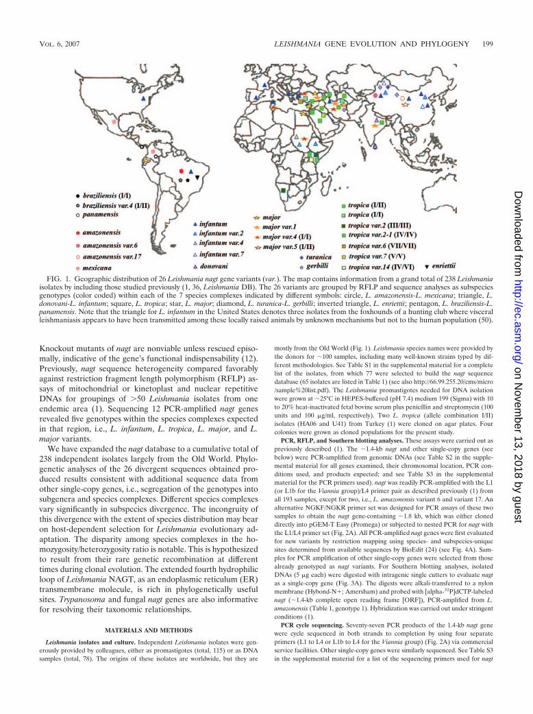
FIG. 1. Geographic distribution of 26 Leishmania nagt gene variants (var.). The map contains information from a grand total of 238 Leishmaniaisolates by including those studied previously (1, 36, Leishmania DB). The 26 variants are grouped by RFLP and sequence analyses as subspeciesgenotypes (color coded) within each of the 7 species complexes indicated by different symbols: circle, L. amazonensis-L. mexicana; triangle, L.donovani-L. infantum; square, L. tropica; star, L. major; diamond, L. turanica-L. gerbilli; inverted triangle, L. enriettii; pentagon, L. braziliensis-L.panamensis. Note that the triangle for L. infantum in the United States denotes three isolates from the foxhounds of a hunting club where visceralleishmaniasis appears to have been transmitted among these locally raised animals by unknown mechanisms but not to the human population (50).
VOL. 6, 2007 LEISHMANIA GENE EVOLUTION AND PHYLOGENY 199
on Novem
ber 13, 2018 by guesthttp://ec.asm
.org/D
ownloaded from

and other single-copy genes encoding ferrochelatase, prostaglandin F2 alpha-synthase, zeta-crystalline/NADPH oxidoreductase, and dihydrofolate reductase-thymidylate synthase, i.e., genes fc, pgfs, p36, and dhfr-ts, respectively. The nagtgene-containing �1.8 kb (Lnagt�; see Table S2 in the supplemental material)cloned in pGEM-T was sequenced with T7 and SP6 vector-specific primers plusL2 and L3. Clones of dhfr-ts and dhfr-ts� (see Table S2 and S3 in the supple-mental material) were similarly obtained and sequenced. The DNA sequencesobtained were verified against their electrochromatograms for both strands,assembled, and aligned by using Clustal X (57).
Phylogenetic analyses. nagt sequences were used in equal lengths of 1,324 bp(nucleotide [nt] 37 to 1360) or 441 amino acids (aa 13 to 453), except for two, i.e.,the L. turanica and L. enriettii nagt genes with a codon deletion and insertion,respectively. Phylogenetic analyses of the aligned sequences were done as de-scribed previously (17, 18) by using different algorithms of Clustal X (57),BioEdit (24), and/or MEGA (version 2.1) (35), i.e., the Kimura 2-parameterneighbor-joining method, maximum likelihood, maximum parsimony, UPGMA(unweighted pair group method with arithmetic mean), and minimum evolution.Bootstrapping was done with 1,000 replicas where applicable. The minimum-evolution and maximum-parsimony algorithms in MEGA were used for phylo-
genetic analyses of the nagt sequences from 26 Leishmania genotypes togetherwith four Trypanosoma and several other parasitic protozoa and also from 17fungal species from GenBank (see below). For genealogical analyses, statisticalparsimony in TCS (v. 1.21) (13) and median joining in Network (v. 4.1.1.2)software were used (3). Genetic distances were calculated using the Kimura2-parameter model in MEGA. Phylogenetic analyses of other single-copy genes,i.e., fc, pgfs, p36, and dhfr-ts, were similarly carried out by using one or more ofthe above-mentioned programs.
Hypothetical model and motif predictions of Leishmania NAGT as an ERtransmembrane protein. This model was constructed by using five differenttopology prediction programs (DAS, HMMTOP, TMHMM, TMpred, andTOPpred) in ExPASy proteomics tools (http://ca.expasy.org/tools/). Topology ofthe Leishmania NAGT is defined by the �10 transmembrane hydrophobic do-mains and manually fine-tuned according to the hamster NAGT model (66). Theprotein families (Pfam) database (http://www.sanger.ac.uk/Software/Pfam/) wassearched for consensus motifs of Leishmania NAGT, yielding 24 representativeproteins of the eukaryotic glycosyltransferase 4 family. Conserved and noncon-served amino acid substitutions were determined using Blosum-80 or Blosum-62
TABLE 1. Twenty six nagt variants within known species complexes of Leishmania recognized by analyses of 193 independent isolatesa
nagt genotypesb WHO Codec Diseased nagt genotypesb WHO Codec Diseased
L. amazonensis/mexicana HOM/TN/90/FMH CLcomplex (5)e 13 Variant 5 (I/II) (1) HOM/CN/99/Gansu-Wang g VL
1 L. amazonensis (1) RAT/BR/72/LV78g CL2 Variant 6 (1) HOM/CN/75/Kashi-BTO13 VL L. turanica/gerbilli3 Variant 17 (1) HOM/CN/97/Gansu-LUO VL complex (21)4 L. mexicana (2) HOM/CO/94/1182g CL 14 L. turanica (15) RHO/CN/99/KMA2g CL
HOM/MX/84/ISTE GS CL RHO/CN/92/Qi Daig CLL. donovani/infantum RHO/IR/04?/IR14 CL
complex (91) 15 L. gerbilli (6) RHO/CN/62/20g CL5 L. infantum (63) HOM/TR/00/OG-VLg VL IMON/CN/97/KMP3 N/A
HOM/TR/03/Adana #7 CLHOM/GR/70?/GH5 CL L. tropica complex (48)CANL/IR/04/IR2B VL? 16 L. tropica (I/II) (22) HOM/TR/95/MACg CLHOM/FR/80/189 VL? HOM/SY/00/Am CLHOM/ES/81/260 VL? HOM/AZ/74/K27 CLHOM/CN/80/801 VL HOM/IQ/73/LRC L32 CLHOM/TN/90/DREP13 CL HOM/IR/94/X54 CLHOM/BR/82/BA-2, C1 VL HOM/SA/91/WR2044 VL (asymptomatic)
6 Variant 2 (15) HOM/CN/50?/Bman VL HOM/JO/90-91?/Jh33 CLHOM/SD/03?/VL2 VL HOM/SU/60/BTO11 CL
7 Variant 4 (1) HOM/KE/84/NLB323g VL HOM/KE/81/NLB 030Bg VL8 Variant 7 (7) HOM/CN/93/KXG-LIUg CL HOM/IN/76/UR6g VL
IWUI/CN/87/KXG65 N/A 17 L. tropica (I/I) (9) HOM/TR/04/EP94 CLHOM/CN/89/Shandong VL HOM/IR/03/IR1 HIV-CL/VL
9 L. donovani (5) HOM/IN/96/JD VL CAN/IR/02/IR3C VL ?HOM/LK/03/H9 CL 18 Variant 2 (III/III) (7) IROS/NA/87/8688g N/A
L. major complex (23) HOM/NA/84/K1 CL10 L. major (9) HOM/IL/80/Friedling CL 19 Variant 2-1 (IV/IV) (1) HOM/KE/84/NLB297Ag ?
HOM/TR/96/DK CL 20 Variant 6 (VII/VII) (6) HOM/TR/03/EP82g CLHOM/EG/95/Dr. B CL 21 Variant 7 (V/V) (1) HOM/IQ/91/WR1095g VL (asymptomatic)HOM/IR/??/IR173 CL 22 Variant 14 (IV/VI) (2) HOM/KE/85/NLB545g ?
HOM/EQ/87/G-09 CL L. braziliensis/panamensis (4)11 Variant 1 (6) HOM/TR/94/HKg CL 23 L. braziliensis (I/I) (1)f HOM/BR/75/M2904g CL
HOM/IR/02/Iran 9 CL 24 Variant 4 (I/II) (1) HOM/BR/75/M2903 CLHOM/IN/05?/RMP-240 PKDL 25 L. panamensis (2) HOM/PA/71/LS94 CL
12 Variant 4 (I/I) (7) HOM/TR/93/HA VL HOM/CO/00?/140 CLHOM/TR/93/SYg CL L. enriettii (1)HOM/IR/64/Iran8 CL 26 enriettii (1) CAV/BR/45/enriettiig CLRHO/SD/02?/CL1 CL
a Listed are 65 representatives of 77 sequenced.b Variant 1 to 17, the number of nucleotide substitutions against an arbitrarily selected reference sequence in each species complex. I to VII, different alleles in L.
major, L. tropica, and L. braziliensis complex. Numbers in parentheses represent the actual number of isolates.c Host/reservoirs: CAN, dog (Canis familiaris); CANL, wolf (C. lupus pallipos); CAV, guinea pig (Cavia sp.); HOM, Homo sapiens; IMON, sand fly (Phlebotomus
mongolensis); IROS, sand fly (P. rossi); IWUI, sand fly (P. major wui); RAT, Rattus rattus; RHO, great gerbil (Rhobomys opimus). Countries of origin: AZ, Azerbaijan;BR, Brazil; CN, China; CO, Colombia; EG, Egypt; EQ, Ecuador; ES, Spain; FR, France; GR, Greece; IL, Israel; IN, India; IQ, Iraq; IR, Iran; JO, Jordan; KE, Kenya;LK, Sri Lanka; MX, Mexico; NA, Namibia; PA, Panama; SA, Saudi Arabia; SD, Sudan; SU, Soviet Union; SY, Syria; TN, Tunisia; TR, Turkey. 45 to 05, year ofisolation. ?, information not provided in writing by the donors.
d PKDL, post-kala-azar dermal leishmaniasis; N/A, not applicable.e Numbers of isolates examined by RFLP analyses and/or sequencing.f Sequence obtained from GeneDB.g Strains used for sequencing the fc gene and other single-copy genes. fc gene sequences were also obtained from L. donovani (HOM/IN/00/IN0041J) and L. infantum
variant 2 (HON/CN/90/901), which are not listed.
200 WAKI ET AL. EUKARYOT. CELL
on Novem
ber 13, 2018 by guesthttp://ec.asm
.org/D
ownloaded from
for Leishmania NAGT proteins alone as a group or together with other NAGTproteins mentioned (see Fig. 5).
GenBank accession numbers. The 26 Leishmania nagt sequences are M96635(36), AF205930-34 (1), AF291678, DQ836147-64 and LbrM35.3940 (L. brazilien-sis; GeneDB, http://www.genedb.org). Sequences of other Leishmania single-copy genes made available in this study for analyses include those for (i) 24 fcgenes, DQ834284-301, DQ974212-3, EF088400-2, and LbrM17.1230; (ii) 13 pgfsgenes, DQ834276-83, DQ981498-9, LmjF31.2150, LinJ31.2550, andLbrM31.2270; (iii) 16 p36 genes, L11705 (37), DQ834268-75, DQ974215-18,LmjF36.4170, LinJ36.6970, and LbrM35.3930; and (iv) 16 dhfr-ts genes,AF289072-3, AY122331, AY123971, DQ834262-6, DQ974214, M12734, X51733,X51735, LmjF06.0860, LinJ06.0890, and LbrM06.0760. The nagt sequences fromfour Trypanosoma and five other protozoa included for phylogenetic analyses arefrom the genome project (http://www.genedb.org) as follows: Trypanosoma cruzi(Tc00.1047053510283.140), T. brucei (Tb11.01.2220), T. gambiense (Tgamb.39611), T. vivax (tviv1098d08.q1k_5), Plasmodium berghei (PB000880.00.0), P.falciparum (PFC0935c), P. knowlesi (PK3_1860c), Entamoeba histolytica(142.m00140), and Theileria annulata (TA18965). The 17 fungal nagt sequencesinclude five members of Euascomycota (AAL78196, EAA58397, EAA77141,EAL93419, CAF06073), seven members of Hemiascomycota (AAS53335,CAA68324, CAG58156, CAG83233, CAG86196, CAG98095, EAK97019), onemember of Archeascomycota (AAA92799), three members of Basidiomycota(AAW46080, CAA67366, EAL17964), and one member of Ustilaginomycota(EAK84857).
RESULTS
PCR amplification of nagt genes from 193 independent iso-lates and other single-copy genes from selected samples.(i) nagt. Using the L1/L4 primer set (Fig. 2A) for PCR, a singleproduct of the expected size (�1.4 kb) was amplified from allsamples of the Leishmania subgenus examined (Fig. 2B), ex-cept for L. amazonensis variant 6 and variant 17 (Table 1,genotypes 2 and 3). Interestingly, the inability of the L1/L4primer to PCR-amplify the nagt sequence from these two sam-ples is not due to sequence heterogeneity in their 5� and 3�regions, corresponding to the primers used. Rather, it is ap-parently due to a diversion of the primers to spurious anneal-
ing sites in the non-nagt region. For these two samples, nagtwas obtained with the alternative primers NGKF and NGKR,designed from the 5� and 3� flanking regions of the L. ama-zonensis nagt gene (Table 1, genotype 1, and Fig. 2A) (36). Wefocused on an �1.8-kb band among the multiple PCR productsamplified with this set of primers (data not shown) because thissize was expected for the specific product (Lnagt�; see TableS2 in the supplemental material) (36). Using this template fornested PCR amplification with the L1/L4 primers, the 1.4-kbnagt DNA fragment was obtained from both samples. Theauthenticity of the obtained products was verified by directsequencing of the nested PCR product from L. amazonensisvariant 17 and by sequencing of the �1.8-kb PCR productfrom L. amazonensis variant 6 after cloning into the pGEM-Tvector. The latter thus provided a complete ORF of nagt plusits flanking regions. Notably, the 5� and 3� ends of this variant6 nagt are almost identical in sequence to L1 and L4, except fora single base substitution at nt 1399 of the ORF (within the
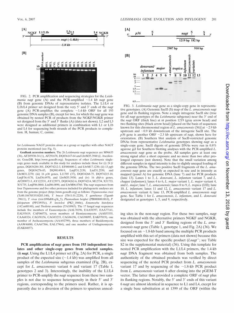
FIG. 2. PCR amplification and sequencing strategies for the Leish-mania nagt gene (A) and the PCR-amplified �1.4 kb nagt gene(B) from genomic DNAs of representative isolates. The L1/L4 orL1b/L4 primer set designed from the very 5� and 3� ends of the nagtgene (A) PCR-amplifies the complete �1.4-kb ORF for all 193genomic DNA samples (B), except for two, for which the nagt gene wasobtained by nested PCR of products from the NGKF/NGKR primerset designed from the 5� and 3� flanks (A) (data not shown). L2 and L3were designed as additional primers in combination with L1 or L1band L4 for sequencing both strands of the PCR products to comple-tion. H, human; C, canine.
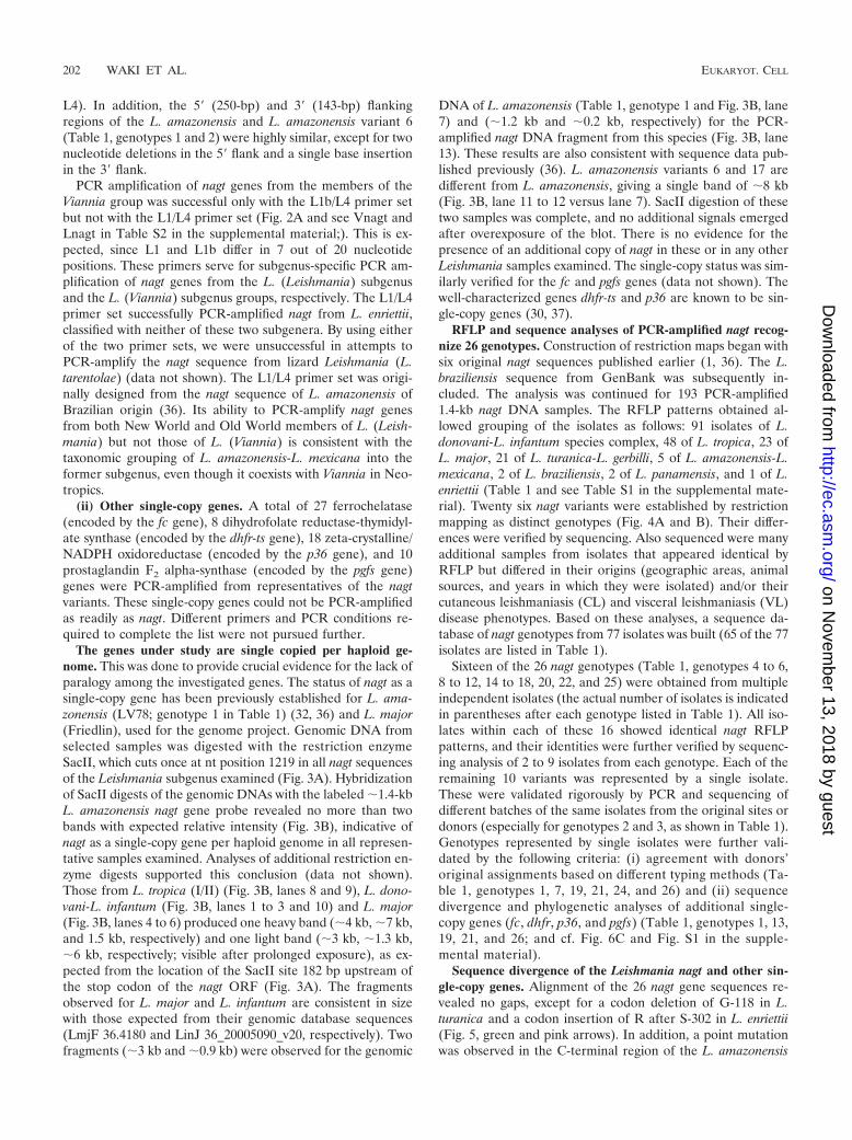
FIG. 3. Leishmania nagt gene as a single-copy gene in representa-tive genotypes. (A) Genomic SacII (S) map of the L. amazonensis nagtgene and its flanking regions. Note a single intragenic SacII site (truefor all nagt genotypes of the Leishmania subgenus) near the 3� end ofthe nagt ORF (thick line) at nt position 1219 (gray arrow head) andtwo flanking sites (black arrow head) [placed on the basis of sequencesknown for this chromosomal region of L. amazonensis (36)] at �2.9 kbupstream and �0.9 kb downstream of the intragenic SacII site. Thep36 gene is another ORF �2.3 kb upstream of nagt, shown here fororientation. (B) Southern blot analysis of SacII-restricted genomicDNAs from representative Leishmania genotypes showing nagt as asingle-copy gene. SacII digests of genomic DNAs were run in 0.8%agarose gel for Southern blotting analyses with the PCR-amplified L.amazonensis nagt gene as the probe. All samples gave at least onestrong signal after a short exposure and no more than two after pro-longed exposure (not shown). Note that the small variation amongdifferent samples in signal intensity is due to slightly unequal loading ofthe genomic DNAs. The two positive SacII fragments of the L. ama-zonensis nagt gene are exactly as expected in size and in intensity asmapped (panel A) for genomic DNA (lane 7) and for PCR products(lane 13). Lanes 1 to 3, L. donovani, L. infantum variant 7, and L.chagasi (infantum); lanes 4 to 6, L. major variant 1, L. major variant 4,and L. major; lane 7, L. amazonensis; lanes 8 to 9, L. tropica (I/II); lane10, L. infantum; lanes 11 and 12, L. amazonensis variant 17 and L.amazonensis variant 6; lane 13, PCR-amplified L. amazonensis nagtgene. See Table 1 for L. amazonensis, L. infantum, and L. donovanidesignated as genotypes 1, 5, and 9, respectively.
VOL. 6, 2007 LEISHMANIA GENE EVOLUTION AND PHYLOGENY 201
on Novem
ber 13, 2018 by guesthttp://ec.asm
.org/D
ownloaded from
L4). In addition, the 5� (250-bp) and 3� (143-bp) flankingregions of the L. amazonensis and L. amazonensis variant 6(Table 1, genotypes 1 and 2) were highly similar, except for twonucleotide deletions in the 5� flank and a single base insertionin the 3� flank.
PCR amplification of nagt genes from the members of theViannia group was successful only with the L1b/L4 primer setbut not with the L1/L4 primer set (Fig. 2A and see Vnagt andLnagt in Table S2 in the supplemental material;). This is ex-pected, since L1 and L1b differ in 7 out of 20 nucleotidepositions. These primers serve for subgenus-specific PCR am-plification of nagt genes from the L. (Leishmania) subgenusand the L. (Viannia) subgenus groups, respectively. The L1/L4primer set successfully PCR-amplified nagt from L. enriettii,classified with neither of these two subgenera. By using eitherof the two primer sets, we were unsuccessful in attempts toPCR-amplify the nagt sequence from lizard Leishmania (L.tarentolae) (data not shown). The L1/L4 primer set was origi-nally designed from the nagt sequence of L. amazonensis ofBrazilian origin (36). Its ability to PCR-amplify nagt genesfrom both New World and Old World members of L. (Leish-mania) but not those of L. (Viannia) is consistent with thetaxonomic grouping of L. amazonensis-L. mexicana into theformer subgenus, even though it coexists with Viannia in Neo-tropics.
(ii) Other single-copy genes. A total of 27 ferrochelatase(encoded by the fc gene), 8 dihydrofolate reductase-thymidyl-ate synthase (encoded by the dhfr-ts gene), 18 zeta-crystalline/NADPH oxidoreductase (encoded by the p36 gene), and 10prostaglandin F2 alpha-synthase (encoded by the pgfs gene)genes were PCR-amplified from representatives of the nagtvariants. These single-copy genes could not be PCR-amplifiedas readily as nagt. Different primers and PCR conditions re-quired to complete the list were not pursued further.
The genes under study are single copied per haploid ge-nome. This was done to provide crucial evidence for the lack ofparalogy among the investigated genes. The status of nagt as asingle-copy gene has been previously established for L. ama-zonensis (LV78; genotype 1 in Table 1) (32, 36) and L. major(Friedlin), used for the genome project. Genomic DNA fromselected samples was digested with the restriction enzymeSacII, which cuts once at nt position 1219 in all nagt sequencesof the Leishmania subgenus examined (Fig. 3A). Hybridizationof SacII digests of the genomic DNAs with the labeled �1.4-kbL. amazonensis nagt gene probe revealed no more than twobands with expected relative intensity (Fig. 3B), indicative ofnagt as a single-copy gene per haploid genome in all represen-tative samples examined. Analyses of additional restriction en-zyme digests supported this conclusion (data not shown).Those from L. tropica (I/II) (Fig. 3B, lanes 8 and 9), L. dono-vani-L. infantum (Fig. 3B, lanes 1 to 3 and 10) and L. major(Fig. 3B, lanes 4 to 6) produced one heavy band (�4 kb, �7 kb,and 1.5 kb, respectively) and one light band (�3 kb, �1.3 kb,�6 kb, respectively; visible after prolonged exposure), as ex-pected from the location of the SacII site 182 bp upstream ofthe stop codon of the nagt ORF (Fig. 3A). The fragmentsobserved for L. major and L. infantum are consistent in sizewith those expected from their genomic database sequences(LmjF 36.4180 and LinJ 36_20005090_v20, respectively). Twofragments (�3 kb and �0.9 kb) were observed for the genomic
DNA of L. amazonensis (Table 1, genotype 1 and Fig. 3B, lane7) and (�1.2 kb and �0.2 kb, respectively) for the PCR-amplified nagt DNA fragment from this species (Fig. 3B, lane13). These results are also consistent with sequence data pub-lished previously (36). L. amazonensis variants 6 and 17 aredifferent from L. amazonensis, giving a single band of �8 kb(Fig. 3B, lane 11 to 12 versus lane 7). SacII digestion of thesetwo samples was complete, and no additional signals emergedafter overexposure of the blot. There is no evidence for thepresence of an additional copy of nagt in these or in any otherLeishmania samples examined. The single-copy status was sim-ilarly verified for the fc and pgfs genes (data not shown). Thewell-characterized genes dhfr-ts and p36 are known to be sin-gle-copy genes (30, 37).
RFLP and sequence analyses of PCR-amplified nagt recog-nize 26 genotypes. Construction of restriction maps began withsix original nagt sequences published earlier (1, 36). The L.braziliensis sequence from GenBank was subsequently in-cluded. The analysis was continued for 193 PCR-amplified1.4-kb nagt DNA samples. The RFLP patterns obtained al-lowed grouping of the isolates as follows: 91 isolates of L.donovani-L. infantum species complex, 48 of L. tropica, 23 ofL. major, 21 of L. turanica-L. gerbilli, 5 of L. amazonensis-L.mexicana, 2 of L. braziliensis, 2 of L. panamensis, and 1 of L.enriettii (Table 1 and see Table S1 in the supplemental mate-rial). Twenty six nagt variants were established by restrictionmapping as distinct genotypes (Fig. 4A and B). Their differ-ences were verified by sequencing. Also sequenced were manyadditional samples from isolates that appeared identical byRFLP but differed in their origins (geographic areas, animalsources, and years in which they were isolated) and/or theircutaneous leishmaniasis (CL) and visceral leishmaniasis (VL)disease phenotypes. Based on these analyses, a sequence da-tabase of nagt genotypes from 77 isolates was built (65 of the 77isolates are listed in Table 1).
Sixteen of the 26 nagt genotypes (Table 1, genotypes 4 to 6,8 to 12, 14 to 18, 20, 22, and 25) were obtained from multipleindependent isolates (the actual number of isolates is indicatedin parentheses after each genotype listed in Table 1). All iso-lates within each of these 16 showed identical nagt RFLPpatterns, and their identities were further verified by sequenc-ing analysis of 2 to 9 isolates from each genotype. Each of theremaining 10 variants was represented by a single isolate.These were validated rigorously by PCR and sequencing ofdifferent batches of the same isolates from the original sites ordonors (especially for genotypes 2 and 3, as shown in Table 1).Genotypes represented by single isolates were further vali-dated by the following criteria: (i) agreement with donors’original assignments based on different typing methods (Ta-ble 1, genotypes 1, 7, 19, 21, 24, and 26) and (ii) sequencedivergence and phylogenetic analyses of additional single-copy genes (fc, dhfr, p36, and pgfs) (Table 1, genotypes 1, 13,19, 21, and 26; and cf. Fig. 6C and Fig. S1 in the supple-mental material).
Sequence divergence of the Leishmania nagt and other sin-gle-copy genes. Alignment of the 26 nagt gene sequences re-vealed no gaps, except for a codon deletion of G-118 in L.turanica and a codon insertion of R after S-302 in L. enriettii(Fig. 5, green and pink arrows). In addition, a point mutationwas observed in the C-terminal region of the L. amazonensis
202 WAKI ET AL. EUKARYOT. CELL
on Novem
ber 13, 2018 by guesthttp://ec.asm
.org/D
ownloaded from
variant 6 gene corresponding to the stop codon in other iso-lates, resulting in a C-terminal tetrapeptide (QSQV) exten-sion.
Pair-wise comparisons of the 26 nagt nt and aa sequencesreveal their divergences according to species complexes basedon the percentage of nt identity as well as the values of theirgenetic distances (see Table S4 in the supplemental material).Similar results were obtained from such analyses of four addi-tional single-copy genes available, i.e., 24 fc, 16 dhfr-ts, 13 pgfs,and 16 p36 (data not shown).
Subspecies divergences of nagt genes vary significantly with
different species complexes (Table 2). The nucleotide polymor-phism seen among the subspecies variants within each com-plex is as follows: L. amazonensis-L. mexicana and L. tropica(29 sites) � L. braziliensis-L. panamensis and L. turanica-L.gerbilli (15 sites) � L. donovani-L. infantum and L. major (6 to8 sites) (Table 2). A similar intraspecies divergence within eachcomplex is noted also from the values of their genetic dis-tances, as follows: L. tropica � L. amazonensis-L. mexicana �L. turanica-L. gerbilli � L. braziliensis-L. panamensis � L.donovani-L. infantum � L. major (Table 2). Intraspecies di-vergence of the fc gene sequences measured by distance anal-
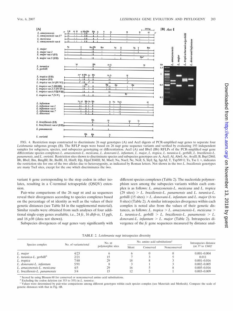
FIG. 4. Restriction maps constructed to discriminate 26 nagt genotypes (A) and AccI digests of PCR-amplified nagt genes to separate fourLeishmania subgenus groups (B). The RFLP maps were based on 26 nagt gene sequence variants and verified by evaluating 193 independentsamples for subgenera, species, and subspecies genotyping or differentiation. AccI (A) and BbeI (Bb) RFLPs of the PCR-amplified nagt genedifferentiate species complexes L. amazonensis-L. mexicana, L. donovani-L. infantum, L. major, L. tropica, L. turanica-L. gerbilli, L. braziliensis-L.panamensis, and L. enriettii. Restriction sites shown to discriminate species and subspecies genotypes are A, AccI; Al, AlwI; Av, AvaII; B, Bsp1286I;Bb, BbeI; Bm, BmgBI; Br, BsrBI, H, HinfI; Hp, HpyCH4III; M, MseI; Na, NaeI; Nc, NciI; S, StyI; Sg, SgrAI; T, Tsp509 I; Ts, Tse I. �, indicatesthe restriction site for one of the two alleles due to heterozygosity, as indicated by Roman letters. Not shown in the two L. braziliensis genotypesare many TseI sites, except for the one which discriminates the two.
TABLE 2. Leishmania nagt intraspecies diversity
Species complex No. of variants/total No. ntpolymorphic sites
No. amino acid substitutionsaIntraspecies distance
(nt 37 to 1360)cSilent Conserved Nonconserved
L. major 4/23 6 6 0 0 0.001–0.004L. turanica-L. gerbillib 2/21 15 7 3 5 0.011L. tropica 7/48 29 18 8 3 0.001–0.016L. donovani-L. infantum 5/91 8 3 1 3 0.002–0.005L. amazonensis-L. mexicana 4/5 29 16 5 8 0.005–0.016L. braziliensis-L. panamensis 3/4 15 12 2 1 0.003–0.009
a Scored by using Blosum-80 for conserved or nonconserved amino acid substitutions.b Excluding the codon deletion (nt 353 to 355) in L. turanica.c Values were determined by pair-wise comparisons among different genotypes within each species complex (see Materials and Methods). Compare the scale of
genetic distances with that in Fig. 6B.
VOL. 6, 2007 LEISHMANIA GENE EVOLUTION AND PHYLOGENY 203
on Novem
ber 13, 2018 by guesthttp://ec.asm
.org/D
ownloaded from

yses also showed the highest and the lowest values for L.tropica and L. donovani-L. infantum, respectively (data notshown). The intraspecies nucleotide polymorphisms resultedlargely in silent mutations, especially in the case of L. major(Table 2).
Allelic sequence heterogeneity (heterozygosity) of nagt isprominent and apparently genome wide in the L. tropica com-plex. Direct PCR cycle sequencing of nagt from 20 isolatesclassified as L. tropica revealed seven diplotype variants (Table1, genotypes 16 to 22). In two cases, i.e., L. tropica (I/II) andvariant 14 (IV/VI), the DNA sequence traces contained 7 and14 overlapping nucleotide positions, respectively (see Table S5in the supplemental material, allele combinations are shown asI/II and IV/VI). These double peaks were subsequently foundto persist in samples from cloned cell populations of L. tropica(I/II). The seven heterozygous sites are thus present in thesame individual cells but not in different cells of a mixedpopulation. Since nagt is present as a single-copy gene perhaploid genome and Leishmania are diploid, the double peaksrepresent allelic differences (heterozygosity). No overlappingpeaks appeared in the remaining five diplotypes, hence indic-ative of their nagt homozygosity, i.e., L. tropica I/I, III/III,
IV/IV, V/V, and VII/VII (Table 1, genotypes 17 to 21). Thefindings of homozygous isolates, i.e., L. tropica I/I and IV/IV,allow us to infer the sequences of alleles II and VI in theheterozygous allelic “recombinants” of I/II and IV/VI. It is alsopossible to predict the potential existence of homozygous “re-combinants” of II/II and VI/VI, although they have not beenencountered so far. Hypothetical combinations of all sevenhaplotypes increase the total to 28 diplotypes in this speciescomplex. The 7 and 14 heterozygous sites in L. tropica (I/II)and variant 14 (IV/VI), respectively (see Table S5 in the sup-plemental material), result in three and four aa substitutions,respectively, between the alleles in these genotypes (Fig. 5,double black and gray arrows). Allelic nucleotide differenceswere also observed in two to four sites of nagt sequences in L.major variant 5 (I/II) and L. braziliensis variant 4 (I/II) (datanot shown and cf. Fig. 6D). Heterozygosity was also noted insequences of dhfr-ts and fc from L. tropica (I/II) and variant 14(IV/VI) (not shown), indicating that it is not limited to nagt, atleast in this species complex.
Possible chimeric nagt in L. amazonensis variant 17. In thisisolate, the bulk of the 5� region of the nagt gene (1,132 bpfrom nt position 37 to nt position 1178, excluding the PCR
FIG. 5. Phylogenetically useful residues in the hydrophilic loops predicted from a hypothetical model of Leishmania NAGT as a trans-ERmembrane molecule. A Protein Family Database (Pfam) search places Leishmania NAGT in the glycosyltransferase 4 family. Circled letter,identical residues in NAGTs of all 26 Leishmania variants; solid blue/red circles, conserved/nonconserved aa substitutions among 26 Leishmaniavariants, respectively; black and gray arrows, heterozygous sites of the two L. tropica variants (I/II and IV/VI), resulting in three and four aasubstitutions, respectively (double arrow); dark and light green circled residues, glycosyltransferase 4 family identical and conserved aa residues,respectively; gray circled letters, L. amazonensis NAGT sequences corresponding to the PCR primer sequences; octagonal sequence, NAGTconsensus catalytic sites; letters circled pink/blue, eukaryotic NAGT consensus residues and conserved substitutions, respectively. Note thatTrypanosoma as well as other eukaryotic NAGT-conserved residues are recognized in the cytosolic loops (third, fifth, and ninth) and the ninthtransmembrane segment of the Leishmania sequence (blue-shaded and pink and blue circles). The motif of CPRHR (aa 396–400 in octagon) wasexperimentally proved to be crucial for enzymatic activity of the hamster NAGTs and conserved in all eukaryotic NAGTs examined. Absent fromLeishmania NAGT is the potential dolichol recognition sequence [F-(I/V)-X-(F/Y)-X-X-I-P-F-X-(F/Y)] predicted for yeast, hamster, (66) andmammalian NAGTs as two separate sites in the second and seventh transmembrane regions (the second site only in mammalian enzymes).Leishmania NAGT dolichol phosphate binding sites appear to differ from the others.
204 WAKI ET AL. EUKARYOT. CELL
on Novem
ber 13, 2018 by guesthttp://ec.asm
.org/D
ownloaded from
primer-corresponding region) is most similar to other nagtgenes in the L. amazonensis-L. mexicana complex, with only 5to 10 nucleotide substitutions among them. In contrast, the last182 bp extending from nt position 1179 to nt position 1360 arecompletely identical to the corresponding 3�-end region of thenagt gene in three variants of the L. donovani-L. infantumcomplex and have only two to three substitutions in the re-maining two variants of this species complex. As expected, thisvariant 17 nagt is segregated into the clades of L. amazonen-sis-L. mexicana (not shown, but see Fig. 6B) and L. dono-vani-L. infantum (see Fig. S3 in the supplemental material),respectively, when subjected to phylogenetic analyses of therespective 5� and 3� regions in question, together with thecorresponding regions of the other 25 nagt genotypes. Samplemix ups and technical errors were ruled out as factors account-ing for the emergence of such an apparently chimeric se-quence. The chimeric nagt cannot possibly be created by PCRor by cycle-sequencing errors with the L1/L4 primers, judgingfrom their positions (Fig. 2A and see Table S2 and S3 in thesupplemental material) relative to the “crossover site” of thechimeric sequence. In addition, had the variant 17 DNA beenmixed with other samples, PCR with the L1/L4 primer setwould have produced the 1.4-kb nagt. Most importantly, theresults were verified by repeated PCR for DNA analysesfrom different batches of the same isolate reacquired fromthe original donor.
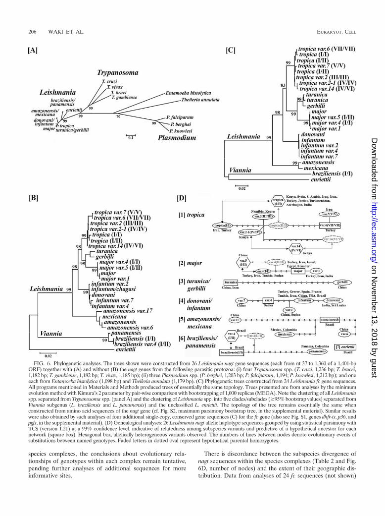
Phylogenetic analyses of the 26 nagt sequences allow group-ing of the variants according to their hierarchical taxonomiclevels and evaluating subspecies divergence. Phylogeneticanalyses of the Leishmania and Trypanosoma nagt genes showthat the 26 Leishmania sequences are clustered together as agroup well separated from Trypanosoma (Fig. 6A and see Fig.S2 in the supplemental material), as reported previously, byusing different markers (25, 28, 56). The topology of thetrypanosomatid trees remains essentially unchanged when sub-jected to further analyses together with the nagt genes fromother parasitic protozoa, such as Plasmodium spp. (Fig. 6A andsee Fig. S2 in the supplemental material). The nagt sequencesare thus sufficiently divergent to distinguish the two genera oftrypanosomatids. The 26 Leishmania nagt sequences formedthree distinct groups according to their subgeneric status, i.e.,L. (Leishmania), L. (Viannia), and the branch of L. enriettiiwhich does not belong to either subgenus (Fig. 6B). Those inthe subgenus L. (Leishmania) are grouped into five knownspecies complexes, i.e., L. (L.) amazonensis-L. mexicana, L.(L.) donovani-L. infantum, L. (L.) tropica, L. (L.) major, and L.(L.) turanica-L. gerbilli (Fig. 6B). Analyses of additional sin-gle-copy genes available, i.e., dhfr-ts, fc, p36, and pgfs pro-duced trees with similar topology at the species level (seeFig. 6C for fc and see Fig. S1 in the supplemental materialfor the rest). The separation of the Leishmania and Vianniagroups, as well as the species complexes therein, agreescompletely with the data from similar analyses of otherprotein-coding genes (14, 27, 39, 44) and internal-tran-scribed-spacer (16) sequences.
Incongruence between the Leishmania nagt gene-basedgrouping and the CL/VL disease phenotype of the isolates isnoted, i.e., the coexistence of subspecies variants with differentdisease phenotypes in the same species complex and, morestrikingly, the same genotype from different disease pheno-
types (Table 1). In the L. donovani-L. infantum complex, forexample, we observed a nagt genotype of L. infantum with bothCL and VL phenotypes, isolated from southern Turkey (Table1, genotype 5, strains OG-VL and Adana #7) (54). Anotherone is L. infantum variant 7, which shows CL exclusively inwestern China (38) but VL in eastern China (Table 1, genotype8, strains KXG-LIU and Shandong).
Phylogenetically useful sites of Leishmania nagt sequences.These are unevenly distributed, as noted from amino acidsubstitutions in the NAGT polypeptide chain presented as aputative ER transmembrane model (Fig. 5). Of the �130 aasubstitutions present among the 26 sequences (Fig. 5, solidcircle), a majority, that is, two-thirds (�93), appears to occur inthe hydrophilic loops, especially the longest fourth and theadjacent one. The fourth loop is unique to Leishmania, since itis truncated in the NAGT of Trypanosoma (Fig. 5) and othereukaryotes (not shown). The remaining one-third of the sub-stitutions (�37) are dispersed throughout the hydrophobic do-mains. In addition, nonconserved substitutions are almostthree times more frequent in the hydrophilic loops (�66) thanin the transmembrane segments (�24) (Fig. 5, solid red circle).
Leishmania intraspecies divergence. The 26 nagt haplotypesequences were further subjected to genealogy analyses bystatistical parsimony (13) (Fig. 6D) and median-joining analy-ses (3) (see Fig. S4 in the supplemental material). Both algo-rithms sorted the 26 sequences into independent networks oftheir respective species complexes (Fig. 6D and see Fig. S4 inthe supplemental material). This result provides further con-firmation of the phylogenetic grouping of the 26 variants pre-sented in Fig. 6A through C. There are four heterozygousgenotypes or “recombinants” (Fig. 6D, subspecies [1], [2], and[6], and see Fig. S4, [1] and [2], hexagon, in the supplementalmaterial), i.e., L. tropica (I/II), L. tropica variant 14 (IV/VI), L.major variant 5 (I/II), and L. braziliensis variant 4 (I/II). Ho-mozygous genotypes with some of the above-mentioned allele-specific sequences were found, including L. tropica (I/I), L.tropica variant 2-1 (IV/IV), L. major variant 4 (I/I), and L.braziliensis (I/I) (Fig. 6D and see Fig. S4 in the supplementalmaterial), but not those with the remaining alleles, such asputative L. tropica (II/II), L. tropica variant 14 (VI/VI), L.major variant 4 (II/II), and L. braziliensis variant 4 (II/II) (Fig.6D, dotted oval, and see Fig. S4, faded letters, in the supple-mental material). The presumed “parental” variants for the“recombinants” (lined arrows) are not necessarily close to eachother evolutionarily (Fig. 6D and see Fig. S4 in the supple-mental material), but they colocalize geographically, as shownby the coexistence of the L. tropica variant 2-1 (IV/IV) andvariant 14 (IV/VI) from Kenya and by L. tropica (I/I) and (I/II)from Asia and the Middle East (Fig. 6D) (1). Preliminarygenealogical analyses of the eight fc sequences available for themost divergent L. tropica complex yielded results which sup-ported the observation of a distant relationship between the“parental” variants, although the genealogy of the subspeciesvariants is not entirely identical in topology to that derivedfrom the nagt sequences (not shown). This situation was notedpreviously in similar analyses of five different single-copy genesfor the subspecies genealogy of L. donovani-L. infantum (41).In the present study, while the levels of intraspecies divergenceare clearly reflected in the number of polymorphic sites andthe values of genetic distance observed in the investigated
VOL. 6, 2007 LEISHMANIA GENE EVOLUTION AND PHYLOGENY 205
on Novem
ber 13, 2018 by guesthttp://ec.asm
.org/D
ownloaded from
species complexes, the conclusions about evolutionary rela-tionships of genotypes within each complex remain tentative,pending further analyses of additional sequences for moreinformative sites.
There is discordance between the subspecies divergence ofnagt sequences within the species complexes (Table 2 and Fig.6D, number of nodes) and the extent of their geographic dis-tribution. Data from analyses of 24 fc sequences (not shown)
FIG. 6. Phylogenetic analyses. The trees shown were constructed from 26 Leishmania nagt gene sequences (each from nt 37 to 1,360 of a 1,401-bpORF) together with (A) and without (B) the nagt genes from the following parasitic protozoa: (i) four Trypanosoma spp. (T. cruzi, 1,236 bp; T. brucei,1,182 bp; T. gambiense, 1,182 bp; T. vivax, 1,185 bp); (ii) three Plasmodium spp. (P. berghei, 1,203 bp; P. falciparum, 1,194; P. knowlesi, 1,212 bp); and oneeach from Entamoeba histolytica (1,098 bp) and Theileria annulata (1,179 bp). (C) Phylogenetic trees constructed from 24 Leishmania fc gene sequences.All programs mentioned in Materials and Methods produced trees of essentially the same topology. Trees presented are from analyses by the minimumevolution method with Kimura’s 2 parameter by pair-wise comparison with bootstrapping of 1,000 replicas (MEGA). Note the clustering of all Leishmaniaspp. separated from Trypanosoma spp. (panel A) and the clustering of Leishmania spp. into five clades/subclades (�95% bootstrap values) separated fromViannia subgenus (L. braziliensis and L. panamensis) and the unclassified L. enriettii. The topology of the tree remains essentially the same whenconstructed from amino acid sequences of the nagt gene (cf. Fig. S2, maximum parsimony bootstrap tree, in the supplemental material). Similar resultswere also obtained by such analyses of four additional single-copy, conserved gene sequences (C) for the fc gene (also see Fig. S1, genes dhfr-ts, p36, andpgfs, in the supplemental material). (D) Genealogical analyses: 26 Leishmania nagt allelic haplotype sequences grouped by using statistical parsimony withTCS (version 1.21) at a 93% confidence level, indicative of relatedness among subspecies variants and predictive of a hypothetical ancestor for eachnetwork (square box). Hexagonal box, allelically heterogeneous variants observed. The numbers of lines between nodes denote evolutionary events ofsubstitutions between named genotypes. Faded letters in dotted oval represent hypothetical parental homozygotes.
206 WAKI ET AL. EUKARYOT. CELL
on Novem
ber 13, 2018 by guesthttp://ec.asm
.org/D
ownloaded from
support this observation from nagt data discussed below. TheL. donovani-L. infantum complex is geographically very widespread (Fig. 1), but it is relatively homogeneous genetically,with the range of intraspecies genetic distances from 0.002 to0.005 and only eight polymorphic sites (Table 2) found amongfive homozygous genotypes (see Table 1, Fig. 6D, [4], and Fig.S4, [3], in the supplemental material). In contrast, the L.tropica complex is geographically more restricted (Fig. 1), butit is the most divergent, with intraspecies genetic distancesvarying from 0.001 to 0.016 and 29 polymorphic sites (Table 2)among two “recombinants” (see Table 1, Fig. 6D, [1], and Fig.S4 [1], hexagon, in the supplemental material) and five ho-mozygous genotypes. The remaining species complexes present apicture intermediate between these two species complexes. TheL. major complex is nearly as widely spread (Fig. 1) as L. dono-vani-L. infantum but is less divergent (Fig. 6D, [2], and Fig. S4 [2],with its four variants including one “recombinant” [hexagon], inthe supplemental material). Closely related to L. major is the L.turanica-L. gerbilli complex, which contains nonpathogenic para-sites of the great gerbil (Rhombomys opimus) (Fig. 6B). Thiscomplex is rather restricted in distribution (Fig. 1) and consists oftwo quite divergent members (Fig. 6D, [3]). Restricted to Neo-tropics is the L. braziliensis-L. panamensis complex (Fig. 1), whichincludes both types of genotypes (Fig. 6D, [6]). Genetic diversityof the Viannia group is underscored by a high level of sequenceheterogeneity obvious by examining only a few isolates. Mostinteresting is the finding that the L. amazonensis-L. mexicanacomplex has two genotypes from Asia (Fig. 1 and Table 1, geno-types 2 and 3), including what appears to be a chimeric hybridwith L. donovani-L. infantum, in addition to the two genotypesfrom Neotropics (Table 1, genotypes 1 and 4) (Fig. 6D, [5], andFig. S4, [4], in the supplemental material). It should be emphat-ically stressed that exceptional caution was exercised to excludeany technical or human errors, such as sample mix up, to ascertainthat the Asian origin of the two L. amazonensis variants was real.In fact, this finding should not be that surprising, except for thenomenclature of this species complex, considering the phyloge-netic grouping of L. amazonensis-L. mexicana with other speciescomplexes of the Old World (Leishmania).
DISCUSSION
Leishmania nagt sequence database for phylogenetic analy-ses. We have identified 26 nagt genotypes (19 from this work)by sequencing 77 PCR-amplified nagt genes selected from atotal of 238 independent isolates (193 from this study). Thesegenotypes represent true nagt gene divergence of the samplesexamined, since data collection and analyses are greatly facil-itated by several favorable factors, i.e., the ease of nagt PCRamplification (Fig. 2), the absence of paralogous genes (Fig. 3),the precision of sequence-based RFLP analyses of the PCRproducts (Fig. 4), and the unambiguous alignment of nagtsequences (Fig. 5). Although the sample size examined here isnot small, sample collection is inevitably biased by its accessi-bility or availability, which favors Asian isolates from the OldWorld, in this case. Thus, analyses of additional samples willundoubtedly uncover new genotypes, especially among thosethat are under-represented from the Viannia group in thepresent study. The database presented for the Old World
Leishmania subgenus is sufficiently robust, especially in con-junction with those from additional single-copy genes.
Genes encoding transmembrane molecules, such as nagt,are useful, albeit previously untapped phylogenetic markers.Consistent with previous findings based on other protein-cod-ing (39) and nonprotein-coding sequences (16, 47, 64), phylo-genetic analyses of the 26 nagt sequences used segregate theisolates into expected genera and species complexes (Fig. 6Aand B and see Fig. S2 in the supplemental material). Thephylogenetic trees obtained are identical in topology to thoseobtained from additional single-copy genes from selected sam-ples in the present study, i.e., dhfr-ts, fc, p36, and pgfs (Fig. 6Cand Fig. S1 in the supplemental material). The location ofthese additional genes on different chromosomes (see Table S2in the supplemental material) strongly implies that the phylo-genetic relationship of the genotypes deduced is genome widebut not limited to the nagt site.
The phylogenetically useful sites of Leishmania nagt are lo-calized mostly in a �500-bp section of this �1.4-kb gene,corresponding to the putative fourth and sixth hydrophilicloops of the trans-ER membrane enzyme (Fig. 5). Divergentsites of the nagt sequences in other single-cell eukaryotes, suchas fungi, are also informative for phylogenetic analyses (seeFig. S5 in the supplemental material). In other genes encodingcytosolic enzymes, e.g., dhfr-ts, p36, fc, and pgfs, the phyloge-netically useful sites are dispersed randomly throughout theORF. The advantage of the Leishmania nagt gene is, thus, itsutility for PCR amplification of only a small section of thissequence (�500 bp) to yield the maximal number of informa-tive sites. This is of practical importance in avoiding in vitroisolation and cultivation of parasites, which is cumbersome, notalways successful, and potentially selective for cultivablestocks. It is possible to envision the use of a battery of genesencoding similar transmembrane molecules in this way forphylogenetic analyses of single-cell eukaryotes toward the goalof genome taxonomy.
Leishmania subspecies divergence and evolutionary adapta-tion. Within the Leishmania subgenus, there is a lack of cor-relation between intraspecies divergence and geographic dis-tribution of the species complexes. Namely, the less divergentL. donovani-L. infantum and L. major complexes are widelydistributed, whereas the more divergent L. tropica and L.turanica-L. gerbilli complexes are geographically restricted (seeFig. 1, Fig. 6D, and Fig. S4 in the supplemental material; alsosee Table 2). The latter scenario appears true for the Neotro-pic-restricted L. (Viannia) subgenus on account of the greatintraspecies divergence seen among the few samples examinedin this study. The observation may have relevance to the evo-lutionary adaptability of Leishmania to a new environmentsubsequent to the organism’s dispersal but not to the dispersalmechanisms per se. Since Leishmania are vector-borne obli-gate endoparasites, their clonal evolution may be driven by twosets of direct pressures: (i) primarily, the internal microenvi-ronments of their long-term residence (months to years) in-traphagolysosomally in the macrophages of the mammalianhosts/reservoirs; and (ii) secondarily, their short-term resi-dence (up to several weeks) in the fly gut. Vector- or reservoir-parasite specificity has long been considered to be the apparentfactor limiting the distributions of leishmaniasis. The externalmacroenvironments are relevant to the evolution of reservoirs
VOL. 6, 2007 LEISHMANIA GENE EVOLUTION AND PHYLOGENY 207
on Novem
ber 13, 2018 by guesthttp://ec.asm
.org/D
ownloaded from
and vectors but exert no direct pressures on Leishmania, es-pecially when they are in homeothermal mammalian hosts.While the precise nature of the host-dependent selective pres-sures remains to be defined, reservoirs are most pertinent toLeishmania evolution, since leishmaniasis is fundamentally azoonotic disease. Reported cases of anthroponosis are basedon the negative finding of animal reservoirs and are limited tofew specific geographic sites, e.g., kala-azar, caused by IndianL. donovani, and CL by L. tropica in Asia but not in Africa.Genetic changes of isolates due to geographic isolation orgenetic drift are demonstrable more readily in the fast-evolvingsequences, e.g., microsatellite DNA markers (45, 52).
Contributions of genetic recombination to Leishmania evo-lution? There is a notable disparity of different species com-plexes in the frequency of the heterozygosity encountered.Heterozygosity is more prominent among the extant genotypesof L. tropica, as observed here and previously (51, 53), thanamong those of L. donovani-L. infantum (41). This may resultfrom different timelines for the event of “recombination” tooccur in different species. Genetic recombination is thought tooccur at a low rate in Leishmania (48), as found experimentallyin Trypanosoma spp. (19, 20). Leishmania sequence analysesshow hybrid and nonhybrid genotypes reminiscent of thoseseen in T. cruzi (40, 63), suggestive of “genetic exchange.” Itscontribution to Leishmania evolution may be suggested, asheterozygosity results in amino acid substitutions in multiplegenes of functionally important enzymes, as shown here atleast for L. tropica (Fig. 5).
Interspecies recombination is hinted at by the observation ofa chimeric nagt sequence in a single isolate as a hybrid of L.amazonensis-L. mexicana and L. donovani-L. infantum, i.e., L.amazonensis variant 17 (Fig. 6B and see Fig. S3 in the supple-mental material). Heterologous species hybrids have also beenreported to occur between L. major and L. arabica (33) andbetween L. braziliensis and other Viannia species (4, 6, 8, 61).Such hybrids could result in a chimeric sequence by anomalouschromosomal breakage/joining (8). Meaningful genetic impli-cations of such interspecies hybridization and chimeras awaitfurther investigation.
Incongruity of Leishmania speciation and disease pheno-types. This observation has long been reported for the Leish-mania subgenus (1, 2, 5, 22, 31, 34, 46). Although host geneticsmay be involved (9, 43), the inherent differences of the caus-ative agents remain to be significant contributing factors. Dif-ferential acquisition of “pathogenic islands” laterally by thesame genotype is a possibility, but there is no evidence for this.Although no genetic marker is available to discriminate CL-from VL-specific isolates (60), A2 (65) and K39 (21) repeatshave been related to “visceralization” of Leishmania relevantto immunopathology (11). Of interest is the elucidation of themechanism by which the expression of these and other pheno-type-specific genes is differentially regulated in VL and CLisolates of an identical genotype apparently independent ofLeishmania speciation.
We demonstrate here for the first time the potential utility ofsingle-copy genes encoding transmembrane versus soluble en-zymes for phylogenetic analyses of eukaryotic protists. Suchanalyses of trans-ER nagt genes from �200 Leishmania sam-ples readily group them into taxa of an expected hierarchy,revealing considerable variations in subspecies divergence and
evidence of heterozygosity in L. tropica, resulting in changes atthe protein level. Further analyses of this and additional se-quence databases will bear on Leishmania evolution and mech-anisms of their recombination crucial for elucidating the clin-ical epidemiology of leishmaniasis and the control measuresagainst these wide-spread diseases.
ACKNOWLEDGMENTS
This work is partially supported by NIH AI-20486 to K.P.C.We thank Leyla (Akman) Anderson for initiating the hypothetical
model presented in Fig. 5 and colleagues who generously providedLeishmania isolates or their DNA samples (see a complete list athttp://66.99.255.20/cms/micro/sample%20list.pdf).
ADDENDUM
Two articles, which came to our attention after the accep-tance of this report, contain information of relevance to thepresent discussion. One article describes the L. major-L. infan-tum hybrid strains isolated originally from human immunode-ficiency virus (HIV)-positive patients in Portugal (49a). Thisfinding is supportive of our proposed existence of putative L.infantum-L. amazonensis hybrids, although our cases involvedifferent parental species and have no record of coinfectionwith HIV. The very low subspecies divergence among differentgenotypes in the L. donovani-L. infantum complex that wenoted is consistent with data presented in another article basedon the examination of a different set of protein-coding genesequences (64a). In that article, subspecies variants were foundgeographically segregated into Indian, African, and Mediter-ranean groups within this species complex (64a). This conclu-sion is not inconsistent with our data presented in Fig. 6D, [4],by excluding some isolates from China, Brazil, and the UnitedStates.
REFERENCES
1. Akman, L., H. S. Aksu, R. Q. Wang, S. Ozensoy, Y. Ozbel, Z. Alkan, M. A.Ozcel, G. Culha, K. Ozcan, S. Uzun, H. R. Memisoglu, and K. P. Chang.2000. Multi-site DNA polymorphism analyses of Leishmania isolates definetheir genotypes predicting clinical epidemiology of leishmaniasis in a specificregion. J. Eukaryot. Microbiol. 47:545–554.
2. Alborzi, A., M. Rasouli, and A. Shamsizadeh. 2006. Leishmania tropica-isolated patient with visceral leishmaniasis in southern Iran. Am. J. Trop.Med. Hyg. 74:306–307.
3. Bandelt, H. J., P. Forster, and A. Rohl. 1999. Median-joining networks forinferring intraspecific phylogenies. Mol. Biol. Evol. 16:37–48.
4. Banuls, A. L., F. Guerrini, F. Le Pont, C. Barrera, I. Espinel, R. Guderian,R. Echeverria, and M. Tibayrenc. 1997. Evidence for hybridization by mul-tilocus enzyme electrophoresis and random amplified polymorphic DNAbetween Leishmania braziliensis and Leishmania panamensis/guyanensis inEcuador. J. Eukaryot. Microbiol. 44:408–411.
5. Barral, A., R. Badaro, M. Barral-Netto, G. Grimaldi, Jr., H. Momem, andE. M. Carvalho. 1986. Isolation of Leishmania mexicana amazonensis fromthe bone marrow in a case of American visceral leishmaniasis. Am. J. Trop.Med. Hyg. 35:732–734.
6. Belli, A. A., M. A. Miles, and J. M. Kelly. 1994. A putative Leishmaniapanamensis/Leishmania braziliensis hybrid is a causative agent of humancutaneous leishmaniasis in Nicaragua. Parasitology 109:435–442.
7. Beverley, S. M., R. B. Ismach, and D. M. Pratt. 1987. Evolution of the genusLeishmania as revealed by comparisons of nuclear DNA restriction fragmentpatterns. Proc. Natl. Acad. Sci. USA 84:484–488.
8. Britto, C., C. Ravel, P. Bastien, C. Blaineau, M. Pages, J. P. Dedet, and P.Wincker. 1998. Conserved linkage groups associated with large-scale chro-mosomal rearrangements between Old World and New World Leishmaniagenomes. Gene 222:107–117.
9. Bucheton, B., L. Abel, M. M. Kheir, A. Mirgani, S. H. El-Safi, C. Chevillard,and A. Dessein. 2003. Genetic control of visceral leishmaniasis in a Sudanesepopulation: candidate gene testing indicates a linkage to the NRAMP1region. Genes. Immun. 4:104–109.
10. Chance, M. L., W. Peters, and L. Shnur. 1974. Biochemical taxonomy of
208 WAKI ET AL. EUKARYOT. CELL
on Novem
ber 13, 2018 by guesthttp://ec.asm
.org/D
ownloaded from

Leishmania. I. Observations on DNA. Ann. Trop. Med. Parasitol. 68:307–316.
11. Chang, K. P., S. G. Reed, B. S. McGwire, and L. Soong. 2003. Leishmaniamodel for microbial virulence: the relevance of parasite multiplication andpathoantigenicity. Acta Trop. 85:375–390.
12. Chen, D. Q., H. Lu, and K. P. Chang. 1999. Replacement of LeishmaniaN-acetylglucosamine-1-phosphate transferase gene requires episomal res-cue. Mol. Biochem. Parasitol. 100:223–227.
13. Clement, M., D. Posada, and K. A. Crandall. 2000. TCS: a computer pro-gram to estimate gene genealogies. Mol. Ecol. 9:1657–1659.
14. Croan, D. G., D. A. Morrison, and J. T. Ellis. 1997. Evolution of the genusLeishmania revealed by comparison of DNA and RNA polymerase genesequences. Mol. Biochem. Parasitol. 89:149–159.
15. Cupolillo, E., G. Grimaldi, Jr., and H. Momen. 1994. A general classificationof New World Leishmania using numerical zymotaxonomy. Am. J. Trop.Med. Hyg. 50:296–311.
16. Davila, A. M., and H. Momen. 2000. Internal-transcribed-spacer (ITS) se-quences used to explore phylogenetic relationships within Leishmania. Ann.Trop. Med. Parasitol. 94:651–654.
17. Du, Y., and K. P. Chang. 1994. Phylogenetic heterogeneity of three Crithidiaspp. vs. Crithidia fasciculata. Mol. Biochem. Parasitol. 66:171–174.
18. Du, Y., D. A. Maslov, and K. P. Chang. 1994. Monophyletic origin of beta-division proteobacterial endosymbionts and their coevolution with insecttrypanosomatid protozoa Blastocrithidia culicis and Crithidia spp. Proc. Natl.Acad. Sci. USA 91:8437–8441.
19. Gaunt, M. W., M. Yeo, I. A. Frame, J. R. Stothard, H. J. Carrasco, M. C.Taylor, S. S. Mena, P. Veazey, G. A. Miles, N. Acosta, A. R. de Arias, andM. A. Miles. 2003. Mechanism of genetic exchange in American trypano-somes. Nature 421:936–939.
20. Gibson, W., and J. Stevens. 1999. Genetic exchange in the trypanosomatidae.Adv. Parasitol. 43:1–46.
21. Goto, Y., R. N. Coler, J. Guderian, R. Mohamath, and S. G. Reed. 2006.Cloning, characterization, and serodiagnostic evaluation of Leishmania in-fantum tandem repeat proteins. Infect. Immun. 74:3939–3945.
22. Gramiccia, M., L. Gradoni, and E. Pozio. 1987. Leishmania infantum sensulato as an agent of cutaneous leishmaniasis in Abruzzi region (Italy). Trans.R. Soc. Trop. Med. Hyg. 81:235–237.
23. Grimaldi, G., and D. McMahon-Pratt. 1996. Monoclonal antibodies for theidentification of New World Leishmania species. Mem. Inst. Oswaldo Cruz91:37–42.
24. Hall, T. A. 1999. BioEdit: a user-friendly biological sequence alignmenteditor and analysis program for Windows 95/98/NT. Nucl. Acids Symp. Ser.41:95–98.
25. Hamilton, P. B., J. R. Stevens, M. W. Gaunt, J. Gidley, and W. C. Gibson.2004. Trypanosomes are monophyletic: evidence from genes for glyceralde-hyde phosphate dehydrogenase and small subunit ribosomal RNA. Int. J.Parasitol. 34:1393–1404.
26. Hanafi, R., M. Barhoumi, S. B. Ali, and I. Guizani. 2001. Molecular analysesof Old World Leishmania RAPD markers and development of a PCR assayselective for parasites of the L. donovani species complex. Exp. Parasitol.98:90–99.
27. Hannaert, V., F. R. Opperdoes, and P. A. Michels. 1998. Comparison andevolutionary analysis of the glycosomal glyceraldehyde-3-phosphate dehy-drogenase from different Kinetoplastida. J. Mol. Evol. 47:728–738.
28. Hughes, A. L., and H. Piontkivska. 2003. Phylogeny of Trypanosomatidaeand Bodonidae (Kinetoplastida) based on 18S rRNA: evidence for paraphylyof Trypanosoma and six other genera. Mol. Biol. Evol. 20:644–652.
29. Jamjoom, M. B., R. W. Ashford, P. A. Bates, S. J. Kemp, and H. A. Noyes.2002. Towards a standard battery of microsatellite markers for the analysis ofthe Leishmania donovani complex. Ann. Trop. Med. Parasitol. 96:265–270.
30. Kapler, G. M., K. Zhang, and S. M. Beverley. 1990. Nuclease mapping andDNA sequence analysis of transcripts from the dihydrofolate reductase-thymidylate synthase (R) region of Leishmania major. Nucleic. Acids Res.18:6399–6408.
31. Karunaweera, N. D., F. Pratlong, H. V. Siriwardane, R. L. Ihalamulla, andJ. P. Dedet. 2003. Sri Lankan cutaneous leishmaniasis is caused by Leishma-nia donovani zymodeme MON-37. Trans. R. Soc. Trop. Med. Hyg. 97:380–381.
32. Kawazu, S., H. G. Lu, and K. P. Chang. 1997. Stage-independent splicing oftranscripts from two heterogeneous neighboring genes in Leishmania ama-zonensis. Gene 196:49–59.
33. Kelly, J. M., J. M. Law, C. J. Chapman, G. J. Van Eys, and D. A. Evans. 1991.Evidence of genetic recombination in Leishmania. Mol. Biochem. Parasitol.46:253–263.
34. Kreutzer, R. D., M. Grogl, F. A. Neva, D. J. Fryauff, A. J. Magill, and M. M.Aleman-Munoz. 1993. Identification and genetic comparison of leishmanialparasites causing viscerotropic and cutaneous disease in soldiers returningfrom Operation Desert Storm. Am. J. Trop. Med. Hyg. 49:357–363.
35. Kumar, S., K. Tamura, I. B. Jakobsen, and M. Nei. 2001. MEGA2: molec-ular evolutionary genetics analysis software. Bioinformatics 17:1244–1245.
36. Liu, X., and K. P. Chang. 1992. The 63-kilobase circular amplicon of tuni-camycin-resistant Leishmania amazonensis contains a functional N-acetylglu-
cosamine-1-phosphate transferase gene that can be used as a dominantselectable marker in transfection. Mol. Cell. Biol. 12:4112–4122.
37. Liu, X., and K. P. Chang. 1994. Identification by extrachromosomal ampli-fication and overexpression of a zeta-crystallin/NADPH-oxidoreductase ho-mologue constitutively expressed in Leishmania spp. Mol. Biochem. Parasi-tol. 66:201–210.
38. Lu, H. G., L. Zhong, L. R. Guan, J. Q. Qu, X. S. Hu, J. J. Chai, Z. B. Xu, C. T.Wang, and K. P. Chang. 1994. Separation of Chinese Leishmania isolatesinto five genotypes by kinetoplast and chromosomal DNA heterogeneity.Am. J. Trop. Med. Hyg. 50:763–770.
39. Luyo-Acero, G. E., H. Uezato, M. Oshiro, K. Takei, K. Kariya, K. Katakura,E. Gomez-Landires, Y. Hashiguchi, and S. Nonaka. 2004. Sequence varia-tion of the cytochrome b gene of various human infecting members of thegenus Leishmania and their phylogeny. Parasitology 128:483–491.
40. Machado, C. A., and F. J. Ayala. 2001. Nucleotide sequences provide evi-dence of genetic exchange among distantly related lineages of Trypanosomacruzi. Proc. Natl. Acad. Sci. USA 98:7396–7401.
41. Mauricio, I. L., M. Yeo, M. Baghaei, D. Doto, F. Pratlong, E. Zemanova, J. P.Dedet, J. Lukes, and M. A. Miles. 2006. Towards multilocus sequence typingof the Leishmania donovani complex: resolving genotypes and haplotypes forfive polymorphic metabolic enzymes (ASAT, GPI, NH1, NH2, PGD). Int. J.Parasitol. 36:757–769.
42. McGwire, B. S., and K. P. Chang. 1996. Posttranslational regulation of aLeishmania HEXXH metalloprotease (gp63). The effects of site-specificmutagenesis of catalytic, zinc binding, N-glycosylation, and glycosyl phos-phatidylinositol addition sites on N-terminal end cleavage, intracellular sta-bility, and extracellular exit. J. Biol. Chem. 271:7903–7909.
43. Mohamed, H. S., M. E. Ibrahim, E. N. Miller, J. K. White, H. J. Cordell,J. M. Howson, C. S. Peacock, E. A. Khalil, A. M. El Hassan, and J. M.Blackwell. 2004. SLC11A1 (formerly NRAMP1) and susceptibility to visceralleishmaniasis in The Sudan. Eur. J. Hum. Genet. 12:66–74.
44. Noyes, H. A., D. A. Morrison, M. L. Chance, and J. T. Ellis. 2000. Evidencefor a neotropical origin of Leishmania. Mem. Inst. Oswaldo Cruz 95:575–578.
45. Ochsenreither, S., K. Kuhls, M. Schaar, W. Presber, and G. Schonian. 2006.Multilocus microsatellite typing as a new tool for discrimination of Leish-mania infantum MON-1 strains. J. Clin. Microbiol. 44:495–503.
46. Oliveira Neto, M. P., G. Grimaldi, Jr., H. Momen, R. S. Pacheco, M. C.Marzochi, and D. McMahon Pratt. 1986. Active cutaneous leishmaniasis inBrazil, induced by Leishmania donovani chagasi. Mem. Inst. Oswaldo Cruz81:303–309.
47. Orlando, T. C., M. A. Rubio, N. R. Sturm, D. A. Campbell, and L. M.Floeter-Winter. 2002. Intergenic and external transcribed spacers of ribo-somal RNA genes in lizard-infecting Leishmania: molecular structure andphylogenetic relationship to mammal-infecting Leishmania in the subgenusLeishmania (Leishmania). Mem. Inst. Oswaldo Cruz 97:695–701.
48. Panton, L. J., R. B. Tesh, K. C. Nadeau, and S. M. Beverley. 1991. A test forgenetic exchange in mixed infections of Leishmania major in the sand flyPhlebotomus papatasi. J. Protozool. 38:224–228.
49. Pratlong, F., J.-A. Rioux, P. Marty, F. Faraut-Gambarelli, J. Dereure, G.Lanotte, and J.-P. Dedet. 2004. Isoenzymatic analysis of 712 strains of Leish-mania infantum in the south of France and relationship of enzymatic poly-morphism to clinical and epidemiological features. J. Clin. Microbiol. 42:4077–4082.
49a.Ravel, C., S. Cortes, F. Pratlong, F. Morio, J. P. Dedet, and L. Campino.2006. First report of genetic hybrids between two very divergent Leishmaniaspecies: Leishmania infantum and Leishmania major. Int. J. Parasitol. 36:1383–1388.
50. Rosypal, A. C., G. C. Troy, A. M. Zajac, R. B. Duncan, Jr., K. Waki, K. P.Chang, and D. S. Lindsay. 2003. Emergence of zoonotic canine leishmaniasisin the United States: isolation and immunohistochemical detection of Leish-mania infantum from foxhounds from Virginia. J. Eukaryot. Microbiol. 50:691–693.
51. Schonian, G., M. El Fari, S. Lewin, C. Schweynoch, and W. Presber. 2001.Molecular epidemiology and population genetics in Leishmania. Med. Mi-crobiol. Immunol. (Berlin) 190:61–63.
52. Schwenkenbecher, J. M., C. Frohlich, F. Gehre, L. F. Schnur, and G.Schonian. 2004. Evolution and conservation of microsatellite markers forLeishmania tropica. Infect. Genet. Evol. 4:99–105.
53. Schwenkenbecher, J. M., T. Wirth, L. F. Schnur, C. L. Jaffe, H. Schallig, A.Al-Jawabreh, O. Hamarsheh, K. Azmi, F. Pratlong, and G. Schonian. 2006.Microsatellite analysis reveals genetic structure of Leishmania tropica. Int. J.Parasitol. 36:237–246.
54. Serin, M. S., K. Daglioglu, M. Bagirova, A. Allahverdiyev, S. Uzun, Z. Vural,B. Kayar, S. Tezcan, M. Yetkin, G. Aslan, G. Emekdas, and F. Koksal. 2005.Rapid diagnosis and genotyping of Leishmania isolates from cutaneous andvisceral leishmaniasis by microcapillary cultivation and polymerase chainreaction-restriction fragment length polymorphism of miniexon region.Diagn. Microbiol. Infect. Dis. 53:209–214.
55. Shaw, J. J. 2002. New world Leishmaniasis: the ecology of leishmaniasis andthe diversity of leishmanial species in Central and South America. p. 11–32.
VOL. 6, 2007 LEISHMANIA GENE EVOLUTION AND PHYLOGENY 209
on Novem
ber 13, 2018 by guesthttp://ec.asm
.org/D
ownloaded from
In J. P. Farrell, (ed.), World class parasites, vol. 4, Leishmania. KluwerAcademic Publishers, Boston, MA.
56. Simpson, A. G., J. R. Stevens, and J. Lukes. 2006. The evolution and diver-sity of kinetoplastid flagellates. Trends. Parasitol. 22:168–174.
57. Thompson, J. D., T. J. Gibson, F. Plewniak, F. Jeanmougin, and D. G.Higgins. 1997. The CLUSTAL_X windows interface: flexible strategies formultiple sequence alignment aided by quality analysis tools. Nucleic AcidsRes. 25:4876–4882.
58. Tibayrenc, M., and F. J. Ayala. 1999. Evolutionary genetics of Trypanosomaand Leishmania. Microbes. Infect. 1:465–472.
59. Tibayrenc, M., F. Kjellberg, and F. J. Ayala. 1990. A clonal theory of parasiticprotozoa: the population structures of Entamoeba, Giardia, Leishmania, Naegle-ria, Plasmodium, Trichomonas, and Trypanosoma and their medical and taxo-nomical consequences. Proc. Natl. Acad. Sci. USA 87:2414–2418.
60. Toledo, A., J. Martin-Sanchez, B. Pesson, C. Sanchiz-Marin, and F. Morillas-Marquez. 2002. Genetic variability within the species Leishmania infantum byRAPD. A lack of correlation with zymodeme structure. Mol. Biochem. Parasi-tol. 119:257–264.
61. Torrico, M. C., S. De Doncker, J. Arevalo, D. Le Ray, and J. C. Dujardin.1999. In vitro promastigote fitness of putative Leishmania (Viannia) bra-ziliensis/Leishmania (Viannia) peruviana hybrids. Acta Trop. 72:99–110.
62. van Belkum, A., M. Struelens, A. de Visser, H. Verbrugh, and M. Tibayrenc.2001. Role of genomic typing in taxonomy, evolutionary genetics, and mi-crobial epidemiology. Clin. Microbiol. Rev. 14:547–560.
63. Westenberger, S. J., C. Barnabe, D. A. Campbell, and N. R. Sturm. 2005.Two hybridization events define the population structure of Trypanosomacruzi. Genetics 171:527–543.
64. Zelazny, A. M., D. P. Fedorko, L. Li, F. A. Neva, and S. H. Fischer. 2004.Evaluation of 7SL RNA gene sequences for the identification of Leishmaniaspp. Am. J. Trop. Med. Hyg. 72:415–420.
64a.Zemanova, E., M. Jirku, I. L. Mauricio, A. Horak, M. A. Miles, and J. Lukes.18 Sept. 2006, posting date. The Leishmania donovani complex: genotypes offive metabolic enzymes (ICD, ME, PI, G6PDH, and FH), new targets formultilocus sequence typing. Int. J. Parasitol. [Epub ahead of print.] doi:10.1016/j.ijpara.2006.08.008.
65. Zhang, W. W., and G. Matlashewski. 2001. Characterization of the A2-A2relgene cluster in Leishmania donovani: involvement of A2 in visceralizationduring infection. Mol. Microbiol. 39:935–948.
66. Zhu, X. Y., and M. A. Lehrman. 1990. Cloning, sequence, and expression ofa cDNA encoding hamster UDP-GlcNAc:dolichol phosphate N-acetylglu-cosamine-1-phosphate transferase. J. Biol. Chem. 265:14250–14255.
210 WAKI ET AL. EUKARYOT. CELL
on Novem
ber 13, 2018 by guesthttp://ec.asm
.org/D
ownloaded from