Embed Size (px)
Citation preview
Arq Neuropsiquiatr 2011;69(6):892-895
892
Article
Transcranial sonography as a diagnostic tool for Parkinson’s diseaseA pilot study in the city of Rio de Janeiro, Brazil
Rita de Cássia Leite Fernandes1, Ana Lucia Zuma de Rosso2, Maurice Borges Vincent3, Kátia Silveira da Silva4, Claudia Bonan4, Nordeval Cavalcante Araújo5, Daniela Berg6
ABSTRACTIn Brazil there is no systematic study on Transcranial Sonography (TCS), a neuroimaging method that depicts echogenic deep brain structures using ultrasound. Objective: To establish the percentage of subjects with permissive temporal windows and to address the ability of TCS of the substantia nigra (SN) to distinguish parkinsonian patients in a Brazilian sample. Method: We performed TCS using the Acuson X300 (Siemens, Germany) in 37 individuals: 23 with Parkinson’s disease (PD) and 14 healthy controls. Results: 10.8% of subjects had insufficient temporal acoustic bone windows. SN echogenic areas were larger in patients (mean±SD, 0.31±0.08cm2) compared to controls (mean±SD, 0.17±0.02cm2). TCS accurately identified 88.2% of PD patients. Conclusion: A large proportion of Brazilians seem to be eligible for TCS. An expressive number of PD patients could be diagnosed by TCS based on an expanded SN echogenic area. However, the current data is preliminary and must be corroborated by larger studies.Key words: diagnostic imaging, ultrasonography, Parkinson’s disease.
A Ultrassonografia transcraniana como método diagnóstico para a doença de Parkinson: um estudo piloto na cidade do Rio de Janeiro, Brasil
RESUMONo Brasil não há estudos sistemáticos sobre a Ultrassonografia Transcraniana (USTC), modalidade de neuroimagem que visualiza estruturas ecogênicas profundas do parênquima cerebral utilizando ultrassom. Objetivo: Determinar a porcentagem de indivíduos com janelas ósseas adequadas e a capacidade da USTC da substância negra (SN) de discernir pacientes parkinsonianos em amostra brasileira. Método: USTC realizada com equipamento AcusonX300 (Siemens, Germany) em 37 indivíduos: 23 com doença de Parkinson (DP) e 14 controles saudáveis. Resultados: 10,8% dos participantes apresentaram janelas acústicas temporais inadequadas. As áreas de ecogenicidade da SN foram maiores nos pacientes (média±desvio padrão, 0,31±0,08 cm2) do que nos controles (média±desvio padrão, 0,17±0,02 cm2). A USTC identificou 88,2% dos pacientes com DP. Conclusão: Grande proporção de brasileiros parece ser elegível para a realização de USTC. Um número expressivo dos pacientes com DP poderia ser diagnosticado com base no aumento da área ecogênica da SN. Contudo, esses dados preliminares devem ser corroborados com amostra mais numerosa.Palavras-Chave: diagnóstico por imagem, ultrassonografia, doença de Parkinson.
CorrespondenceRita C.L. Fernandes Rua Marquês de Abrantes 171 / 502 22230-060 Rio de Janeiro RJ - BrasilE-mail: [email protected]
Conflict of interestThe authors report no conflicts of interest
Received 16 May 2011Received in final form 30 June 2011Accepted 7 July 2011
Serviço de Neurologia Prof. Sérgio Novis, Hospital Universitário Clementino Fraga Filho/Universidade Federal do Rio de Janeiro (HUCFF/UFRJ), Rio de Janeiro RJ, Brazil: 1Doutoranda do Serviço de Neurologia Prof. Sérgio Novis, HUCFF/UFRJ; 2Chefe do Setor de Distúrbios do Movimento, HUCFF/UFRJ; 3Professor Adjunto de Neurologia, HUCFF/UFRJ; 4Professora da Pós-Graduação do Instituto Fernandes Figueira/Fundação Oswaldo Cruz, Rio de Janeiro RJ, Brazil; 5Doutor em Ciências, Hospital Universitário Pedro Ernesto/Universidade do Estado do Rio de Janeiro, Rio de Janeiro RJ, Brazil; 6Professor, Department of Neurodegeneration, Hertie Institute of Clinical Brain Research and German Center of Neurodegenerative Diseases (DZNE) University of Tübingen, Germany.
Arq Neuropsiquiatr 2011;69(6)
893
Transcranial sonography: ParkinsonFernandes et al.
Transcranial sonography (TCS) is a promising neuro-imaging technique for investigating movement disorders1.It is based on reflection and scattering of ultrasound waves at interfaces with diverse acoustic impedance and depicts the brain structures near the midline in most de-tail such as the mesencephalon and the basal ganglia2. Displaying the echo pattern (echogenicity) of brain tissue TCS may provide new and complementary information to other neuroimaging methods3. Over the past 16 years, various independent studies have documented a substan-tial echogenic area detected by TCS at the mesencephalic substantia nigra (SN) of patients with Parkinson’s dis-ease (PD)1,4. The alteration in SN signal, seen as an area of increased signal intensity and extent (termed hyper-echogenicity) is observed in up to 90% of patients with PD and is suggested to be clinically useful in diagnosing this disorder1-8. Nevertheless, SN hyperechogenicity can also be observed in a proportion of individuals without parkinsonian signs4,5,7.
The exam is non-invasive, inexpensive, widely avail-able, and quick to perform in moving patients. Its lim-itations include an inadequate bone window in some patients and its dependency on qualified personnel. Ref-erence values must also be generated for each ultrasound system1,6. So far, there exists primarily data of PD patients from Europe, North America and Asia. In Brazil there has been no systematic study addressing TCS for diag-nostic testing of PD. Hence, it needs to be shown how the bone window and echogenicity of the SN is distrib-uted in Brazilian population.
The objectives of this research were: [a] to establish the percentage of eligible subjects (those with permis-sible temporal windows), and [b] to assess the capability of TCS to distinguish Parkinsonian patients from healthy controls in a sampling of the Brazilian population.
METHODFor this pilot cross-sectional study on TCS of the SN,
37 participants were divided in two groups: patients di-agnosed with PD (n=23) for at least two years based on the UK Brain Bank criteria9, which was confirmed by a movement disorder specialist (ALZR), and control sub-jects (n=14), who were either caregivers, staff members, or medical students from the Neurological Unit of the Federal University of Rio de Janeiro. The study was ap-proved by the ethical committee from Hospital Univer-sitário Clementino Fraga Filho. All individuals provided informed consent.
TCS was performed bilaterally through the acoustic temporal windows by a qualified neurologist (RCLF) using the Acuson X300 (Siemens, Germany). Equipment was set according to standards reached at a consensus conference of the European Society of Neurosonology and Cerebral Hemodynamics, including a 1.6-2.5 MHz phased-array transducer, penetration depth of 14.0-16.0 cm, dynamic range of 45-55 dB, and moderate suppres-sion of low echo signals1.
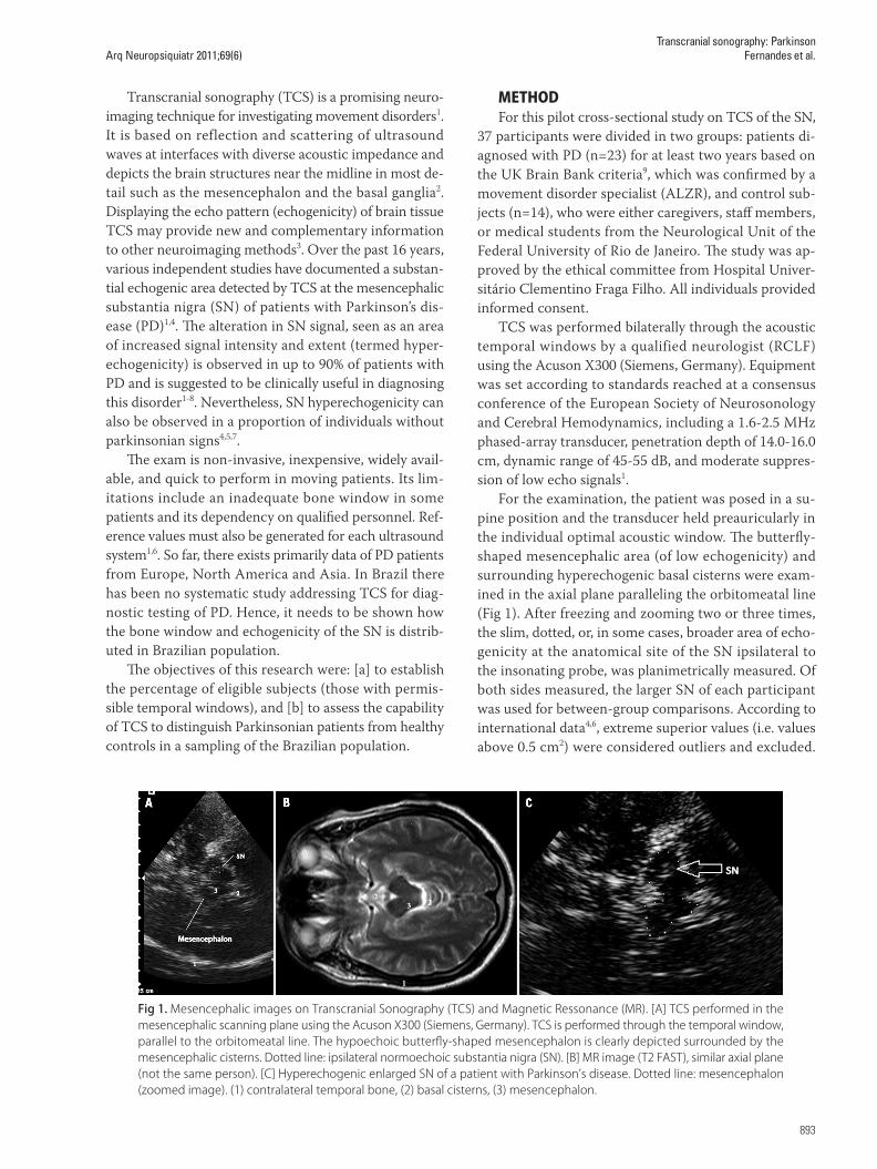
For the examination, the patient was posed in a su-pine position and the transducer held preauricularly in the individual optimal acoustic window. The butterfly-shaped mesencephalic area (of low echogenicity) and surrounding hyperechogenic basal cisterns were exam-ined in the axial plane paralleling the orbitomeatal line (Fig 1). After freezing and zooming two or three times, the slim, dotted, or, in some cases, broader area of echo-genicity at the anatomical site of the SN ipsilateral to the insonating probe, was planimetrically measured. Of both sides measured, the larger SN of each participant was used for between-group comparisons. According to international data4,6, extreme superior values (i.e. values above 0.5 cm2) were considered outliers and excluded.
Fig 1. Mesencephalic images on Transcranial Sonography (TCS) and Magnetic Ressonance (MR). [A] TCS performed in the mesencephalic scanning plane using the Acuson X300 (Siemens, Germany). TCS is performed through the temporal window, parallel to the orbitomeatal line. The hypoechoic butterfly-shaped mesencephalon is clearly depicted surrounded by the mesencephalic cisterns. Dotted line: ipsilateral normoechoic substantia nigra (SN). [B] MR image (T2 FAST), similar axial plane (not the same person). [C] Hyperechogenic enlarged SN of a patient with Parkinson’s disease. Dotted line: mesencephalon (zoomed image). (1) contralateral temporal bone, (2) basal cisterns, (3) mesencephalon.
Arq Neuropsiquiatr 2011;69(6)
894
Transcranial sonography: ParkinsonFernandes et al.
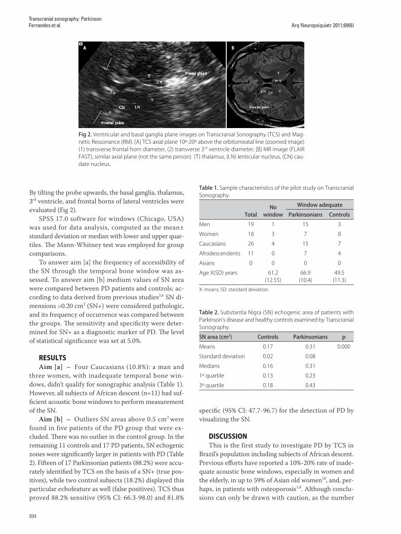
By tilting the probe upwards, the basal ganglia, thalamus, 3rd ventricle, and frontal horns of lateral ventricles were evaluated (Fig 2).
SPSS 17.0 software for windows (Chicago, USA) was used for data analysis, computed as the mean± standard deviation or median with lower and upper quar-tiles. The Mann-Whitney test was employed for group comparisons.
To answer aim [a] the frequency of accessibility of the SN through the temporal bone window was as-sessed. To answer aim [b] medium values of SN area were compared between PD patients and controls; ac-cording to data derived from previous studies2,6 SN di-mensions >0.20 cm2 (SN+) were considered pathologic, and its frequency of occurrence was compared between the groups. The sensitivity and specificity were deter-mined for SN+ as a diagnostic marker of PD. The level of statistical significance was set at 5.0%.
RESULTSAim [a] – Four Caucasians (10.8%): a man and
three women, with inadequate temporal bone win-dows, didn’t qualify for sonographic analysis (Table 1). However, all subjects of African descent (n=11) had suf-ficient acoustic bone windows to perform measurement of the SN.
Aim [b] – Outliers SN areas above 0.5 cm2 were found in five patients of the PD group that were ex-cluded. There was no outlier in the control group. In the remaining 11 controls and 17 PD patients, SN echogenic zones were significantly larger in patients with PD (Table 2). Fifteen of 17 Parkinsonian patients (88.2%) were accu-rately identified by TCS on the basis of a SN+ (true pos-itives), while two control subjects (18.2%) displayed this particular echofeature as well (false positives). TCS thus proved 88.2% sensitive (95% CI: 66.3-98.0) and 81.8%
specific (95% CI: 47.7-96.7) for the detection of PD by visualizing the SN.
DISCUSSIONThis is the first study to investigate PD by TCS in
Brazil’s population including subjects of African descent. Previous efforts have reported a 10%-20% rate of inade-quate acoustic bone windows, especially in women and the elderly, in up to 59% of Asian old women10, and, per-haps, in patients with osteoporosis1,8. Although conclu-sions can only be drawn with caution, as the number
Fig 2. Ventricular and basal ganglia plane images on Transcranial Sonography (TCS) and Mag-netic Ressonance (RM). [A] TCS axial plane 10º-20º above the orbitomeatal line (zoomed image) (1) transverse frontal horn diameter, (2) transverse 3rd ventricle diameter; [B] MR image (FLAIR FAST), similar axial plane (not the same person) (T) thalamus, (LN) lenticular nucleus, (CN) cau-date nucleus.
Table 1. Sample characteristics of the pilot study on Transcranial Sonography.
TotalNo
window
Window adequate
Parkinsonians Controls
Men 19 1 15 3
Women 18 3 7 8
Caucasians 26 4 15 7
Afrodescendents 11 0 7 4
Asians 0 0 0 0
Age X(SD) years 61.2 (12.55)
66.9(10.4)
49.5(11.3)
X: means; SD: standard deviation.
Table 2. Substantia Nigra (SN) echogenic area of patients with Parkinson’s disease and healthy controls examined by Transcranial Sonography.
SN area (cm2) Controls Parkinsonians p
Means 0.17 0.31 0.000
Standard deviation 0.02 0.08
Medians 0.16 0.31
1º quartile 0.13 0.23
3º quartile 0.18 0.43

Arq Neuropsiquiatr 2011;69(6)
895
Transcranial sonography: ParkinsonFernandes et al.
of participants was rather small in this pilot study, only 10.8% of our participants lacked adequate bone windows. Among them, females were more prevalent, but none were of African descent. Our findings, if corroborated, therefore suggest that a large proportion of Brazilians would be eligible for TCS and that the frequency of an insufficient bone window is similar to, for example, the European population.
Since the discovery, in 1995, of a characteristic ab-normal hyperechogenic appearance of the SN on TCS of parkinsonian patients11, the currently most widely used clinical application of TCS in movement disorders is the early and differential diagnosis of PD8. There is a progres-sively growing pool of published studies worldwide dem-onstrating the characteristic finding of SN enlarged areas (above 0.20 cm3) in 80-90% of patients with PD4. Still, the diagnostic assessment of SN echogenicity needs well-trained investigators, but the intra-rater and inter-rater reliability of the ultrasound measures of SN is very good when conducted by experienced sonographers2. In addi-tion, SN areas depicted by TCS have been shown to cor-relate well with MRI T2-hypointensity and T2-relaxation times in the same brainstem region3,12. Therefore, the dis-crimination between normal echogenic and abnormal hyperechogenic (enlarged) SN can nowadays be regarded as reliable provided adequate performance of TCS3.
Similar to prior publications1,4, an expressive number of our subjects (88%) with clinically confirmed PD could be diagnosed by TCS, based on an expanded SN echo-genic area. Only two individuals (18%) of the control group displayed this echofeature as well. Other studies have shown that hyperechogenic SN can be found in 8% to 14% of the general population, suggesting a large number of false-positive findings4. However, there is some evidence that the SN hyperechogenicity in healthy controls is related to a slight motor impairment12,13 and that SPECT scans are also abnormal in up to 60% of the asymptomatic individuals with an abnormal TCS14,15. These findings raised the question whether the TCS finding of SN hyperechogenicity alone in a healthy indi-vidual might be a predictor for subsequent occurrence of PD15. The results of ongoing longitudinal studies will give further insight into this question16.
Although data to date suggest that this simple and in-expensive imaging modality may be useful in improving diagnosis of movement disorders, the present knowl-edge about the mechanism leading to the changes in SN echomorphology is limited7. It has been speculated that SN hyperechogenicity reflects histopathological changes or displays an alteration in tissue impedance due to an abnormal iron accumulation, a process associated with neurodegeneration in PD17.
Difference in age and gender between the groups
need to be named as limitations of this pilot study. How-ever, although distributed unevenly between the groups, these differences are not of importance to evaluate the prevalence of an adequate acoustic bone window in the whole cohort of subjects investigated. Still, it needs to be considered that female sex and higher age are associated with a higher prevalence of insufficient bone windows. Future studies, therefore, need to include even older sub-jects, especially women.
Results of this pilot study indicate that TCS enables the depiction of the substantia nigra in the Caucasian and African Brazilian population to the same percentage as the European Caucasian population. The prevalence of SN hyperechogenicity among PD patients in Brazil is similar to the prevalence reported in other popula-tions. The current data, however, is preliminary and must be confirmed in larger studies with less participant heterogeneity.
REFERENCES1. Walter U, Behnke S, Eyding J, et al. Transcranial brain parenchyma so-
nography in movement disorders: state of the art. Ultrasound Med Biol 2007;33:15-25.
2. Školoudík D, Walter U. Method and validity of Transcranial Sonography in movement disorders. Internat Rev Neurobiol 2010;90:7-34.
3. Niehaus L, Boelmans K. Diagnosis of Parkinson’s disease: Transcranial So-nography in relation to MRI. Internat Rev Neurobiol 2010;90:63-79.
4. Vlaar AMM, Bouwmans A, Mess WH, Tromp SC, Weber WEJ. Transcranial duplex in the differential diagnosis of parkinsonian syndromes: a system-atic review. J Neurol 2009;256:530-538.
5. Berg D. Transcranial ultrasound as a risk marker for Parkinson’s disease. Mov Disord 2009;24(Suppl):S677-S683.
6. Berg D, Godau J, Walter U. Transcranial Sonography in movement disor-ders. Lancet Neurol 2008;7:1044-1055.
7. Double KL, Todd G, Duma SR. Pathophysiology of Transcranial Sonography signal changes in the human substantia nigra. Internat Rev Neurobiol 2010;90:107-120.
8. Godau J, Berg D. Role of Transcranial Ultrasound in the diagnosis of move-ment disorders. Neuroimag Clin N Am 2010;20:87-101.
9. Hughes AJ, Daniel SE, Kilford L, Lees AJ. Accuracy of clinical diagnosis of idiopathic Parkinson’s disease: a clinic-pathological study of 100 cases. J Neurol Neurosurg Psychiatry 1992;55:181-184.
10. Okawa M, Miwa H, Kajimoto Y, et al. Transcranial Sonography of the sub-stantia nigra in Japanese patients with Parkinson’s disease or atypical par-kinsonism: clinical potential and limitations. Intern Med 2007;46:1527-1531.
11. Becker G, Seufert J, Bogdahn U, Reichmann H, Reiners K. Degeneration of the substantia nigra in chronic Parkinson’s disease visualized by Tran-scranial color-coded real-time Sonography. Neurology 1985;45:182-184.
12. Behnke S, Schroeder U, Dillmann U, et al. Hyperechogenicity of the sub-stantia nigra in healthy controls is related to MRI changes and to neu-ronal loss as determined by F-Dopa PET. Neuroimage 2009;47:1237-1243.
13. Berg D, Siefker C, Ruprecht-Dörfler P, Becker G. Relationship of substantia nigra echogenicity and motor function in elderly subjects. Neurology 2001;56:13-17.
14. Sommer U, Hummel T, Cormann K, et al. Detection of presymptomatic Parkinson’s disease: combining smell tests, Transcranial Sonography and SPECT. Mov Disord 2004;19:1196-1202.
15. Berg D. Ultrasound in the (premotor) diagnosis of Parkinson’s disease. Parkinsonism Relat Disord 2007;13(Suppl 3):S429-S433.
16. Behnke S, Schröder U, Berg D. Transcranial Sonography in the premotor diagnosis of Parkinson’s disease. Internat Rev Neurobiol 2010;90:93-106.
17. Berg D, Hochstrasser H, Schweitzer KJ, Riess O. Disturbance of iron metabolism in Parkinson’s disease: ultrasonography as a biomarker. Neurotox Res 2006;9:1-13.