Embed Size (px)
Citation preview
J A C C : C A R D I O V A S C U L A R I N T E R V E N T I O N S VO L . 1 1 , N O . 7 , 2 0 1 8
P U B L I S H E D B Y E L S E V I E R O N B E H A L F O F T H E AM E R I C A N
C O L L E G E O F C A R D I O L O G Y F O U N D A T I O N
STRUCTURAL
Transcatheter Laceration ofAortic Leaflets to PreventCoronary Obstruction DuringTranscatheter Aortic Valve Replacement
Concept to First-in-HumanJaffar M. Khan, BM BCH,a Danny Dvir, MD,b Adam B. Greenbaum, MD,c Vasilis C. Babaliaros, MD,d
Toby Rogers, PHD, BM BCH,a Gabriel Aldea, MD,b Mark Reisman, MD,b G. Burkhard Mackensen, MD,b
Marvin H.K. Eng, MD,c Gaetano Paone, MD,c Dee Dee Wang, MD,c Robert A. Guyton, MD,d
Chandan M. Devireddy, MD,d William H. Schenke, BS,a Robert J. Lederman, MDa
ABSTRACT
ISS
Fro
of
Ca
Ce
Ins
Blo
OBJECTIVES This study sought to develop a novel technique called bioprosthetic or native aortic scallop intentional
laceration to prevent coronary artery obstruction (BASILICA).
BACKGROUND Coronary artery obstruction is a rare but fatal complication of transcatheter aortic valve replacement
(TAVR).
METHODS We lacerated pericardial leaflets in vitro using catheter electrosurgery, and tested leaflet splaying after benchtop
TAVR.Theprocedurewas tested in swine.BASILICAwas thenoffered topatients athigh riskof coronaryobstruction fromTAVR
and ineligible for surgical aortic valve replacement. BASILICA used marketed devices. Catheters directed an electrified
guidewire to traverse and lacerate the aortic leaflet down the center line. TAVR was performed as usual.
RESULTS TAVR splayed lacerated bovine pericardial leaflets. BASILICA was successful in pigs, both to left and right
cusps. Necropsy revealed full length lacerations with no collateral thermal injury. Seven patients underwent BASILICA on
a compassionate basis. Six had failed bioprosthetic valves, both stented and stent-less. Two had severe aortic stenosis,
including 1 patient with native disease, 3 had severe aortic regurgitation, and 2 had mixed aortic valve disease. One
patient required laceration of both left and right coronary cusps. There was no hemodynamic compromise in any patient
following BASILICA. All patients had successful TAVR, with no coronary obstruction, stroke, or any major complications.
All patients survived to 30 days.
CONCLUSIONS BASILICA may durably prevent coronary obstruction from TAVR. The procedure was successful
across a range of presentations, and requires further evaluation in a prospective trial. Its role in treatment of
degenerated TAVR devices remains untested. (J Am Coll Cardiol Intv 2018;11:677–89) Published by Elsevier on behalf of
the American College of Cardiology Foundation.
N 1936-8798/$36.00 https://doi.org/10.1016/j.jcin.2018.01.247
m the aCardiovascular Branch, Division of Intramural Research, National Heart, Lung, and Blood Institute, National Institutes
Health, Bethesda, Maryland; bUniversity of Washington, Seattle, Washington; cCenter for Structural Heart Disease, Division of
rdiology, and Division of Cardiac Surgery, Henry Ford Health System, Detroit, Michigan; and the dStructural Heart and Valve
nter, Emory University Hospital, Atlanta, Georgia. Supported by the National Heart, Lung, and Blood Institute, National
titutes of Health (Z01-HL006040-7), and by the intramural programs of the participating centers. National Heart, Lung, and
od Institute has a collaborative research and development agreement with Edwards Lifesciences on transcatheter modification
ABBR EV I A T I ON S
AND ACRONYMS
BASILICA = bioprosthetic or
native aortic scallop intentional
laceration to prevent iatrogenic
coronary artery obstruction
CT = computed tomography
TAVR = transcatheter aortic
valve replacement
of the mitra
for Edward
and his em
sciences, A
Dr. Paone i
research co
and Boston
investigati
Medical, an
paper to di
Manuscript
Khan et al. J A C C : C A R D I O V A S C U L A R I N T E R V E N T I O N S V O L . 1 1 , N O . 7 , 2 0 1 8
BASILICA Procedure Before TAVR to Avoid Coronary Obstruction A P R I L 9 , 2 0 1 8 : 6 7 7 – 8 9
678
T ranscatheter aortic valve replace-ment (TAVR) is an effective alterna-tive to surgical aortic valve
replacement in intermediate- and high-riskpatients with native aortic stenosis (1,2).TAVR is also an effective treatment for fail-ure of bioprosthetic surgical aortic valves, atreatment known as valve-in-valve TAVR(3,4). Coronary artery obstruction is a devas-
tating complication of TAVR, with a >50% mortality(5). Coronary artery obstruction occurs when thetranscatheter heart valve displaces the underlyingsurgical or native aortic valve leaflets outward andobstructs the coronary artery ostia, either by sealingthe sinus of Valsalva at the sinotubular junction orby the leaflet itself covering the coronary ostiabecause of low-lying coronary ostia and inadequatesinus width (Figure 1). Coronary artery obstructionis 4 times as common during valve-in-valveTAVR as during TAVR for native aortic stenosis (6),
SEE PAGE 690
likely because most surgical prostheses are supra-annular in design, lowering coronary heights relativeto the valve leaflets, and because valve suturingdraws the coronaries closer, decreasing sinus width.The risk of coronary obstruction is highest duringTAVR for surgical bioprothesis designs intended tomaximize effective aortic orifice area (“stented” bio-prostheses that have externally mounted leaflets,and “stent-less” surgical bioprostheses) (5). Treat-ment requires bail-out percutaneous coronary inter-vention, which may not be possible with a valveleaflet obstructing the coronary artery, or emergencybypass surgery. Pre-emptive coronary protectionwith a guidewire, with or without a coronary balloonor stent prepositioned down the coronary artery, isvariably successful (7,8) in the short and intermediateterm. One-third of coronary obstruction events maymanifest after the TAVR is concluded (5).
We propose a solution based on the LAMPOONprocedure (9,10), which uses catheters to split the
l valve. Dr. Dvir is a consultant for Edwards Lifesciences, Medtron
s Lifesciences and Abbott St. Jude Medical. Dr. Babaliaros is a cons
ployer has research contracts for clinical investigation of transca
bbott Vascular, Medtronic, St. Jude Medical, and Boston Scient
s a proctor for Edwards Lifesciences. Dr. Wang is a consultant fo
ntracts for clinical investigation of aortic and mitral devices from
Scientific. Dr. Devireddy is a consultant for Medtronic; and
on of transcatheter aortic and mitral devices from Edwards L
d Boston Scientific. All other authors have reported that they h
sclose. Drs. Khan and Dvir contributed equally to this work.
received October 20, 2017; revised manuscript received Decemb
mitral valve leaflet and prevent obstruction of the leftventricular outflow tract during transcatheter mitralvalve replacement. Here we report a technique tosplit aortic valve leaflets, whether bioprosthetic ornative, to prevent coronary artery obstruction afterTAVR. The new technique is called bioprosthetic ornative aortic scallop intentional laceration to preventcoronary artery obstruction (BASILICA).
We developed the technique in vitro and in ani-mals, and then offered the procedure to patientsexperiencing aortic valve failure who were ineligiblefor conventional surgical aortic valve replacement,and high or prohibitive risk of coronary arteryobstruction from TAVR.
METHODS
We set out to demonstrate several key technicalprinciples. First, that an aortic leaflet scallop can betraversed in situ by an electrified guidewire betweenthe sinus of Valsalva and the left ventricular outflowtract. Second, that the traversed leaflet, whethernative or bioprosthetic, can be lacerated in situ by themid-shaft of an electrified guidewire. Third, that thelacerated leaflets splay after TAVR to allow blood flowacross them toward otherwise obstructed coronaryostia. Fourth, whether partial (mid-scallop vs. basalleaflet) lacerations extend lengthwise when stretchedby an implanted valve, which may influence therequired spatial precision of the procedure. Fifth, thatboth left and right coronary cusps can be laceratedsimultaneously in vivo.
IN VITRO. We tested radiofrequency-assistedtranscatheter perforation and laceration of exterior-mounted bovine pericardial leaflets on a representa-tive bioprosthetic heart valve (19-mm Trifecta valve,Abbott St. Jude Medical, St. Paul, Minnesota) sub-merged in a 0.9% saline bath with a remote dispersiveelectrode (Online Figure 1). Two lacerations wereattempted on the bioprosthetic heart valve. One leafletwas lacerated from base to tip and the second from
ic, and St. Jude Medical. Dr. Greenbaum is a proctor
ultant for Edwards Lifesciences and Abbott Vascular;
theter aortic and mitral devices from Edwards Life-
ific. Dr. Eng is a proctor for Edwards Lifesciences.
r Edwards Lifesciences. Dr. Guyton’s employer has
Edwards Lifesciences, Abbott Vascular, Medtronic,
his employer has research contracts for clinical
ifesciences, Abbott Vascular, Medtronic, St. Jude
ave no relationships relevant to the contents of this
er 19, 2017, accepted January 1, 2018.
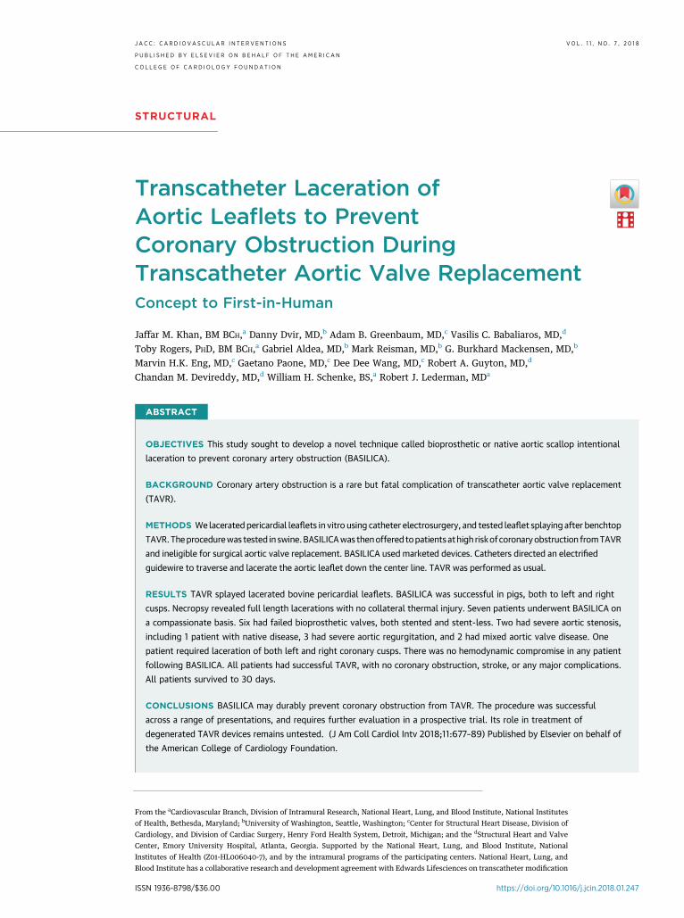
FIGURE 1 Illustrations of Coronary Obstruction and Prevention by BASILICA
In normal transcatheter aortic valve replacement performed in a capacious aortic root, blood flows unrestricted around valve leaflets into
coronary arteries. In patients with a crowded sinus and low-lying coronary arteries, coronary blood flow is obstructed by the bioprosthetic
valve leaflets after transcatheter aortic valve replacement. After BASILICA, blood flows through the open cells of the transcatheter heart valve
unimpeded into the coronary artery. BASILICA ¼ bioprosthetic or native aortic scallop intentional laceration to prevent iatrogenic coronary
artery obstruction; LCA ¼ left coronary artery; RCA ¼ right coronary artery.
J A C C : C A R D I O V A S C U L A R I N T E R V E N T I O N S V O L . 1 1 , N O . 7 , 2 0 1 8 Khan et al.A P R I L 9 , 2 0 1 8 : 6 7 7 – 8 9 BASILICA Procedure Before TAVR to Avoid Coronary Obstruction
679
mid-point to tip. A third scallop was left intact andserved as a control.
Balloon expandable (20-mm Sapien 3, EdwardsLifesciences, Irvine, California) and self-expandingvalves (23-mm Evolut Pro, Medtronic, Minneapolis,Minnesota) were deployed in the bioprosthetic valveto test splaying of split leaflet around the open cells ofthe transcatheter heart valve and propagation of thesplit in the leaflet. A second valve (25-mm Mitroflow,Sorin Livanova, London, England) was cut with ascalpel and leaflet splaying was also tested withappropriately sized balloon expanding and self-expanding valves.
ANIMALS. Animal experiments on naive Yorkshireand Yucatan pigs were approved by the institutionalanimal care and use committee and conducted percontemporary National Institutes of Health guide-lines. Anesthesia was induced and maintained withmechanical ventilation and inhaled isoflurane, 2femoral arterial sheaths of 6-F catheter and a 9-Fcatheter femoral venous sheath were placed
percutaneously, and heparin and amiodarone wereadministered. The BASILICA procedure without TAVRwas performed using catheters directed underbiplane x-ray fluoroscopy and intracardiac echocar-diography guidance. Pre-procedural cardiac magneticresonance imaging was performed at 1.5-T (Aera,Siemens, Erlangen, Germany) to plan fluoroscopyprojection angles. Hemodynamics were recorded for 1h after laceration until euthanasia. The length ofscallop laceration relative to the overall length of thescallop was measured using calipers at necropsy. Theheart was carefully inspected for evidence ofbystander electrical or mechanical injury.
CLINICAL. Pat ients . Patients with high or prohibi-tive risk for surgical aortic valve replacement andhigh risk of coronary artery obstruction with TAVRunderwent TAVR with BASILICA at 3 medical centers(University of Washington, Henry Ford, and EmoryUniversity Hospitals). All consented to clinical treat-ment on a compassionate basis, despite explicitlyhigh risk, after consensus from the local
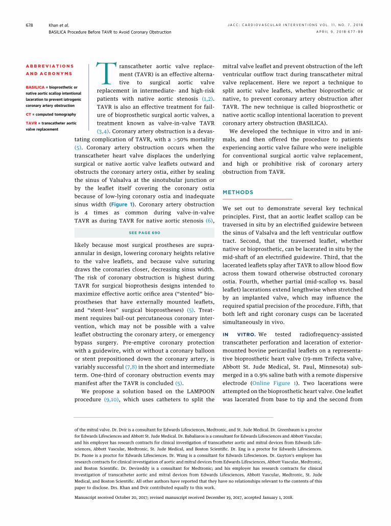
FIGURE 2 Illustration of the BASILICA Procedure
A catheter directs an electrified guidewire through the base of
the left aortic cusp into a snare in the left ventricular outflow
tract (A). After snare retrieval (B), the mid-shaft of the guide-
wire is electrified to lacerate the leaflet (C). The leaflet splays
after transcatheter aortic valve replacement permitting cor-
onary flow (D). Abbreviation as in Figure 1.
Khan et al. J A C C : C A R D I O V A S C U L A R I N T E R V E N T I O N S V O L . 1 1 , N O . 7 , 2 0 1 8
BASILICA Procedure Before TAVR to Avoid Coronary Obstruction A P R I L 9 , 2 0 1 8 : 6 7 7 – 8 9
680
multidisciplinary heart teams. The institutionalethics review boards of all participating institutionsapproved this retrospective report.
The local heart teams determined coronaryobstruction risk based on manufacturer-described
geometry of the specific implanted bioprostheticvalve; and computed tomography (CT) and angio-graphic measurements of the coronary ostia heights,sinus of Valsalva width, presence and type of bio-prosthetic valve, and virtual transcatheter heart valveto coronary distance (Figure 1) (5).BASIL ICA procedure . The procedure was plannedusing electrocardiogram-gated contrast-enhancedCT, performed under general anesthesia, and guidedby fluoroscopy and transesophageal echocardiogra-phy. Catheter access was obtained typically via 3femoral arterial (2 typically ipsilateral for BASILICA,and 1 for TAVR) and at least 1 venous (for temporarytransvenous pacing) introducer sheaths. Heparinanticoagulation achieved an activated clotting time>300 s.
A pair of coaxial catheters (typically a 5-F mammarydiagnostic catheter inside a 6-F extra backup shape-guiding catheter) was positioned in the targetedaortic leaflet scallop to direct a guidewire across it,near the scallop hinge point, by echocardiographicand angiographic guidance. These aimed at a snarepositioned immediately below the leaflet using aseparate retrograde catheter (Figure 2, Online Video 1).
To traverse the aortic leaflet scallop, a 0.014-inchguidewire (Astato XS 20, Asahi-Intecc, Santa Ana,California) sheathed in an insulated polymer jacket(Piggyback Wire Convertor, Vascular Solutions Tele-flex, Minneapolis, Minnesota) was electrified,advanced, and snare-retrieved. The wire was electri-fied using a short burst of “cutting” radiofrequencyenergy (w30 W) by clamping to an electrosurgerypencil (Valleylab FX, Covidien Medtronic, Minneap-olis, Minnesota).
After externalization of the free guidewire end,the guidewire straddles across the leaflet scallopbetween 2 catheters. The scallop was lacerated byapplying radiofrequency energy at approximately 70W while tensioning both free ends of the guidewire.A pigtail catheter was pre-positioned in the leftventricle to allow TAVR to be performed immedi-ately afterward.
TAVR was performed using established techniques.Coronary artery stent systems were positioned pro-phylactically at the discretion of the operator.Cracking of a failed bioprosthetic heart valve frame,using a high-pressure balloon (11), was performed atoperator discretion to achieve an optimum hemody-namic result. Coronary artery patency was assessedusing angiography and post-TAVR CT. Antiplateletand anticoagulation therapy were prescribed atoperator discretion. Complications were assessed ac-cording to the Valve Academic Research Consortium-2 Consensus Document (12).
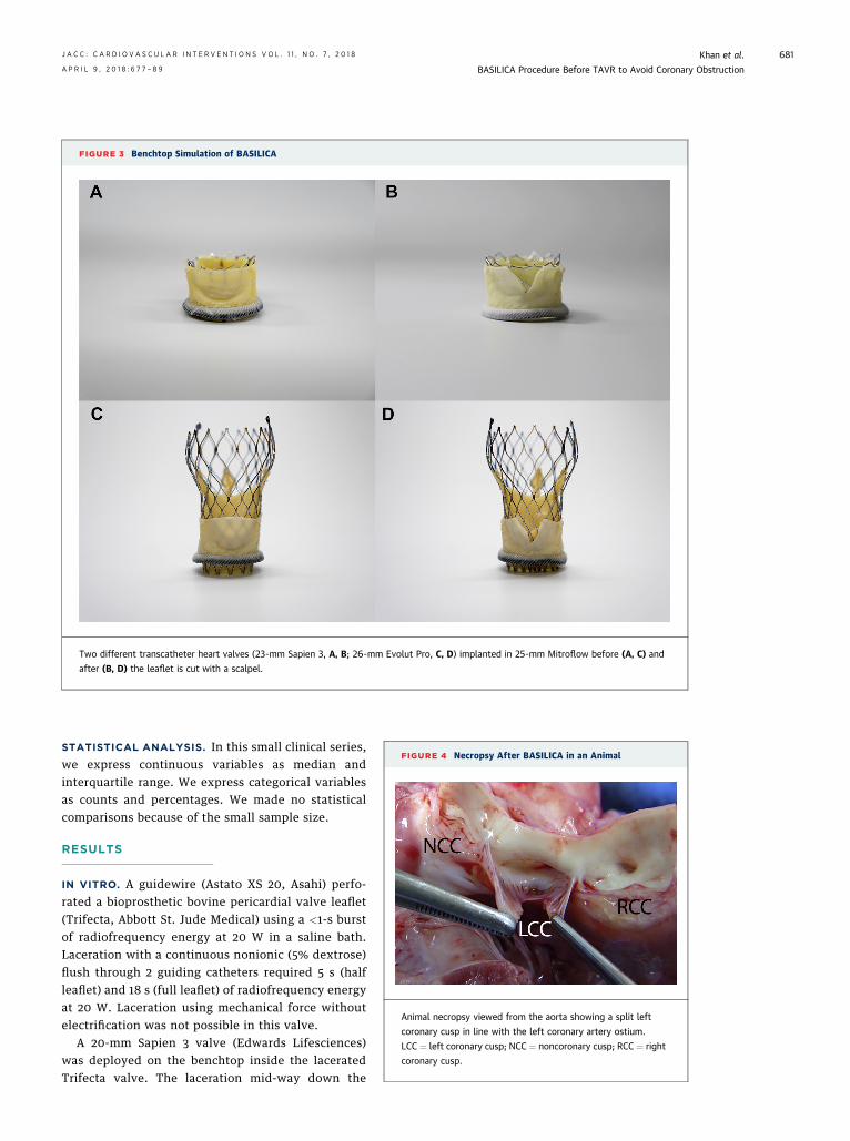
FIGURE 3 Benchtop Simulation of BASILICA
Two different transcatheter heart valves (23-mm Sapien 3, A, B; 26-mm Evolut Pro, C, D) implanted in 25-mm Mitroflow before (A, C) and
after (B, D) the leaflet is cut with a scalpel.
FIGURE 4 Necropsy After BASILICA in an Animal
Animal necropsy viewed from the aorta showing a split left
coronary cusp in line with the left coronary artery ostium.
LCC ¼ left coronary cusp; NCC ¼ noncoronary cusp; RCC ¼ right
coronary cusp.
J A C C : C A R D I O V A S C U L A R I N T E R V E N T I O N S V O L . 1 1 , N O . 7 , 2 0 1 8 Khan et al.A P R I L 9 , 2 0 1 8 : 6 7 7 – 8 9 BASILICA Procedure Before TAVR to Avoid Coronary Obstruction
681
STATISTICAL ANALYSIS. In this small clinical series,we express continuous variables as median andinterquartile range. We express categorical variablesas counts and percentages. We made no statisticalcomparisons because of the small sample size.
RESULTS
IN VITRO. A guidewire (Astato XS 20, Asahi) perfo-rated a bioprosthetic bovine pericardial valve leaflet(Trifecta, Abbott St. Jude Medical) using a <1-s burstof radiofrequency energy at 20 W in a saline bath.Laceration with a continuous nonionic (5% dextrose)flush through 2 guiding catheters required 5 s (halfleaflet) and 18 s (full leaflet) of radiofrequency energyat 20 W. Laceration using mechanical force withoutelectrification was not possible in this valve.
A 20-mm Sapien 3 valve (Edwards Lifesciences)was deployed on the benchtop inside the laceratedTrifecta valve. The laceration mid-way down the
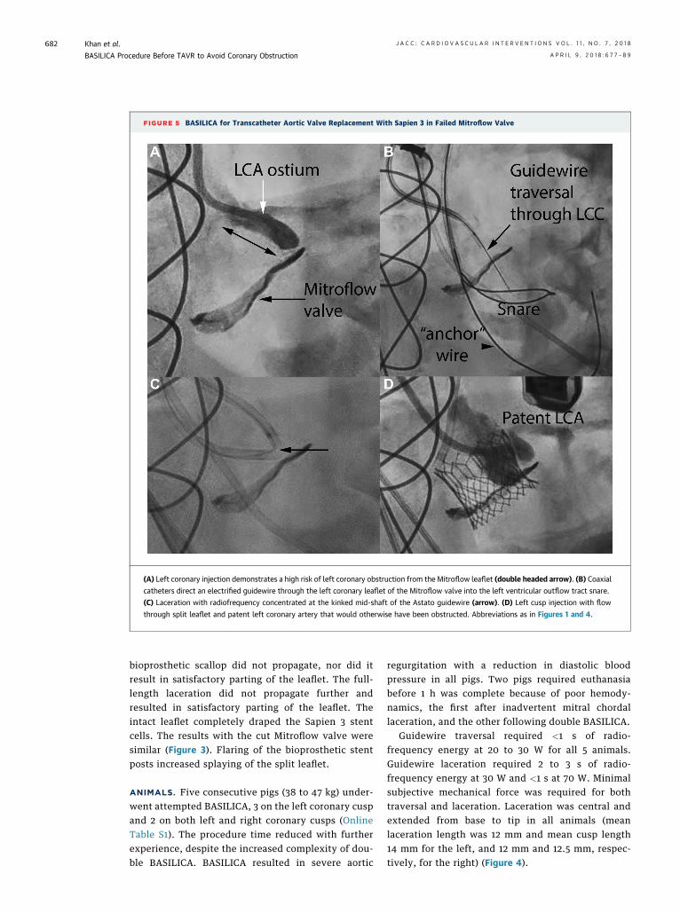
FIGURE 5 BASILICA for Transcatheter Aortic Valve Replacement With Sapien 3 in Failed Mitroflow Valve
(A) Left coronary injection demonstrates a high risk of left coronary obstruction from the Mitroflow leaflet (double headed arrow). (B) Coaxial
catheters direct an electrified guidewire through the left coronary leaflet of the Mitroflow valve into the left ventricular outflow tract snare.
(C) Laceration with radiofrequency concentrated at the kinked mid-shaft of the Astato guidewire (arrow). (D) Left cusp injection with flow
through split leaflet and patent left coronary artery that would otherwise have been obstructed. Abbreviations as in Figures 1 and 4.
Khan et al. J A C C : C A R D I O V A S C U L A R I N T E R V E N T I O N S V O L . 1 1 , N O . 7 , 2 0 1 8
BASILICA Procedure Before TAVR to Avoid Coronary Obstruction A P R I L 9 , 2 0 1 8 : 6 7 7 – 8 9
682
bioprosthetic scallop did not propagate, nor did itresult in satisfactory parting of the leaflet. The full-length laceration did not propagate further andresulted in satisfactory parting of the leaflet. Theintact leaflet completely draped the Sapien 3 stentcells. The results with the cut Mitroflow valve weresimilar (Figure 3). Flaring of the bioprosthetic stentposts increased splaying of the split leaflet.
ANIMALS. Five consecutive pigs (38 to 47 kg) under-went attempted BASILICA, 3 on the left coronary cuspand 2 on both left and right coronary cusps (OnlineTable S1). The procedure time reduced with furtherexperience, despite the increased complexity of dou-ble BASILICA. BASILICA resulted in severe aortic
regurgitation with a reduction in diastolic bloodpressure in all pigs. Two pigs required euthanasiabefore 1 h was complete because of poor hemody-namics, the first after inadvertent mitral chordallaceration, and the other following double BASILICA.
Guidewire traversal required <1 s of radio-frequency energy at 20 to 30 W for all 5 animals.Guidewire laceration required 2 to 3 s of radio-frequency energy at 30 W and <1 s at 70 W. Minimalsubjective mechanical force was required for bothtraversal and laceration. Laceration was central andextended from base to tip in all animals (meanlaceration length was 12 mm and mean cusp length14 mm for the left, and 12 mm and 12.5 mm, respec-tively, for the right) (Figure 4).
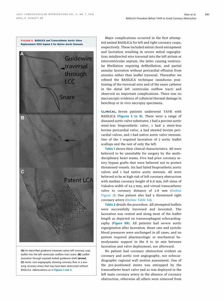
FIGURE 6 BASILICA and Transcatheter Aortic Valve
Replacement With Sapien 3 for Native Aortic Stenosis
(A) An electrified guidewire traverses native left coronary cusp
leaflet into the left ventricular outflow tract snare. (B) Leaflet
laceration through exposed kinked guidewire shaft (arrow).
(C) Aortic root angiography showing coronary flow in a low-
lying coronary artery that may have been obstructed without
BASILICA. Abbreviations as in Figures 1 and 4.
J A C C : C A R D I O V A S C U L A R I N T E R V E N T I O N S V O L . 1 1 , N O . 7 , 2 0 1 8 Khan et al.A P R I L 9 , 2 0 1 8 : 6 7 7 – 8 9 BASILICA Procedure Before TAVR to Avoid Coronary Obstruction
683
Major complications occurred in the first attemp-ted animal BASILICA for left and right coronary cusps,respectively. These included mitral chord entrapmentand laceration resulting in severe mitral regurgita-tion; misdirected wire traversal into the left atrium orinterventricular septum, the latter causing ventricu-lar fibrillation requiring defibrillation; and partialannular laceration without pericardial effusion fromannulus rather than leaflet traversal. Thereafter werefined the BASILICA technique (assiduous posi-tioning of the traversal wire and of the snare catheterin the distal left ventricular outflow tract) andobserved no important complications. There was nomacroscopic evidence of collateral thermal damage inbenchtop or in vivo necropsy specimens.
CLINICAL. Seven patients underwent TAVR withBASILICA (Figures 5 to 9). There were a range ofdiseased aortic valve substrates: 1 had a porcine aorticstent-less bioprosthetic valve, 1 had a stent-lessbovine pericardial valve, 4 had stented bovine peri-cardial valves, and 1 had native aortic valve stenosis.One of the 7 required laceration of 2 aortic leafletscallops and the rest of only the left.
Table 1 shows their clinical characteristics. All werebelieved to be unsuitable for surgery by the multi-disciplinary heart teams. Five had prior coronary ar-tery bypass grafts that were believed not to protectthreatened vessels. Six had failed bioprosthetic aorticvalves and 1 had native aortic stenosis. All werebelieved to be at high risk of left coronary obstructionwith median coronary height of 6.8 mm, left sinus ofValsalva width of 24.3 mm, and virtual transcathetervalve to coronary distance of 2.8 mm (OnlineFigure 2). One patient also had a threatened rightcoronary artery (Online Table S2).
Table 2 details the procedure. All attempted leafletswere successfully traversed and lacerated. Thelaceration was central and along most of the leafletlength as depicted on transesophageal echocardiog-raphy (Figure 8B). All patients had severe aorticregurgitation after laceration. Heart rate and systolicblood pressures were unchanged in all cases, and nopatient required pharmacologic or mechanical he-modynamic support in the 8 to 30 min betweenlaceration and valve deployment, nor afterward.
No patient had coronary obstruction evident oncoronary and aortic root angiography, nor echocar-diographic regional wall motion assessment. One ofthe pre-positioned stents was entrapped by thetranscatheter heart valve and so was deployed in theleft main coronary artery in the absence of coronaryobstruction, otherwise all others were removed from
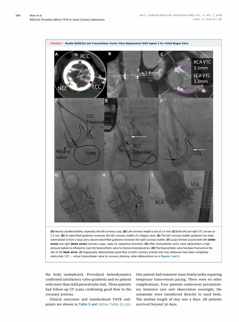
FIGURE 7 Double BASILICA and Transcatheter Aortic Valve Replacement With Sapien 3 for Failed Magna Valve
(A) Heavily calcified leaflets, especially the left coronary cusp. (B) Left coronary height is low at 3.4 mm. (C) Both left and right VTC are low at
3.3 mm. (D) An electrified guidewire traverses the left coronary leaflet of a Magna valve. (E) The left coronary leaflet guidewire has been
externalized to form a loop and a second electrified guidewire traverses the right coronary leaflet. (F) Loops formed around both left (white
arrow) and right (black arrow) coronary cusps, ready for sequential laceration. (G) After transcatheter aortic valve replacement a high-
pressure balloon is inflated to crack the bioprosthetic valve to improve hemodynamics. (H) The bioprosthetic valve has been fractured at the
site of the black arrow. (I) Angiography demonstrates good flow to both coronary arteries that may otherwise have been completely
obstructed. VTC ¼ virtual transcatheter valve to coronary distance; other abbreviations as in Figures 1 and 4.
Khan et al. J A C C : C A R D I O V A S C U L A R I N T E R V E N T I O N S V O L . 1 1 , N O . 7 , 2 0 1 8
BASILICA Procedure Before TAVR to Avoid Coronary Obstruction A P R I L 9 , 2 0 1 8 : 6 7 7 – 8 9
684
the body undeployed. Procedural hemodynamicsconfirmed satisfactory valve gradients and no patientwith more than mild paravalvular leak. Three patientshad follow-up CT scans confirming good flow in thecoronary arteries.
Clinical outcomes and standardized TAVR end-points are shown in Table 3 and Online Table S3 (12).
One patient had transient sinus bradycardia requiringtemporary transvenous pacing. There were no othercomplications. Four patients underwent precaution-ary intensive care unit observation overnight; theremainder were transferred directly to ward beds.The median length of stay was 4 days. All patientssurvived beyond 30 days.
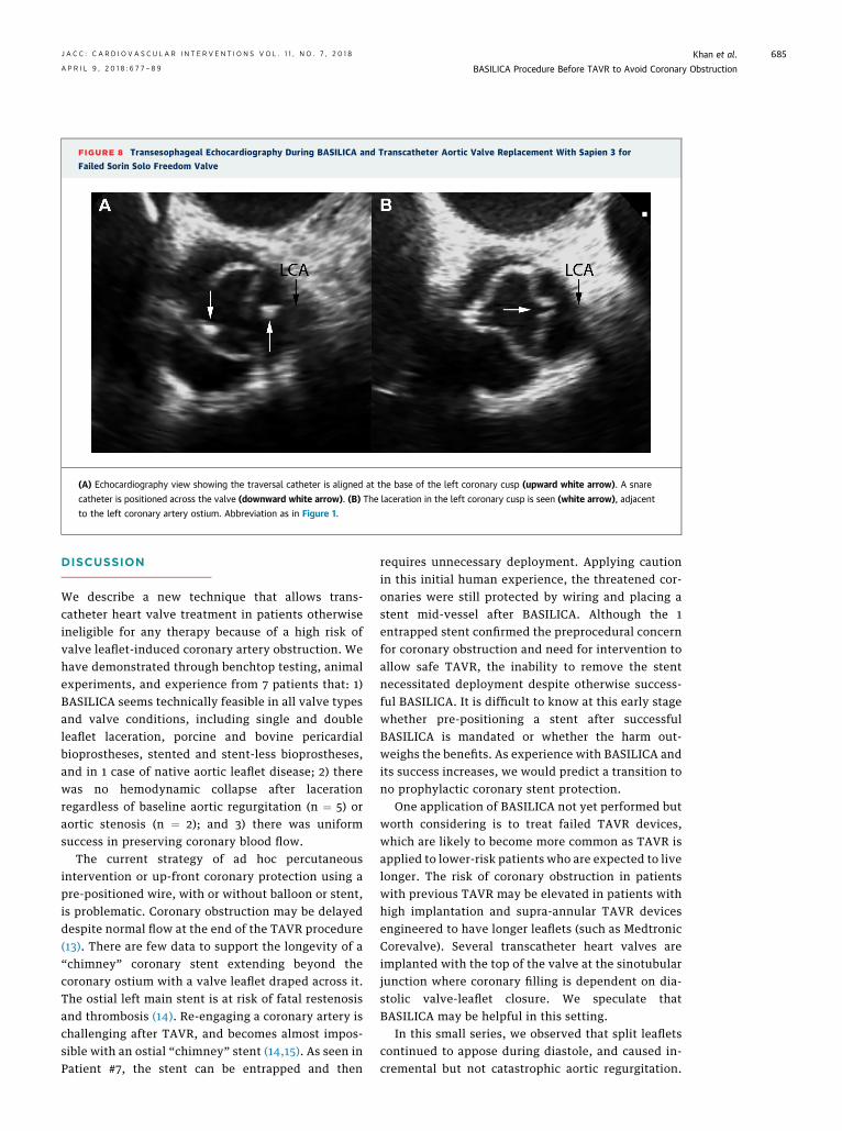
FIGURE 8 Transesophageal Echocardiography During BASILICA and Transcatheter Aortic Valve Replacement With Sapien 3 for
Failed Sorin Solo Freedom Valve
(A) Echocardiography view showing the traversal catheter is aligned at the base of the left coronary cusp (upward white arrow). A snare
catheter is positioned across the valve (downward white arrow). (B) The laceration in the left coronary cusp is seen (white arrow), adjacent
to the left coronary artery ostium. Abbreviation as in Figure 1.
J A C C : C A R D I O V A S C U L A R I N T E R V E N T I O N S V O L . 1 1 , N O . 7 , 2 0 1 8 Khan et al.A P R I L 9 , 2 0 1 8 : 6 7 7 – 8 9 BASILICA Procedure Before TAVR to Avoid Coronary Obstruction
685
DISCUSSION
We describe a new technique that allows trans-catheter heart valve treatment in patients otherwiseineligible for any therapy because of a high risk ofvalve leaflet-induced coronary artery obstruction. Wehave demonstrated through benchtop testing, animalexperiments, and experience from 7 patients that: 1)BASILICA seems technically feasible in all valve typesand valve conditions, including single and doubleleaflet laceration, porcine and bovine pericardialbioprostheses, stented and stent-less bioprostheses,and in 1 case of native aortic leaflet disease; 2) therewas no hemodynamic collapse after lacerationregardless of baseline aortic regurgitation (n ¼ 5) oraortic stenosis (n ¼ 2); and 3) there was uniformsuccess in preserving coronary blood flow.
The current strategy of ad hoc percutaneousintervention or up-front coronary protection using apre-positioned wire, with or without balloon or stent,is problematic. Coronary obstruction may be delayeddespite normal flow at the end of the TAVR procedure(13). There are few data to support the longevity of a“chimney” coronary stent extending beyond thecoronary ostium with a valve leaflet draped across it.The ostial left main stent is at risk of fatal restenosisand thrombosis (14). Re-engaging a coronary artery ischallenging after TAVR, and becomes almost impos-sible with an ostial “chimney” stent (14,15). As seen inPatient #7, the stent can be entrapped and then
requires unnecessary deployment. Applying cautionin this initial human experience, the threatened cor-onaries were still protected by wiring and placing astent mid-vessel after BASILICA. Although the 1entrapped stent confirmed the preprocedural concernfor coronary obstruction and need for intervention toallow safe TAVR, the inability to remove the stentnecessitated deployment despite otherwise success-ful BASILICA. It is difficult to know at this early stagewhether pre-positioning a stent after successfulBASILICA is mandated or whether the harm out-weighs the benefits. As experience with BASILICA andits success increases, we would predict a transition tono prophylactic coronary stent protection.
One application of BASILICA not yet performed butworth considering is to treat failed TAVR devices,which are likely to become more common as TAVR isapplied to lower-risk patients who are expected to livelonger. The risk of coronary obstruction in patientswith previous TAVR may be elevated in patients withhigh implantation and supra-annular TAVR devicesengineered to have longer leaflets (such as MedtronicCorevalve). Several transcatheter heart valves areimplanted with the top of the valve at the sinotubularjunction where coronary filling is dependent on dia-stolic valve-leaflet closure. We speculate thatBASILICA may be helpful in this setting.
In this small series, we observed that split leafletscontinued to appose during diastole, and caused in-cremental but not catastrophic aortic regurgitation.
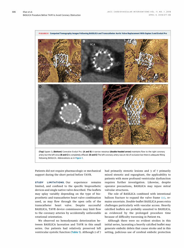
FIGURE 9 Computed Tomography Images Following BASILICA and Transcatheter Aortic Valve Replacement With Sapien 3 and Evolut Pro
(Top) Sapien 3; (Bottom) Corevalve Evolut Pro. (A and B) A narrow neosinus (double-headed arrow) maintains flow to the right coronary
artery but the left sinus (B and C) is completely effaced. (D and E) The left coronary artery was at risk of occlusion but there is adequate filling
following BASILICA. Abbreviations as in Figure 1.
Khan et al. J A C C : C A R D I O V A S C U L A R I N T E R V E N T I O N S V O L . 1 1 , N O . 7 , 2 0 1 8
BASILICA Procedure Before TAVR to Avoid Coronary Obstruction A P R I L 9 , 2 0 1 8 : 6 7 7 – 8 9
686
Patients did not require pharmacologic or mechanicalsupport during the short period before TAVR.
STUDY LIMITATIONS. Our experience remainslimited, and confined to the specific bioprostheticdevices and single native valve described. The leafletsmay splay variably depending on the type of bio-prosthetic and transcatheter heart valve combinationused, as may flow through the open cells of thetranscatheter heart valve. Despite successfulBASILICA, TAVR device commissures may limit flowto the coronary arteries by accidentally unfavorablerotational orientation.
We observed no hemodynamic deterioration be-tween BASILICA laceration and TAVR in this smallseries. Our patients had relatively preserved leftventricular systolic function (Table 1). Although 2 of 7
had primarily stenotic lesions and 3 of 7 primarilymixed stenotic and regurgitant, the applicability topatients with more profound ventricular dysfunctionrequires further investigation. Likewise, despiteoperator precautions, BASILICA may injure mitralvalvular structures.
The role of BASILICA combined with intentionalballoon fracture to expand the valve frame (11), re-mains uncertain. Double-leaflet BASILICA poses extrachallenges particularly with vascular access. Heavilycalcified leaflets are probably unsuited to BASILICA,as evidenced by the prolonged procedure timebecause of difficulty traversing in Patient #4.
Although there were no evident strokes in thisinitial series, lacerating a heavily calcified leaflet maygenerate embolic debris that cause stroke and in thissetting, judicious use of cerebral embolic protection
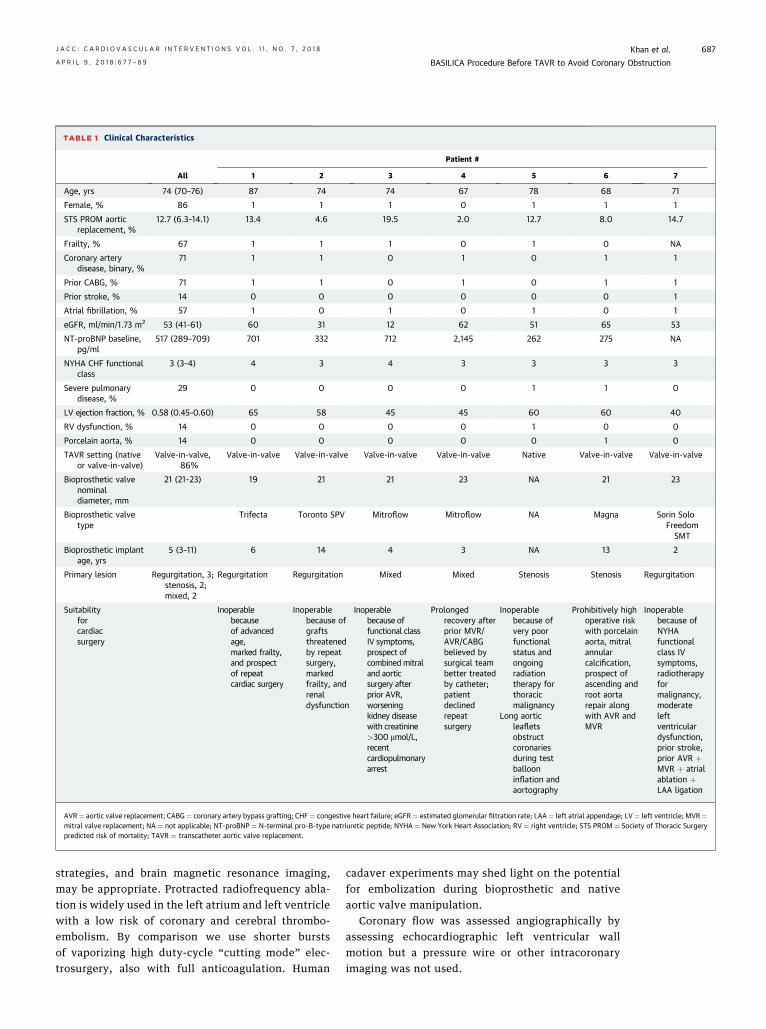
TABLE 1 Clinical Characteristics
All
Patient #
1 2 3 4 5 6 7
Age, yrs 74 (70–76) 87 74 74 67 78 68 71
Female, % 86 1 1 1 0 1 1 1
STS PROM aorticreplacement, %
12.7 (6.3–14.1) 13.4 4.6 19.5 2.0 12.7 8.0 14.7
Frailty, % 67 1 1 1 0 1 0 NA
Coronary arterydisease, binary, %
71 1 1 0 1 0 1 1
Prior CABG, % 71 1 1 0 1 0 1 1
Prior stroke, % 14 0 0 0 0 0 0 1
Atrial fibrillation, % 57 1 0 1 0 1 0 1
eGFR, ml/min/1.73 m2 53 (41–61) 60 31 12 62 51 65 53
NT-proBNP baseline,pg/ml
517 (289–709) 701 332 712 2,145 262 275 NA
NYHA CHF functionalclass
3 (3–4) 4 3 4 3 3 3 3
Severe pulmonarydisease, %
29 0 0 0 0 1 1 0
LV ejection fraction, % 0.58 (0.45–0.60) 65 58 45 45 60 60 40
RV dysfunction, % 14 0 0 0 0 1 0 0
Porcelain aorta, % 14 0 0 0 0 0 1 0
TAVR setting (nativeor valve-in-valve)
Valve-in-valve,86%
Valve-in-valve Valve-in-valve Valve-in-valve Valve-in-valve Native Valve-in-valve Valve-in-valve
Bioprosthetic valvenominaldiameter, mm
21 (21–23) 19 21 21 23 NA 21 23
Bioprosthetic valvetype
Trifecta Toronto SPV Mitroflow Mitroflow NA Magna Sorin SoloFreedomSMT
Bioprosthetic implantage, yrs
5 (3–11) 6 14 4 3 NA 13 2
Primary lesion Regurgitation, 3;stenosis, 2;mixed, 2
Regurgitation Regurgitation Mixed Mixed Stenosis Stenosis Regurgitation
Suitabilityforcardiacsurgery
Inoperablebecauseof advancedage,marked frailty,and prospectof repeatcardiac surgery
Inoperablebecause ofgraftsthreatenedby repeatsurgery,markedfrailty, andrenaldysfunction
Inoperablebecause offunctional classIV symptoms,prospect ofcombined mitraland aorticsurgery afterprior AVR,worseningkidney diseasewith creatinine>300 mmol/L,recentcardiopulmonaryarrest
Prolongedrecovery afterprior MVR/AVR/CABGbelieved bysurgical teambetter treatedby catheter;patientdeclinedrepeatsurgery
Inoperablebecause ofvery poorfunctionalstatus andongoingradiationtherapy forthoracicmalignancy
Long aorticleafletsobstructcoronariesduring testballooninflation andaortography
Prohibitively highoperative riskwith porcelainaorta, mitralannularcalcification,prospect ofascending androot aortarepair alongwith AVR andMVR
Inoperablebecause ofNYHAfunctionalclass IVsymptoms,radiotherapyformalignancy,moderateleftventriculardysfunction,prior stroke,prior AVR þMVR þ atrialablation þLAA ligation
AVR ¼ aortic valve replacement; CABG¼ coronary artery bypass grafting; CHF ¼ congestive heart failure; eGFR ¼ estimated glomerular filtration rate; LAA¼ left atrial appendage; LV ¼ left ventricle; MVR¼mitral valve replacement; NA ¼ not applicable; NT-proBNP ¼ N-terminal pro–B-type natriuretic peptide; NYHA ¼ New York Heart Association; RV ¼ right ventricle; STS PROM ¼ Society of Thoracic Surgerypredicted risk of mortality; TAVR ¼ transcatheter aortic valve replacement.
J A C C : C A R D I O V A S C U L A R I N T E R V E N T I O N S V O L . 1 1 , N O . 7 , 2 0 1 8 Khan et al.A P R I L 9 , 2 0 1 8 : 6 7 7 – 8 9 BASILICA Procedure Before TAVR to Avoid Coronary Obstruction
687
strategies, and brain magnetic resonance imaging,may be appropriate. Protracted radiofrequency abla-tion is widely used in the left atrium and left ventriclewith a low risk of coronary and cerebral thrombo-embolism. By comparison we use shorter burstsof vaporizing high duty-cycle “cutting mode” elec-trosurgery, also with full anticoagulation. Human
cadaver experiments may shed light on the potentialfor embolization during bioprosthetic and nativeaortic valve manipulation.
Coronary flow was assessed angiographically byassessing echocardiographic left ventricular wallmotion but a pressure wire or other intracoronaryimaging was not used.
TABLE 2 Procedure Characteristics and Hemodynamics
All
Patient #
1 2 3 4 5 6 7
Transcatheter heart valve Sapien 3, 6;Evolut Pro, 1
Sapien 3 Evolut Pro Sapien 3 Sapien 3 Sapien 3 Sapien 3 Sapien 3
Transcatheter heart valve size, mm 23 (22–23) 20 23 23 23 26 20 23
Transcatheter heart valvepost-dilatation
14% 0 0 0 0 0 1 0
Invasive hemodynamics baseline
Aortic regurgitation severity(0 ¼ none, 1 ¼ trace, 2 ¼ mild,3 ¼ moderate, 4 ¼ severe)
4 (3–4) 4 4 4 3 2 2 4
Aortic valve peak-to-peakgradient, mm Hg
43 (14–64) 12 8 43 72 56 135 15
HR 75 (71–80) 84 72 67 77 69 83 75
SBP 126 (96–148) 151 126 93 95 166 145 97
DBP 44 (39–50) 32 47 35 53 73 42 44
LVEDP 31 (22–34) 23 21 35 36 31 32 16
Invasive hemodynamics completion
Aortic regurgitation severity(0 ¼ none, 1 ¼ trace, 2 ¼ mild,3 ¼ moderate, 4 ¼ severe)
0 (0–1) 0 0 1 0 0 1 2
Aortic valve peak-to-peakgradient, mm Hg
1 (1–7) 1 10 1 12 0 0 3
HR 81 (79–84) 80 82 85 79 62 87 40 (sinus brady,paced at 80)
SBP 175 (151–179) 177 151 175 120 181 197 150
DBP 68 (64–72) 64 63 79 68 72 57 71
LVEDP 27 (26–30) 34 28 26 26 27 18 31
Echocardiography, baseline
Aortic regurgitation severity(0 ¼ none, 1 ¼ trace, 2 ¼ mild,3 ¼ moderate, 4 ¼ severe)
4 (3–4) 4 4 3.5 4 3 2 4
Aortic valve peak velocity, m/s 3.4 (3.2–4.6) 3.3 3.1 5.6 4.1 3.4 5.0 1.6
Aortic valve mean gradient, mm Hg 24 (22–48) 24.0 22.0 67.0 22.6 45.4 51.0 4.8
Indexed effective orifice area, m2/m2 0.62 (0.49–1.00) 1.0 1.6 0.48 0.31 0.49 0.62 1.0
LVEF, % 58 (45–60) 65 58 45 45 60 60 35
Echocardiography, pre-discharge
Aortic regurgitation severity(0 ¼ none, 1 ¼ trace, 2 ¼ mild,3 ¼ moderate, 4 ¼ severe)
0 (0–0) 0 0 1 0 0 0 0
Aortic valve peak velocity, m/s 2.9 (2.7–3.2) 3.3 2.7 3.6 2.6 3.1 2.9 1.6
Aortic valve mean gradient, mm Hg 18 (17–21) 17.0 16.0 28.2 17.6 21.0 20.0 4.8
LVEF, % 61 (56–65) 71 64 61 51 60 65 35
DBP ¼ diastolic blood pressure; HR ¼ heart rate; LVEDP ¼ left ventricular end diastolic pressure; LVEF ¼ left ventricular ejection fraction; SBP ¼ systolic blood pressure.
TABLE 3 Clinical Outcomes
All
Patient #
1 2 3 4 5 6 7
Length of stay after TAVR, days 4 (4–5) 4 4 6 1 5 5 3
ICU stay, days 1 (0–2) 2 2 0 0 1 1 0
Survival to hospital discharge 100% 1 1 1 1 1 1 1
Survival 30 days 100% 1 1 1 1 1 1 1
Survival ascertainment, days 116 (109–153) 154 154 151 116 109 109 95
NYHA functional class at latest follow-up 2.0 (1.5–2.0) 2 2 1 2 1 2 2
Values are median (interquartile range) or %. ICU ¼ intensive care unit; other abbreviations as in Table 1.
Khan et al. J A C C : C A R D I O V A S C U L A R I N T E R V E N T I O N S V O L . 1 1 , N O . 7 , 2 0 1 8
BASILICA Procedure Before TAVR to Avoid Coronary Obstruction A P R I L 9 , 2 0 1 8 : 6 7 7 – 8 9
688
Finally, there was no comparator and so coronaryartery obstruction was not certain but predicted usingprevailing standards, which have their limitations.The potential risk and benefit of BASILICA should beweighed before applying it to any patient, includingthe risk of embolization and, in patients with severeventricular dysfunction, the risk of acute severeaortic regurgitation.
We believe technical descriptions are no substitutefor live observation, and we recommend BASILICAonly be undertaken with appropriate training.
PERSPECTIVES
WHAT IS KNOWN? Coronary obstruction following TAVR
carries up to 50% mortality, and CT-predicted coronary
obstruction may deprive patients of TAVR as a therapeutic
option. Current methods of pre-emptive or bail-out coronary
stenting are suboptimal.
WHAT IS NEW? We describe a catheter technique (BASILICA)
to lacerate aortic leaflets that otherwise threaten to obstruct a
coronary artery during TAVR. After TAVR, which is performed
immediately after BASILICA, blood is able to flow across the
lacerated aortic leaflets into the coronary arteries.
WHAT IS NEXT? BASILICA may have value in the future as
more patients have bioprosthetic surgical and even transcatheter
aortic valves likely to degenerate. BASILICA warrants further
prospective evaluation in a larger number of patients.
J A C C : C A R D I O V A S C U L A R I N T E R V E N T I O N S V O L . 1 1 , N O . 7 , 2 0 1 8 Khan et al.A P R I L 9 , 2 0 1 8 : 6 7 7 – 8 9 BASILICA Procedure Before TAVR to Avoid Coronary Obstruction
689
CONCLUSIONS
Bioprosthetic and native aortic leaflet lacerationseems feasible and may reduce the risk of coronaryartery obstruction following TAVR in patients at highrisk. No patient had a drop in blood pressurefollowing BASILICA. The technique offers a promisingalternative to “chimney” stenting to provide durableprevention against coronary obstruction from TAVR.BASILICA needs careful prospective investigation,which begins with a Food and Drug Administration–approved trial in early 2018.
ACKNOWLEDGMENTS The authors thank AlanHoofring for medical illustrations; KatherineLucas for animal care; Daniel Herzka, Merdim Son-mez, and Dmitri Levin for technical assistance; Nor-ihiko Kamioka, Lauren Wheeler, and Patricia Keeganfor data assistance; and Elena Grant and JamesMcCabe for thoughtful advice. They thank RichardOlson of Abbott for supplying a sample Trifecta valvein anticipation of helping the first patient.
ADDRESS FOR CORRESPONDENCE: Dr. Robert J.Lederman, Cardiovascular Branch, Division of
Intramural Research, National Heart, Lung, and BloodInstitute, National Institutes of Health, Building 10,Room 2c713, MSC 1538, Bethesda, Maryland 20892-1538. E-mail: [email protected].
RE F E RENCE S
1. Leon MB, Smith CR, Mack MJ, et al. Trans-catheter or surgical aortic-valve replacement inintermediate-risk patients. N Engl J Med 2016;374:1609–20.
2. Reardon MJ, Van Mieghem NM, Popma JJ, et al.Surgical or transcatheter aortic-valve replacementin intermediate-risk patients. N Engl J Med 2017;376:1321–31.
3. Dvir D, Webb JG, Bleiziffer S, et al. Trans-catheter aortic valve implantation in failed bio-prosthetic surgical valves. JAMA 2014;312:162–70.
4. Webb JG, Mack MJ, White JM, et al. Trans-catheter aortic valve implantation within degen-erated aortic surgical bioprostheses: PARTNER 2Valve-in-Valve Registry. J Am Coll Cardiol 2017;69:2253–62.
5. Ribeiro HB, Rodés-Cabau J, Blanke P, et al.Incidence, predictors and clinical outcomes ofcoronary obstruction following transcatheteraortic valve replacement for degenerative bio-prosthetic surgical valves: insights from the VIVIDRegistry. Eur Heart J 2018;39:687–95.
6. Ribeiro HB, Webb JG, Makkar RR, et al. Pre-dictive factors, management, and clinical out-comes of coronary obstruction followingtranscatheter aortic valve implantation: insightsfrom a large multicenter registry. J Am Coll Cardiol2013;62:1552–62.
7. Yamamoto M, Shimura T, Kano S, et al. Impactof preparatory coronary protection in patients athigh anatomical risk of acute coronary obstructionduring transcatheter aortic valve implantation. IntJ Cardiol 2016;217:58–63.
8. Abramowitz Y, Chakravarty T, Jilaihawi H, et al.Clinical impact of coronary protection duringtranscatheter aortic valve implantation: first re-ported series of patients. EuroIntervention 2015;11:572–81.
9. Khan JM, Rogers T, Schenke WH, et al. Inten-tional laceration of the anterior mitral valve leafletto prevent left ventricular outflow tract obstruc-tion during transcatheter mitral valve replace-ment: pre-clinical findings. J Am Coll Cardiol Intv2016;9:1835–43.
10. Babaliaros VC, Greenbaum AB, Khan JM, et al.Intentional percutaneous laceration of the anteriormitral leaflet to prevent outflow obstruction dur-ing transcatheter mitral valve replacement: first-in-human experience. J Am Coll Cardiol Intv2017;10:798–809.
11. Chhatriwalla AK, Allen KB, Saxon JT, et al. Bio-prosthetic valve fracture improves the hemodynamicresults of valve-in-valve transcatheter aortic valvereplacement. Circ Cardiovasc Interv 2017;10.
12. Kappetein AP, Head SJ, Genereux P, et al.Updated standardized endpoint definitions for
transcatheter aortic valve implantation: the ValveAcademic Research Consortium-2 consensusdocument. J Am Coll Cardiol 2012;60:1438–54.
13. Dvir D, Webb J, Brecker S, et al. Transcatheteraortic valve replacement for degenerative bio-prosthetic surgical valves: results from the globalvalve-in-valve registry. Circulation 2012;126:2335–44.
14. Dvir D. Transcatheter aortic valve-in-valveimplantations: lessons from bench to bedside[oral presentation]. Presented at: TranscatheterValve Therapies; June 14 to 17, 2017; Chicago, IL.
15. Valsecchi O, Vassileva A. The chimney tech-nique during valve in valve TAVR in stentlessfreestyle [oral presentation]. Presented at: JointInterventional Meeting Milan (JIM); February 9 to11, 2017; Milan, Italy.
KEY WORDS bioprosthetic heart valvefailure, coronary artery obstruction,structural heart disease, transcatheter aorticvalve replacement, transcatheterelectrosurgery
APPENDIX For supplemental tables,figures, and a video, please see the onlineversion of this paper.