Embed Size (px)
Citation preview
Proc. Natl. Acad. Sci. USAVol. 90, pp. 3216-3220, April 1993Cell Biology
Transactivation of gene expression by Myc is inhibited by mutationat the phosphorylation sites Thr-58 and Ser-62SHASHI GUPTA*, ALPNA SETH*, AND ROGER J. DAVIS*ttHoward Hughes Medical Institute, *Program in Molecular Medicine, and the Department of Biochemistry and Molecular Biology, University ofMassachusetts Medical School, Worcester, MA 01605
Communicated by Leon Heppel, January 4, 1993
ABSTRACT The product of the human c-myc protoonco-gene (Myc) is a sequence-specific DNA binding protein. Here,we demonstrate that the placement of the specific Myc DNAbinding site CACGTG upstream of a luciferase reporter geneconferred Myc-stimulated expression that was inhibited by theoverexpression of the basic-helix4oop-helix/leucine zipperprotein Max. It was observed that Myc was phosphorylated invivo within the NH2-terminal domain at Thr-58 and Ser-62.Replacement of these phosphorylation sites with Ala residuescaused a marked decrease in Myc-stimulated reporter geneexpression. In contrast, the replacement of Thr-58 or Ser-62with an acidic residue (Glu) caused only a small inhibition oftransactivation. Together, these data demonstrate that theNH2-terminal phosphorylation sites Thr-58 and Ser-62 arerequired for high levels of transactivation of gene expression byMyc.
It has been established that the product of the c-myc gene(Myc) is a short-lived nuclear phosphoprotein that has animportant functional role during cellular proliferation, differ-entiation, and neoplasia (1). The structural organization ofMyc indicates the presence of subdomains that are found inseveral transcriptional regulators. A transcriptional activa-tion region is located within the NH2-terminal domain (2), andbasic-helix-loop-helix (bHLH) and leucine zipper (Zip) mo-tifs (associated with dimerization and DNA binding activi-ties) are found within the COOH-terminal domain ofMyc (1).Furthermore, a specific DNA binding site for Myc has beenidentified as the E-box sequence CACGTG (3-6). These datasuggest that Myc may function as a sequence-specific regu-lator of gene expression (7).The presence of dimerization motifs (HLH and Zip) in Myc
suggests that this protein may function as a homo- or hetero-oligomer. A functional assay has failed to detect Myc homo-oligomers in intact cells (8), but evidence for the formation ofheterodimeric complexes ofMyc with the bHLH/Zip proteinMax has been obtained (9). The overall structure of Max issimilar to Myc, except that Max lacks the transcriptionalactivation domain that is present within the NH2-terminalregion of Myc (9-11). Significantly, the formation of Myc-Max heterodimeric complexes increases the specific bindingofMyc to the DNA sequence CACGTG (9-13). It is thereforelikely that the Myc-Max complex represents the functionalform of Myc in vivo (1).The Max protein has a long half-life and is expressed
constitutively (9, 11). In contrast, the expression of Myc isregulated at both transcriptional and posttranscriptional lev-els. An increase in Myc expression is observed during thegrowth factor-stimulated transition of growth-arrested cellsfrom the Go to the G1 phase of the cell cycle (14). However,in proliferating cells, the level of Myc expression is invariantduring the cell cycle (15). Therefore, Myc activity may be
The publication costs of this article were defrayed in part by page chargepayment. This article must therefore be hereby marked "advertisement"in accordance with 18 U.S.C. §1734 solely to indicate this fact.
regulated by posttranslational modification in growing cells.The activity of many transcription factors is regulated byphosphorylation. For example, phosphorylation regulatesthe DNA binding activity of Myb, Max, SRF, Jun, E2F, andE4F (16). Phosphorylation also regulates the transactivationof gene expression by cAMP response element bindingprotein (CREB) (17) and Jun (18-20). As Myc is a phospho-protein (21), it is therefore possible that Myc function is alsoregulated by phosphorylation.
Recently, we have demonstrated that Myc is an in vitrosubstrate for phosphorylation by mitogen-activated proteinkinase (MAP kinases) at Ser-62 (22). This phosphorylationsite is located within a proline-rich region of the transacti-vation domain that is conserved in the Myc family. Thelocation of Ser-62 within the transcriptional activation do-main of Myc suggests that it may be a regulatory site ofphosphorylation. Evidence consistent with this hypothesiswas obtained in experiments using an artificial GAL4-Mycfusion protein (23). Thus, phosphorylation may be an impor-tant mechanism of regulation of Myc function in vivo. Ex-periments reported here test this hypothesis. We show thatSer-62 is a site ofphosphorylation ofthe Myc protein in intactcells. An additional site of in vivo phosphorylation of Mycwas identified as Thr-58. Mutational analysis demonstratedthat the replacement of Thr-58 and Ser-62 with Ala caused amarked decrease in the Myc-dependent transactivation ofgene expression. These data demonstrate that the function ofthe Myc NH2-terminal transactivation domain may be regu-lated in vivo by phosphorylation at Thr-58 and Ser-62.
MATERIALS AND METHODSPlasmids. The plasmids pCH110 and pUC13 were from
Pharmacia LKB. The plasmid pGEM-Luc was fromPromega. The expression vectors pCMV2 and pCMV5 (24)were from D. Russell (University of Texas SouthwesternMedical School). The plasmid pM21 containing the humanc-myc gene (25) was obtained from C. V. Dang (JohnsHopkins University School of Medicine). The followingplasmids were constructed:
(i) Point mutations were introduced at codons Thr-58 andSer-62 by using PCR and the human c-myc gene (26). The526-bp Dra III-BstEII restriction fragments (containing thepoint mutations) were cloned into pM21 at the Dra III andBstEII sites. A simian virus 40 origin ofDNA replication wascloned into these plasmids by replacing the murine leukemiavirus long terminal repeat of pM21 (BamHI fragment) withthe simian virus 40 promoter and origin of replication frompCMV2 (1064-bp BamHI fragment) to create pMyc.
(ii) The plasmid pG5ElbLuc was prepared frompG5E1bCAT (23) by replacing the chloramphenicol acetyl-transferase gene (EcoRI and Sty I fragment) with the firefly
Abbreviations: bHLH, basic-helix-loop-helix; Zip, leucine zipper;CREB, cAMP response element binding protein; MAP kinase,mitogen-activated protein kinase.
3216
Proc. Natl. Acad. Sci. USA 90 (1993) 3217
luciferase gene (from pGEM-Luc) as a 1745-bp HindIII-StuI blunt-end fragment. The GAL4 binding sites were removedfrom pG5ElbLuc as a Pst I and Kpn I fragment and replacedwith a double-stranded 20-mer oligonucleotide that containsthe Elb promoter TATA element (5'-GAGGGTATATAAT-GTGGTAC-3' and 5'-CACATTATATACCCTCTGCA-3') tocreate the plasmid pMycOElbLuc. Specific Myc binding siteswere inserted in pMycOElbLuc as double-stranded oligonu-cleotides at the HindIII site. To prepare plasmids with one,two, three, and four Myc binding sites (underlined), a 26-merdouble-stranded oligonucleotide (5'-AGCTTAACTGAC-CACGTGGTCAACTA-3' and 5'-AGCTTAGTTGAC-CACGTGGTCAGTTA-3') was employed in a ligation reac-tion to obtain the plasmids pMyclElbLuc, pMyc2ElbLuc,pMyc3ElbLuc, and pMyc4ElbLuc.
(iii) The cDNAs encoding p21 Max and p22 Max (10) wereprovided by E. Blackwood and R. N. Eisenman (FredHutchinson Cancer Center, Seattle). These cDNAs werecloned in the polylinker of the pCMV5 expression vector asa HindIII-Sal I fragment to create the plasmids pCMV-p2lMax and pCMV-p22Max.
Cell Culture and Transient Expression Assays. COS-7 andCV-1 monkey cells were maintained in Dulbecco's modifiedEagle's medium supplemented with 10% fetal calf serum
(GIBCO/BRL). Transfections of COS-7 cells were per-formed by using the DEAE-dextran method (22) with 2 ug ofexpression vector (pMyc), 2 jug of control /3-galactosidaseexpression vector (pCH110), and 2 ,g of pUC13 carrierDNA. The cells were harvested after 48 hr of incubation at37°C. CV-1 cells were cotransfected using the calcium phos-phate method (23) with 2 ,ug of a luciferase reporter plasmid,0-15 ,g of the Myc expression vector, and 3 ,ug of a controlplasmid (pCH110). The total DNA was maintained at 20 ,gusing pUC13 as carrier DNA. In experiments using the Maxexpression vector, 2 ,ug of pCMV-Max (or 2 ,ug of pUC13carrier DNA) was included in the transfection (a total of 22,ug ofDNA). The cells were harvested 36 hr posttransfection,and cell extracts were prepared for measurement ofluciferaseand S3-galactosidase activity (27).
Analysis of Metabolically Labeled Proteins. Metabolic la-beling oftransfected cells in 100-mm dishes was performed 40hr posttransfection by transferring the cells into culturemedium (Flow Laboratories) supplemented with 1% fetal calfserum. The culture media used were (i) methionine-freemodified Eagle's medium containing [35S]methionine (Amer-sham) at 200 ,uCi/ml (1 Ci = 37 GBq) or (ii) phosphate-freemodified Eagle's medium containing [32P]phosphate (Du-Pont/NEN) at 2 mCi/ml. After 3 hr of additional incubation,the cells were lysed in 0.01 M Tris (pH 7.5), 0.05 M NaCl,0.5% (vol/vol) Nonidet P-40, 0.5% (wt/vol) SDS, 0.5%(wt/vol) sodium deoxycholate, 1 mM phenylmethylsulfonylfluoride, and leupeptin at 10 ,g/ml. The Myc proteins were
immunoprecipitated with 10 ug of sheep anti-Myc IgG pre-
bound to Pansorbin (Calbiochem) and resolved by SDS/PAGE. The sheep anti-Myc IgG was purified from serum thatwas obtained after immunization with a bacterially expressedhuman Myc protein (residues 2-243) as an antigen (22).Phosphopeptide mapping was performed by two-dimensionalseparation on 100-,um cellulose thin-layer plates (Machery &Nagel) as described (22). Phosphoamino acid analysis was
performed as described (22).Immunocytochemistry. Laser scanning confocal micros-
copy of cells stained with anti-Myc monoclonal antibody(Ab-2; 5 ,g/ml; Oncogene Sciences, Mineola, NY) andfluoroscein isothiocyanate-conjugated goat anti-mouse IgGantibody (ICN) was performed as described by Seth et al.(27).
RESULTS
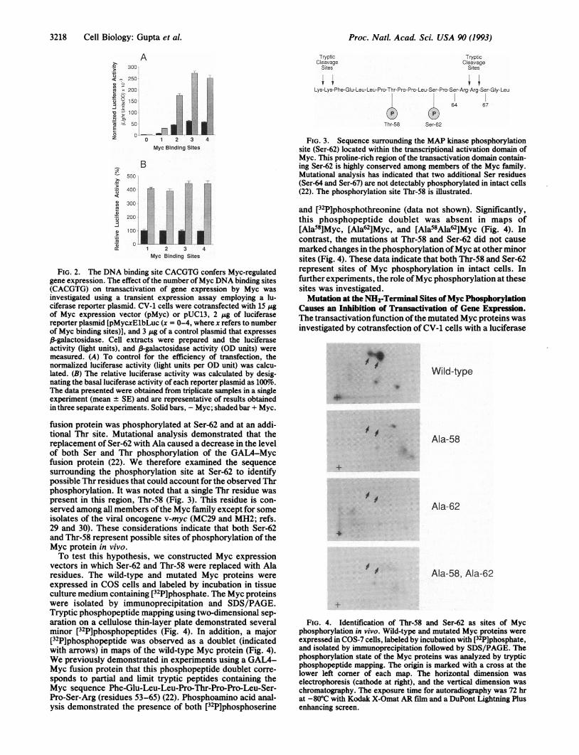
The DNA Sequence CACGTG Confers Myc Regulation ofGene Expression. To investigate the transactivation of geneexpression by Myc, we used a reporter plasmid containingspecific Myc DNA binding sites. The experimental strategythat we employed is illustrated schematically in Fig. 1. Aplasmid containing the firefly luciferase gene and a minimalElb promoter (TATA) element was constructed. Transfec-tion of this plasmid into CV-1 cells caused a very low level ofluciferase activity (Fig. 2). However, the insertion ofthe Mycbinding site CACGTG into this plasmid (Fig. 1) resulted in theobservation of Myc-stimulated luciferase expression (Fig. 2).Examination of the dose-response indicated that luciferaseexpression was increased in assays using 2 ,ug of the Mycexpression vector and that 15 ,ug caused the maximumobserved increase (data not shown). The level of Myc-stimulated luciferase activity was increased when multiplebinding sites were inserted into the reporter plasmid (Fig.2A). However, the basal luciferase activity was also in-creased when a larger number of Myc binding sites werepresent in the reporter plasmid. [The increased basal activityis likely to be caused by endogenous E-box binding proteins(including Myc) present in CV-1 cells.] The fold increase inluciferase activity caused by Myc was similar when differentnumbers ofbinding sites (from one to four) were inserted intothe reporter plasmid (Fig. 2B). Several genes have beenidentified as putative targets for transactivation by Myc thatcontain only a single Myc binding site (c-sis, mouse HI9, andU3B RNA gene; ref. 4). Therefore, in further experiments,we used a reporter plasmid containing one Myc binding siteto study Myc-stimulated gene expression.The active form ofMyc has been proposed to be a complex
with Max (1). We therefore investigated the effect of theexpression of exogenous Max on the Myc-stimulated lu-ciferase expression in CV-1 cells. Expression of p21 Maxcaused an inhibition of the Myc-stimulated gene expression(data not shown). An alternatively spliced variant of p21Max, p22 Max (10), also caused an inhibition of Myc-stimulated luciferase activity (data not shown). An inhibitionof a different function of Myc (rat embryo fibroblast trans-formation) caused by the expression of exogenous Max haspreviously been reported (13,- 27). It is likely that the ob-served inhibition of Myc function is the result of Maxhomodimers that compete with Myc complexes for binding tothe DNA sequence CACGTG (1). Thus, the level of endog-enous Max (9, 11) is likely to be sufficient to form complexeswith Myc introduced into CV-1 cells (1). Therefore, in furtherexperiments, we examined the transactivation function ofMyc without expressing exogenous Max.Myc Is Phosphorylated at Thr-58 and Ser-62 in Vivo. In
previous studies we established that the Myc protein isphosphorylated in vitro at Ser-62 byMAP kinases (22, 23, 28).Phosphorylation of Myc at this site in vivo has not beenreported. However, we have investigated the phosphoryla-tion ofthe Myc transactivation domain in intact cells by usinga GAL4-Myc fusion protein (22). Phosphoamino acid anal-ysis and phosphopeptide mapping demonstrated that this
c-Myc Elb Luciferase poly ABinding PromoterSites
FIG. 1. Schematic illustration of the Myc transactivation assay.The structure of the Myc reporter plasmids [pMycxElbLuc (x =1-4), where x refers to the number of Myc binding sites] employedfor transient transfection assays are presented. The Myc binding sites(CACGTG) are located upstream ofa minimal Elb promoter (TATA)element and the firefly luciferase gene.
Cell Biology: Gupta et al.
Proc. Natl. Acad. Sci. USA 90 (1993)
A- 300
< , 250
2 200
3i 9 1503 -
0 D 100
F z 50-
o 0-z
-Rec
I 1 2 3 4Myc Binding Sites
B500
400
300
200
100
I
1 2 3 4Myc Binding Sites
FIG. 2. The DNA binding site CACGTG confers Myc-regulatedgene expression. The effect of the number ofMyc DNA binding sites(CACGTG) on transactivation of gene expression by Myc wasinvestigated using a transient expression assay employing a lu-ciferase reporter plasmid. CV-1 cells were cotransfected with 15 'Ugof Myc expression vector (pMyc) or pUC13, 2 ,ug of luciferasereporter plasmid [pMycxElbLuc (x = 0-4, where x refers to numberof Myc binding sites)], and 3 Mg of a control plasmid that expresses,B-galactosidase. Cell extracts were prepared and the luciferaseactivity (light units), and 3-galactosidase activity (OD units) weremeasured. (A) To control for the efficiency of transfection, thenormalized luciferase activity (light units per OD unit) was calcu-lated. (B) The relative luciferase activity was calculated by desig-nating the basal luciferase activity of each reporter plasmid as loo10.The data presented were obtained from triplicate samples in a singleexperiment (mean ± SE) and are representative of results obtainedin three separate experiments. Solid bars, - Myc; shaded bar + Myc.
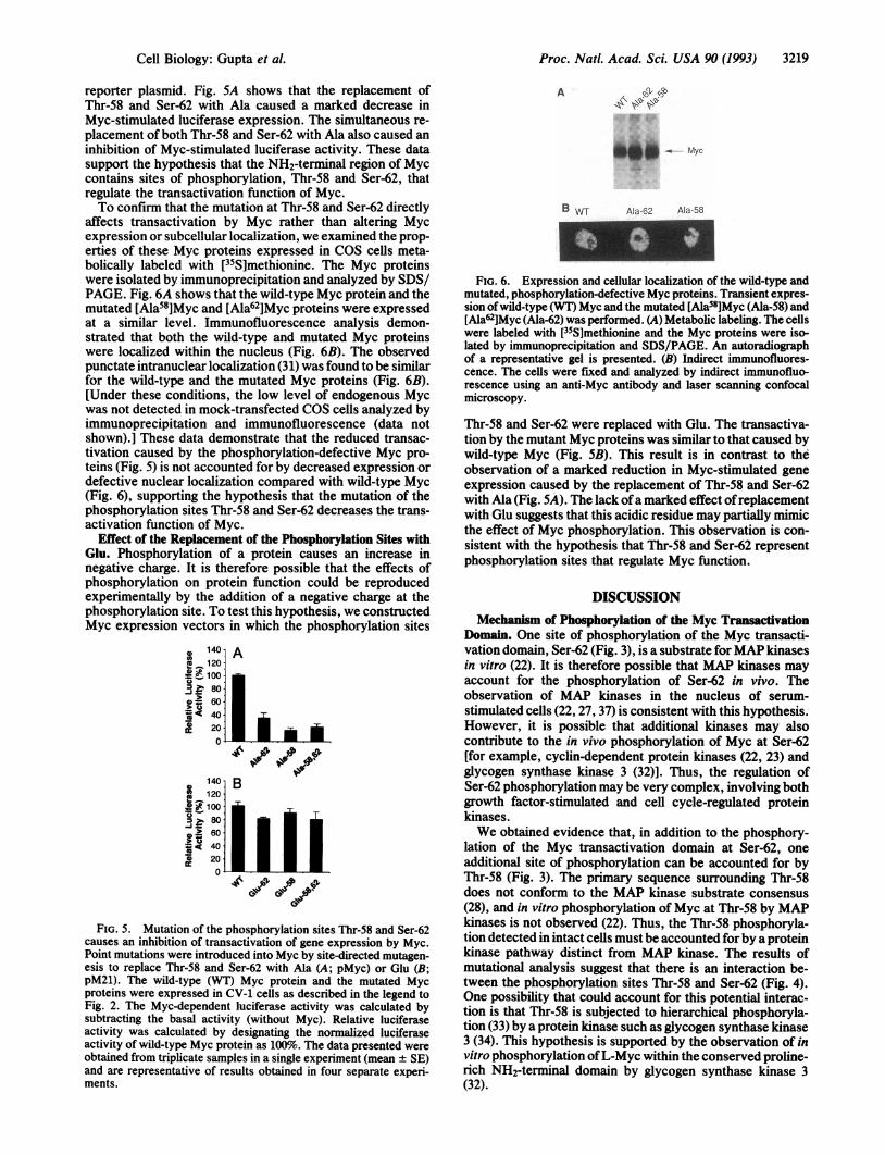
fusion protein was phosphorylated at Ser-62 and at an addi-tional Thr site. Mutational analysis demonstrated that thereplacement of Ser-62 with Ala caused a decrease in the levelof both Ser and Thr phosphorylation of the GAL4-Mycfusion protein (22). We therefore examined the sequencesurrounding the phosphorylation site at Ser-62 to identifypossible Thr residues that could account for the observed Thrphosphorylation. It was noted that a single Thr residue waspresent in this region, Thr-58 (Fig. 3). This residue is con-served among all members ofthe Myc family except for someisolates of the viral oncogene v-myc (MC29 and MH2; refs.29 and 30). These considerations indicate that both Ser-62and Thr-58 represent possible sites of phosphorylation of theMyc protein in vivo.To test this hypothesis, we constructed Myc expression
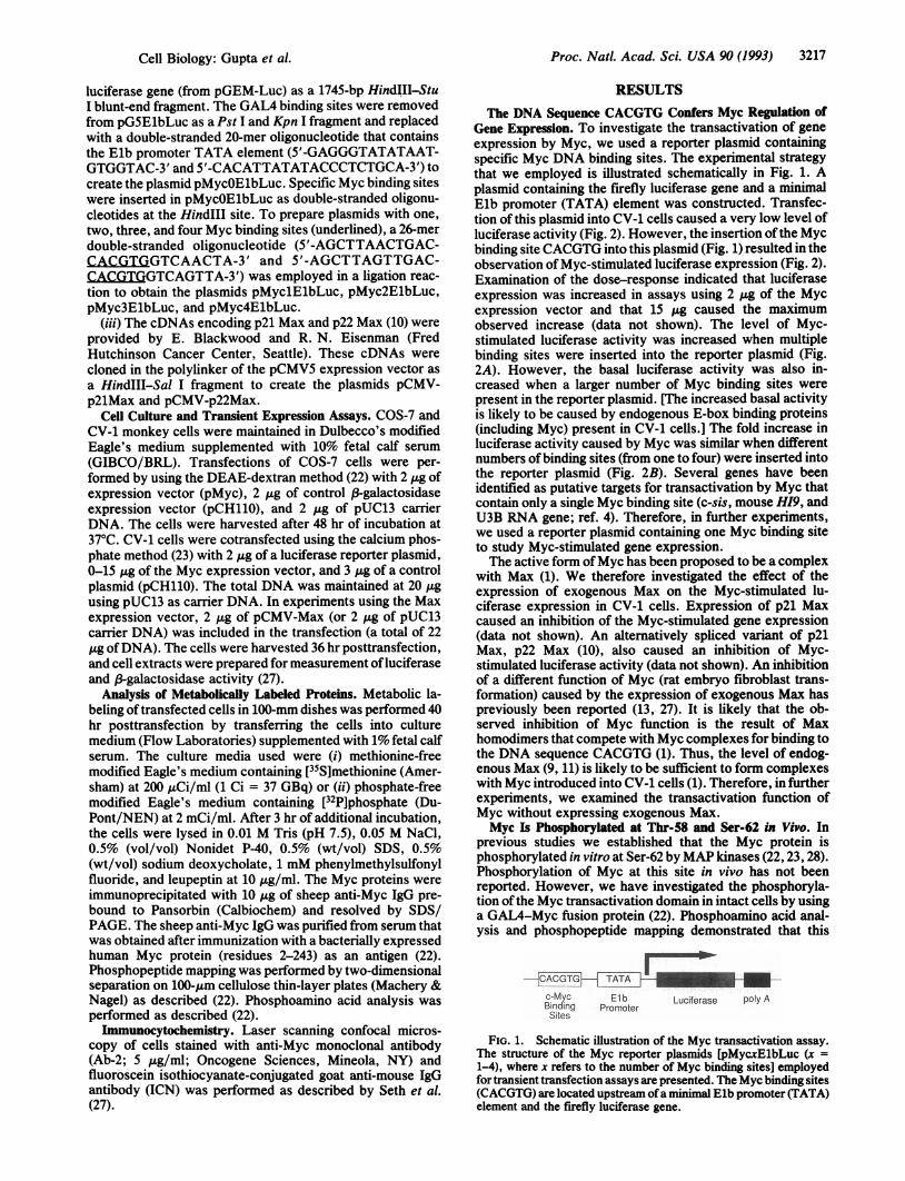
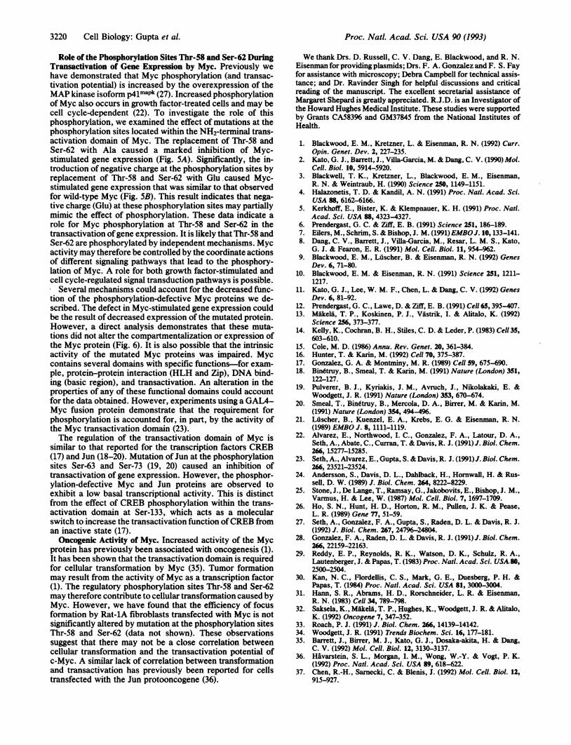
vectors in which Ser-62 and Thr-58 were replaced with Alaresidues. The wild-type and mutated Myc proteins wereexpressed in COS cells and labeled by incubation in tissueculture medium containing [32P]phosphate. The Myc proteinswere isolated by immunoprecipitation and SDS/PAGE.Tryptic phosphopeptide mapping using two-dimensional sep-aration on a cellulose thin-layer plate demonstrated severalminor [32P]phosphopeptides (Fig. 4). In addition, a major[32P]phosphopeptide was observed as a doublet (indicatedwith arrows) in maps of the wild-type Myc protein (Fig. 4).We previously demonstrated in experiments using a GAL4-Myc fusion protein that this phosphopeptide doublet corre-sponds to partial and limit tryptic peptides containing theMyc sequence Phe-Glu-Leu-Leu-Pro-Thr-Pro-Pro-Leu-Ser-Pro-Ser-Arg (residues 53-65) (22). Phosphoamino acid anal-ysis demonstrated the presence of both [32P]phosphoserine
Tryptic TrypticCleavage Cleavage
Sites Sites
Lys-Lys- Phe-GI u-Leu- Leu-Pro-Th r-Pro-Pro-Leu-Ser- Pro-Ser-Arg-Arg-Ser-GIy-Leu
64 67
Thr-58 Ser-62
FIG. 3. Sequence surrounding the MAP kinase phosphorylationsite (Ser-62) located within the transcriptional activation domain ofMyc. This proline-rich region of the transactivation domain contain-ing Ser-62 is highly conserved among members of the Myc family.Mutational analysis has indicated that two additional Ser residues(Ser-64 and Ser-67) are not detectably phosphorylated in intact cells(22). The phosphorylation site Thr-58 is illustrated.
and [32P]phosphothreonine (data not shown). Significantly,this phosphopeptide doublet was absent in maps of[Ala58]Myc, [Ala62]Myc, and [Ala58Ala62]Myc (Fig. 4). Incontrast, the mutations at Thr-58 and Ser-62 did not causemarked changes in the phosphorylation ofMyc at other minorsites (Fig. 4). These data indicate that both Thr-58 and Ser-62represent sites of Myc phosphorylation in intact cells. Infurther experiments, the role ofMyc phosphorylation at thesesites was investigated.Mutation at the NH2-Terminal Sites ofMyc Phosphorylation
Causes an Inhibition of Transactivation of Gene Expression.The transactivation function ofthe mutated Myc proteins wasinvestigated by cotransfection of CV-1 cells with a luciferase
*9 Wild-type*
Ala-58
+
.1
1,1
Ala-62
Ala-58, Ala-62
FIG. 4. Identification of Thr-58 and Ser-62 as sites of Mycphosphorylation in vivo. Wild-type and mutated Myc proteins wereexpressed in COS-7 cells, labeled by incubation with [32P]phosphate,and isolated by immunoprecipitation followed by SDS/PAGE. Thephosphorylation state of the Myc proteins was analyzed by trypticphosphopeptide mapping. The origin is marked with a cross at thelower left corner of each map. The horizontal dimension waselectrophoresis (cathode at right), and the vertical dimension waschromatography. The exposure time for autoradiography was 72 hrat -80°C with Kodak X-Omat AR film and a DuPont Lightning Plusenhancing screen.
3218 Cell Biology: Gupta et al.
v
L
Proc. Natl. Acad. Sci. USA 90 (1993) 3219
reporter plasmid. Fig. 5A shows that the replacement ofThr-58 and Ser-62 with Ala caused a marked decrease inMyc-stimulated luciferase expression. The simultaneous re-placement of both Thr-58 and Ser-62 with Ala also caused aninhibition of Myc-stimulated luciferase activity. These datasupport the hypothesis that the NH2-terminal region of Myccontains sites of phosphorylation, Thr-58 and Ser-62, thatregulate the transactivation function of Myc.To confirm that the mutation at Thr-58 and Ser-62 directly
affects transactivation by Myc rather than altering Mycexpression or subcellular localization, we examined the prop-erties of these Myc proteins expressed in COS cells meta-bolically labeled with [35S]methionine. The Myc proteinswere isolated by immunoprecipitation and analyzed by SDS/PAGE. Fig. 6A shows that the wild-type Myc protein and themutated [Ala58]Myc and [Ala6iMyc proteins were expressedat a similar level. Immunofluorescence analysis demon-strated that both the wild-type and mutated Myc proteinswere localized within the nucleus (Fig. 6B). The observedpunctate intranuclear localization (31) was found to be similarfor the wild-type and the mutated Myc proteins (Fig. 6B).[Under these conditions, the low level of endogenous Mycwas not detected in mock-transfected COS cells analyzed byimmunoprecipitation and immunofluorescence (data notshown).] These data demonstrate that the reduced transac-tivation caused by the phosphorylation-defective Myc pro-teins (Fig. 5) is not accounted for by decreased expression ordefective nuclear localization compared with wild-type Myc(Fig. 6), supporting the hypothesis that the mutation of thephosphorylation sites Thr-58 and Ser-62 decreases the trans-activation function of Myc.
Effect of the Replacement of the Phosphorylation Sites withGlu. Phosphorylation of a protein causes an increase innegative charge. It is therefore possible that the effects ofphosphorylation on protein function could be reproducedexperimentally by the addition of a negative charge at thephosphorylation site. To test this hypothesis, we constructedMyc expression vectors in which the phosphorylation sites
FIG. 5. Mutation of the phosphorylation sites Thr-58 and Ser-62causes an inhibition of transactivation of gene expression by Myc.Point mutations were introduced into Myc by site-directed mutagen-esis to replace Thr-58 and Ser-62 with Ala (A; pMyc) or Glu (B;pM21). The wild-type (WT) Myc protein and the mutated Mycproteins were expressed in CV-1 cells as described in the legend toFig. 2. The Myc-dependent luciferase activity was calculated bysubtracting the basal activity (without Myc). Relative luciferaseactivity was calculated by designating the normalized luciferaseactivity of wild-type Myc protein as 100%o. The data presented were
obtained from triplicate samples in a single experiment (mean ± SE)and are representative of results obtained in four separate experi-ments.
A > Fb
C{iof? :..
*,
B WT Ala-62 Ala-58
FIG. 6. Expression and cellular localization of the wild-type andmutated, phosphorylation-defective Myc proteins. Transient expres-sion ofwild-type (WT) Myc and the mutated [Ala58]Myc (Ala-58) and[Ala62]Myc (Ala-62) was performed. (A) Metabolic labeling. The cellswere labeled with [35S]methionine and the Myc proteins were iso-lated by immunoprecipitation and SDS/PAGE. An autoradiographof a representative gel is presented. (B) Indirect immunofluores-cence. The cells were fixed and analyzed by indirect immunofluo-rescence using an anti-Myc antibody and laser scanning confocalmicroscopy.
Thr-58 and Ser-62 were replaced with Glu. The transactiva-tion by the mutant Myc proteins was similar to that caused bywild-type Myc (Fig. SB). This result is in contrast to theobservation of a marked reduction in Myc-stimulated geneexpression caused by the replacement of Thr-58 and Ser-62with Ala (Fig. 5A). The lack ofa marked effect ofreplacementwith Glu suggests that this acidic residue may partially mimicthe effect of Myc phosphorylation. This observation is con-sistent with the hypothesis that Thr-58 and Ser-62 representphosphorylation sites that regulate Myc function.
DISCUSSIONMechanism of Phosphorylation of the Myc Transactivation
Domain. One site of phosphorylation of the Myc transacti-vation domain, Ser-62 (Fig. 3), is a substrate forMAP kinasesin vitro (22). It is therefore possible that MAP kinases mayaccount for the phosphorylation of Ser-62 in vivo. Theobservation of MAP kinases in the nucleus of serum-stimulated cells (22, 27, 37) is consistent with this hypothesis.However, it is possible that additional kinases may alsocontribute to the in vivo phosphorylation of Myc at Ser-62[for example, cycin-dependent protein kinases (22, 23) andglycogen synthase kinase 3 (32)]. Thus, the regulation ofSer-62 phosphorylation may be very complex, involving bothgrowth factor-stimulated and cell cycle-regulated proteinkinases.We obtained evidence that, in addition to the phosphory-
lation of the Myc transactivation domain at Ser-62, oneadditional site of phosphorylation can be accounted for byThr-58 (Fig. 3). The primary sequence surrounding Thr-58does not conform to the MAP kinase substrate consensus(28), and in vitro phosphorylation of Myc at Thr-58 by MAPkinases is not observed (22). Thus, the Thr-58 phosphoryla-tion detected in intact cells must be accounted for by a proteinkinase pathway distinct from MAP kinase. The results ofmutational analysis suggest that there is an interaction be-tween the phosphorylation sites Thr-58 and Ser-62 (Fig. 4).One possibility that could account for this potential interac-tion is that Thr-58 is subjected to hierarchical phosphoryla-tion (33) by a protein kinase such as glycogen synthase kinase3 (34). This hypothesis is supported by the observation of invitro phosphorylation ofL-Myc within the conserved proline-rich NH2-terminal domain by glycogen synthase kinase 3(32).
Cell Biology: Gupta et al.
4.o,3?;. oo .e0
Proc. Natl. Acad. Sci. USA 90 (1993)
Role of the Phosphorylation Sites Thr-58 and Ser-62 DuringTransactivation of Gene Expression by Myc. Previously wehave demonstrated that Myc phosphorylation (and transac-tivation potential) is increased by the overexpression of theMAP kinase isoform p4lmaPk (27). Increased phosphorylationof Myc also occurs in growth factor-treated cells and may becell cycle-dependent (22). To investigate the role of thisphosphorylation, we examined the effect of mutations at thephosphorylation sites located within the NH2-terminal trans-activation domain of Myc. The replacement of Thr-58 andSer-62 with Ala caused a marked inhibition of Myc-stimulated gene expression (Fig. 5A). Significantly, the in-troduction of negative charge at the phosphorylation sites byreplacement of Thr-58 and Ser-62 with Glu caused Myc-stimulated gene expression that was similar to that observedfor wild-type Myc (Fig. 5B). This result indicates that nega-tive charge (Glu) at these phosphorylation sites may partiallymimic the effect of phosphorylation. These data indicate arole for Myc phosphorylation at Thr-58 and Ser-62 in thetransactivation of gene expression. It is likely that Thr-58 andSer-62 are phosphorylated by independent mechanisms. Mycactivity may therefore be controlled by the coordinate actionsof different signaling pathways that lead to the phosphory-lation of Myc. A role for both growth factor-stimulated andcell cycle-regulated signal transduction pathways is possible.
Several mechanisms could account for the decreased func-tion of the phosphorylation-defective Myc proteins we de-scribed. The defect in Myc-stimulated gene expression couldbe the result of decreased expression of the mutated protein.However, a direct analysis demonstrates that these muta-tions did not alter the compartmentalization or expression ofthe Myc protein (Fig. 6). It is also possible that the intrinsicactivity of the mutated Myc proteins was impaired. Myccontains several domains with specific functions-for exam-ple, protein-protein interaction (HLH and Zip), DNA bind-ing (basic region), and transactivation. An alteration in theproperties of any of these functional domains could accountfor the data obtained. However, experiments using a GAL4-Myc fusion protein demonstrate that the requirement forphosphorylation is accounted for, in part, by the activity ofthe Myc transactivation domain (23).The regulation of the transactivation domain of Myc is
similar to that reported for the transcription factors CREB(17) and Jun (18-20). Mutation of Jun at the phosphorylationsites Ser-63 and Ser-73 (19, 20) caused an inhibition oftransactivation of gene expression. However, the phosphor-ylation-defective Myc and Jun proteins are observed toexhibit a low basal transcriptional activity. This is distinctfrom the effect of CREB phosphorylation within the trans-activation domain at Ser-133, which acts as a molecularswitch to increase the transactivation function ofCREB froman inactive state (17).Oncogenic Activity of Myc. Increased activity of the Myc
protein has previously been associated with oncogenesis (1).It has been shown that the transactivation domain is requiredfor cellular transformation by Myc (35). Tumor formationmay result from the activity of Myc as a transcription factor(1). The regulatory phosphorylation sites Thr-58 and Ser-62may therefore contribute to cellular transformation caused byMyc. However, we have found that the efficiency of focusformation by Rat-lA fibroblasts transfected with Myc is notsignificantly altered by mutation at the phosphorylation sitesThr-58 and Ser-62 (data not shown). These observationssuggest that there may not be a close correlation betweencellular transformation and the transactivation potential ofc-Myc. A similar lack of correlation between transformationand transactivation has previously been reported for cellstransfected with the Jun protooncogene (36).
We thank Drs. D. Russell, C. V. Dang, E. Blackwood, and R. N.Eisenman for providing plasmids; Drs. F. A. Gonzalez and F. S. Fayfor assistance with microscopy; Debra Campbell for technical assis-tance; and Dr. Ravinder Singh for helpful discussions and criticalreading of the manuscript. The excellent secretarial assistance ofMargaret Shepard is greatly appreciated. R.J.D. is an Investigator ofthe Howard Hughes Medical Institute. These studies were supportedby Grants CA583% and GM37845 from the National Institutes ofHealth.
1. Blackwood, E. M., Kretzner, L. & Eisenman, R. N. (1992) Curr.Opin. Genet. Dev. 2, 227-235.
2. Kato, G. J., Barrett, J., Villa-Garcia, M. & Dang, C. V. (1990) Mol.Cel. Biol. 10, 5914-5920.
3. Blackwell, T. K., Kretzner, L., Blackwood, E. M., Eisenman,R. N. & Weintraub, H. (1990) Science 250, 1149-1151.
4. Halazonetis, T. D. & Kandil, A. N. (1991) Proc. Natl. Acad. Sci.USA 88, 6162-6166.
5. Kerkhoff, E., Bister, K. & Klempnauer, K. H. (1991) Proc. Natl.Acad. Sci. USA 88, 4323-4327.
6. Prendergast, G. C. & Ziff, E. B. (1991) Science 251, 186-189.7. Eilers, M., Schrim, S. & Bishop, J. M. (1991) EMBOJ. 10, 133-141.8. Dang, C. V., Barrett, J., Villa-Garcia, M., Resar, L. M. S., Kato,
G. J. & Fearon, E. R. (1991) Mol. Cell. Biol. 11, 954-962.9. Blackwood, E. M., Luscher, B. & Eisenman, R. N. (1992) Genes
Dev. 6, 71-80.10. Blackwood, E. M. & Eisenman, R. N. (1991) Science 251, 1211-
1217.11. Kato, G. J., Lee, W. M. F., Chen, L. & Dang, C. V. (1992) Genes
Dev. 6, 81-92.12. Prendergast, G. C., Lawe, D. & Ziff, E. B. (1991) Ce!! 65, 395-407.13. Makela, T. P., Koskinen, P. J., Vastrik, I. & Alitalo, K. (1992)
Science 256, 373-377.14. Kelly, K., Cochran, B. H., Stiles, C. D. & Leder, P. (1983) Cell35,
603-610.15. Cole, M. D. (1986) Annu. Rev. Genet. 20, 361-384.16. Hunter, T. & Karin, M. (1992) Cel! 70, 375-387.17. Gonzalez, G. A. & Montminy, M. R. (1989) Ce!l 59, 675-690.18. Binetruy, B., Smeal, T. & Karin, M. (1991) Nature (London) 351,
122-127.19. Pulverer, B. J., Kyriakis, J. M., Avruch, J., Nikolakaki, E. &
Woodgett, J. R. (1991) Nature (London) 353, 670-674.20. Smeal, T., Binetruy, B., Mercola, D. A., Birrer, M. & Karin, M.
(1991) Nature (London) 354, 494-496.21. Luscher, B., Kuenzel, E. A., Krebs, E. G. & Eisenman, R. N.
(1989) EMBO J. 8, 1111-1119.22. Alvarez, E., Northwood, I. C., Gonzalez, F. A., Latour, D. A.,
Seth, A., Abate, C., Curran, T. & Davis, R. J. (1991) J. Biol. Chem.266, 15277-15285.
23. Seth, A., Alvarez, E., Gupta, S. & Davis, R. J. (1991)J. Biol. Chem.266, 23521-23524.
24. Andersson, S., Davis, D. L., Dahlback, H., Hornwall, H. & Rus-sell, D. W. (1989) J. Biol. Chem. 264, 8222-8229.
25. Stone, J., De Lange, T., Ramsay, G., Jakobovits, E., Bishop, J. M.,Varmus, H. & Lee, W. (1987) Mol. Cell. Biol. 7, 1697-1709.
26. Ho, S. N., Hunt, H. D., Horton, R. M., Pullen, J. K. & Pease,L. R. (1989) Gene 77, 51-59.
27. Seth, A., Gonzalez, F. A., Gupta, S., Raden, D. L. & Davis, R. J.(1992) J. Biol. Chem. 267, 24796-24804.
28. Gonzalez, F. A., Raden, D. L. & Davis, R. J. (1991) J. Biol. Chem.266, 22159-22163.
29. Reddy, E. P., Reynolds, R. K., Watson, D. K., Schulz, R. A.,Lautenberger, J. & Papas, T. (1983) Proc. Natl. Acad. Sci. USA8O,2500-2504.
30. Kan, N. C., Flordelis, C. S., Mark, G. E., Duesberg, P. H. &Papas, T. (1984) Proc. Natl. Acad. Sci. USA 81, 3000-3004.
31. Hann, S. R., Abrams, H. D., Rorschneider, L. R. & Eisenman,R. N. (1983) Cell 34, 789-798.
32. Saksela, K., Mikela, T. P., Hughes, K., Woodgett, J. R. & Alitalo,K. (1992) Oncogene 7, 347-352.
33. Roach, P. J. (1991) J. Biol. Chem. 266, 14139-14142.34. Woodgett, J. R. (1991) Trends Biochem. Sci. 16, 177-181.35. Barrett, J., Birrer, M. J., Kato, G. J., Dosaka-akita, H. & Dang,
C. V. (1992) Mol. Ce!!. Biol. 12, 3130-3137.36. Hivarstein, S. L., Morgan, I. M., Wong, W.-Y. & Vogt, P. K.
(1992) Proc. Natl. Acad. Sci. USA 89, 618-622.37. Chen, R.-H., Sarnecki, C. & Blenis, J. (1992) Mol. Cell. Biol. 12,
915-927.
3220 CeH Biology: Gupta et al.