Embed Size (px)
Citation preview

Tracking Brain Plasticity in Cochlear Implant Patients Using the Event-Related Optical Signal (EROS)
Chun-Yu TSE PhD
Department of Psychology
The Chinese University of Hong Kong (CUHK)

Introduction
• Hearing ability deteriorates with age
– Loss of hair cells in the cochlea diminishes the transduction of auditory signals
• Cochlear implants by-pass the transduction process by stimulating the auditory nerve
– Induce changes in the auditory cortex

Cochlear Implant
© Mayo Foundation for Medical Education and Research
External component
Internal component

Monitoring of Brain Plasticity in Cochlear Implant Patients
• MRI
– Safety issue
• EEG/ERP
– with artifact correction methods (e.g., ICA; Gilley et al., 2006; Debener et al., 2008; Viola et al.,
2011)

Aim
• Using the Event-related Optical Signal (EROS), an imaging method without interference from electrical stimulation, to track brain plasticity in cochlear implant patients prior to and after the implantation
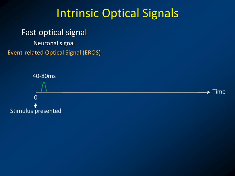
Intrinsic Optical Signals
Fast optical signal Neuronal signal
Event-related Optical Signal (EROS)
Time 0
40-80ms
Stimulus presented

Intrinsic Optical Signals
Fast optical signal Neuronal signal
Event-related Optical Signal (EROS)
Time 0
40-80ms
Stimulus presented
Active Rest
More light scattering Less light scattering

Intrinsic Optical Signals
Fast optical signal Neuronal signal
Event-related Optical Signal (EROS)
Time 0
40-80ms
Stimulus presented
Slow optical signal Hemodynamic signal
Near Infra-Red Spectroscopy (NIRS)
Oxygen
Deoxy Hb Oxy Hb 0.5-4sec 2-12sec
Active Rest
More light scattering Less light scattering

Intrinsic Optical Signals
Fast optical signal Neuronal signal
Event-related Optical Signal (EROS)
Time 0
Deoxy Hb Oxy Hb 0.5-4sec 2-12sec 40-80ms
EROS and NIRS can be measured simultaneously
Stimulus presented
Slow optical signal Hemodynamic signal
Near Infra-Red Spectroscopy (NIRS)
(Huettel, Song, & McCarthy, 2009)

Non-invasive Functional Brain Imaging Methods
EEG, MEG
fMRI
Temporal localization
Spatial localization
Millisecond (High)
Second (Low)
Event-Related Optical Signal (EROS)
Millimeter (High)
Centimeter (Low)
NIRS

Recording Optical Signals
Source Optical Fiber (e.g., 830nm)
Detector Optical Fiber
The “banana
-shaped” path
followed by light
that reaches the
detector.

Scattering in the Brain

Locating Optical Signals

Detector
Source
Recording Optical Signals
Recording System Recording Montage
Source Fibers
Detector Fibers Optical Imaging Machine
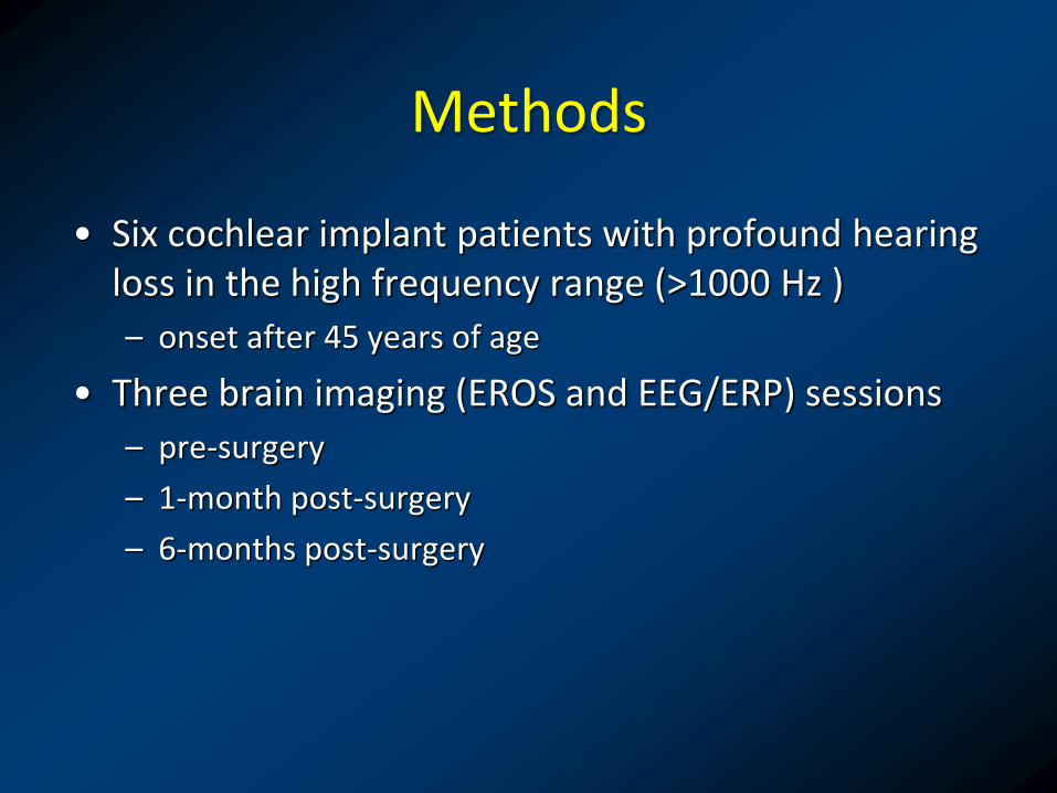
Methods
• Six cochlear implant patients with profound hearing loss in the high frequency range (>1000 Hz )
– onset after 45 years of age
• Three brain imaging (EROS and EEG/ERP) sessions
– pre-surgery
– 1-month post-surgery
– 6-months post-surgery

Low Frequency Auditory Block (LA): Tones with frequency within the audible range
High Frequency Auditory Block (HA): Tones with frequency above the audible range of individual patients prior to implant
Task: Classifying the long and short tones by button press
100ms (80%)
50ms (20%)
Methods
Active Oddball Paradigm

Results - All Participants
Behavioral Results
d’ (
Z Sc
ore
)
1 2 30
1
2
3
4
5
Pre-implant 1 Month 6 Months
Low Frequency High Frequency
Low Frequency High Frequency
1 2 30
1
2
3
4
5
1 2 30
1
2
3
4
5
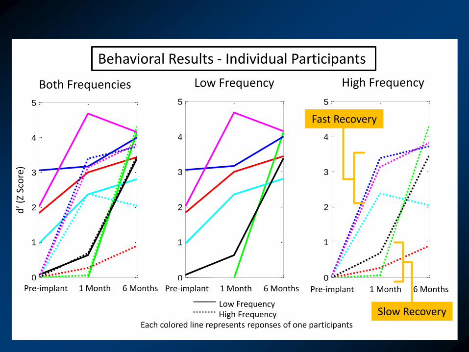
Slow Recovery
Fast Recovery
Pre-implant 1 Month 6 Months
Behavioral Results - Individual Participants
Low Frequency
Pre-implant 1 Month 6 Months
High Frequency
1 2 30
1
2
3
4
5
Pre-implant 1 Month 6 Months
Both Frequencies
d’ (
Z Sc
ore
)
Each colored line represents reponses of one participants

Single Subject – Fast Recovery d
’ (Z
Sco
re)
Behavioral Results
Correct-standard
Incorrect-standard
Miss-standard
Correct-deviant
Incorrect-deviant
Miss-deviant
Pre-implant 1 Month Post-implant 6 Months Post-implant
Low Frequency
High Frequency
ERP Results - Pz (µV)
1 2 30
1
2
3
4
5
1 2 30
1
2
3
4
5
Pre-implant 1 Month 6 Months
Low Frequency High Frequency
0 500 1000
-10
0
10
20
0 500 1000
-10
0
10
20
0 500 1000
-10
0
10
20
0 500 1000
-10
0
10
20
0 500 1000
-10
0
10
20
0 500 1000
-10
0
10
20
0 500 1000
-10
0
10
20
0 500 1000
-10
0
10
20
0 500 1000
-10
0
10
20
(ms)

Single Subject – Slow Recovery d
’ (Z
Sco
re)
Behavioral Results
Pre-implant 1 Month Post-implant 6 Months Post-implant
Low Frequency
High Frequency
ERP Results - Pz
(ms)
Low Frequency High Frequency
1 2 30
1
2
3
4
5
1 2 30
1
2
3
4
5
(µV)
Pre-implant 1 Month 6 Months
Low Frequency High Frequency
0 500 1000
-10
0
10
20
0 500 1000
-10
0
10
20
0 500 1000
-10
0
10
20
0 500 1000
-10
0
10
20
0 500 1000
-10
0
10
20
0 500 1000
-10
0
10
20
0 500 1000
-10
0
10
20
0 500 1000
-10
0
10
20
0 500 1000
-10
0
10
20
Correct-standard
Incorrect-standard
Miss-standard
Correct-deviant
Incorrect-deviant
Miss-deviant

Summary
• The results revealed a correspondence between EROS/ERP and the behavioral response
• This study demonstrated
• the possibility of applying EROS to monitor the reorganization of brain responses associated with recovery of hearing ability
• optical imaging is useful for recoding brain signals during electrical stimulation

Acknowledgments
University of Illinois at Urbana-Champaign
• Monica Fabiani
• Gabriele Gratton
• Ed Maclin
• Chin-Hong Tan
• Benjamin Zimmerman
Carle Foundation Hospital
• Michael A. Novak
• Jennifer Black
Washington University in St. Louis
• Brian A. Gordon
Funding supports
• Beckman Institute, University of Illinois at Urbana-Champaign
• The Carle Foundation

Questions and Answers
Tracking Brain Plasticity in Cochlear Implant Patients Using the Event-Related Optical Signal (EROS)
Chun-Yu TSE PhD
Department of Psychology
The Chinese University of Hong Kong (CUHK)

Recording Optical Signals