Embed Size (px)
Citation preview

Toxicology in Vitro 30 (2015) 309–317
Contents lists available at ScienceDirect
Toxicology in Vitro
j ourna l homepage: www.e lsev ie r .com/ locate / t iv
Gallic acid reduces the effect of LPS on apoptosis and inhibits theformation of neutrophil extracellular traps
Gabriela Viegas Haute a,⁎, Eduardo Caberlon a, Eamim Squizani a, Fernanda Cristina de Mesquita a,Leonardo Pedrazza a, Bianca Andrade Martha a, Denizar Alberto da Silva Melo a, Eduardo Cassel b,Rafael Sanguinetti Czepielewski c, Shanna Bitencourt d, Márcia Inês Goettert d, Jarbas Rodrigues de Oliveira a
a Laboratório de Pesquisa em Biofísica Celular e Inflamação, Pontifícia Universidade Católica do Rio Grande do Sul (PUCRS), Porto Alegre-RS, Brazilb Laboratório de Operações Unitárias, Pontifícia Universidade Católica do Rio Grande do Sul (PUCRS), Porto Alegre, RS, Brazilc Laboratório de Imunologia Celular e Molecular, IPB, Pontifícia Universidade Católica do Rio Grande do Sul (PUCRS), Porto Alegre, RS, Brazild Laboratório de Cultura de Células, Centro de Ciências Biológicas e da Saúde, Centro Universitário UNIVATES, Lajeado, RS, Brazil
⁎ Corresponding author at: Laboratório de Pesquisa emPontifícia Universidade Católica do Rio Grande do Sul (Pprédio 12, bloco C, sala 221, CEP 90619-900, Porto Alegre
E-mail address: [email protected] (G.V. Hau
http://dx.doi.org/10.1016/j.tiv.2015.10.0050887-2333/© 2015 Elsevier Ltd. All rights reserved.
a b s t r a c t
a r t i c l e i n f oArticle history:Received 25 May 2015Received in revised form 28 September 2015Accepted 12 October 2015Available online 23 October 2015
Keywords:InflammationGallic acidNeutrophilsROSNETosisApoptosis
Apoptosis and NETosis of neutrophils are two major mechanisms of programmed cell death that differ in theirmorphological characteristics and effects on the immune system. Apoptosis can be delayed by the presence ofpathogens or chemical components such as lipopolysaccharide (LPS). Neutrophils have other antimicrobialstrategy, called neutrophil extracellular traps (NETs), which contributes to the elimination and control of thepathogen. NETosis is induced by infection, inflammation or trauma and represents an innate immune activationmechanism. The objective of this study was to evaluate the effect of gallic acid (GA) in themodulation of apopto-sis and NETs release. The results show that GA decreased the anti-apoptotic effect of LPS, blocked the induction ofNETs and prevented the formation of free radicals induced by LPS. These findings demonstrate that the GA is anovel therapeutic agent for decreasing the exacerbated response of the body against an infectious agent.
© 2015 Elsevier Ltd. All rights reserved.
1. Introduction
Sepsis is a complex syndrome that results in an exaggerated system-ic inflammatory response against an infectious agent (Bone et al., 1997;Matot and Sprung, 2001). This reaction aims to destroy, dilute or immo-bilize the infectious agent. (Teixeira et al., 2003) Inflammations aredivided into acute and chronic. Acute inflammation is characterized bythe accumulation of fluid, fibrin, leukocytes (especially neutrophils)and red blood cells in the aggression area. Upon arrival in the inflamedsite, neutrophils are already equipped with the necessary proteins todestroy infectious agents (Brinkmann et al., 2004). The encounterwith the pathogen causes the activation of the cells with the immersionof the microorganism in a phagosome (Brinkmann et al., 2004;Guimarães-Costa et al., 2012; Fuchs et al., 2007). In the phagosometwo events occur: first, there is great generation of reactive oxygenspecies (ROS), and second, the granules of neutrophils merge thephagosome, and unload antimicrobial peptides and enzymes. Together
Biofísica Celular e Inflamação,UCRS), Avenida Ipiranga 6681,, Rio Grande do Sul, Brazil.te).
these two events lead to microbial death (Vaughan, 2013; Liu et al.,2000). The inflammatory reaction is mediated endogenously by activesubstances, called “inflammatory mediators” and excessive productionof these mediators leads to an increase in host response, causing a met-abolic imbalance that can propagate the inflammatory response(Teixeira et al., 2003; Fuchs et al., 2007; Esmann et al., 2010; Mello,2012). Chronic inflammation is the sumof the reactions of the organismas consequence of the offending agent residence, which was noteliminated by the mechanisms of acute inflammation (Mello, 2012).
Septic shock is an example of the increase of uncontrolled inflamma-tory response that results in a metabolic imbalance (Radic, 2014). Theshock and the complications aremainly related to the release of compo-nents of the bacterial wall. The endotoxin of gram-negative bacteria,lipopolysaccharide (LPS), and teichoic acid of gram-positive bacteriaindirectly trigger the inflammatory cascade via induction of cytokineproduction by activatedmacrophages andmonocytes, and sequentially,produce tumor necrosis factor-alpha (TNF-α), interleukin-1β (IL-1β),interleukin-6 (IL-6) and interleukin-8 (IL-8). These cytokinesinteract with other cells and cellular elements (polymorphonuclearcells, endothelial cells, fibroblast cells, platelets and monocytes), induc-ing production and release of secondary mediators, which contributeto a delayed inflammatory response. (Sulowska et al., 2005) The
310 G.V. Haute et al. / Toxicology in Vitro 30 (2015) 309–317
overproduction or inappropriate expression of these factors can lead toa variety of pathological conditions, including septic shock and systemictoxicity (Mello, 2012; Radic, 2014; Kirchner et al., 2013).
NETosis and apoptosis of neutrophils are two major mechanisms ofprogrammed cell death that differ in theirmorphological characteristicsand their effects on the immune system (Brinkmann and Zychlinsky,2012). Apoptosis is characterized by packaging of nuclear chromatinand nuclear fragments, subsequently occurring absorption of apoptoticcells by phagocytes, which generally suppress the immune response.Neutrophils under physiological conditions suffer apoptosis in 20 h.However, in infected tissues this can be delayed by microbial compo-nents such as LPS and pro-inflammatory stimuli (Teixeira et al., 2003;Esmann et al., 2010). Apoptosis of neutrophils is an important point inthe physiological control of the immune response, playing an importantrole in the resolution of inflammation. In this context, apoptosis shouldbe delayed until the essential functions of pathogen are completelyphagocyted, then these cells must die to undo the inflammationand prevent tissue damage (Vaughan, 2013; Saffarzadeh and Preissner,2013).
Recent studies have shown that neutrophils have another antimicro-bial mechanism called NETosis, which can be induced by infection, in-flammation or trauma and represents an innate immune activationmechanism (Brinkmann and Zychlinsky, 2012). When neutrophils areactivated by phorbol myristate acetate (PMA), IL-8, LPS or fungi, theyrelease means for the chromatin that are associated with different pro-teins, forming a complex called neutrophil extracellular traps (NETs),which capture and kill pathogens (Fuchs et al., 2007; Meng et al.,2012). NETs are abundant in inflamed sites, as found in patients withappendicitis, preeclampsia and infection by Streptococcus pneumoniae(Fuchs et al., 2007). Some studies suggest a pathophysiological role ofNETs and their components in autoimmune diseases such as smallvessel vasculitis, lupus nephritis, systemic lupus erythematosus (SLE),psoriasis, and rheumatoid arthritis (Meng et al., 2012; Bone, 1991;Thijs and Hack, 1995). Recent studies suggest that this action maycause tissue damage and the control of NETs release can result in bene-ficial effects in autoimmune diseases (Meng et al., 2012; Vilcek and Lee,1991).
Gallic acid (GA) is a phenolic compound found in various plants, fruitand food, and it has antioxidant, anti-carcinogenic and anti-viralproperties (You et al., 2011; Chandramohan Reddy et al., 2012). Otherstudies report that GA also has antibacterial, antifungal, anti-inflammatory, anti-malarial and anti-herpetic effects and is present insome of the most consumed beverages in the world, such as green tea(You et al., 2011; Chandramohan Reddy et al., 2012; Eslami et al., 2010).Hence, the objective of this studywas to evaluate the effect of GA control-ling apoptosis and formation of NETs in primary cultures of humanneutrophils.
2. Materials & methods
2.1. Ethics statement
Study experimental protocol (443.648) was approved by the EthicsResearch Committee of Pontifícia Universidade Católica do Rio Grandedo Sul (PUCRS).
2.2. Peripheral blood polymorphonuclear cells preparation
The peripheral blood polymorphonuclear cells (PMNs)were isolatedfrom whole blood obtained from healthy human donors by Ficoll-Paque™ PLUS (GE Healthcare) density gradient centrifugation. Briefly,12 mL of blood samples were collected by venipuncture in heparin-containing tubes. Plasma was discarded and blood cells were diluted1:2 with saline solution. After, 4 mL Ficoll-Paque™ PLUS were addedto cell solution and centrifuged at 720 ×g at room temperature for20 min. After centrifugation, the supernatant was removed and the
cells were washed with hypotonic lysis buffer, containing 0.83%NH4Cl, to lyse red blood cells (erythrocytes). The solution was centri-fuged at 200 ×g at 4 °C for 10 min and this procedure was repeatedtwice. The resultant pellet was washed with phosphate-buffered saline(PBS). Following isolation, neutrophils (2.0 × 10⁵ / 200 μL) were main-tained in RPMI 1640 medium supplemented with 10% autologousserum and 0.15% garamycin (Schering-Plough) in 96-well flat-bottomplates (Nunc™-ImmunoModules) at 37 °C in a humidified atmospherecontaining 5% CO2 (Morita et al., 2014; Czerwinska et al., 2013). Cell vi-ability was assessed by trypan blue exclusion assay. Purity of this prep-arationwas ≥95%of neutrophils. All reagents usedwerefiltered througha disposable sterile filter unit 0.22 μM (Millex). All human subjects readand signed an informed consent.
2.3. Peripheral blood mononuclear cells (PBMCs) preparation
PBMCs were isolated from whole blood of healthy human donors(12 mL of heparinized blood) using Ficoll-Paque™ PLUS densitygradient centrifugation. Briefly, plasma was discarded and the bloodcells were diluted 1:2 with saline solution. After, 4 mL Ficoll-Paque™PLUS were added to cell solution and centrifuged at 720 ×g at roomtemperature for 20 min. PBMCs were removed from the interfaceformed by centrifugation and washed with PBS. Cells were maintainedin RPMI 1640 medium supplemented with 10% autologous serum and0.15% garamycin (Schering-Plough) in 96-well flat bottom plates at37 °C in a humidified atmosphere containing 5% CO2. Cell viability wasassessed by trypan blue exclusion assay. All reagents used were filteredthrough a disposable sterile filter unit 0.22 μM (Millex). All humansubjects read and signed an informed consent.
2.4. Cytotoxicity assay
1.088 mg GA (MW 170.12) (Sigma Aldrich) was weighed anddissolved directly into 1 mL serum-free RPMI 1640 medium. Aftervortexing for a couple ofminutes, GA is totally dissolved in themedium.From a stock solution of 6400 μM, the serial dilution was prepared asfollows: 3200, 1600, 800, 400, 200, 100, 50, 25, and 12.5 μM. 100 μL ofeach concentration was added to wells containing 100 μL of RPMI1640 medium + cells. Thus, the final concentrations in the wells were1600, 800, 400, 200, 100, 50, 25, 12.5 and 6.25 μM, respectively.Neutrophils (2.0 × 10⁵ cells/200 μL) were incubated for 16 h. Controlgroup was composed of neutrophils in RPMI 1640 medium. LPS group(Escherichia coli 026: B6), the drug was diluted in RPMI 1640 mediumand added directly to cell culture at the stated concentration (25, 50and 100 ng/mL). GA group was composed by a GA serial dilution (6.25to 1600 μM) and GA + LPS group was composed by the sameserial dilution of GA + LPS (50 ng/mL), the drugs were diluted inmedium and then added to the cell culture. All groups were made intriplicate and the viability was performed by trypan blue exclusionassay.
PBMCs (1.6 × 10⁵ cells/200 μL) were incubated for 96 h. Controlgroup was composed of PBMCs in RPMI 1640. GA group was com-posed by a GA serial dilution (6.25 to 1600 μM). All groups weremade in triplicate and the viability was performed by trypan blue exclu-sion assay.
2.5. Apoptosis assay
Neutrophils were incubated in the presence of GA (25, 50and 100 μM), LPS (50 ng/mL) or GA (25, 50 and 100 μM) + LPS(50 ng/mL) for 16 h. Untreated cells represented the control group.Apoptosis was evaluated by flow cytometry using Annexin-V Kitassay. Annexin-V and 7-AAD were added to 1 × 10⁵ cell suspensionaccording to the manufacturer's instructions (BD Biosciences) andthen incubated for 15 min at room temperature in the dark. Subse-quently 2 × 10⁴ cells were analyzed by flow cytometry (FACS Canto

311G.V. Haute et al. / Toxicology in Vitro 30 (2015) 309–317
II, BD Bioscience) within 1 h. Early apoptotic cells were stained withAnnexin V alone, whereas necrotic cells and late apoptotic cells werestained with both Annexin V and 7-AAD. Data were analyzed withFlowJo software v. 7.2.5 (Tree Star Inc., USA).
Morphological assessment of neutrophil apoptosis was evaluated bylight microscopy analysis. Cells were spin down on a glass slide by acytospin. Cells were fixed with methanol and stained with May–Grunwald–Giemsa staining solution.
2.6. Western blot
Neutrophils (1.0 × 10⁶ cells/mL) were incubated in the presence ofLPS (50 ng/mL) or GA (25, 50 and 100 μM) + LPS (50 ng/mL) for 16 hand re-suspended in a lysis buffer (10 mM pH 7.5 Tris-HCl, 100 mMNaCl, 0,3% CHAPS, 50mMNaF,β-glicerol phosphate and protease inhib-itors). The lysates (50 μg) were subjected to sodium dodecyl sulfate-polyacrylamide gel electrophoresis (SDS-PAGE) and transferred to ni-trocellulose membranes. Themembranes were blocked with a blockingbuffer (1M Trizma pH 7.5, 5MNaCl, 0.8% Tween 20 and 10% skimmilk)for 2 h and stained with primary polyclonal antibodies – HumanAnti-Caspase-3 – (1:500) in a blocking buffer overnight at 4 °C. Themembranes were incubated with the secondary antibodies – RabbitAnti-Mouse IgG – (1:7500) for 2 h at 4 °C. The bands were detectedusing Opti-4 CN revelator solution and the band intensities wereobtained using the program ImageJ 3.0.
Fig. 1. Effect of different concentrations of GA on cell viability. Neutrophils were challenged w(50 ng/mL) for 16 h and cell viability was assessed by trypan blue exclusion assay. (D) PBMCs weblue exclusion assay. Results were expressed as the percentage of viable cells. All data represent t
2.7. Induction and detection of NETs
NETs formation of neutrophils was quantitated in the supernatants(2.0 × 10⁵ cells/200 μL). The cells were incubated in the presence ofGA (25, 50, 100 μM), LPS (50 ng/mL) or GA (25, 50 and 100 μM) + LPS(50 ng/mL) for 16 h. Untreated cells represented the control group. Toquantify levels of NETs (DNA), the Quant-iT™ PicoGreen® dsDNA Kitassaywas used according to themanufacturer's instructions (Invitrogen).The fluorescence intensity of DNA was measured at excitation and emis-sion wavelengths of 485 nm and 530 nm, respectively, in a microplatereader (Victor 3, PerkinElmer). A standard calibration curve was used.(Luo et al., 2014)
2.8. Immunofluorescence staining of NETs
The NETs formation was analyzed using the Kit Falcon™ CultureSlide (BD Biosciences). For immunofluorescence, freshly isolatedPMNswere seeded on Poly-L-lysin coated cover slips, allowed to adhere(1 h), and stimulated with LPS (50 ng/mL) for NETs induction. Toevaluate the effect of GA on the NETS formation, we incubated thecells with LPS (50 ng/mL) + GA (100 μM). After 16 h of incubation,cells were fixed with formaldehyde 4% for 2 h and permeabilized withPBS containing 0.03% Triton X-100 and 10% fetal bovine serum(Gibco — Life Technologies) for 30 min. To stain the NETs, sampleswere incubated with a primary monoclonal antibody – Mouse Anti-Myeloperoxidase – (1:200) for 30 min and then with a secondary
ith (A) GA (6.25 to 1600 μM), (B) LPS (50 ng/mL) or (C) GA (6.25 to 1600 μM) + LPSre challengedwith GA (6.25 to 1600 μM) for 96 h and cell viability was assessed using trypanhe mean ± SD (n = 5). ***P b 0.001, **P b 0.01 and *P b 0.05 compared with control group.

Fig. 3.Morphological changes of neutrophils after LPS stimulation visualized by opticalmicroscopy. In this image,we could verify themorphological differences of cell in apoptosis. Arrowsshowmorphological detail of an apoptotic cell which lost its original shape. 50 ng/mL LPS-treated cells show predominance of normal neutrophils, with fine granularity of chromatin andnormal lobulated nucleus. Cells exposed to GA (25, 50 and 100 μM) and GA (50 and 100 μM)+ LPS have the same pattern as control cells. Magnification ×400.
Fig. 2. Effects of GA, LPS and GA+ LPS on apoptosis of neutrophils. (A) Cells were exposed to GA (25, 50 and 100 μM). (B) Cells were exposed to GA (25, 50 and 100 μM), LPS (50 ng/mL)and GA (25, 50 and 100 μM)+ LPS (50 ng/mL). Results were expressed as percentage of apoptotic cells. Data represent the mean ± SD (n = 5). *P b 0.05 compared with control group.(C) Representative flow cytometric scatter plots of Annexin V (x axis)/7-AAD (y axis) stained control and GA- and/or LPS-treated cells for 16 h. The lower left quadrant shows viable cells,which are annexin V− and 7-AAD−. The lower right quadrant represents the apoptotic cells, annexin V+ and 7-AAD−. The upper right quadrant shows the late apoptotic or dead cells thatare annexin V+ and 7-AAD+.
312 G.V. Haute et al. / Toxicology in Vitro 30 (2015) 309–317
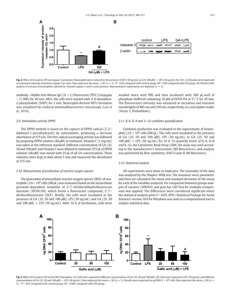
Fig. 4. Effect of GA and/or LPS on caspase-3 activation. Neutrophilswere cultured in the presence of LPS (50 ng/mL) or GA (100 μM)+ LPS (50 ng/mL) for 16 h. (A) Results were expressedas expression intensity of protein caspase-3/β-actin. Data represent themean± SD (n= 3). *P b 0.05 comparedwith control group; #P b 0.05 comparedwith LPS group. (B)Western blotanalysis of extracts of neutrophils cultured for cleaved caspase-3 and β-actin proteins. Representative experiments are depicted (n = 3).
313G.V. Haute et al. / Toxicology in Vitro 30 (2015) 309–317
antibody – Rabbit Anti-Mouse IgG (H+ L) Fluorescein (FITC) Conjugate– (1:500) for 30 min. After, the cells were stained with 4′,6-diamidino-2-phenylindole (DAPI) for 2 min. Neutrophil-derived NETs formationwas visualized by confocal immunofluorescence microscopy (Luo etal., 2014).
2.9. Antioxidant activity DPPH
The DPPH method is based on the capture of DPPH radical (2′,2′-diphenyl-1-picrylhydrazyl) by antioxidants, producing a decreaseabsorbance at 515 nm. The free radical scavenging activitywas followedby preparing DPPH solution (60 μM) in methanol. Vitamin C (1mg/mL)was taken as the reference standard. Different concentration of GA (25,50 and 100 μM)andVitamin Cwere diluted inmethanol. 975 μL of DPPHsolution (60 μM) was mixed with 25 μL of all GA concentrations. Thesemixtures were kept in dark about 5 min and measured the absorbanceat 515 nm.
2.10. Measurement of production of reactive oxygen species
The generation of intracellular reactive oxygen species (ROS) of neu-trophils (2.0× 10⁵ cells/200 μL)was evaluated based on the intracellularperoxide-dependent oxidation of 2′,7′-dichlorodihydrofluoresceindiacetate (DCFH-DA) which forms a fluorescent compound, 2′,7′-dichlorofluorescein (DCF). Briefly, the cells were incubated in thepresence of GA (25, 50 and 100 μM), LPS (50 ng/mL) and GA (25, 50and 100 μM) + LPS (50 ng/mL). After 16 h of incubation, cells were
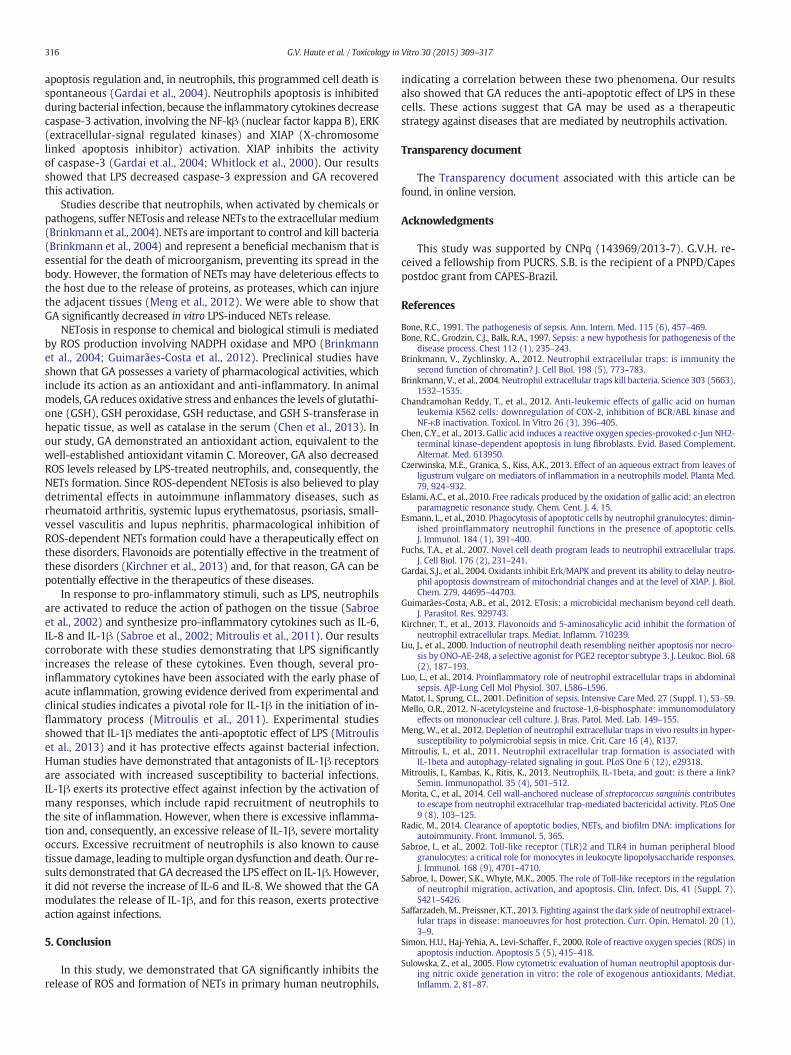
Fig. 5. Effect of GA and/or LPS on theNETs formation. (A) Cells were exposed to different concenconcentrations of GA (25, 50 and 100 μM)+ LPS (50 ng/mL). Data represent themean± SD (n5). **P b 0.01 compared with control group; #P b 0.001 compared with LPS group.
washed twice with PBS and then incubated with 200 μL/well ofphosphate-buffered containing 10 μM of DCFH-DA at 37 °C for 30 min.The fluorescence intensity was measured at excitation and emissionwavelengths of 485 nmand530 nm, respectively, in amicroplate reader(Victor 3, PerkinElmer).
2.11. IL-6, IL-8 and 1L-1β cytokines quantification
Cytokines production was evaluated in the supernatants of neutro-phils (2.0 × 10⁵ cells/200 μL). The cells were incubated in the presenceof GA (25, 50 and 100 μM), LPS (50 ng/mL) or GA (25, 50 and100 μM) + LPS (50 ng/mL) for 16 h. To quantify levels of IL-6, IL-8and IL-1β, the Cytometric Bead Array (CBA) Kit assay was used accord-ing to the manufacturer's instructions (BD Biosciences) and analysiswas performed by flow cytometry (FACS Canto II, BD Bioscience).
2.12. Statistical analysis
All experiments were done in triplicates. The normality of the datawas analyzed by the Shapiro–Wilk test. The measures were parametricand then we calculated the mean and standard deviation of the meanfor each of the variables analyzed. For comparison between groups anal-ysis of variance (ANOVA) and post hoc LSD Test for multiple compari-sons was applied. The differences were considered significant whenthe statistical analysis gives P b 0.05. SPSS (Statistical Package for SocialSciences) version 18.0 forWindowswas used as a computational tool toanalyze statistical data.
trations of GA (25, 50 and 100 μM). (B) Cells were exposed to LPS (50 ng/mL) and different= 5). Results were expressed as ng DNA/2 × 105 cells. Data represent themean± SD (n=

Fig. 7. Antioxidant effect of GA (25, 50 and 100 μM) and vitamin C (1 mg/mL). Data wereexpressed as percentage of control group. ***P b 0.001 compared with control group.
Fig. 6. NETs formation after LPS stimulation visualized by fluorescence. The image of con-trol group shows the nuclear localization of DNA (blue fluorescence) and the granular pat-terns of myeloperoxidase (MPO) (green fluorescence). The LPS group shows the changesduring NETs formation that can be determined with loss of granular integrity of MPO andnuclear lobules. GA + LPS group presents a similar morphology to the control group.Magnification ×630.
314 G.V. Haute et al. / Toxicology in Vitro 30 (2015) 309–317
3. Results
3.1. Cytotoxic effect of GA, GA + LPS and LPS in human neutrophils andPBMCs
To evaluate cytotoxic effect of GA in human neutrophils, cells wereexposed to different concentrations of GA (6.25 to 1600 μM). After16 h of treatment, it was verified that GA did not decrease cell viability(Fig. 1A). We performed an apoptosis curve with LPS at concentrationsof 25, 50 and 100 ng/mL (data not show). The concentration of 25ng/mLhad no significant anti-apoptotic effect, unlike the concentrations of 50and 100 ng/mL. So, we tested the toxicity of LPS in neutrophils and ver-ified that any concentration (25, 50 and 100 ng/mL) showed decrease incell viability (Fig. 1B). Therefore, we chose the lowest concentration thatpossessed anti-apoptotic effect for the next experiments.
When we associate the LPS (50 ng/mL) with different concentra-tions of GA (6.25 to 1600 μM), only 1600 μM GA demonstrated toxicity(Fig. 1C). To evaluate cytotoxic effect of GA in other blood cells,PBMC cells were exposed to different concentrations of GA (6.25to 1600 μM). The concentrations 200, 400, 800 and 1600 μM of GAdecreased the cell viability (Fig. 1D).
3.2. Effect of GA, GA + LPS and LPS on apoptosis of human neutrophils
To analyze apoptosis induction, cells were exposed to different con-centrations of GA (25, 50 and 100 μM) and LPS (50 ng/mL). GA alone didnot induce apoptosis (Fig. 2A and C). However, LPS showed significantanti-apoptotic effect when compared with control, and GA inhibitedthis effect (Fig. 2B and C). Apoptosis was also evaluated by opticalmicroscopy to verify morphological differences between apoptoticcells (the nucleus loses its original shape) and normal cells confirmingthe results by flow cytometry (Fig. 3).
3.3. Effect of LPS and GA + LPS on caspase 3 activation
As GA decreases anti-apoptotic effect of LPS, we decided to evaluatethe activation of caspase-3. Cells treated with LPS (50 ng/mL) exhibiteda significant reduction of caspase-3 when compared to control groupand 100 μM GA + LPS (50 ng/mL) increased significantly the caspase-3 activationwhen compared to the cells treatedwith LPS (Fig. 4A andB).
3.4. Effect of GA, GA + LPS and LPS in the release of NETs of humanneutrophils
The GA (25, 50 and 100 μM) alone did not induce the formation ofNETs (Fig. 5A). LPS (50 ng/mL) increased the NETs formation and theGA decreased the LPS effect (Fig. 5B). This effect was visualized usingimmunofluorescence with confocal microscopy (Fig. 6).
3.1. Antioxidant effect of GA and Vitamin C
We observed that all concentrations of GA (25, 50 and 100 μM)decreased the free radical DPPH, showing a similar effect to Vitamin C(Fig. 7).
3.6. Effect of GA, GA + LPS and LPS on ROS generation
We observed that GA (25, 50 and 100 μM) alone did not induce theformation of ROS (Fig. 8A). 100 μM GA decreased significantly the ROSrelease when compared to LPS group (Fig. 8B).
3.7. Effect of GA + LPS and LPS on cytokine release
Cytokines have a key role in the resolution of inflammation, for thisreason we decided to quantify the cytokines released by activated neu-trophils. Cells stimulated with LPS increased IL-6, IL-8 and IL-1β levels
when compared with control group. GA (50 and 100 μM) treatmentdecreased this effect only on IL-1β release (Fig. 9A, B and C).
4. Discussion
Neutrophils are the first line of defense of our body and are the firstcells to reach the focus of inflammation. In response to inflammatorystimuli, they migrate from the peripheral blood to infected tissues,where they efficiently bind, engulf, and inactivate bacteria (Brinkmannet al., 2004). These cells have a short half-life and die by apoptosis in a

Fig. 8. Effect ofGA, and/or LPS onneutrophils ROS release. (A) Cellswere exposed to GA (25, 50 and 100 μM). (B) Cellswere exposed to GA (25, 50 and100 μM), LPS (50ng/mL) andGA (25,50 and 100 μM)+ LPS (50 ng/mL). Data represent the mean ± SD (n = 3). Results were expressed as DCF Fluorescence/mg protein. #P b 0.05 compared with LPS group.
315G.V. Haute et al. / Toxicology in Vitro 30 (2015) 309–317
few hours. The presence of pathogens contributes to prolonging the lifeof neutrophils in the infected site. The permanence of these cells in theinflamed site, for a certain time, is beneficial to the host because ithelps against the invasion, but on the other hand can lead to tissuedamage by excessive release of toxic products. Neutrophils alsohave another antimicrobial strategy, called NETosis, which resultsin the death of these cells, and contributes to the elimination ofthe pathogens (Guimarães-Costa et al., 2012; Simon et al., 2000;Wallach-Dayan et al., 2006; Luo et al., 2014). Our study aimed to in-vestigate the in vitro action of GA on LPS-induced apoptosis andNETosis of human neutrophils.
Our initial results showed that GA is not cytotoxic in neutrophils. Inorder to verify their possible cytotoxicity in other blood cells, we didexperiments in primary cultures of human mononuclear cells. Wefound that the concentrations of 200, 400, 800 and 1600 μM of GAcould decrease cell viability, thus they were not suitable for therapeutic
Fig. 9. Effect of GA on inflammatory cytokines release after 16 h treatmentwith GA (25, 50 and 1in neutrophils supernatant. Cytokines IL-6 (A), IL-8 (B) and IL-1β (C)were analyzed. Data represwith control group.
purposes. For this reason,we chose 25, 50 and 100 μMconcentrations tofollow the study.
Our study showed that LPS decreased apoptosis in neutrophils. It isreported that LPS acts on TLR4 receptor and for consequence of thisbinding, occurs activation and increased lifetime of the cells. Neutrophilapoptosis is essential to regulate adult cell populations and in the reso-lution of inflammation. When the organism is under attack, these cellsdie slowly in order to control the infection, however they should dieby apoptosis immediately after the combat against the pathogen.When LPS binds to TLR4 receptor, these cells release cytokines and pro-duce ROS, and these inflammation mediators cause tissue damage(Brinkmann et al., 2004; Sabroe et al., 2005; Sabroe et al., 2002). Inthis study, GAdecreased the anti-apoptotic effect of LPS,which indicatesthat GA has a protective role against infection-induced tissue damage.
The above-mentioned findings raised the question whether thecaspase-3 was changed in GA-treated cells. Caspases play a key role of
00 μM) and/or LPS (50 ng/mL). Flow cytometric analyses of (A) IL-6, (B) IL-8 and (C) IL-1βent themean±SD (n=5). Results were expressed as pg/2× 105 cells. *P b 0.05 compared
316 G.V. Haute et al. / Toxicology in Vitro 30 (2015) 309–317
apoptosis regulation and, in neutrophils, this programmed cell death isspontaneous (Gardai et al., 2004). Neutrophils apoptosis is inhibitedduring bacterial infection, because the inflammatory cytokines decreasecaspase-3 activation, involving the NF-kβ (nuclear factor kappa B), ERK(extracellular-signal regulated kinases) and XIAP (X-chromosomelinked apoptosis inhibitor) activation. XIAP inhibits the activityof caspase-3 (Gardai et al., 2004; Whitlock et al., 2000). Our resultsshowed that LPS decreased caspase-3 expression and GA recoveredthis activation.
Studies describe that neutrophils, when activated by chemicals orpathogens, suffer NETosis and release NETs to the extracellularmedium(Brinkmann et al., 2004). NETs are important to control and kill bacteria(Brinkmann et al., 2004) and represent a beneficial mechanism that isessential for the death of microorganism, preventing its spread in thebody. However, the formation of NETs may have deleterious effects tothe host due to the release of proteins, as proteases, which can injurethe adjacent tissues (Meng et al., 2012). We were able to show thatGA significantly decreased in vitro LPS-induced NETs release.
NETosis in response to chemical and biological stimuli is mediatedby ROS production involving NADPH oxidase and MPO (Brinkmannet al., 2004; Guimarães-Costa et al., 2012). Preclinical studies haveshown that GA possesses a variety of pharmacological activities, whichinclude its action as an antioxidant and anti-inflammatory. In animalmodels, GA reduces oxidative stress and enhances the levels of glutathi-one (GSH), GSH peroxidase, GSH reductase, and GSH S-transferase inhepatic tissue, as well as catalase in the serum (Chen et al., 2013). Inour study, GA demonstrated an antioxidant action, equivalent to thewell-established antioxidant vitamin C. Moreover, GA also decreasedROS levels released by LPS-treated neutrophils, and, consequently, theNETs formation. Since ROS-dependent NETosis is also believed to playdetrimental effects in autoimmune inflammatory diseases, such asrheumatoid arthritis, systemic lupus erythematosus, psoriasis, small-vessel vasculitis and lupus nephritis, pharmacological inhibition ofROS-dependent NETs formation could have a therapeutically effect onthese disorders. Flavonoids are potentially effective in the treatment ofthese disorders (Kirchner et al., 2013) and, for that reason, GA can bepotentially effective in the therapeutics of these diseases.
In response to pro-inflammatory stimuli, such as LPS, neutrophilsare activated to reduce the action of pathogen on the tissue (Sabroeet al., 2002) and synthesize pro-inflammatory cytokines such as IL-6,IL-8 and IL-1β (Sabroe et al., 2002; Mitroulis et al., 2011). Our resultscorroborate with these studies demonstrating that LPS significantlyincreases the release of these cytokines. Even though, several pro-inflammatory cytokines have been associated with the early phase ofacute inflammation, growing evidence derived from experimental andclinical studies indicates a pivotal role for IL-1β in the initiation of in-flammatory process (Mitroulis et al., 2011). Experimental studiesshowed that IL-1β mediates the anti-apoptotic effect of LPS (Mitrouliset al., 2013) and it has protective effects against bacterial infection.Human studies have demonstrated that antagonists of IL-1β receptorsare associated with increased susceptibility to bacterial infections.IL-1β exerts its protective effect against infection by the activation ofmany responses, which include rapid recruitment of neutrophils tothe site of inflammation. However, when there is excessive inflamma-tion and, consequently, an excessive release of IL-1β, severe mortalityoccurs. Excessive recruitment of neutrophils is also known to causetissue damage, leading tomultiple organ dysfunction and death. Our re-sults demonstrated that GA decreased the LPS effect on IL-1β. However,it did not reverse the increase of IL-6 and IL-8. We showed that the GAmodulates the release of IL-1β, and for this reason, exerts protectiveaction against infections.
5. Conclusion
In this study, we demonstrated that GA significantly inhibits therelease of ROS and formation of NETs in primary human neutrophils,
indicating a correlation between these two phenomena. Our resultsalso showed that GA reduces the anti-apoptotic effect of LPS in thesecells. These actions suggest that GA may be used as a therapeuticstrategy against diseases that are mediated by neutrophils activation.
Transparency document
The Transparency document associated with this article can befound, in online version.
Acknowledgments
This study was supported by CNPq (143969/2013-7). G.V.H. re-ceived a fellowship from PUCRS. S.B. is the recipient of a PNPD/Capespostdoc grant from CAPES-Brazil.
References
Bone, R.C., 1991. The pathogenesis of sepsis. Ann. Intern. Med. 115 (6), 457–469.Bone, R.C., Grodzin, C.J., Balk, R.A., 1997. Sepsis: a new hypothesis for pathogenesis of the
disease process. Chest 112 (1), 235–243.Brinkmann, V., Zychlinsky, A., 2012. Neutrophil extracellular traps: is immunity the
second function of chromatin? J. Cell Biol. 198 (5), 773–783.Brinkmann, V., et al., 2004. Neutrophil extracellular traps kill bacteria. Science 303 (5663),
1532–1535.Chandramohan Reddy, T., et al., 2012. Anti-leukemic effects of gallic acid on human
leukemia K562 cells: downregulation of COX-2, inhibition of BCR/ABL kinase andNF-κB inactivation. Toxicol. In Vitro 26 (3), 396–405.
Chen, C.Y., et al., 2013. Gallic acid induces a reactive oxygen species-provoked c-Jun NH2-terminal kinase-dependent apoptosis in lung fibroblasts. Evid. Based Complement.Alternat. Med. 613950.
Czerwinska, M.E., Granica, S., Kiss, A.K., 2013. Effect of an aqueous extract from leaves ofligustrum vulgare on mediators of inflammation in a neutrophils model. Planta Med.79, 924–932.
Eslami, A.C., et al., 2010. Free radicals produced by the oxidation of gallic acid: an electronparamagnetic resonance study. Chem. Cent. J. 4, 15.
Esmann, L., et al., 2010. Phagocytosis of apoptotic cells by neutrophil granulocytes: dimin-ished proinflammatory neutrophil functions in the presence of apoptotic cells.J. Immunol. 184 (1), 391–400.
Fuchs, T.A., et al., 2007. Novel cell death program leads to neutrophil extracellular traps.J. Cell Biol. 176 (2), 231–241.
Gardai, S.J., et al., 2004. Oxidants inhibit Erk/MAPK and prevent its ability to delay neutro-phil apoptosis downstream of mitochondrial changes and at the level of XIAP. J. Biol.Chem. 279, 44695–44703.
Guimarães-Costa, A.B., et al., 2012. ETosis: a microbicidal mechanism beyond cell death.J. Parasitol. Res. 929743.
Kirchner, T., et al., 2013. Flavonoids and 5-aminosalicylic acid inhibit the formation ofneutrophil extracellular traps. Mediat. Inflamm. 710239.
Liu, J., et al., 2000. Induction of neutrophil death resembling neither apoptosis nor necro-sis by ONO-AE-248, a selective agonist for PGE2 receptor subtype 3. J. Leukoc. Biol. 68(2), 187–193.
Luo, L., et al., 2014. Proinflammatory role of neutrophil extracellular traps in abdominalsepsis. AJP-Lung Cell Mol Physiol. 307, L586–L596.
Matot, I., Sprung, C.L., 2001. Definition of sepsis. Intensive Care Med. 27 (Suppl. 1), S3–S9.Mello, O.R., 2012. N-acetylcysteine and fructose-1,6-bisphosphate: immunomodulatory
effects on mononuclear cell culture. J. Bras. Patol. Med. Lab. 149–155.Meng, W., et al., 2012. Depletion of neutrophil extracellular traps in vivo results in hyper-
susceptibility to polymicrobial sepsis in mice. Crit. Care 16 (4), R137.Mitroulis, I., et al., 2011. Neutrophil extracellular trap formation is associated with
IL-1beta and autophagy-related signaling in gout. PLoS One 6 (12), e29318.Mitroulis, I., Kambas, K., Ritis, K., 2013. Neutrophils, IL-1beta, and gout: is there a link?
Semin. Immunopathol. 35 (4), 501–512.Morita, C., et al., 2014. Cell wall-anchored nuclease of streptococcus sanguinis contributes
to escape from neutrophil extracellular trap-mediated bactericidal activity. PLoS One9 (8), 103–125.
Radic, M., 2014. Clearance of apoptotic bodies, NETs, and biofilm DNA: implications forautoimmunity. Front. Immunol. 5, 365.
Sabroe, I., et al., 2002. Toll-like receptor (TLR)2 and TLR4 in human peripheral bloodgranulocytes: a critical role for monocytes in leukocyte lipopolysaccharide responses.J. Immunol. 168 (9), 4701–4710.
Sabroe, I., Dower, S.K., Whyte, M.K., 2005. The role of Toll-like receptors in the regulationof neutrophil migration, activation, and apoptosis. Clin. Infect. Dis. 41 (Suppl. 7),S421–S426.
Saffarzadeh,M., Preissner, K.T., 2013. Fighting against the dark side of neutrophil extracel-lular traps in disease: manoeuvres for host protection. Curr. Opin. Hematol. 20 (1),3–9.
Simon, H.U., Haj-Yehia, A., Levi-Schaffer, F., 2000. Role of reactive oxygen species (ROS) inapoptosis induction. Apoptosis 5 (5), 415–418.
Sulowska, Z., et al., 2005. Flow cytometric evaluation of human neutrophil apoptosis dur-ing nitric oxide generation in vitro: the role of exogenous antioxidants. Mediat.Inflamm. 2, 81–87.

317G.V. Haute et al. / Toxicology in Vitro 30 (2015) 309–317
Teixeira, C.F., et al., 2003. Inflammatory effects of snake venom myotoxic phospholipasesA2. Toxicon 42 (8), 947–962.
Thijs, L.G., Hack, C.E., 1995. Time course of cytokine levels in sepsis. Intensive Care Med.21 (Suppl. 2), S258–S263.
Vaughan, R.K., 2013. Inhibition of neutrophil apoptosis by ATP is mediated by the P2Y11receptor. In: Stokes, L. (Ed.)The Journal of Immunology, pp. 8544–8553.
Vilcek, J., Lee, T.H., 1991. Tumor necrosis factor. New insights into the molecularmechanisms of its multiple actions. J. Biol. Chem. 266 (12), 7313–7316.
Wallach-Dayan, S.B., et al., 2006. Bleomycin initiates apoptosis of lung epithelial cells byROS but not by Fas/FasL pathway. Am. J. Physiol. Lung Cell. Mol. Physiol. 290 (4),L790–L796.
Whitlock, B.B., et al., 2000. Differential roles for αᴹβ2 integrin clustering or activation inthe control of apoptosis via regulation of Akt and ERK survival mechanisms. J. CellBiol. 151, 1305–1320.
You, B.R., et al., 2011. Gallic acid-induced lung cancer cell death is accompanied by ROSincrease and glutathione depletion. Mol. Cell. Biochem. 357 (1–2), 295–303.