Embed Size (px)
Citation preview
Review began 08/05/2021 Review ended 08/12/2021 Published 08/16/2021
© Copyright 2021Bakir et al. This is an open access articledistributed under the terms of theCreative Commons Attribution LicenseCC-BY 4.0., which permits unrestricteduse, distribution, and reproduction in anymedium, provided the original author andsource are credited.
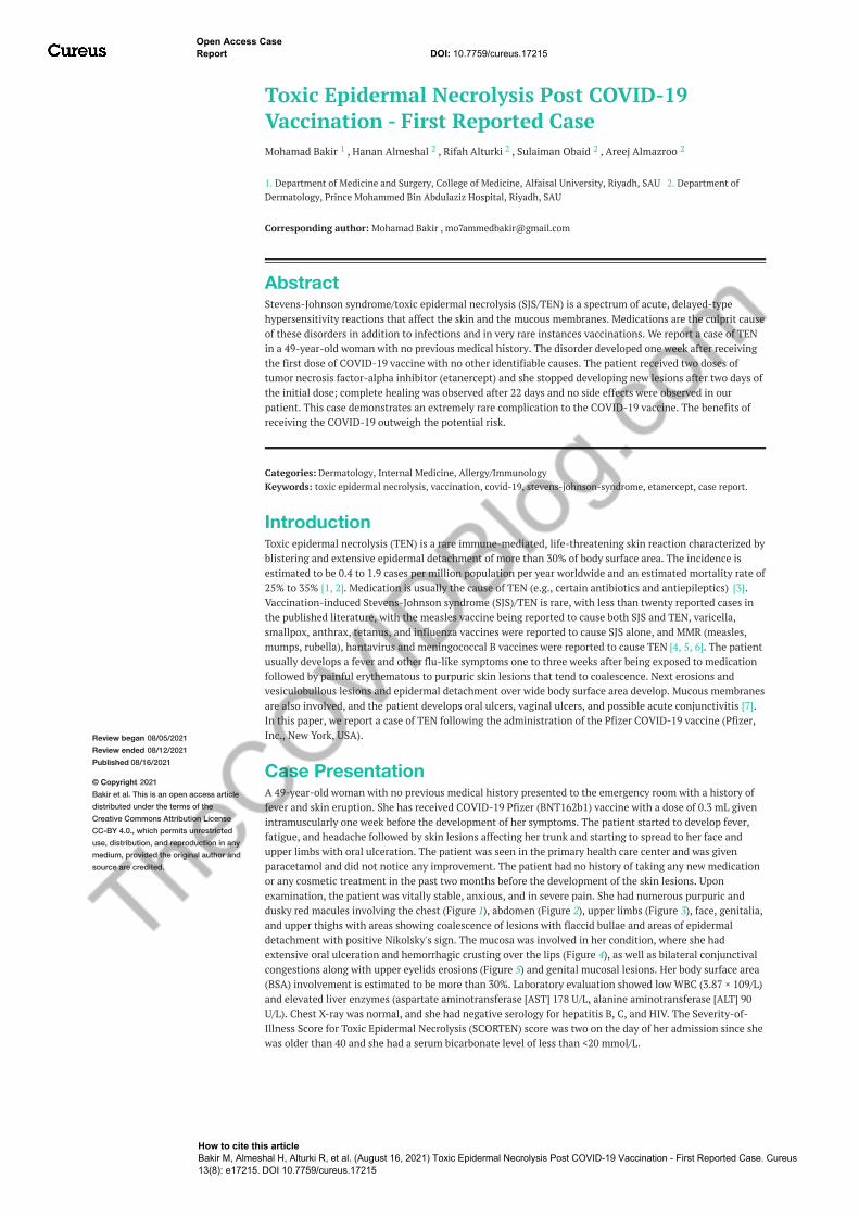
Toxic Epidermal Necrolysis Post COVID-19Vaccination - First Reported CaseMohamad Bakir , Hanan Almeshal , Rifah Alturki , Sulaiman Obaid , Areej Almazroo
1. Department of Medicine and Surgery, College of Medicine, Alfaisal University, Riyadh, SAU 2. Department ofDermatology, Prince Mohammed Bin Abdulaziz Hospital, Riyadh, SAU
Corresponding author: Mohamad Bakir , [email protected]
AbstractStevens-Johnson syndrome/toxic epidermal necrolysis (SJS/TEN) is a spectrum of acute, delayed-typehypersensitivity reactions that affect the skin and the mucous membranes. Medications are the culprit causeof these disorders in addition to infections and in very rare instances vaccinations. We report a case of TENin a 49-year-old woman with no previous medical history. The disorder developed one week after receivingthe first dose of COVID-19 vaccine with no other identifiable causes. The patient received two doses oftumor necrosis factor-alpha inhibitor (etanercept) and she stopped developing new lesions after two days ofthe initial dose; complete healing was observed after 22 days and no side effects were observed in ourpatient. This case demonstrates an extremely rare complication to the COVID-19 vaccine. The benefits ofreceiving the COVID-19 outweigh the potential risk.
Categories: Dermatology, Internal Medicine, Allergy/ImmunologyKeywords: toxic epidermal necrolysis, vaccination, covid-19, stevens-johnson-syndrome, etanercept, case report.
IntroductionToxic epidermal necrolysis (TEN) is a rare immune-mediated, life-threatening skin reaction characterized byblistering and extensive epidermal detachment of more than 30% of body surface area. The incidence isestimated to be 0.4 to 1.9 cases per million population per year worldwide and an estimated mortality rate of25% to 35% [1, 2]. Medication is usually the cause of TEN (e.g., certain antibiotics and antiepileptics) [3].Vaccination-induced Stevens-Johnson syndrome (SJS)/TEN is rare, with less than twenty reported cases inthe published literature, with the measles vaccine being reported to cause both SJS and TEN, varicella,smallpox, anthrax, tetanus, and influenza vaccines were reported to cause SJS alone, and MMR (measles,mumps, rubella), hantavirus and meningococcal B vaccines were reported to cause TEN [4, 5, 6]. The patientusually develops a fever and other flu-like symptoms one to three weeks after being exposed to medicationfollowed by painful erythematous to purpuric skin lesions that tend to coalescence. Next erosions andvesiculobullous lesions and epidermal detachment over wide body surface area develop. Mucous membranesare also involved, and the patient develops oral ulcers, vaginal ulcers, and possible acute conjunctivitis [7].In this paper, we report a case of TEN following the administration of the Pfizer COVID-19 vaccine (Pfizer,Inc., New York, USA).
Case PresentationA 49-year-old woman with no previous medical history presented to the emergency room with a history offever and skin eruption. She has received COVID-19 Pfizer (BNT162b1) vaccine with a dose of 0.3 mL givenintramuscularly one week before the development of her symptoms. The patient started to develop fever,fatigue, and headache followed by skin lesions affecting her trunk and starting to spread to her face andupper limbs with oral ulceration. The patient was seen in the primary health care center and was givenparacetamol and did not notice any improvement. The patient had no history of taking any new medicationor any cosmetic treatment in the past two months before the development of the skin lesions. Uponexamination, the patient was vitally stable, anxious, and in severe pain. She had numerous purpuric anddusky red macules involving the chest (Figure 1), abdomen (Figure 2), upper limbs (Figure 3), face, genitalia,and upper thighs with areas showing coalescence of lesions with flaccid bullae and areas of epidermaldetachment with positive Nikolsky's sign. The mucosa was involved in her condition, where she hadextensive oral ulceration and hemorrhagic crusting over the lips (Figure 4), as well as bilateral conjunctivalcongestions along with upper eyelids erosions (Figure 5) and genital mucosal lesions. Her body surface area(BSA) involvement is estimated to be more than 30%. Laboratory evaluation showed low WBC (3.87 × 10⁹/L)and elevated liver enzymes (aspartate aminotransferase [AST] 178 U/L, alanine aminotransferase [ALT] 90U/L). Chest X-ray was normal, and she had negative serology for hepatitis B, C, and HIV. The Severity-of-Illness Score for Toxic Epidermal Necrolysis (SCORTEN) score was two on the day of her admission since shewas older than 40 and she had a serum bicarbonate level of less than <20 mmol/L.
1 2 2 2 2
Open Access CaseReport DOI: 10.7759/cureus.17215
How to cite this articleBakir M, Almeshal H, Alturki R, et al. (August 16, 2021) Toxic Epidermal Necrolysis Post COVID-19 Vaccination - First Reported Case. Cureus13(8): e17215. DOI 10.7759/cureus.17215

FIGURE 1: Overall image of the chest before initiating the treatmentThe image shows multiple purpuric patches with epidermal detachment affecting the chest.
2021 Bakir et al. Cureus 13(8): e17215. DOI 10.7759/cureus.17215 2 of 10
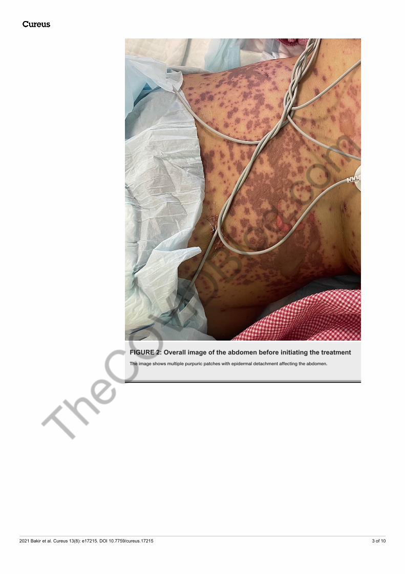
FIGURE 2: Overall image of the abdomen before initiating the treatmentThe image shows multiple purpuric patches with epidermal detachment affecting the abdomen.
2021 Bakir et al. Cureus 13(8): e17215. DOI 10.7759/cureus.17215 3 of 10
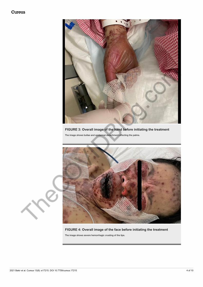
FIGURE 3: Overall image of the hand before initiating the treatmentThe image shows bullae and epidermal detachment affecting the palms.
FIGURE 4: Overall image of the face before initiating the treatmentThe image shows severe hemorrhagic crusting of the lips.
2021 Bakir et al. Cureus 13(8): e17215. DOI 10.7759/cureus.17215 4 of 10
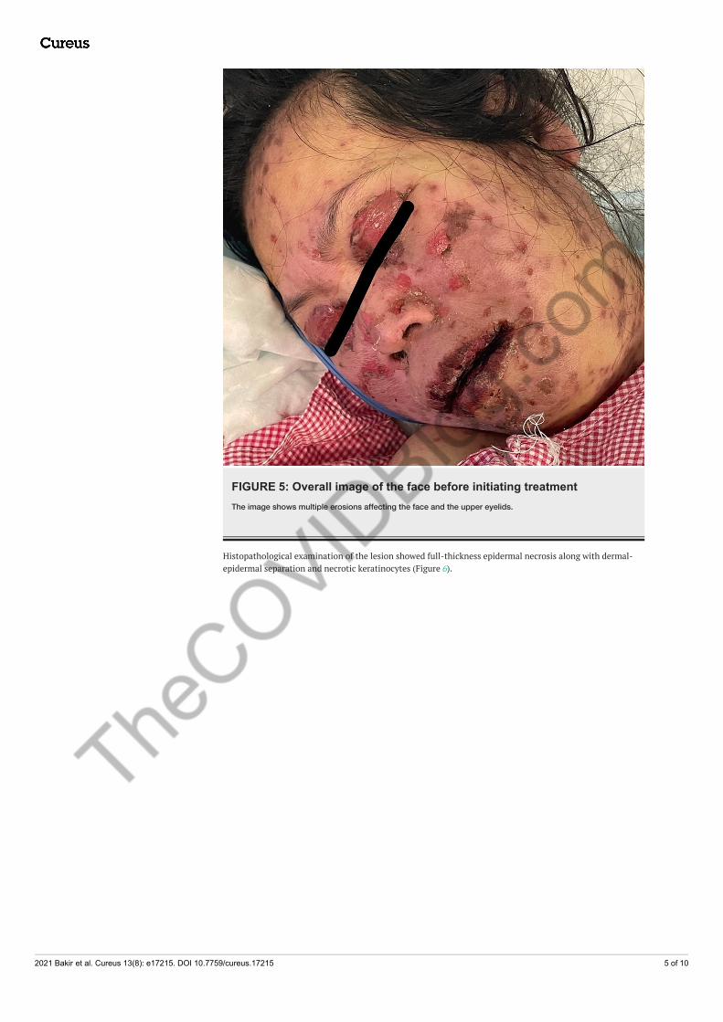
FIGURE 5: Overall image of the face before initiating treatmentThe image shows multiple erosions affecting the face and the upper eyelids.
Histopathological examination of the lesion showed full-thickness epidermal necrosis along with dermal-epidermal separation and necrotic keratinocytes (Figure 6).
2021 Bakir et al. Cureus 13(8): e17215. DOI 10.7759/cureus.17215 5 of 10
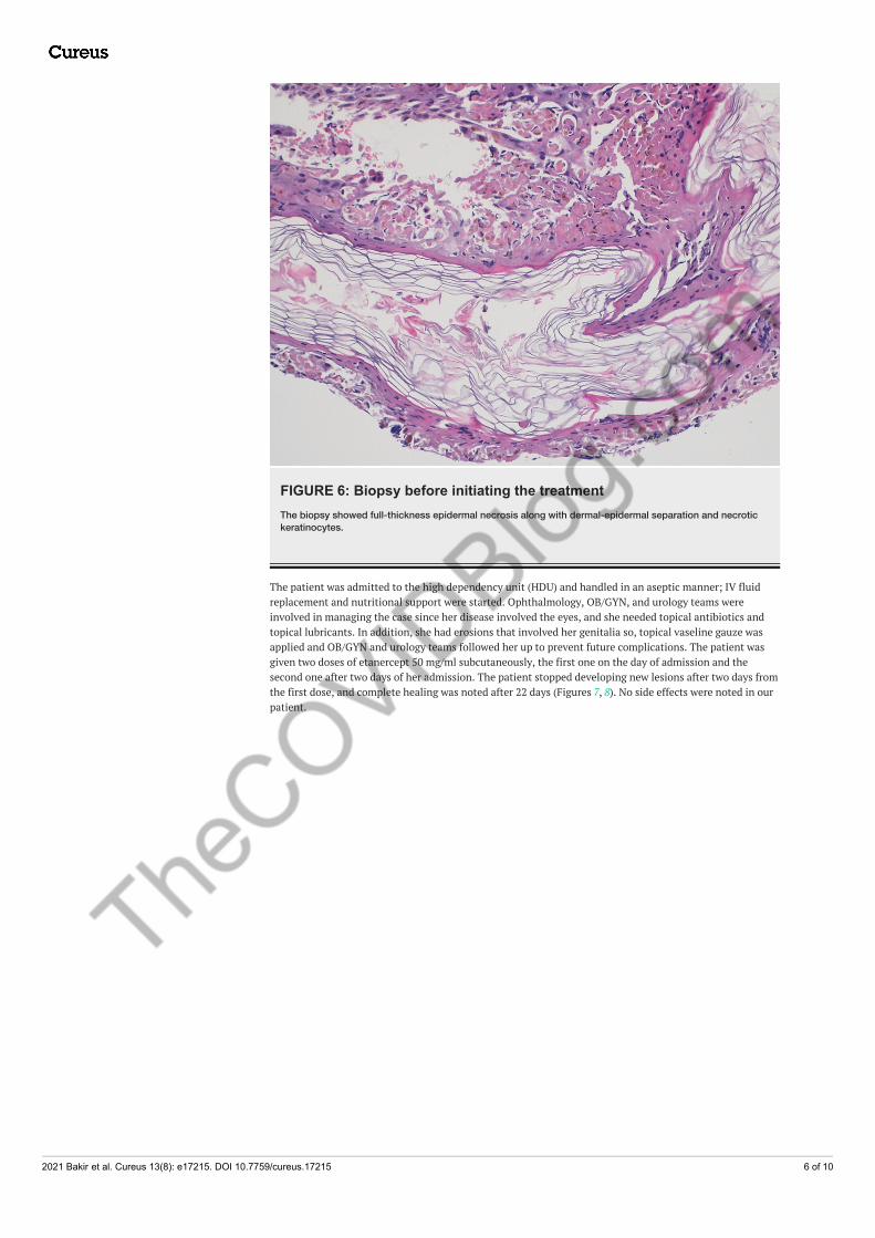
FIGURE 6: Biopsy before initiating the treatmentThe biopsy showed full-thickness epidermal necrosis along with dermal-epidermal separation and necrotickeratinocytes.
The patient was admitted to the high dependency unit (HDU) and handled in an aseptic manner; IV fluidreplacement and nutritional support were started. Ophthalmology, OB/GYN, and urology teams wereinvolved in managing the case since her disease involved the eyes, and she needed topical antibiotics andtopical lubricants. In addition, she had erosions that involved her genitalia so, topical vaseline gauze wasapplied and OB/GYN and urology teams followed her up to prevent future complications. The patient wasgiven two doses of etanercept 50 mg/ml subcutaneously, the first one on the day of admission and thesecond one after two days of her admission. The patient stopped developing new lesions after two days fromthe first dose, and complete healing was noted after 22 days (Figures 7, 8). No side effects were noted in ourpatient.
2021 Bakir et al. Cureus 13(8): e17215. DOI 10.7759/cureus.17215 6 of 10
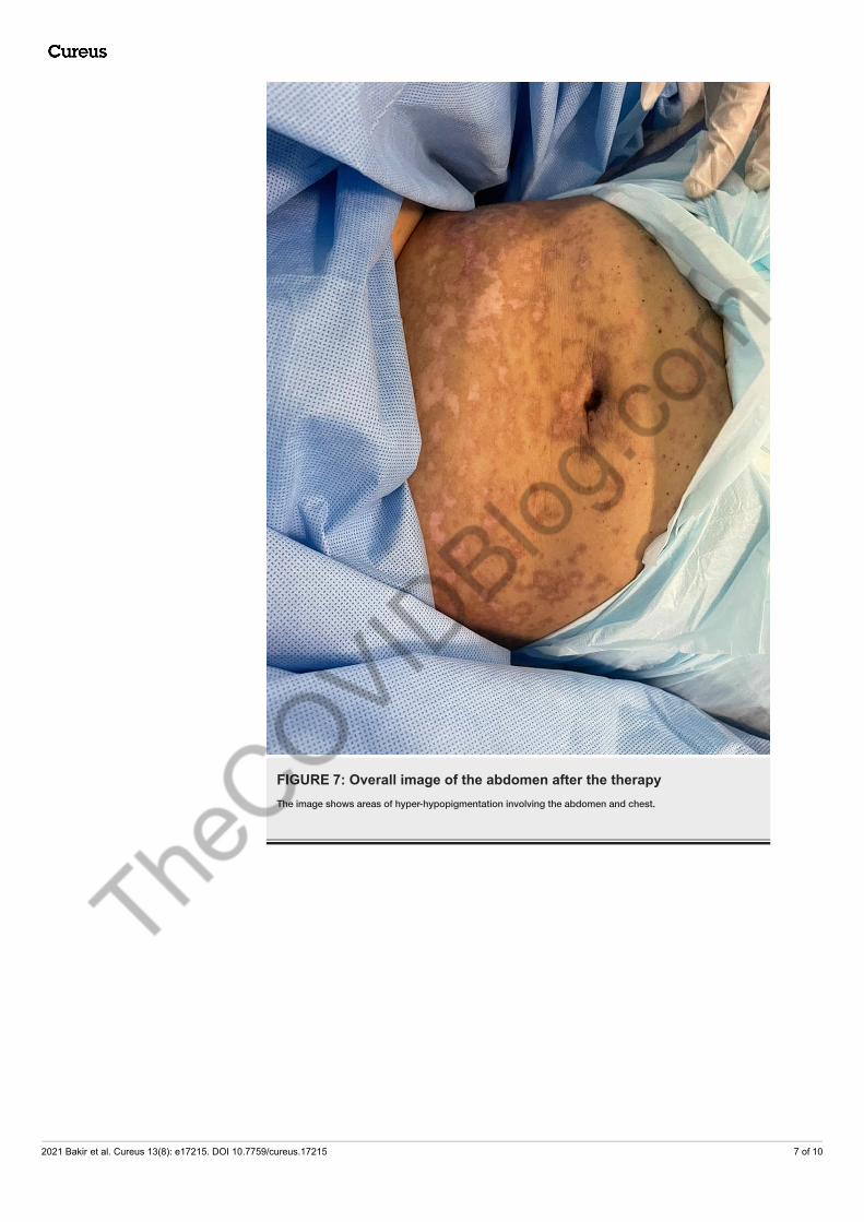
FIGURE 7: Overall image of the abdomen after the therapyThe image shows areas of hyper-hypopigmentation involving the abdomen and chest.
2021 Bakir et al. Cureus 13(8): e17215. DOI 10.7759/cureus.17215 7 of 10
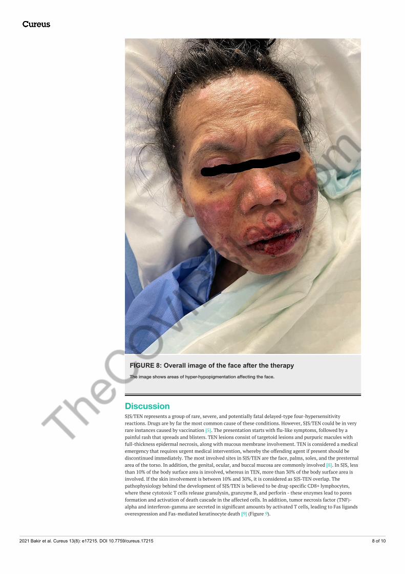
FIGURE 8: Overall image of the face after the therapyThe image shows areas of hyper-hypopigmentation affecting the face.
DiscussionSJS/TEN represents a group of rare, severe, and potentially fatal delayed-type four-hypersensitivityreactions. Drugs are by far the most common cause of these conditions. However, SJS/TEN could be in veryrare instances caused by vaccination [5]. The presentation starts with flu-like symptoms, followed by apainful rash that spreads and blisters. TEN lesions consist of targetoid lesions and purpuric macules withfull-thickness epidermal necrosis, along with mucous membrane involvement. TEN is considered a medicalemergency that requires urgent medical intervention, whereby the offending agent if present should bediscontinued immediately. The most involved sites in SJS/TEN are the face, palms, soles, and the presternalarea of the torso. In addition, the genital, ocular, and buccal mucosa are commonly involved [8]. In SJS, lessthan 10% of the body surface area is involved, whereas in TEN, more than 30% of the body surface area isinvolved. If the skin involvement is between 10% and 30%, it is considered as SJS-TEN overlap. Thepathophysiology behind the development of SJS/TEN is believed to be drug-specific CD8+ lymphocytes,where these cytotoxic T cells release granulysin, granzyme B, and perforin - these enzymes lead to poresformation and activation of death cascade in the affected cells. In addition, tumor necrosis factor (TNF)-alpha and interferon-gamma are secreted in significant amounts by activated T cells, leading to Fas ligandsoverexpression and Fas-mediated keratinocyte death [9] (Figure 9).
2021 Bakir et al. Cureus 13(8): e17215. DOI 10.7759/cureus.17215 8 of 10
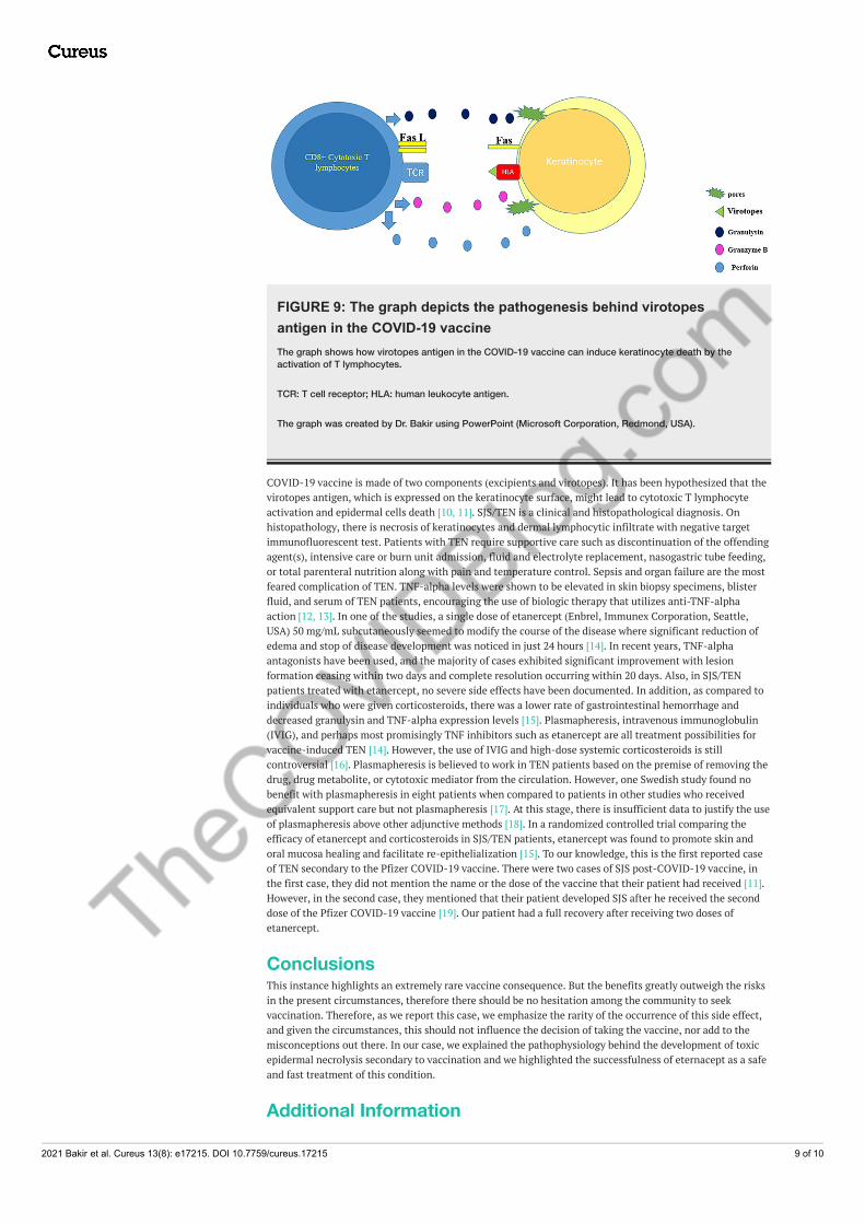
FIGURE 9: The graph depicts the pathogenesis behind virotopesantigen in the COVID-19 vaccineThe graph shows how virotopes antigen in the COVID-19 vaccine can induce keratinocyte death by theactivation of T lymphocytes.
TCR: T cell receptor; HLA: human leukocyte antigen.
The graph was created by Dr. Bakir using PowerPoint (Microsoft Corporation, Redmond, USA).
COVID-19 vaccine is made of two components (excipients and virotopes). It has been hypothesized that thevirotopes antigen, which is expressed on the keratinocyte surface, might lead to cytotoxic T lymphocyteactivation and epidermal cells death [10, 11]. SJS/TEN is a clinical and histopathological diagnosis. Onhistopathology, there is necrosis of keratinocytes and dermal lymphocytic infiltrate with negative targetimmunofluorescent test. Patients with TEN require supportive care such as discontinuation of the offendingagent(s), intensive care or burn unit admission, fluid and electrolyte replacement, nasogastric tube feeding,or total parenteral nutrition along with pain and temperature control. Sepsis and organ failure are the mostfeared complication of TEN. TNF-alpha levels were shown to be elevated in skin biopsy specimens, blisterfluid, and serum of TEN patients, encouraging the use of biologic therapy that utilizes anti-TNF-alphaaction [12, 13]. In one of the studies, a single dose of etanercept (Enbrel, Immunex Corporation, Seattle,USA) 50 mg/mL subcutaneously seemed to modify the course of the disease where significant reduction ofedema and stop of disease development was noticed in just 24 hours [14]. In recent years, TNF-alphaantagonists have been used, and the majority of cases exhibited significant improvement with lesionformation ceasing within two days and complete resolution occurring within 20 days. Also, in SJS/TENpatients treated with etanercept, no severe side effects have been documented. In addition, as compared toindividuals who were given corticosteroids, there was a lower rate of gastrointestinal hemorrhage anddecreased granulysin and TNF-alpha expression levels [15]. Plasmapheresis, intravenous immunoglobulin(IVIG), and perhaps most promisingly TNF inhibitors such as etanercept are all treatment possibilities forvaccine-induced TEN [14]. However, the use of IVIG and high-dose systemic corticosteroids is stillcontroversial [16]. Plasmapheresis is believed to work in TEN patients based on the premise of removing thedrug, drug metabolite, or cytotoxic mediator from the circulation. However, one Swedish study found nobenefit with plasmapheresis in eight patients when compared to patients in other studies who receivedequivalent support care but not plasmapheresis [17]. At this stage, there is insufficient data to justify the useof plasmapheresis above other adjunctive methods [18]. In a randomized controlled trial comparing theefficacy of etanercept and corticosteroids in SJS/TEN patients, etanercept was found to promote skin andoral mucosa healing and facilitate re-epithelialization [15]. To our knowledge, this is the first reported caseof TEN secondary to the Pfizer COVID-19 vaccine. There were two cases of SJS post-COVID-19 vaccine, inthe first case, they did not mention the name or the dose of the vaccine that their patient had received [11].However, in the second case, they mentioned that their patient developed SJS after he received the seconddose of the Pfizer COVID-19 vaccine [19]. Our patient had a full recovery after receiving two doses ofetanercept.
ConclusionsThis instance highlights an extremely rare vaccine consequence. But the benefits greatly outweigh the risksin the present circumstances, therefore there should be no hesitation among the community to seekvaccination. Therefore, as we report this case, we emphasize the rarity of the occurrence of this side effect,and given the circumstances, this should not influence the decision of taking the vaccine, nor add to themisconceptions out there. In our case, we explained the pathophysiology behind the development of toxicepidermal necrolysis secondary to vaccination and we highlighted the successfulness of eternacept as a safeand fast treatment of this condition.
Additional Information
2021 Bakir et al. Cureus 13(8): e17215. DOI 10.7759/cureus.17215 9 of 10
DisclosuresHuman subjects: Consent was obtained or waived by all participants in this study. Our hospital does notrequire an ethical approval for case report or case series issued approval N/A. Written informed consent wasobtained from the patient for publication of this case report and accompanying images. A copy of thewritten consent is available for review on request. Conflicts of interest: In compliance with the ICMJEuniform disclosure form, all authors declare the following: Payment/services info: All authors havedeclared that no financial support was received from any organization for the submitted work. Financialrelationships: All authors have declared that they have no financial relationships at present or within theprevious three years with any organizations that might have an interest in the submitted work. Otherrelationships: All authors have declared that there are no other relationships or activities that could appearto have influenced the submitted work.
References1. Schwartz RA, McDonough PH, Lee BW: Toxic epidermal necrolysis: Part I. Introduction, history,
classification, clinical features, systemic manifestations, etiology, and immunopathogenesis. J Am AcadDermatol. 2013, 69:173.e1-13; quiz 185-6. 10.1016/j.jaad.2013.05.003
2. Schwartz RA, McDonough PH, Lee BW: Toxic epidermal necrolysis: Part II. Prognosis, sequelae, diagnosis,differential diagnosis, prevention, and treatment. J Am Acad Dermatol. 2013, 69:187.e1-16; quiz 203-4.10.1016/j.jaad.2013.05.002
3. Mockenhaupt M: Epidemiology of cutaneous adverse drug reactions. Chem Immunol Allergy. 2012, 97:1-17.10.1159/000335612
4. Rosenblatt AE, Stein SL: Cutaneous reactions to vaccinations. Clin Dermatol. 2015, 33:327-32.10.1016/j.clindermatol.2014.12.009
5. Ball R, Ball LK, Wise RP, Braun MM, Beeler JA, Salive ME: Stevens-Johnson syndrome and toxic epidermalnecrolysis after vaccination: reports to the vaccine adverse event reporting system. Pediatr Infect Dis J.2001, 20:219-23. 10.1097/00006454-200102000-00022
6. Dobrosavljevic D, Milinkovic MV, Nikolic MM: Toxic epidermal necrolysis following morbilli-parotitis-rubella vaccination. J Eur Acad Dermatol Venereol. 1999, 13:59-61.
7. Harr T, French LE: Stevens-Johnson syndrome and toxic epidermal necrolysis. Chem Immunol Allergy.2012, 97:149-66. 10.1159/000335627
8. Grazina I, Mannocci A, Meggiolaro A, La Torre G: Is there an association between Stevens-JohnsonSyndrome and vaccination? A systematic review. Ann Ig. 2020, 32:81-96. 10.7416/ai.2020.2333
9. Oakley AM, Krishnamurthy K: Stevens Johnson Syndrome. StatPearls [Internet]. StatPearls Publishing (ed):Treasure Island, 2021; 2021 Jan-.
10. Stone CA Jr, Rukasin CR, Beachkofsky TM, Phillips EJ: Immune-mediated adverse reactions to vaccines. Br JClin Pharmacol. 2019, 85:2694-706. 10.1111/bcp.14112
11. Dash S, Sirka CS, Mishra S, Viswan P: Covid-19 vaccine induced Steven-Johnson syndrome: a case report .Clin Exp Dermatol. 2021, 10.1111/ced.14784
12. Paquet P, Paquet F, Al Saleh W, Reper P, Vanderkelen A, Piérard GE: Immunoregulatory effector cells indrug-induced toxic epidermal necrolysis. Am J Dermatopathol. 2000, 22:413-7. 10.1097/00000372-200010000-00005
13. Posadas SJ, Padial A, Torres MJ, et al.: Delayed reactions to drugs show levels of perforin, granzyme B, andFas-L to be related to disease severity. J Allergy Clin Immunol. 2002, 109:155-61. 10.1067/mai.2002.120563
14. Chahal D, Aleshin M, Turegano M, Chiu M, Worswick S: Vaccine-induced toxic epidermal necrolysis: a caseand systematic review. Dermatol Online J. 2018241, 13030-7. 10.5070/D3241037941
15. Wang CW, Yang LY, Chen CB, et al.: Randomized, controlled trial of TNF-α antagonist in CTL-mediatedsevere cutaneous adverse reactions. J Clin Invest. 2018, 128:985-96. 10.1172/JCI93349
16. St John J, Ratushny V, Liu KJ, et al.: Successful use of cyclosporin a for Stevens-Johnson syndrome and toxicepidermal necrolysis in three children. Pediatr Dermatol. 2017, 34:540-6. 10.1111/pde.13236
17. Furubacke A, Berlin G, Anderson C, Sjöberg F: Lack of significant treatment effect of plasma exchange in thetreatment of drug-induced toxic epidermal necrolysis?. Intensive Care Med. 1999, 25:1307-10.10.1007/s001340051063
18. Fernando SL: The management of toxic epidermal necrolysis . Australas J Dermatol. 2012, 53:165-71.10.1111/j.1440-0960.2011.00862.x
19. Elboraey MO, Essa EE: Stevens-Johnson syndrome post second dose of Pfizer COVID-19 vaccine: a casereport. Oral Surg Oral Med Oral Pathol Oral Radiol. 2021, 10.1016/j.oooo.2021.06.019
2021 Bakir et al. Cureus 13(8): e17215. DOI 10.7759/cureus.17215 10 of 10

![[Product Monograph Template - Standard] - Pfizer … (Phenytoin Sodium) Product Monograph Page 5 of 28 Stevens-Johnson Syndrome and Toxic Epidermal Necrolysis Serious and …](https://img.dokumen.tips/doc/110x75/5afc162a7f8b9a8b4d8bac14/product-monograph-template-standard-pfizer-phenytoin-sodium-product.jpg)