Embed Size (px)
Citation preview
ArcheoSciencesRevue d'archéométrie 35 | 2011Varia / International ArBoCo Workshop
Towards a Better Understanding of AlterationPhenomena of Archaeological Bone by a CloserLook at the Organic/Mineral Association at Micro-and Nanoscale. Preliminary Results on NeolithicSamples from Chalain Lake Site 19, Jura, FranceVers une meilleure compréhension des phénomènes d’altération des ossementsarchéologiques par une observation fine de l’association organique-minérale àmicro- et nanoéchelle. Premiers résultats obtenus sur des échantillonsnéolithiques de la station 19 du lac de Chalain, Jura, France
Ina Reiche, Céline Chadefaux, Katharina Müller and Aurélien Gourrier
Electronic versionURL: http://journals.openedition.org/archeosciences/3075DOI: 10.4000/archeosciences.3075ISBN: 978-2-7535-1849-0ISSN: 2104-3728
PublisherPresses universitaires de Rennes
Printed versionDate of publication: 30 April 2011Number of pages: 143-158ISBN: 978-2-7535-1847-6ISSN: 1960-1360
Electronic referenceIna Reiche, Céline Chadefaux, Katharina Müller and Aurélien Gourrier, “Towards a BetterUnderstanding of Alteration Phenomena of Archaeological Bone by a Closer Look at the Organic/Mineral Association at Micro- and Nanoscale. Preliminary Results on Neolithic Samples from ChalainLake Site 19, Jura, France”, ArcheoSciences [Online], 35 | 2011, Online since 30 April 2013, connectionon 16 February 2021. URL: http://journals.openedition.org/archeosciences/3075 ; DOI: https://doi.org/10.4000/archeosciences.3075
Article L.111-1 du Code de la propriété intellectuelle.
rec. jun. 2011 ; acc. nov. 2011 ArcheoSciences, revue d’archéométrie, 35, 2011, p. 143-158
Towards a Better Understanding of Alteration
Phenomena of Archaeological Bone by
a Closer Look at the Organic/Mineral Association
at Micro- and Nanoscale.
Preliminary Results on Neolithic Samples
from Chalain Lake Site 19, Jura, France
Vers une meilleure compréhension des phénomènes d’altération
des ossements archéologiques par une observation fi ne de l’association
organique-minérale à micro- et nanoéchelle. Premiers résultats obtenus
sur des échantillons néolithiques de la station 19 du lac de Chalain, Jura, France
Ina Reiche *, **, Céline Chadefaux *, Katharina Müller *et Aurélien Gourrier ***, ****
Abstract: In this paper we present the extension of existing analytical schemes for the precise evaluation of archaeological bone preservation states. Th e new methodological developments concerning the study of the morphological and structural features of archaeological bones at micro- and nanoscale are emphasized in order to elucidate fi ne diagenetic modifi cations and to better understand the underlying alteration mechanisms. A combination of synchrotron X-ray microtomography, infrared micro-spectroscopy imaging and quantitative scanning small-angle X-ray scatte-ring (sSAXS) imaging allowed studying micromorphological changes and the distribution of the organic and mineral components of bone at a histological level. Transmission electron microscopy gives precise information on changes of apatite crystal dimensions, orientations, periodicity of collagen fi ber arrangement and crystal distributions within the fi bers in archaeological bone. It allows establishing criteria to evaluate more precisely the preservation state of archaeological bone material with respect to modern references. Th e potential of these methods is highlighted on the basis o f the study of some macroscopically well preserved archaeological bone samples from the Chalain lake station 19, Neolithic period, Jura, France. Th e investigations allowed a diff erentiation in preservation state between these samples and the postulation of an alteration sequence in such humid chalk-rich burial environments.
* UMR 171 CNRS, Laboratoire du Centre de Recherche et de Restauration des Musées de France, Palais du Louvre, 14 quai François-Mitterrand, 75001 Paris, France.** Present address: Biomaterials department, Max-Planck-Institute of Colloids and Interfaces, Am Mühlenberg 1, 14476 Potsdam-Golm, Germany*** Laboratoire de Physique des Solides, UMR 8502 CNRS Université Paris-Sud, 91405 Orsay, France.**** European Synchrotron Radiation Facility, 38043 Grenoble, France.
144 Ina REICHE et al.
ArcheoSciences, revue d’archéométrie, 35, 2011, p. 143-158
1. INTRODUCTION
Bones or objects made of bone material are an important part of the archaeological witnesses and contribute largely to the understanding of ancient societies. In archaeological studies they are, among others, used to estimate the number of individuals that lived at a given archaeological site, [1] and to get information on the exploitation of animal troops by humans of past societies (mode of use, treating with pri-mary materials, fabrication techniques) [2-5]. As biological materials they also register in their chemical and isotopic composition a wealth of information on the life style of individuals [6-10]. More recently ancient DNA studies have also been used to extract information on past civilizations [11, 12].
Generally, archaeological materials are buried in soils or sediments and feature complex alteration phenomena. It is indispensable to study these modifi cations in order to better assess their original state, and thus to gather as much information as possible from them. Th ey are par-ticularly important for prehistoric times as there are no written sources and all information obtained on the past basically needs to be deduced from the material discov-ered and its spatial distribution on an archaeological site. Many studies have already been devoted to the assessment and the understanding of the evolution of bone materials in diff erent environments because of the importance of osseous remains in archaeology and paleontology [13-23]. Th ese studies allowed defi ning key “diagenetic parameters” (% collagen, mineral crystallinity index, CO3
2-/PO43- ratio,
Oxford Histological index (OHI), Cracking index, ‘s’, ‘m’ ‘l’ porosity, bulk density, skeletal density, amino acid
contents) through microscopic observations and physico-chemical measurements and, as a consequence, the esti-mation of the bone’s conservation state. Some “diagenetic trajectories” and even long term prediction models for the bone’s evolution in special burial conditions could be pos-tulated. General tendencies of alteration processes could be established and categorized as a function of the soil chemistry and the state of the bone remains before burial by means of a statistical approach [20, 21]. Important parameters to be considered for alteration phenomena are: taphonomic and hydrological conditions, soil composi-tion, biological activity and temperature. Th is approach is powerful to generalize the alteration phenomena but does not take into account the intrinsic particularities of each site. In particular, trace element concentrations, isotopic ratios, ancient DNA preservation, crystallinity state and the presence of special crystalline phases and their distri-butions in the bone material might be highly variable as a function of the biogeochemical conditions for each site, and even for diff erent stratigraphical units. Some cases of exceptional bone conservation, especially of its organic content, have been reported [24, 25]. Th erefore, it seems appropriate to study the conservation state of bone remains for each archaeological context and stratigraphical unit, where physico-chemical analyses are performed such as stable isotopic or carbon-14 dating studies etc. Th is allows estimating more reliably their informative potential.
Bone properties are essentially determined by its hierar-chical structure and the strong linkage of the organic and mineral phase at the nanoscale [26]. Consequently, it seems important to study the modifi cations induced by alteration phenomena of archaeological bone on the micro- and nano-
Résumé : Dans cet article nous présentons une extension de méthodes analytiques existantes pour évaluer précisément l’état de conversation des os archéologiques. Les nouveaux développements méthodologiques concernent particulièrement l’étude des caractéristiques structurales et morphologiques des ossements archéologiques aux échelles micro- et nanoscopiques afi n de pouvoir élucider des modifi cations diagénetiques fi nes et de mieux comprendre les mécanismes d’altération sous-jacents. Une combinaison de microtomographie X, de micro-spectroscopie infrarouge et de micro-imagerie SAXS à balayage au synchrotron a permis d’étudier les altérations micromorphologiques et la répartition spatiale des composés organiques et minéraux dans les os à l’échelle histologiques. La microscopie électronique à transmission donne des informations précises sur les modifi cations de la taille des cristaux, leur orientation, la périodicité des arrangements des fi bres collagéniques ainsi que la répartition des cristaux au sein des fi bres dans l’os archéologique. Ces observations ont permis d’établir des paramétres permettant l’évaluation fi ne des modifi cations diagénetiques de la structure des os archéologiques en comparaison aux références modernes. Le potentiel de ces méthodes est illustré grâce à l’étude d’os archéologiques qui sont bien préservés à l’échelle macroscopique provenant du site néolithique de Chalain 19, Jura, France. Cette investigation a permis de montrer des diff érences dans l’état de conservation de ces os et permet de proposer une séquence d’altération pour les os enfouis dans des environnements humides et riches en craie.
Keywords: archaeological bone, diagenesis, synchrotroCTmicroCT, synchrotron microFTIR, TEM, scanning-SAXS imaging.
Mots clés : os archéologique, diagenèse, microCT au synchrotron, imagerie microFTIR au synchrotron, MET, micro-imagerie SAXS.
Towards a Better Understanding of Alteration Phenomena of Archaeological Bone… 145
ArcheoSciences, revue d’archéométrie, 35, 2011, p. 143-158
level and to link these observations to those made on higher hierarchical levels.
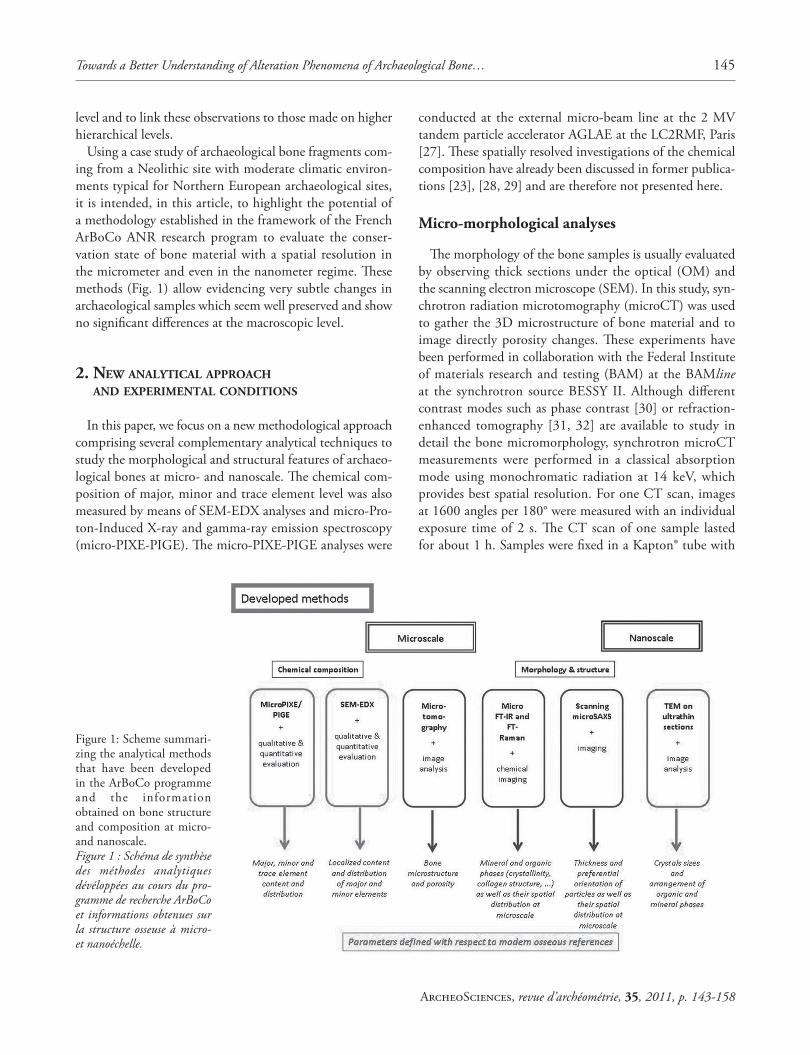
Using a case study of archaeological bone fragments com-ing from a Neolithic site with moderate climatic environ-ments typical for Northern European archaeological sites, it is intended, in this article, to highlight the potential of a methodology established in the framework of the French ArBoCo ANR research program to evaluate the conser-vation state of bone material with a spatial resolution in the micrometer and even in the nanometer regime. Th ese methods (Fig. 1) allow evidencing very subtle changes in archaeological samples which seem well preserved and show no signifi cant diff erences at the macroscopic level.
2. NEW ANALYTICAL APPROACH AND EXPERIMENTAL CONDITIONS
In this paper, we focus on a new methodological approach comprising several complementary analytical techniques to study the morphological and structural features of archaeo-logical bones at micro- and nanoscale. Th e chemical com-position of major, minor and trace element level was also measured by means of SEM-EDX analyses and micro-Pro-ton-Induced X-ray and gamma-ray emission spectroscopy (micro-PIXE-PIGE). Th e micro-PIXE-PIGE analyses were
conducted at the external micro-beam line at the 2 MV tandem particle accelerator AGLAE at the LC2RMF, Paris [27]. Th ese spatially resolved investigations of the chemical composition have already been discussed in former publica-tions [23], [28, 29] and are therefore not presented here.
Micro-morphological analyses
Th e morphology of the bone samples is usually evaluated by observing thick sections under the optical (OM) and the scanning electron microscope (SEM). In this study, syn-chrotron radiation microtomography (microCT) was used to gather the 3D microstructure of bone material and to image directly porosity changes. Th ese experiments have been performed in collaboration with the Federal Institute of materials research and testing (BAM) at the BAMline at the synchrotron source BESSY II. Although diff erent contrast modes such as phase contrast [30] or refraction-enhanced tomography [31, 32] are available to study in detail the bone micromorphology, synchrotron microCT measurements were performed in a classical absorption mode using monochromatic radiation at 14 keV, which provides best spatial resolution. For one CT scan, images at 1600 angles per 180° were measured with an individual exposure time of 2 s. Th e CT scan of one sample lasted for about 1 h. Samples were fi xed in a Kapton® tube with
Figure 1: Scheme summari-zing the analytical methods that have been developed in the ArBoCo programme and the information obtained on bone structure and composition at micro- and nanoscale.Figure 1 : Schéma de synthèse des méthodes analytiques dévéloppées au cours du pro-gramme de recherche ArBoCo et informations obtenues sur la structure osseuse à micro- et nanoéchelle.
146 Ina REICHE et al.
ArcheoSciences, revue d’archéométrie, 35, 2011, p. 143-158
a diameter of 1 mm adapted to the size of the bone frag-ment or object under investigation. Th e set-up is explained in detail in the following references [33, 34]. Th e voxel data were reconstructed with a fi ltered-backprojection algo-rithm and visualised using the software VGStudio Max 2.1 (VolumeGraphics GmbH, Heidelberg/Germany). Th e reconstructed volumes have a voxel size of about 0.4 μm [35, 36]. Generally, virtual sections (2D images) of the samples are represented in this paper.
Micro-structural investigations
Fourier-transform Infrared Spectroscopy (FT-IR) analysis and X-ray Diff raction (XRD) generally allow the structural analysis at the atomic level of the main components present in the bone mineral. Th ese techniques permit the determi-nation of a special index indicating the degree of mineral alteration in bone described by the “splitting factor” (SF) and the “crystallinity index” (CI), respectively [14, 37]. Th e principal organic phase of bone, collagen, can also be ana-lyzed at the molecular level by FT-IR by means of the detec-tion of the amid absorption bands. Th ese bands refl ect amid polypeptide groups and the lateral chains of amino acids. Th e secondary protein structure can also be analyzed. Th us, it is possible to detect organic residues in ancient bones and to evaluate the integrity of the preserved organic phase [38].
Within the ArBoCo research programme, we developed a method in order to obtain spatially resolved informa-tion of both, the bone organic and mineral phases. For this issue, synchrotron microFT-IR seemed to be well suited for studying the heterogeneity of the preservation state of archaeological bones at microscale. Th e experiments were performed at the IRIS beamline (BESSY II, HZB, Berlin, Germany) on a Th ermo Nicolet ContinuumTM microscope equipped with a MCT detector. Th e sample was mounted on a 1 mm thick BaF2 pellet on a motorized microscope stage and raster scanned through the synchrotron beam with a diameter of 10 μm collecting a grid-like pattern of
IR spectra with increments of 7 μm. Measurements were performed in transmission mode at a magnifi cation of x32 using confocal objectives. Infrared spectra have been regis-tered between 4000 and 650 cm-1 with a spectral resolution of 8 cm-1. For each measuring point, 128 scans were accu-mulated. Background spectra were collected under identical conditions in constant intervals. Th e spectrum acquisition has been performed by using the OMNIC AtlμsTM soft-ware [39]. Spectral data analysis was performed using the OMNIC 7.4 (Th ermo) and OPUS (Bruker Optic GmbH) software.
Th e spectra were baseline corrected (by means of the concave rubberband correction) in the spectral region between 2000-800 cm-1. Subsequently, the contribution of the impregnation resin (indicator = vibration band at around 1730 cm-1) was subtracted as indicated in [40]. Th e table 1 shows the IR parameters used for imaging. Th e visualization of IR parameter distributions in the scanned zone was performed with the help of MATLAB 7.7 software (MathsWorks).
Scanning-SAXS measurements were performed at MySpot beamline at BESSY II, Berlin, Germany. SAXS patterns were obtained with a 30x30 μm2 X-ray microbeam on 40 μm thick sections cut in the central part of the archaeological bone specimens [41]. Experimental conditions are analo-gous to those described in [41, 43]. A W/Si (311) multilayer monochromator was used to monochromatize X-rays to an energy of 15keV (λ=0.8265 nm). Th e SAXS patterns were recorded with a 16 bit CCD area detector (MarMosaic 225, Rayonix, Evanston, USA) with a 225 x 225 mm2 square converter screen. Th e images were recorded with a readout time of ~ 2.5 s at full resolution (3072 x 3072 pixels with a pixel size of 73.24 x 73.24 μm2) and the sample-detector distance as well as the detector tilt and center were determi-ned by measuring a silver behenate standard.
Prior to the SAXS measurements a transmission scan of the sample is performed using a photodiode.
IR parameters Intensity ratio of vibration bands Baseline / cm-1
Collagen/PO4 I (max 1670-1630) / I 1095 1800-1300 / 1175-900CO3/PO4 I 1415 (max 1428-1398) / I 1095 1800-1300 /1175-900Crystallinity I 1030 / I 1020 1175-900HPO4/PO4 I 1106 / I 1095 1175-900Collagen integrity I 1690 / I 1660 1800-1300Random coil / α-Helix I 1645 / I 1660 1800-1300
Table 1: Selected IR parameters for imaging.Tableau 1 : Paramètres IR défi nis pour l’imagerie.
Towards a Better Understanding of Alteration Phenomena of Archaeological Bone… 147
ArcheoSciences, revue d’archéométrie, 35, 2011, p. 143-158
Th e SAXS data analysis included the spherical integra-tion along the azimuthal direction of the two-dimensional SAXS patterns using the FIT2D software package [42]. Th e one-dimensional profi les were subsequently analyzed using a dedicated SAXS analysis library written in Python language by A. Gourrier optimized for large data sets [43, 44]. Th e structural parameters relating to the mineral nanoparticle thickness can thus be determined following procedures esta-blished by Fratzl et al. for bone studies (see, e.g. [45]). Th e average chord length (so-called T parameter) can be consi-dered as a standard parameter in the SAXS analysis of bone from archaeological contexts [46].
Nanostructural investigations
Transmission Electron Microscopy coupled with an Energy-Dispersive X-ray System (TEM-EDX) was used to characterize specifi c apatite crystal shapes and sizes as well as the localization of the crystals within the collagen fi ber matrix [47]. Transmission Electron Microscopy gives loca-lized information whereas 2D scanning Small-Angle X-ray Scattering (sSAXS) imaging provides a more global view on the texture and the crystal size distribution in the bone material.
Sample observation using TEM, giving direct evidence of the preservation state of the organic and mineral bone phases, was carried out by means of a Philips EM208 micro-scope at 80 kV at the Common centre of electron micros-copy (CCME) UMR 8080 CNRS, Orsay, France.
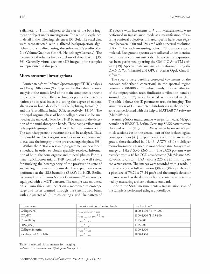
According to a study by Rubin et al. (2003) [48] in longi-tudinal ultrathin sections of normal and osteoporotic human bone the apatite crystals are oriented parallel following the collagen fi bers. To estimate the degree of disorientation and alteration we decided to prepare ultrathin longitudinal sec-tions for the direct observation of the mineralized collagen.
Parameters have been established to characterize at the nanoscale the state of preservation of the organic-mineral arrangement in the archaeological material: Ls represents the width of the deposited crystals at the surface of the fi bers; Lf the width of collagen fi bers; Λ the spatial periodicity of the crystals in the fi bers; e the thickness of the crystallites and l the width of the crystals (fi g. 2a and b).
Preparation of archaeological bone sections and thin sections
It has to be pointed out that the sample preparation procedures were crucial steps for all analyses at the micro-
Figure 2: a) Electron micrographs on a micrograph of an ultrathin section of a modern antler MA. b) Indications of characteristic param-eters (Ls: width of deposited crystals at the surface of the fi bers, Lf: width of collagen fi bers, Λ: spatial periodicity of the crystals in the fi bers, e: thickness of the crystallites, l: width of the crystals).Figure 2 : a) Micrographie électronique d’une coupe ultrafi ne de l’os bovin moderne de référence MA. b) Indications des paramètres caractéristiques (Ls: largeur des cristaux déposés à la surface des fi bres, Lf: largeur des fi bres de collagène, Λ: periodicité spatiale des cristaux dans les fi bres, e: épaisseur des cristallites, l: largeur des cristaux) sur une micrographie électronique d’une coupe ultrafi ne d’un bois de renne moderne.

148 Ina REICHE et al.
ArcheoSciences, revue d’archéométrie, 35, 2011, p. 143-158
and the nanoscale. Indeed, the preparation procedure com-prised partly embedding, ultramicrotomy and polishing in order to keep all necessary information on the structure at micro- and nanoscale and on the texture of the material. It is important that the procedure does not change the features of the samples and does not introduce artefacts into the sample.
Fragments of two archaeological cortical bones (AB_CH19nb1 and nb2) were prepared for the analyses. First, thick bone sections were cut with a water-cooled diamond saw. Sections were subsequently ultrasonically rinsed with water, distilled water and ethanol, before they were dried in air. For synchrotron microFTIR, transverse thin sections with a thickness of less than 2 μm were realized on in poly-methyl methacrylate (PMMA) embedded bone material using a ReichertTM ultramicrotome equipped with a glass knife set (at CCME, Orsay) according to the procedure described in [49].
Ultrathin bone sections of approximately 70 nm thick-ness could be directly cut without embedding the bone fragment with a diamond knife on a Reichert Ultracut E Ultramicrotome at CCME, Orsay and picked up on 200-mesh copper grids coated with a membrane of carbon Formwar for TEM observations. Th e cut orientation was chosen to be longitudinal to better visualize the texture of mineralized collagen fi brils in bone. Th ree grids were pre-pared for the bone sample.
For SAXS measurements, bone fragments of about 1 mm thickness were cut and subsequently machine-ground using SiC grained paper of the fi neness of 800, 1200 and 4000 before being rinsed with distilled water. Th e sections were glued with superglue on a glass slide and polished using a LAM PLAN M.M.8027S machine with SiC in water as polishing liquid down to a thickness of about 40 μm. Th e fi nal polishing was obtained by diamond paste polishing to a precision of 0.25 μm.
Possible eff ects of the sample preparation
Sample preparation is a key step in the analyses of slight changes in the preservation state of bone materials. Indeed, it is fundamental to avoid any changes of these fragile and precious remains induced by sample preparation procedures in order to conserve the state of the material. However, most of archaeological bone materials are altered and therefore brittle. Measurements conducted here show that impregna-tion treatment can be avoided in some cases if the mineral and the organic parts are still conserved in the considered bone materials to provide cohesion of the bone structure but generally more reliable results can be obtained on in
PMMA embedded samples. However, the main drawback of the embedding procedure is that the analysis needs to take into account the resin contribution in the spectra.
3. MATERIAL
Th e Neolithic bone material originates from the lacustrine sites of the Chalain lake, Jura, France. At this lake situated in the Franche-Comté region, over thirty archaeological sites were discovered during the excavations and prospec-tion carried out by Pierre Pétrequin and his team [50]. Seven Neolithic villages (3850 to 2650 BC) have been excavated there within the past thirty years. Multidisciplinary research projects have been undertaken in order to take advantage of the archaeological, biological, chemical, and geological information contained in the objects found at these sites (see e.g. [51]). Research on diagenetic modifi cations of bone remains are integrated in the context of these investigations. At Chalain, the bones are generally found to be well pre-served by optical observations. According to archaeologi-cal observations, the bone fragments originating from the preparation of meals (butchering and cooking) were dis-carded in the dumps in front of the only entrance to the houses on pilings, built on fl ood prone ground. Th is refuse (including the bone remains) therefore fell onto humid soil or into shallow water. Th ey were quickly covered by the vegetal litter brought in by humans to stabilize and reclaim the exterior soils during low water periods. After the villages were abandoned, the lake’s level rose again and lake chalk was deposited.
During the excavation campaign conducted in 1998, some fragments of burned and unburned bones from the emerged station 19 were entrusted to us for analysis [28]. Selected results obtained by the new analytical strategy on two compact bone fragments AB_CH19nb1 and nb2, pro-bably Bos taurus [52] (fi g. 3) are presented here in compari-son to those obtained on a modern bovine reference sample
Figure 3: (See colour plate) Archaeological bone samples from lacustrine Chalain site 19.Figure 3 : (Voir planche couleur) échantillons d’os archéologiques du site lacustre 19 de Chalain.

Towards a Better Understanding of Alteration Phenomena of Archaeological Bone… 149
ArcheoSciences, revue d’archéométrie, 35, 2011, p. 143-158
MBB. It is of importance to provide a modern bone control as close as possible to the archaeological bone samples under investigation. Th erefore, a bovine bone reference was chosen in this study, which has been submitted to the same type of thorough investigations as the archaeological specimens in order to diff erentiate variations in the defi ned parameters within the modern sample from possible diagenetic changes at micro- and nanoscale in the archaeological ones. Th e archaeological samples have been selected because they show not only a very good preservation state at macroscale but also little diff erences at the histological level as evidenced by SEM observations on cross sections (fi g. 4). Th e newly esta-blished approach should provide a better description of the samples at micro- and nanoscale in order to better asses their preservation state, which is relevant for the understanding of the underlying alteration phenomena and for a better eva-luation of their informative potential in archaeological stu-dies. Th e archaeological samples (AB_CH19nb1 and nb2) originate from layers H and K of station 19 of Chalain, and are dated to 3033-2886 calBC (86.8 %) and 3141-3002 calBC (41.4%), respectively [53]. Th e layers H and K cor-respond to remains of villages with raised fl oor houses with sediments of anthropic dung that contain a small percentage of calcium carbonates deposited by the lake.
4. RESULTS
Determination of the micromorphological changes using synchrotron microtomography
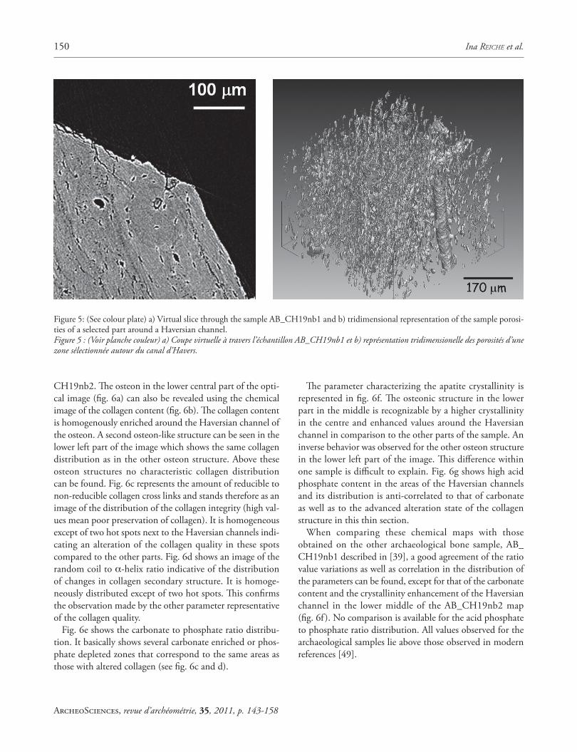
Th e study of the preservation state of the micromor-phology could be performed on one archaeological sample AB_CH19nb1 using synchrotron microCT (fi g. 5). Th ese investigations allowed evidencing the micromorphological structure of this sample. Th e information is equivalent to
those that can be obtained on cross sections by SEM (fi g. 4) but with a tridimensional resolution so that virtual section can be observed in several depths of the sample. A typi-cal bone structure with secondary osteons can be clearly observed in fi g. 5a.
Th e regular structure of a Haversian system can be seen as well as other intrinsic bone porosities in another representa-tion evidencing bone porosities (fi g. 5b). Th ey seem smaller in comparison to the modern bone reference measured. Th e porosity estimates based on the image analysis for the vol-umes selected are summarized in the table 2. Th e volume p of open pores is calculated as a ratio with respect to the total volume according to the following formula. Th ese cal-culations show smaller values for the archaeological sample compared to the modern one.
Spatially resolved analysis of the organic and mineral content at microscale using synchrotron microFTIR in transmission mode
Th e IR maps and the corresponding optical images of the archaeological bone thin sections AB_CH19nb1 and nb2 are reported in [39] and represented in the fi g. 6a-g, respectively. Th e fi gure 6b represents the distribution of the collagen content in the bone thin section of the sample AB_
Figure 4: Electron micrographs in secondary electron mode of the archaeological bone thick sections of a) AB_CH19nb1 and b) AB_CH19nb2.Figure 4 : Micrographies électroniques en mode d’électrons secon-daires des coupes épaisses des os archéologiques : a) AB_CH19nb1 et b) AB_CH19nb2.
Sample Porosity estimation (%)MBB 7.4 ± 0.5
AB_CH19nb1 4.9 ± 0.5
Table 2: Porosity estimated by image analysis of the archaeological bone sample AB_CH19nb1 compared to that of the measured rence AB_CH19nb1 and MBB.Tableau 2 : Porosité estimée par analyse d’image de l’échantillon d’os archéologique AB_CH19nb1 comparé à celle de la référence moderne meurée MBB.
150 Ina REICHE et al.
ArcheoSciences, revue d’archéométrie, 35, 2011, p. 143-158
CH19nb2. Th e osteon in the lower central part of the opti-cal image (fi g. 6a) can also be revealed using the chemical image of the collagen content (fi g. 6b). Th e collagen content is homogenously enriched around the Haversian channel of the osteon. A second osteon-like structure can be seen in the lower left part of the image which shows the same collagen distribution as in the other osteon structure. Above these osteon structures no characteristic collagen distribution can be found. Fig. 6c represents the amount of reducible to non-reducible collagen cross links and stands therefore as an image of the distribution of the collagen integrity (high val-ues mean poor preservation of collagen). It is homogeneous except of two hot spots next to the Haversian channels indi-cating an alteration of the collagen quality in these spots compared to the other parts. Fig. 6d shows an image of the random coil to α-helix ratio indicative of the distribution of changes in collagen secondary structure. It is homoge-neously distributed except of two hot spots. Th is confi rms the observation made by the other parameter representative of the collagen quality.
Fig. 6e shows the carbonate to phosphate ratio distribu-tion. It basically shows several carbonate enriched or phos-phate depleted zones that correspond to the same areas as those with altered collagen (see fi g. 6c and d).
Th e parameter characterizing the apatite crystallinity is represented in fi g. 6f. Th e osteonic structure in the lower part in the middle is recognizable by a higher crystallinity in the centre and enhanced values around the Haversian channel in comparison to the other parts of the sample. An inverse behavior was observed for the other osteon structure in the lower left part of the image. Th is diff erence within one sample is diffi cult to explain. Fig. 6g shows high acid phosphate content in the areas of the Haversian channels and its distribution is anti-correlated to that of carbonate as well as to the advanced alteration state of the collagen structure in this thin section.
When comparing these chemical maps with those obtained on the other archaeological bone sample, AB_CH19nb1 described in [39], a good agreement of the ratio value variations as well as correlation in the distribution of the parameters can be found, except for that of the carbonate content and the crystallinity enhancement of the Haversian channel in the lower middle of the AB_CH19nb2 map (fi g. 6f ). No comparison is available for the acid phosphate to phosphate ratio distribution. All values observed for the archaeological samples lie above those observed in modern references [49].
Figure 5: (See colour plate) a) Virtual slice through the sample AB_CH19nb1 and b) tridimensional representation of the sample porosi-ties of a selected part around a Haversian channel.Figure 5 : (Voir planche couleur) a) Coupe virtuelle à travers l’échantillon AB_CH19nb1 et b) représentation tridimensionelle des porosités d’une zone sélectionnée autour du canal d’Havers.

Towards a Better Understanding of Alteration Phenomena of Archaeological Bone… 151
ArcheoSciences, revue d’archéométrie, 35, 2011, p. 143-158
Figure 6: a) (See colour plate) Optical image of the zone selected on the archaeological bone thin section AB_CH19nb2 from the Chalain Lake site corresponding to an area with visible osteons. Image obtained from the microscope Continuum™ (Nicolet) at the IRIS Beamline (BESSY II, Berlin) and showing the points analyzed. Represented area corresponds to 120×220 μm2. SR microFTIR maps of the same zone b) of the amide I/phosphate ratio (ca. 1660/1095), Chalain Lake site, c) the collagen cross-links represented by the 1690/1660 ratio corresponding to reducible to non-reducible cross-links, d) the ratio of random coils to α-helix subbands (1645/1660), e) the carbonate/phosphate ratio (ca. 1415/1095), f ) the apatite crystallinity (1030/1020) and g) the acid phosphate to phosphate ratio (1106/1095). A false colour scale from red (high intensity) to blue (low intensity) was adopted for all images.Figure 6 : (Voir planche couleur) a) Image optique d’une zone selectionnée d’une coupe fi ne de l’os archéologique AB_CH19nb2 du lac de Chalain correspondant à une zone avec des osteons visibles. Image obtenue grâce au microscope Continuum™ (Nicolet) à la ligne IRIS (BESSY II, Berlin) montrant les points analysés. La zone représentée correspond à 120×220 μm2. Des cartographies en microIRTF de la même zone b) du rapport amide I/phosphate (ca. 1660/1095), c) des cross-links du collagène representés par le rapport 1690/1660 correspondant au rapport entre les cross-links reductibles et non-reductibles, d) du rapport (1645/1660), e) du rapport carbonate sur phosphate (ca. 1415/1095), f ) de la cristallinité de l’apatite (1030/1020) et g) du rapport hydrophosphate au phosphate (1106/1095). Une échelle en fausses couleurs allant du rouge (intensité forte) au bleu (intensité faible) a été adoptée pour toutes les images.
a b c
d e f
g
152 Ina REICHE et al.
ArcheoSciences, revue d’archéométrie, 35, 2011, p. 143-158
Statistical view of crystalline structure of the nanoparticles by means of scanning-SAXS imaging
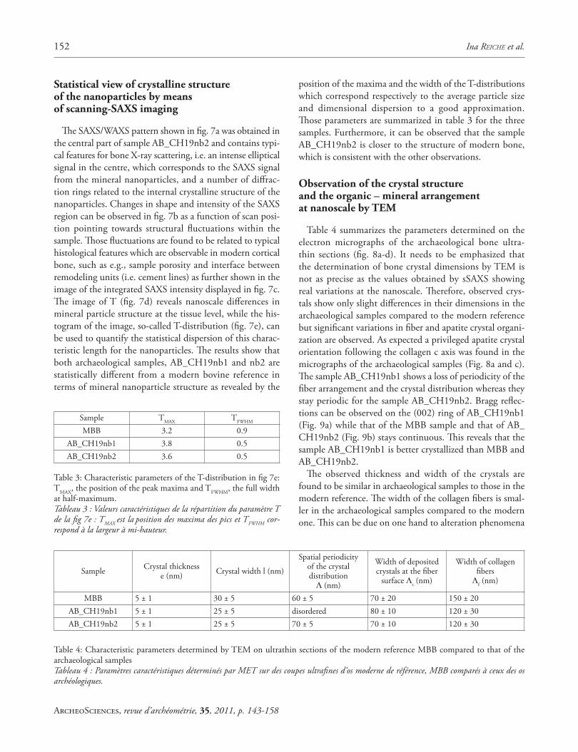
Th e SAXS/WAXS pattern shown in fi g. 7a was obtained in the central part of sample AB_CH19nb2 and contains typi-cal features for bone X-ray scattering, i.e. an intense elliptical signal in the centre, which corresponds to the SAXS signal from the mineral nanoparticles, and a number of diff rac-tion rings related to the internal crystalline structure of the nanoparticles. Changes in shape and intensity of the SAXS region can be observed in fi g. 7b as a function of scan posi-tion pointing towards structural fl uctuations within the sample. Th ose fl uctuations are found to be related to typical histological features which are observable in modern cortical bone, such as e.g., sample porosity and interface between remodeling units (i.e. cement lines) as further shown in the image of the integrated SAXS intensity displayed in fi g. 7c. Th e image of T (fi g. 7d) reveals nanoscale diff erences in mineral particle structure at the tissue level, while the his-togram of the image, so-called T-distribution (fi g. 7e), can be used to quantify the statistical dispersion of this charac-teristic length for the nanoparticles. Th e results show that both archaeological samples, AB_CH19nb1 and nb2 are statistically diff erent from a modern bovine reference in terms of mineral nanoparticle structure as revealed by the
position of the maxima and the width of the T-distributions which correspond respectively to the average particle size and dimensional dispersion to a good approximation. Th ose parameters are summarized in table 3 for the three samples. Furthermore, it can be observed that the sample AB_CH19nb2 is closer to the structure of modern bone, which is consistent with the other observations.
Observation of the crystal structure and the organic – mineral arrangement at nanoscale by TEM
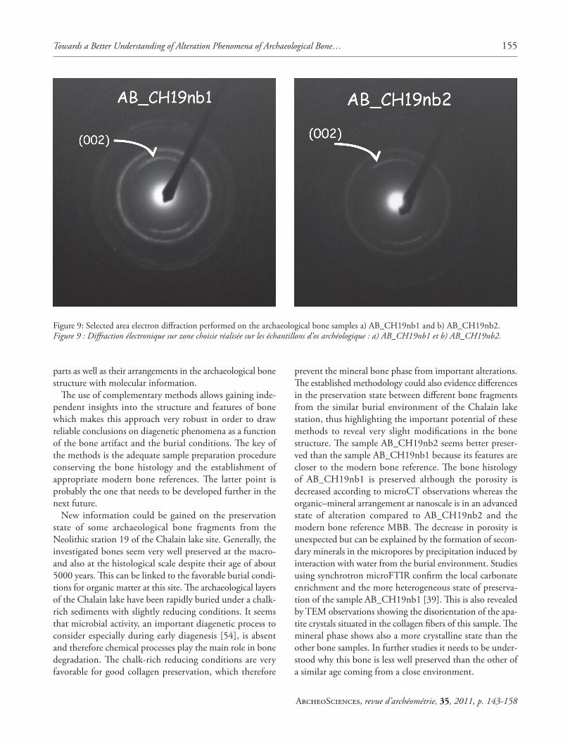
Table 4 summarizes the parameters determined on the electron micrographs of the archaeological bone ultra-thin sections (fi g. 8a-d). It needs to be emphasized that the determination of bone crystal dimensions by TEM is not as precise as the values obtained by sSAXS showing real variations at the nanoscale. Th erefore, observed crys-tals show only slight diff erences in their dimensions in the archaeological samples compared to the modern reference but signifi cant variations in fi ber and apatite crystal organi-zation are observed. As expected a privileged apatite crystal orientation following the collagen c axis was found in the micrographs of the archaeological samples (Fig. 8a and c). Th e sample AB_CH19nb1 shows a loss of periodicity of the fi ber arrangement and the crystal distribution whereas they stay periodic for the sample AB_CH19nb2. Bragg refl ec-tions can be observed on the (002) ring of AB_CH19nb1 (Fig. 9a) while that of the MBB sample and that of AB_CH19nb2 (Fig. 9b) stays continuous. Th is reveals that the sample AB_CH19nb1 is better crystallized than MBB and AB_CH19nb2.
Th e observed thickness and width of the crystals are found to be similar in archaeological samples to those in the modern reference. Th e width of the collagen fi bers is smal-ler in the archaeological samples compared to the modern one. Th is can be due on one hand to alteration phenomena
Sample TMAX TFWHM
MBB 3.2 0.9AB_CH19nb1 3.8 0.5AB_CH19nb2 3.6 0.5
Table 3: Characteristic parameters of the T-distribution in fi g 7e: TMAX, the position of the peak maxima and TFWHM, the full width at half-maximum.Tableau 3 : Valeurs caractéristiques de la répartition du paramètre T de la fi g 7e : TMAX est la position des maxima des pics et TFWHM cor-respond à la largeur à mi-hauteur.
Sample Crystal thickness e (nm) Crystal width l (nm)
Spatial periodicity of the crystal distribution Λ (nm)
Width of deposited crystals at the fi ber
surface Λs (nm)
Width of collagen fi bers Λf (nm)
MBB 5 ± 1 30 ± 5 60 ± 5 70 ± 20 150 ± 20AB_CH19nb1 5 ± 1 25 ± 5 disordered 80 ± 10 120 ± 30AB_CH19nb2 5 ± 1 25 ± 5 70 ± 5 70 ± 10 120 ± 30
Table 4: Characteristic parameters determined by TEM on ultrathin sections of the modern reference MBB compared to that of the archaeological samplesTableau 4 : Paramètres caractéristiques déterminés par MET sur des coupes ultrafi nes d’os moderne de référence, MBB comparés à ceux des os archéologiques.
Towards a Better Understanding of Alteration Phenomena of Archaeological Bone… 153
ArcheoSciences, revue d’archéométrie, 35, 2011, p. 143-158
or to a diff erence in bone type with respect to the modern reference.
5. DISCUSSION AND CONCLUSIONS
Th e newly developed analytical strategy represents an extension of existing analytical schemes for the study of bone diagenesis to fi ner structural hierarchical scales. It allows a precise evaluation of the preservation state of archaeologi-cal bone with respect to an appropriate modern reference. Information is obtained on the microscale on morphological
changes by microCT and the distribution of organic and mineral components by means of synchrotron microFTIR and scanning-SAXS imaging. At the nanolevel, it was pos-sible to study precisely crystal dimensions and orientations, the periodicity of the collagen fi ber arrangement and the crystal distribution variations in the fi bers on a localized scale by means of TEM. Th e high potential of new scan-ning-SAXS imaging results obtained on archaeological bone was highlighted allowing a more statistical view of nanos-cale diff erences in mineral particle structure and sizes at the tissue level. Th erefore, we dispose today of effi cient tools for characterizing fi ne changes of the mineral and organic
Figure 7: Scanning-SAXS imaging results of AB_CH19nb2. a) SAXS/WAXS pattern obtained with a 30x30 μm2 X-ray microbeam in the central part of AB_CH19nb2; b) composite image of the SAXS region indicated by a rectangle in a); c), d), images of the integrated SAXS intensity and T, respectively; e) T-distribution of the modern bovine bone reference (full line) and of the specimens AB_CH19nb1 (dotted line) and AB_CH19nb2 (dashed line). Note that the dotted line corresponds to the histogram of the image in d).Figure 7 : Résultats en imagerie SAXS à balayage de l’échantillon AB_CH19nb2. a) données SAXS/WAXS obtenues avec un microfaisceau X de 30x30 μm2 dans la partie centrale de l’échantillon AB_CH19nb2; b) image composite de la région SAXS indiquée par le rectangle dans a); c), d), images de l’intensité SAXS intégrée et T, respectivement; e) répartition du paramètre T de la référence de l’os bovin moderne (ligne continue) et du spécimen archéologique AB_CH19nb1 (ligne en pointillés larges) et AB_CH19nb2 (ligne en pointillés fi ns). Il est à noter que la ligne en pointillés correspond à l’histogramme de l’image en d).

154 Ina REICHE et al.
ArcheoSciences, revue d’archéométrie, 35, 2011, p. 143-158
Figure 8: Electron micrographs performed on ultrathin sections of the archaeological bone samples investigated: a) bone AB_CH19nb1 and b) bone AB_CH19nb2.Figure 8 : Micrographies électroniques des sections ultrafi nes des échantillons d’os archéologique étudiés : a) os AB_CH19nb1 et b) os AB_CH19nb2.
Towards a Better Understanding of Alteration Phenomena of Archaeological Bone… 155
ArcheoSciences, revue d’archéométrie, 35, 2011, p. 143-158
parts as well as their arrangements in the archaeological bone structure with molecular information.
Th e use of complementary methods allows gaining inde-pendent insights into the structure and features of bone which makes this approach very robust in order to draw reliable conclusions on diagenetic phenomena as a function of the bone artifact and the burial conditions. Th e key of the methods is the adequate sample preparation procedure conserving the bone histology and the establishment of appropriate modern bone references. Th e latter point is probably the one that needs to be developed further in the next future.
New information could be gained on the preservation state of some archaeological bone fragments from the Neolithic station 19 of the Chalain lake site. Generally, the investigated bones seem very well preserved at the macro- and also at the histological scale despite their age of about 5000 years. Th is can be linked to the favorable burial condi-tions for organic matter at this site. Th e archaeological layers of the Chalain lake have been rapidly buried under a chalk-rich sediments with slightly reducing conditions. It seems that microbial activity, an important diagenetic process to consider especially during early diagenesis [54], is absent and therefore chemical processes play the main role in bone degradation. Th e chalk-rich reducing conditions are very favorable for good collagen preservation, which therefore
prevent the mineral bone phase from important alterations. Th e established methodology could also evidence diff erences in the preservation state between diff erent bone fragments from the similar burial environment of the Chalain lake station, thus highlighting the important potential of these methods to reveal very slight modifi cations in the bone structure. Th e sample AB_CH19nb2 seems better preser-ved than the sample AB_CH19nb1 because its features are closer to the modern bone reference. Th e bone histology of AB_CH19nb1 is preserved although the porosity is decreased according to microCT observations whereas the organic–mineral arrangement at nanoscale is in an advanced state of alteration compared to AB_CH19nb2 and the modern bone reference MBB. Th e decrease in porosity is unexpected but can be explained by the formation of secon-dary minerals in the micropores by precipitation induced by interaction with water from the burial environment. Studies using synchrotron microFTIR confi rm the local carbonate enrichment and the more heterogeneous state of preserva-tion of the sample AB_CH19nb1 [39]. Th is is also revealed by TEM observations showing the disorientation of the apa-tite crystals situated in the collagen fi bers of this sample. Th e mineral phase shows also a more crystalline state than the other bone samples. In further studies it needs to be under-stood why this bone is less well preserved than the other of a similar age coming from a close environment.
Figure 9: Selected area electron diff raction performed on the archaeological bone samples a) AB_CH19nb1 and b) AB_CH19nb2.Figure 9 : Diff raction électronique sur zone choisie réalisée sur les échantillons d’os archéologique : a) AB_CH19nb1 et b) AB_CH19nb2.

156 Ina REICHE et al.
ArcheoSciences, revue d’archéométrie, 35, 2011, p. 143-158
Th e following alteration sequence can be postulated for the neolithic bone samples from the station 19 of the Chalain lake based on the micro- and nanoscale analyses and observations: 1) disorganization of collagen fi bers, 2) loss of apatite crystal texture, 3) dissolution of apatite and hydro-lysis of collagen, 4) loss of collagen and increase in apatite crystallinity by dissolution-recrystallization of the mineral phase and 5) uptake and trap of secondary minerals such as calcium carbonates in pores originating from interactions with the burial environment. Th ese processes can take place subsequently but also partly simultaneously. At long term, these processes lead to a complete mineralization of the archaeological bone under such or similar conditions. Th e informative potential for archaeological studies of the altered bones would be relatively limited because many biomarkers are lost during the diagenetic alteration processes. Further research on these samples is in progress in order to correlate the observed changes of the archaeological bones at micro- and nanoscale to bulk parameters, to isotopic values at the same levels and to DNA preservation.
At the moment, these conclusions are based on the study of quite a few archaeological samples and need to be placed on a more statistical basis in order to get reliable infor-mation on diagenetic trajectories as a function of specifi c burial conditions. Additionally, it is very important to study a larger set of modern references as close as possible to the archaeological samples in order to provide the best suited comparative parameters for the evaluation of the preser-vation state. It is nevertheless clear that the new methods established allow a closer look at diagenetic modifi cations indispensable for the understanding of the underlying mechanisms.
AcknowledgementsTh e authors acknowledge Pierre Pétrequin, UMR 6249
CNRS Besancon, for having provided the archaeoloigcal samples and for the detailed explanations on their burial context. Matthieu Lebon, MNHN and C2RMF Paris, is thanked for his advises concerning sample preparation and syn-chrotron FT-IR measurements. Yvan Coquinot, C2RMF Paris, and Danielle Jaillard, CCME Orsay, are acknowledged for help during thin section sample preparation. We acknowledge the Helmholtz-Zentrum Berlin - Electron storage ring BESSY II for the attribution of access to synchrotron radiation beam-time at the BAMline, IRIS and MySpot beamlines and would like to thank Andreas Staude and Heinrich Riesemeier, BAM Berlin, Ullrich Schade and Michael Gensch, IRIS beamline, Ivo Zizak, BESSY II, Oskar Paris, Chenghao Li and Stefan Siegel, MPICI Potsdam-Golm for assistance during the mea-
surements at BESSY II as well as for the data analyses. Eva-Maria Geigl (Institut Jacques Monod Paris) is thanked for the information concerning the identifi cation of the archaeo-logical bone species and Peter Steier and his group (VERA Vienna) for the C-14 dating of the archaeological samples. Th e research leading to these results has received funding from the European Community’s Seventh Framework Programme (FP7/2007-2013) under grant agreement n.°226716 through the following EU contracts: R II 3.CT-2004-506008 (BESSY ID.09.1.80792, 08.1.70139, 08.1.70810, 08.2.80355). Th e French ANR programme ArBoCo ANR-07-JCJC-0149-01 is acknowledged for fi nancial support of this research.
References
1. HUBLIN, J. J., 2009 – Th e origin of Neandertals. Proceedings of the National Academy of Sciences of the United States of America. 106(38): p. 16022-16027.
2. MÜLLER, K. and I. REICHE, 2011 – Diff erentiation of archaeologi-cal ivory and bone materials by micro-PIXE/PIGE with emphasis on two Upper Palaeolithic key sites: Abri Pataud and Isturitz, France. Journal of Archaeological Science. 38: p. 3234-3243.
3. PÉTILLON, J.-M., 2008 – First evidence of a whale bone industry in the Western European Upper Paleolithic: Magdalenian artifacts from Isturitz (Pyrénées-Atlantiques, France). Journal of Human Evolution. 54 (5): p. 720-726.
4. WHITE, R., 1995 – Ivory personal ornaments of Aurignacian Age: technological, social and symbolic perspectives. in Travail et usages de l’ivoire au Paléolithique supérieur. Ravello: Centre universa-rio europeo.
5. VILLA, P. AND F. D’ERRICO, 2001 – Bone and ivory points in the Lower and Middle Palaeolithic of Europe. Journal of Human Evolution. 41: p. 69-112.
6. BOCHERENS, H. et al., 2008 – Grotte Chauvet (Ardèche, France): A “natural experiment” for bone diagenesis in karstic context. Paleogeography, Paleoclimatology, Paleoecology. 266: p. 220-226.
7. BOCHERENS, H. et al., 2006 – Bears and humans in Chauvet Cave (Vallon-Pont-d’Arc, Ardèche, France): Insights from stable isotopes and radiocarbon dating of bone collagen Journal of Human Evolution. 50(3): p. 370-376.
8. PRICE, T. D., J. H. BURTON and R. A. BENTLEY, 2002 – Th e Characterization of biologically available strontium isotope ratios for the the study of prehistoric migration. Archaeometry, 44(1): p. 117-135.
9. RICHARDS, M. P. and E. TRINKAUS, 2009 – Isotopic evidence for the diets of European Neanderthals and early modern humans. Proceedings of the National Academy of Sciences of the United States of America. 106(38): p. 16034-16039.
Towards a Better Understanding of Alteration Phenomena of Archaeological Bone… 157
ArcheoSciences, revue d’archéométrie, 35, 2011, p. 143-158
10. RICHARDS, M. P. and HEDGES R. E. M., 1999 – Stable iso-tope Evidence for similarities in the type of marine foods used by late Mesolithic humans at sites along the atlantic coast of Europe. Journal of Archaeological Sciene. (26): p. 717-722.
11. PÄÄBO, S. et al., 2004 – Genetic Analyses from Ancient DNA. Annu. Rev. Genet., 38: p. 645-79.
12. KRAUSE, J. et al., 2010 – Th e complete mitochondrial DNA genome of an unknown hominin from southern Siberia. Nature, 464: p. 894-897.
13. BEHRENSMEYER, A. K., 1978 – Taphonomic and ecologic infor-mation on bone weathering. Paleobiology, 4: p. 150-162.
14. WEINER, S. AND O. BAR-YOSEF, 1990 – States of Preservation of Bones from Prehistoric Sites in the Near East: A Survey. Journal of Archaeological Science, 17: p. 187-196.
15. KOHN, M. J., M. J. SCHOENINGER, and W. W. BARKER, 1999 – Altered states: Eff ects of diagenesis on fossil tooth chemistry. Geochimica et Cosmochimicaa, 63 n° (18): p. 2737-2747.
16. COLLINS, M. J. et al., 2002 – Th e survival of organic matter in bone: a review. Archaeometry, 44(3): p. 383-394.
17. HEDGES, R. E. M., 2002 – Bone diagenesis: an overview of processes. Archaeometry, 44(3): p. 319-328.
18. TRUEMAN, C. N., K . PRIVAT, and J. FIELD, 2008 – Why do crystallinity values fail to predict the extent of diagenetic alte-ration of bone mineral ? Palaeogegraphy, Palaeoclimatology, Palaeoecology, 266: p. 160-167.
19. TRUEMAN, N. G. et al., 2004 – Mineralogical and compositional changes in bones exposed on soil surfaces in Amboseli National Park, Kenya: diagenetic mechanisms and the role of sediment pore fl uids. Journal of Archaeological Science, (31): p. 721-739.
20. NIELSEN-MARSH, C . M. et al., 2007 – Bone diagenesis in the European Holocene II: taphonomic and environmental conside-rations. Journal of Archaeological Science, 34: p. 1523-1531.
21. SMITH, C. I. et al., 2007 – Bone diagenesis in the European Holocene I: patterns and mechanisms. Journal of Archaeological Science, 34(2007): p. 1485-1493.
22. REICHE, I. et al., 1999 – Trace element composition of archaeolo-gical bones and post-mortem alteration in the burial environment. Nuclear Instruments and Methods in Physics Research B, 150: p. 656-662.
23. REICHE, I. et al., 2003 – A multi-analytical study of bone dia-genesis: the Neolithic site of Bercy (Paris, France). Measurement Science and Technology, 14: p. 1608-1619.
24. SCHWEITZER, M. H. et al., 2008 – Microscopic, chemical and molecular methods for examining fossil preservation. C.R. Palevol., 7(2-3): p. 159-184.
25. SCHWEITZER, M. H. et al., 2007 – Analyses of soft tissue from Tyrannosaurus rex suggest the presence of protein. Science, 316: p. 277-208.
26. WEINER, S. and TRAUB, W., 1992 – Bone structure; from ångs-troms to microns. FASEB Journal, 6: p. 879-885.
27. CALLIGARO, T., et al., 2004 – Review of accelerator gadgets for art and archaeology. Nuclear Instruments and Methods in Physics Research Section B, 226(1-2): p. 29-37.
28. REICHE, I. 2010 – Heatin g and diagenesis-induced heteroge-neities in the chemical composition and structure of archaeologi-cal bones from the Neolithic site of Chalain 19 (Jura, France). In Th e taphonomy of burned organic residues and combustion features in archaeological contexts, Th éry-Parisot I., Chabal L. & Costamagno S. (eds). Proceedings of the round table, Valbonne, May 27-29 2008. P@lethnologie, 2 : p. 129-144.
29. REICHE, I. et al., 2007 – Les matériaux osseux archéologiques Des biomatériaux nanocomposites complexes. l’actualité chimique, 312-313: p. 86-92.
30. TAFFOREAU, P. and T. M. SMITH, 2008 – Nondestructive ima-ging of hominoid dental microstructure using phase contrast X-ray synchrotron microtomography. Journal of Human Evolution, 54: p. 272-278.
31. ZABLER, S. et al., 2006 – Fre snel-propagated imaging for the study of human tooth dentin by partially coherent X-ray tomo-graphy. Optics express, 14(19): p. 8584-8597.
32. Müller, B.R. et al., 2009 – S ynchrotron-Based Micro-CT and Refraction-Enhanced Micro-CT for Non-Destructive materials Characterisation. Advanced Engineering Materials, 11(6): p. 435-440.
33. RACK, A. et al., 2008 – High r esolution synchrotron-based radiography and tomography using hard X-rays at the BAMline (BESSY II). Nuclear Instruments and Methods in Physics Research, A 586: p. 327-344.
34. RIESEMEIER, H. et al., 2009 – J. Phys. Conf. Ser., 186: p. 010247.
35. REICHE, I. et al., 2011. – Sync hrotron radiation and laboratory micro X-ray computed tomography – useful tools for the material’s identifi cation of prehistoric objects made of ivory, bone or antler. Journal of Analytical Atomic Spectroscopy, 26: p. 1802-1812.
36. REICHE, I. et al., 2009. – Synch rotron Mikro-Computertomographie zur zerstörungsfreien Evaluierung des Erhaltungszustandes von archäologischem Knochen- und Geweihmaterial. in Jahrestagung für Archäometrie und Denkmalpfl ege München (Allemagne): Metalla (Bochum) Sonderheft 2: p. 87-89.
37. BARTSIOKAS, A. and A. P. MIDDL ETON, 2009 – Characterization and Dating of Recent and Fossil Bone by X-ray Diff raction. Journal of Archaeo38. Chadefaux, C., et al., Micro-ATR-F TIR studies combined with curve fi tting of the amide I and II bands of type I collagen in archaeological bone materials. e-PS, 6: p. 129-137.
39. REICHE, I. et al., 2010 – Microscale ima ging of the preservation state of 5,000-year-old archaeological bones by synchrotron infra-red microspectroscopy. Analytical and Bioanalytical Chemistry, 397: p. 2491-2499.

158 Ina REICHE et al.
ArcheoSciences, revue d’archéométrie, 35, 2011, p. 143-158
40. LEBON, M. et al., 2010 – New parameters f or the characteriza-tion of diagenetic alterations and heat-induced changes of fos-sil bone mineral using Fourier transform infrared spectrometry. Journal of Archaeological Science, 37: p. 2265-2276.
41. LI, C. et al., 2005 – Th e microfocus beam line at BESSY II.42. HAMMERSLEY, A. P., 1997 – FIT2D: An Introdu ction and
Overview, ESRF.43. GOURRIER, A. et al., 2010 – Scanning smal l angle X-ray scatter-
ing analysis of the size and organization of the mineral nanopar-ticles in fl uorotic bone using a stack of cards model. Journal of Applied Crystallography, 43: p. 1385-1392.
44. GOURRIER, A. et al., 2007 – Scanning X-Ra y imaging with small-angle scattering contrast. Journal of Applied Crystallography, 40 (Supplement): p. s78-s82.
45. FRATZL, P. et al., 1997 – Position-Resolv ed small X-ray scat-tering of complex biological materials. Journal of Applied Crystallography, 30: p. 765-769.
46. GOURRIER, A. et al., 2011 – Artifi cially heated bone at low temperatures: a quantitative scanning small-angle X-ray scatter-ing imaging study of the mineral particle size. Archéociences. 35 (this volume).
47. CHADEFAUX, C. and I. REICHE, 2009 – Archaeo logical bone from macro- to nanoscale. Heat-induced modifi cations at low tempera-tures. Journal of NanoResearch, 8: p. 157-172.
48. RUBIN, M.A. et al., 2003 – TEM analysis of the nanostructure of normal and osteoporotic human trabecular bone. Bone, 33: p. 270-282.
49. LEBON, M. et al., 2011 – Imaging fossil bo ne alterations at the microscale by SR-FTIR microspectroscopy. J. Anal. At. Spectrom., 26(5): p. 922-929.
50. PÉTREQUIN, P. and A. M. PÉTREQUIN, 1988 – Le Néolithique des Lacs. Préhistoire des lacs de Chalain et de Clairvaux (4000-2000 av. J.-C.), Paris: éditions Errance. 285 p.
51. PÉTREQUIN, P. et al., 1998 – Demographic growth , environmen-tal changes and technical adaptations: responses of an agricul-tural community from the 32nd to the 30th centuries BC. World Archaeology, 30 (2): p. 181-192.
52. GEIGL, E. M. – DNA identifi cation of bone s pecies, pers. comm.53. STEIER, P. and K. MAIR – Carbon-14 dates of archaeological
bones from the station 19 of the Chalain lake pers. comm.54. MÜLLER, K. et al., 2011 – Microbial attack of ar chaeological
bones versus high concentrations of heavy metals in the burial environment. A case study of animal bones from a medieval cop-per workshop in Paris. Palaeogeography, Palaeoclimatology, Palaeoecology, 310: p. 39-51.