Embed Size (px)
Citation preview
CASE REPORT Open Access
Torsion of an accessory spleen: a rare casepreoperatively diagnosed and cured bysingle-port surgeryMaiko Ozeki1, Mitsuhiro Asakuma1*, Nakai Go2, Takeshi Ogura3, Yoshihiro Inoue1, Tetsunosuke Shimizu1,Fumitoshi Hirokawa1, Kazuhiro Yamamoto2, Michihiro Hayashi1, Yoshifumi Narumi2, Kazuhide Higuchi3
and Kazuhisa Uchiyama1
Abstract
We report a very rare case of acute abdomen caused by torsion of an accessory spleen that was preoperativelydiagnosed and cured by single-port surgery. A 31-year-old woman was admitted to our hospital with severe leftabdominal pain. Physical examination revealed a left upper quadrant abdominal tenderness with voluntary guarding.Ultrasound demonstrated a well-defined round mass isoechoic to the spleen, measuring 3.0 cm in diameter in the leftupper quadrant adjacent to the spleen. A contrast-enhanced CT scan showed a normally enhanced spleen and a 3.0 ×3.0, hypodense, non-enhancing mass anterior to the spleen with a twisted funicular structure. Torsion of an accessoryspleen was suspected, and emergency single-port surgery was performed. During surgery, a rounded violet massmeasuring 3.0 cm in diameter, suggestive of an accessory spleen, with a 1800° torsion around a long vascular pediclealong the left side of the greater omentum was discovered. The mass was removed and post-operative recovery wasuneventful. A review of the literature revealed 26 cases (including ours) of torsion of an accessory spleen in English. Evenwith the recent advances in radiologic imaging modalities, making a preoperative diagnosis of this is difficult and mostcases are diagnosed during laparotomy. This is the first report preoperatively diagnosed and cured by single-portsurgery. We decided to start the operation by using a single port, not only for cosmetic reasons for this young femalepatient, but also for final confirmation of our diagnosis. We believe that single-port laparoscopy is valuable as a diagnostictool as long as safety is assured for patients with acute abdomen. Although torsion of an accessory spleen is extremelyrare, it should be considered in the differential diagnosis of acute abdomen in children and young adults.
Keywords: Single-port surgery; Laparoscopic surgery; Surgical glove; Accessory spleen; Acute abdomen; Torsion;Preoperative diagnosis
BackgroundAccessory spleen is a congenital anomaly characterized byectopic tissue separated from the main body of the spleen.It is a relatively common condition that appears in 10 to30 % of autopsy findings and is usually asymptomatic [1,2]. It is diagnosed incidentally in radiologic examinationscarried out for other reasons. However, it seldom givesrise to symptoms and very rarely involves torsion. Its clin-ical presentation is characterized by a non-specific acuteonset or recurrent abdominal pain. Surgical removal leads
to prompt recovery, but preoperative diagnosis in anemergency situation is extremely difficult, even with mod-ern imaging techniques [2, 3].Herein we report a rare case of an acute torsion of an
accessory spleen as an emergency acute abdomen casein a young female patient, that was successfully diag-nosed preoperatively. She was subsequently operated onusing laparoscopic single-port surgery which has re-cently been developed. To the best of our knowledge,this is the first acute case of treatment by single-portsurgery following preoperative diagnosis.
* Correspondence: [email protected] of General and Gastroenterological Surgery, Osaka MedicalCollege, 2-7 Daigaku-cho, Takatsuki, Osaka 569-8686, JapanFull list of author information is available at the end of the article
© 2015 Ozeki et al. Open Access This article is distributed under the terms of the Creative Commons Attribution 4.0International License (http://creativecommons.org/licenses/by/4.0/), which permits unrestricted use, distribution, andreproduction in any medium, provided you give appropriate credit to the original author(s) and the source, provide a link tothe Creative Commons license, and indicate if changes were made.
Ozeki et al. Surgical Case Reports (2015) 1:100 DOI 10.1186/s40792-015-0101-x
Case presentationA 31-year-old otherwise healthy woman was admitted asan emergency with intense left abdominal pain. At thetime of admission, she had pyrexia (38.5 °C). Physicalexamination revealed a left upper quadrant abdominal
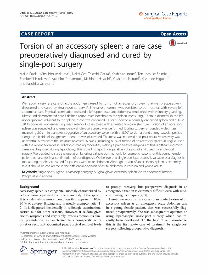
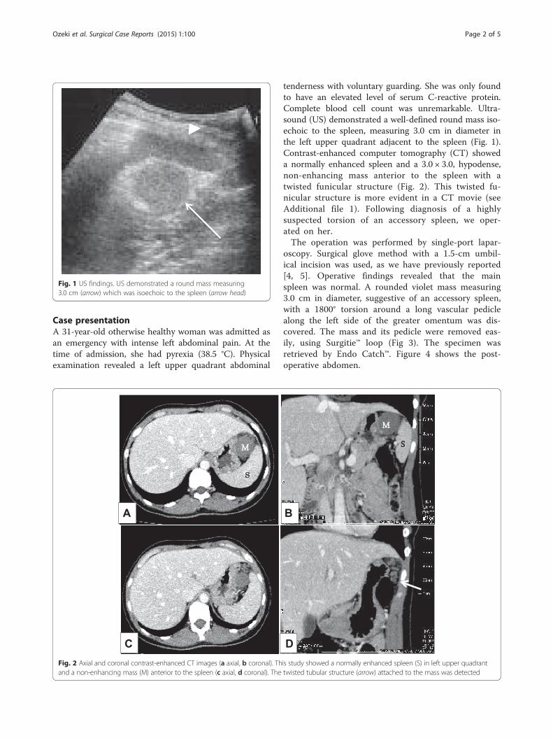
tenderness with voluntary guarding. She was only foundto have an elevated level of serum C-reactive protein.Complete blood cell count was unremarkable. Ultra-sound (US) demonstrated a well-defined round mass iso-echoic to the spleen, measuring 3.0 cm in diameter inthe left upper quadrant adjacent to the spleen (Fig. 1).Contrast-enhanced computer tomography (CT) showeda normally enhanced spleen and a 3.0 × 3.0, hypodense,non-enhancing mass anterior to the spleen with atwisted funicular structure (Fig. 2). This twisted fu-nicular structure is more evident in a CT movie (seeAdditional file 1). Following diagnosis of a highlysuspected torsion of an accessory spleen, we oper-ated on her.The operation was performed by single-port lapar-
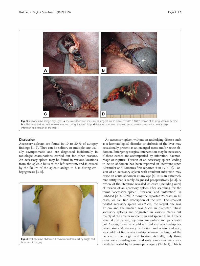
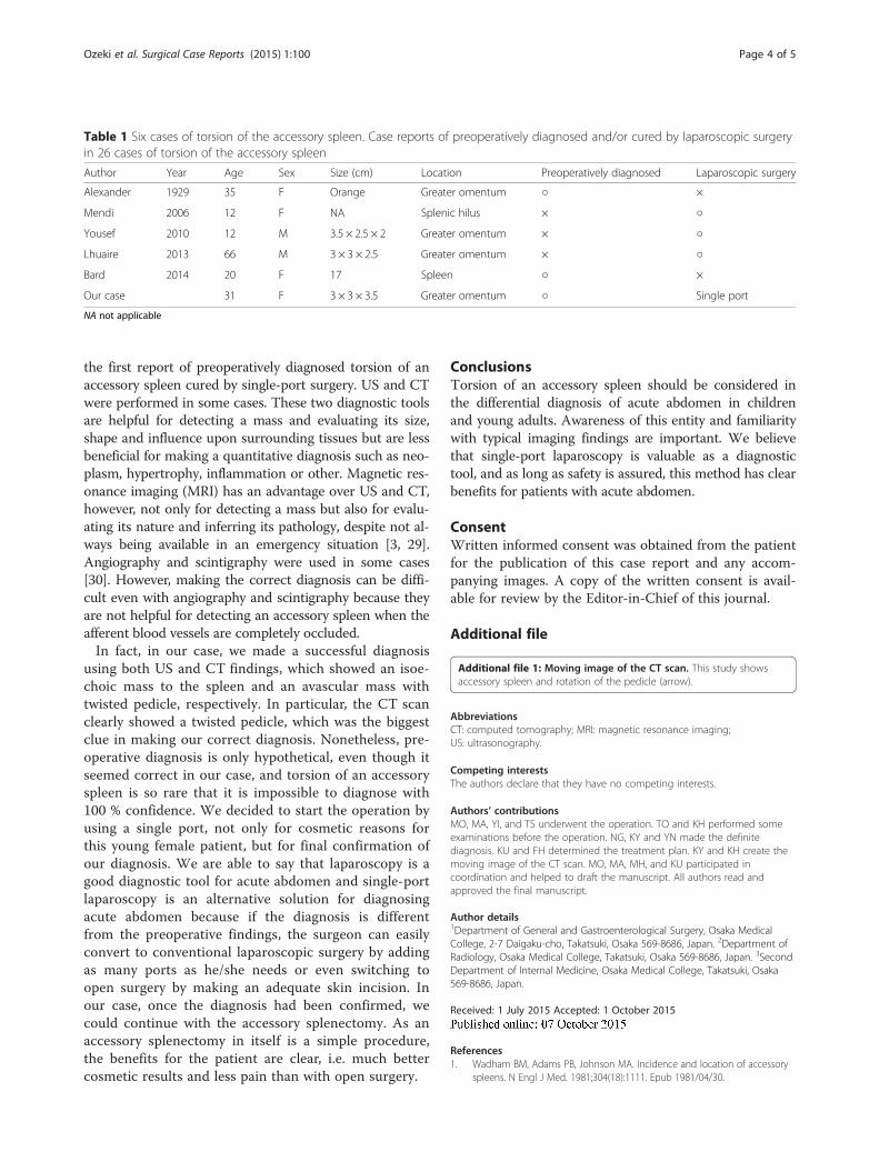
oscopy. Surgical glove method with a 1.5-cm umbil-ical incision was used, as we have previously reported[4, 5]. Operative findings revealed that the mainspleen was normal. A rounded violet mass measuring3.0 cm in diameter, suggestive of an accessory spleen,with a 1800° torsion around a long vascular pediclealong the left side of the greater omentum was dis-covered. The mass and its pedicle were removed eas-ily, using Surgitie™ loop (Fig 3). The specimen wasretrieved by Endo Catch™. Figure 4 shows the post-operative abdomen.
Fig. 1 US findings. US demonstrated a round mass measuring3.0 cm (arrow) which was isoechoic to the spleen (arrow head)
Fig. 2 Axial and coronal contrast-enhanced CT images (a axial, b coronal). This study showed a normally enhanced spleen (S) in left upper quadrantand a non-enhancing mass (M) anterior to the spleen (c axial, d coronal). The twisted tubular structure (arrow) attached to the mass was detected
Ozeki et al. Surgical Case Reports (2015) 1:100 Page 2 of 5
DiscussionAccessory spleens are found in 10 to 30 % of autopsyfindings [1, 2]. They can be solitary or multiple, are usu-ally asymptomatic and are diagnosed incidentally inradiologic examinations carried out for other reasons.An accessory spleen may be found in various locationsfrom the splenic hilus to the left scrotum, and is causedby the failure of the splenic anlage to fuse during em-bryogenesis [3, 6].
An accessory spleen without an underlying disease suchas a haematological disorder or cirrhosis of the liver mayoccasionally present as an enlarged mass and/or acute ab-domen. Emergency surgical intervention may be necessaryif these events are accompanied by infarction, haemor-rhage or rupture. Torsion of an accessory spleen leadingto acute abdomen has been reported in literature sinceAlexander and Romanes first reported it in 1914 [7]. Tor-sion of an accessory spleen with resultant infarction maycause an acute abdomen at any age [8]. It is an extremelyrare entity that is rarely diagnosed preoperatively [2, 3]. Areview of the literature revealed 26 cases (including ours)of torsion of an accessory spleen after searching for theterms “accessory spleen”, “torsion” and “infarction” inPubMed [2, 3, 6–28]. Among the reported 26 cases, in 16cases, we can find description of the size. The smallesttwisted accessory spleen was 2 cm, the largest one was17 cm and the median was 6 cm in diameter. Theseaccessory spleens are originated in various places butmainly at the greater momentum and splenic hilus. Otherswere at the cecum, jejunum, mesentery and pancreatictail. Among them, we could not find any relationship be-tween size and tendency of torsion and origin, and also,we could not find a relationship between the length of thepedicle or the origin and torsion. Actually, only threecases were pre-diagnosed and only four cases were suc-cessfully treated by laparoscopic surgery (Table 1). This is
Fig. 3 Intraoperative image highlights. a The rounded violet mass measuring 3.0 cm in diameter with a 1800° torsion of its long vascular pedicle.b, c The mass and its pedicle were removed using Surgitie™ loop. d Resected specimen showing an accessory spleen with hemorrhagicinfarction and torsion of the stalk
Fig. 4 Post-operative abdomen. It shows a scarless result by single-portlaparoscopic surgery
Ozeki et al. Surgical Case Reports (2015) 1:100 Page 3 of 5
the first report of preoperatively diagnosed torsion of anaccessory spleen cured by single-port surgery. US and CTwere performed in some cases. These two diagnostic toolsare helpful for detecting a mass and evaluating its size,shape and influence upon surrounding tissues but are lessbeneficial for making a quantitative diagnosis such as neo-plasm, hypertrophy, inflammation or other. Magnetic res-onance imaging (MRI) has an advantage over US and CT,however, not only for detecting a mass but also for evalu-ating its nature and inferring its pathology, despite not al-ways being available in an emergency situation [3, 29].Angiography and scintigraphy were used in some cases[30]. However, making the correct diagnosis can be diffi-cult even with angiography and scintigraphy because theyare not helpful for detecting an accessory spleen when theafferent blood vessels are completely occluded.In fact, in our case, we made a successful diagnosis
using both US and CT findings, which showed an isoe-choic mass to the spleen and an avascular mass withtwisted pedicle, respectively. In particular, the CT scanclearly showed a twisted pedicle, which was the biggestclue in making our correct diagnosis. Nonetheless, pre-operative diagnosis is only hypothetical, even though itseemed correct in our case, and torsion of an accessoryspleen is so rare that it is impossible to diagnose with100 % confidence. We decided to start the operation byusing a single port, not only for cosmetic reasons forthis young female patient, but for final confirmation ofour diagnosis. We are able to say that laparoscopy is agood diagnostic tool for acute abdomen and single-portlaparoscopy is an alternative solution for diagnosingacute abdomen because if the diagnosis is differentfrom the preoperative findings, the surgeon can easilyconvert to conventional laparoscopic surgery by addingas many ports as he/she needs or even switching toopen surgery by making an adequate skin incision. Inour case, once the diagnosis had been confirmed, wecould continue with the accessory splenectomy. As anaccessory splenectomy in itself is a simple procedure,the benefits for the patient are clear, i.e. much bettercosmetic results and less pain than with open surgery.
ConclusionsTorsion of an accessory spleen should be considered inthe differential diagnosis of acute abdomen in childrenand young adults. Awareness of this entity and familiaritywith typical imaging findings are important. We believethat single-port laparoscopy is valuable as a diagnostictool, and as long as safety is assured, this method has clearbenefits for patients with acute abdomen.
ConsentWritten informed consent was obtained from the patientfor the publication of this case report and any accom-panying images. A copy of the written consent is avail-able for review by the Editor-in-Chief of this journal.
Additional file
Additional file 1: Moving image of the CT scan. This study showsaccessory spleen and rotation of the pedicle (arrow).
AbbreviationsCT: computed tomography; MRI: magnetic resonance imaging;US: ultrasonography.
Competing interestsThe authors declare that they have no competing interests.
Authors’ contributionsMO, MA, YI, and TS underwent the operation. TO and KH performed someexaminations before the operation. NG, KY and YN made the definitediagnosis. KU and FH determined the treatment plan. KY and KH create themoving image of the CT scan. MO, MA, MH, and KU participated incoordination and helped to draft the manuscript. All authors read andapproved the final manuscript.
Author details1Department of General and Gastroenterological Surgery, Osaka MedicalCollege, 2-7 Daigaku-cho, Takatsuki, Osaka 569-8686, Japan. 2Department ofRadiology, Osaka Medical College, Takatsuki, Osaka 569-8686, Japan. 3SecondDepartment of Internal Medicine, Osaka Medical College, Takatsuki, Osaka569-8686, Japan.
Received: 1 July 2015 Accepted: 1 October 2015
References1. Wadham BM, Adams PB, Johnson MA. Incidence and location of accessory
spleens. N Engl J Med. 1981;304(18):1111. Epub 1981/04/30.
Table 1 Six cases of torsion of the accessory spleen. Case reports of preoperatively diagnosed and/or cured by laparoscopic surgeryin 26 cases of torsion of the accessory spleen
Author Year Age Sex Size (cm) Location Preoperatively diagnosed Laparoscopic surgery
Alexander 1929 35 F Orange Greater omentum ○ ×
Mendi 2006 12 F NA Splenic hilus × ○
Yousef 2010 12 M 3.5 × 2.5 × 2 Greater omentum × ○
Lhuaire 2013 66 M 3 × 3 × 2.5 Greater omentum × ○
Bard 2014 20 F 17 Spleen ○ ×
Our case 31 F 3 × 3 × 3.5 Greater omentum ○ Single port
NA not applicable
Ozeki et al. Surgical Case Reports (2015) 1:100 Page 4 of 5
2. Lhuaire M, Sommacale D, Piardi T, Grenier P, Diebold MD, Avisse C, et al. Arare cause of chronic abdominal pain: recurrent sub-torsions of an accessoryspleen. J Gastrointest Surg. 2013;17(10):1893–6. Epub 2013/06/14.
3. Grinbaum R, Zamir O, Fields S, Hiller N. Torsion of an accessory spleen.Abdom Imaging. 2006;31(1):110–2. Epub 2005/12/01.
4. Asakuma M, Hayashi M, Komeda K, Shimizu T, Hirokawa F, Miyamoto Y,et al. Impact of single-port cholecystectomy on postoperative pain. Br JSurg. 2011;98(7):991–5. Epub 2011/05/04.
5. Hayashi M, Asakuma M, Komeda K, Miyamoto Y, Hirokawa F, Tanigawa N.Effectiveness of a surgical glove port for single port surgery. World J Surg.2010;34(10):2487–9. Epub 2010/08/13.
6. Wacha M, Danis J, Wayand W. Laparoscopic resection of an accessoryspleen in a patient with chronic lower abdominal pain. Surg Endosc.2002;16(8):1242–3. Epub 2002/05/23.
7. Alexander RC, Romanes A. Accessory spleen causing acute attacks ofabdominal pain. Lancet. 1914;184:1089–91.
8. Hems TE, Bellringer JF. Torsion of an accessory spleen in an elderly patient.Postgrad Med J. 1990;66(780):838–9. Epub 1990/10/01.
9. Rc A. Accessory spleen with recurring torsion of its pedicle. Lancet.1929;214:21.
10. Kitchin RJ, Green NA. Torsion of an accessory spleen presenting as acuteappendicitis. Br J Surg. 1962;50:232–3. Epub 1962/09/01.
11. Perrine G. Torsion of the accessory splenic pedicle. A case report. Int Surg.1966;45(2):164–6. Epub 1966/02/01.
12. Bass RT, Yao ST, Freeark RJ. Torsion of an accessory spleen of the cecumpresenting as acute appendicitis. N Engl J Med. 1967;277(22):1190–1. Epub1967/11/30.
13. Babcock TL, Coker DD, Haynes JL, Conklin HB. Infarction of an accessoryspleen causing an acute abdomen. Am J Surg. 1974;127(3):336–7. Epub1974/03/01.
14. Onuigbo WI, Ojukwu JO, Eze WC. Infarction of accessory spleen. J PediatrSurg. 1978;13(2):129–30. Epub 1978/04/01.
15. Grunspan M, Wechsler U, Weintraub S. Torsion of an accessory spleensimulating acute appendicitis. Isr J Med Sci. 1981;17(6):458–9. Epub 1981/06/01.
16. Seo T, Ito T, Watanabe Y, Umeda T. Torsion of an accessory spleenpresenting as an acute abdomen with an inflammatory mass. US, CT, andMRI findings. Pediatr Radiol. 1994;24(7):532–4. Epub 1994/01/01.
17. Dahlin LB, Anagnostaki L, Delshammar M, Fork FT, Genell S. Torsion of anaccessory spleen in an adult. Case report. Eur J Surg. 1995;161(8):607–9.Epub 1995/08/01.
18. Jans R, Vanslembrouck R, Van Hoe L, Sockx L, Demedts I, Baert AL. Torsionof accessory spleen in an adult patient: imaging findings at CT, MRI andangiography. J Belge Radiol. 1997;80(5):229–30. Epub 1997/12/24.
19. Valls C, Mones L, Guma A, Lopez-Calonge E. Torsion of a wanderingaccessory spleen: CT findings. Abdom Imaging. 1998;23(2):194–5. Epub1998/03/28.
20. Padilla D, Ramia JM, Martin J, Pardo R, Cubo T, Hernandez-Calvo J. Acuteabdomen due to spontaneous torsion of an accessory spleen. Am J EmergMed. 1999;17(4):429–30. Epub 1999/08/19.
21. Perez Fontan FJ, Soler R, Santos M, Facio I. Accessory spleen torsion: US, CTand MR findings. Eur Radiol. 2001;11(3):509–12. Epub 2001/04/06.
22. Gardikis S, Pitiakoudis M, Sigalas I, Theocharous E, Simopoulos C. Infarctionof an accessory spleen presenting as acute abdomen in a neonate. Eur Jpediatr Surg. 2005;15(3):203–5. Epub 2005/07/07.
23. Mendi R, Abramson LP, Pillai SB, Rigsby CK. Evolution of the CT imagingfindings of accessory spleen infarction. Pediatr Radiol. 2006;36(12):1319–22.Epub 2006/10/04.
24. Impellizzeri P, Montalto AS, Borruto FA, Antonuccio P, Scalfari G, Arena F,et al. Accessory spleen torsion: rare cause of acute abdomen in childrenand review of literature. J Pediatr Surg. 2009;44(9):e15–8. Epub 2009/09/09.
25. Yousef Y, Cameron BH, Maizlin ZV, Boutross-Tadross O. Laparoscopicexcision of infarcted accessory spleen. J Laparoendosc Adv Surg Tech A.2010;20(3):301–3. Epub 2010/01/12.
26. Ishibashi H, Oshio T, Sogami T, Nii A, Mori H, Shimada M. Torsion of anaccessory spleen with situs inversus in a child. J Med Invest. 2012;59(1–2):220–3. Epub 2012/03/28.
27. Bard V, Goldberg N, Kashtan H. Torsion of a huge accessory spleen in a 20-year-old patient. Int J Surg Case Reports. 2014;5(2):67–9. Epub 2014/01/21.
28. Perin A, Cola R, Favretti F. Accessory wandering spleen: report of a case oflaparoscopic approach in an asymptomatic patient. Int J Surg Case Reports.2014;5(12):887–9. Epub 2014/12/03.
29. Gayer G, Zissin R, Apter S, Atar E, Portnoy O, Itzchak Y. CT findings incongenital anomalies of the spleen. Br J Radiol. 2001;74(884):767–72. Epub2001/08/21.
30. Ohta H, Kohno K, Kojima N, Ihara N, Ishigaki T, Todo G, et al. A case ofdiaphragm hernia containing accessory spleen and great omentumdetected by Tc-99m phytate scintigraphy. Ann Nucl Med. 1999;13(5):347–9.Epub 1999/12/03.
Submit your manuscript to a journal and benefi t from:
7 Convenient online submission
7 Rigorous peer review
7 Immediate publication on acceptance
7 Open access: articles freely available online
7 High visibility within the fi eld
7 Retaining the copyright to your article
Submit your next manuscript at 7 springeropen.com
Ozeki et al. Surgical Case Reports (2015) 1:100 Page 5 of 5