Embed Size (px)
Citation preview

Molecular Biology of the CellVol. 16, 4572–4583, October 2005
TOR Complex 2 Integrates Cell Movement duringChemotaxis and Signal Relay in DictyosteliumSusan Lee,* Frank I. Comer,† Atsuo Sasaki,* Ian X. McLeod,‡ Yung Duong,*Koichi Okumura,§ John R. Yates III,‡ Carole A. Parent,† and Richard A. Firtel*
*Section of Cell and Developmental Biology, Division of Biological Sciences and Center for MolecularGenetics, University of California, San Diego, La Jolla, CA 92093-0380; †Laboratory of Cellular and MolecularBiology, Division of Basic Sciences, NCI/National Institutes of Health, Bethesda, MD 20892-4256;‡Department of Cell Biology, Scripps Research Institute, La Jolla, CA 92037; and §Ludwig Institute for CancerResearch, School of Medicine, University of California, San Diego, La Jolla, CA 92093-0660
Submitted April 25, 2005; Revised July 8, 2005; Accepted July 21, 2005Monitoring Editor: Anne Ridley
Dictyostelium cells form a multicellular organism through the aggregation of independent cells. This process requiresboth chemotaxis and signal relay in which the chemoattractant cAMP activates adenylyl cyclase through the G protein-coupled cAMP receptor cAR1. cAMP is produced and secreted and it activates receptors on neighboring cells, therebyrelaying the chemoattractant signal to distant cells. Using coimmunoprecipitation and mass spectrometric analyses, wehave identified a TOR-containing complex in Dictyostelium that is related to the TORC2 complex of Saccharomycescerevisiae and regulates both chemotaxis and signal relay. We demonstrate that mutations in Dictyostelium LST8, RIP3,and Pia, orthologues of the yeast TORC2 components LST8, AVO1, and AVO3, exhibit a common set of phenotypesincluding reduced cell polarity, chemotaxis speed and directionality, phosphorylation of Akt/PKB and the related PKBR1,and activation of adenylyl cyclase. Further, we provide evidence for a role of Ras in the regulation of TORC2. We proposethat, through the regulation of chemotaxis and signal relay, TORC2 plays an essential role in controlling aggregation bycoordinating the two essential arms of the developmental pathway that leads to multicellularity in Dictyostelium.
INTRODUCTION
TOR, Target of rapamycin, is a member of the PI3K-relatedfamily of serine/threonine protein kinases and a knownregulator of cell growth in eukaryotic cells (Lorberg andHall, 2004). The protein complex that controls this pathway,termed TOR Complex 1 (TORC1), is highly conserved inevolution and contains the conserved proteins LST8 andKOG1/Raptor complexed with TOR (Loewith et al., 2002;Jacinto et al., 2003; Hay and Sonenberg, 2004). In addition,TOR controls other pathways, including regulated polarizedactin assembly in S. cerevisiae (Schmidt et al., 1996). A distinctTOR complex, TORC2, contains TOR, LST8, and three otherproteins: AVO1, AVO2, and AVO3. TORC2 regulates thiscytoskeletal rearrangement (Loewith et al., 2002), and a re-lated TORC2 has been identified in mammalian cells thatcontains, in addition to TOR and LST8, the AVO3 orthologmAVO3/Rictor (Jacinto et al., 2004; Sarbassov dos et al.,2004). AVO1 and AVO3 are orthologues of two previouslyidentified Dictyostelium proteins, RIP3 (Ras interacting pro-tein 3) and Pia (Pianissimo) (Chen et al., 1997; Lee et al.,1999). Disruption of either of these gene products in Dictyo-stelium (pia null [pia�] and rip3 null [rip3�] cells) gives rise toserious developmental defects, because they are unable toactivate the aggregation-stage adenylyl cyclase ACA in re-
sponse to chemoattractant stimulation and are defective inchemotaxis.
In Dictyostelium, the activation of ACA and chemotaxis areclosely integrated responses, although they have beenthought to be independently regulated (Comer et al., 2005).On starvation, Dictyostelium cells initiate a developmentalprogram that requires chemotaxis toward extracellularcAMP emitted by neighboring cells, which leads to theformation of multicellular aggregates (Parent and Dev-reotes, 1996; Aubry and Firtel, 1999; Kimmel and Parent,2003). The chemoattractant cAMP binds to the heterotri-meric G-protein-coupled cAMP receptor cAR1, which acti-vates multiple intracellular signaling events that control che-motaxis, the activation of adenylyl cyclase A (ACA), andgene expression required for aggregation (Aubry and Firtel,1999; Kimmel and Parent, 2003). The activation of ACAproduces more cAMP, which acts both intracellularly toactivate protein kinase A (PKA) and mediated downstreameffectors and extracellularly to initiate chemotaxis and torelay the chemoattractant signal to distal cells (signal relay).The concerted regulation of chemotaxis and signal relaycomprises a system that efficiently culminates with the de-velopment of a true, differentiated multicellular organism.In this article, we demonstrate that Dictyostelium TORC2functions as an integrator of multiple distinct signaling path-ways to control aggregation and the formation of the mul-ticellular organism. Cells lacking any of the TORC2 proteinsLST8, RIP3 (AVO1), and Pia (AVO3, mAVO3, Rictor) exhibitstrong chemotaxis defects including loss of speed, cell po-larity, and directionality, and are unable to fully activateAkt/PKB and the related kinase PKBR1, both of which arerequired for cell polarity and chemotaxis and are defective
This article was published online ahead of print in MBC in Press(http://www.molbiolcell.org/cgi/doi/10.1091/mbc.E05–04–0342)on August 3, 2005.
Address correspondence to: Richard A. Firtel ([email protected]).
4572 © 2005 by The American Society for Cell Biology

in their capacity to activate adenylyl cyclase in response tochemoattractant stimulation. Further, we demonstrate thatRIP3 carrying a point mutation that abrogates Ras-GTPbinding is unable to fully complement the null mutation,suggesting that Ras plays a role in mediating TORC2 func-tion.
MATERIALS AND METHODS
Biochemical and Chemotaxis AssaysTo obtain developmentally competent cells capable of responding to cAMP asa chemoattractant, log-phase vegetative cells were washed twice and resus-pended at a density 5 � 106 cells/ml with 12 mM Na�/K� phosphate buffer,pH 6.2, and pulsed with 30 nM cAMP for 5 h at 6-min intervals. Cells werethen washed and resuspended in 12 mM Na�/K� phosphate buffer.
The chemoattractant receptor-mediated activation of adenylyl cyclase wascarried out as previously described (Parent and Devreotes, 1995). Briefly,differentiated cells were stimulated with 50 �M cAMP and filter lysed at theindicated time points into Tris buffer containing 32P-�ATP diluted withunlabeled ATP to a final concentration of �150 Ci/mol. The reaction wasallowed to proceed for 1 min, stopped with SDS/ATP, and the radiolabeledcAMP was purified by column chromatography. Adenylyl cyclase assaysexhibit considerable variability between experiments. Although the relativeextent of activation of different cell lines or treatment conditions is highlyreproducible between experiments, the absolute activity can vary significantly(2–3-fold at times) from day to day. Therefore we, and others in the field, havechosen to present adenylyl cyclase activation data as representative of resultsfrom at least 3–5 independent experiments, each performed in duplicate on agiven day. Furthermore, comparisons between different cell lines or treatmentconditions are made on samples assayed on the same day.
For direct G-protein stimulation of adenylyl cyclase, differentiated cellswere filter lysed with GTP�S (50 �M final concentration) and, after 5 min,adenylyl cyclase activity was assayed for 2 min. To prepare cytosolic extractsfor the in vitro complementation experiments, cells were suspended at 8 � 107
cells/ml in simple lysis buffer (SLB, 10 mM Tris, pH 7.5, 0.2 mM EGTA, 200mM sucrose), filter lysed, and centrifuged at 9500 � g for 20 min. The in vitrocomplementation was performed in essentially the same manner as the directGTP-�-S-stimulated adenylyl cyclase activation assay, except that cells werelysed directly into cytosolic extract.
The PKB and PKBR1 kinase activities were assayed as described previously(Meili et al., 1999, 2000). Briefly, unstimulated differentiated cells (0 timepoint) and differentiated cells stimulated with cAMP for various times werelysed with an equal volume of 2� NP-40 lysis buffer (2� phosphate-bufferedsaline, 100 mM NaF, 2% NP-40, 4 mM EDTA, 2 mM pyrophosphate, 2 mMNa3VO4, and protease inhibitors leupeptin and aprotinin) on ice for 10 min.The lysate was precleared by centrifugation for 10 min. To immunoprecipitatePKB or PKBR1, the anti-PKB or anti-PKBR1 antibody and 30 �l of slurry ofprotein A-Sepharose were added into the supernatant. The beads werewashed twice with lysis buffer and twice with kinase buffer (25 mM MOPS,pH 7.4, 25 mM �-glycerophosphate, 20 mM MgCl2, and 1 mM dithiothreitol[DTT]). We incubated beads with 75 �l of kinase buffer containing 5 �M coldATP, 10 �Ci of �-32P-ATP, and 5 �g of H2B as substrate at room temperaturefor 15 min. The reaction was stopped by the addition of 25 �l of 5� samplebuffer (250 mM Tris, 500 mM DTT, 10% SDS, 50% glycerol, and 0.5% bromo-phenol blue) and boiled for 2 min. Samples were separated by 12% SDS-PAGE, blotted onto a polyvinylidene difluoride (PVDF) membrane, andexposed to film. After the autoradiography, the lower part of the membranecontaining the phosphorylated H2B was cut off and the upper portion withthe Akt/PKB or PKBR1 was subjected to Western blot analysis to quantify thelevel of Akt/PKB or PKBR1 in each lane. Experiments were repeated inde-pendently at least three times, always assaying wild-type cells as a control forcomparison in each experiment. The wild-type samples were run on the samegel as one of the experimental strains, and all gels and membranes werecoprocessed together.
Ras activation was performed as previously described (Sasaki et al., 2004).F-actin polymerization and myosin II assembly were assayed as describedpreviously (Lee et al., 2004; Park et al., 2004). Experiments were repeatedindependently at least three times, always including a wild-type control forcomparison.
Chemotaxis analysis was performed as described previously (Park et al.,2004; Sasaki et al., 2004) and analyzed using DIAS software (Wessels et al.,1998). Differentiated cells were plated in Na�/K� phosphate buffer at adensity of 6 � 104 cells/cm2 onto a plate with a hole covered by a 0.17-mmglass coverslip. An Eppendorf Patchman micromanipulator with a glasscapillary filled with 150 �M cAMP solution was brought into the field of viewof an inverted microscope. The response of the cells was recorded by time-lapse video. Experiments were performed at least three times on differentdays, always including a wild-type control strain in the analyses.
Cell Growth in the Presence of RapamycinCells growing in log phase (�3–4 � 106 cells/ml) were diluted to 4 � 105
cells/ml in HL5 medium. Rapamycin (Sigma Chemical Co, St. Louis, MO)was dissolved in dimethyl sulfoxide as a 1 mM stock and added to thecultures to the indicated concentration. Cell number in the suspension cul-tures was monitored over 4 d, with no further addition of the drug.
Translocation AssaysThe chemoattractant-mediated recruitment of the CRAC PH domain(PHCRAC-GFP) to the plasma membrane was measured biochemically aspreviously described in response to 20 �M cAMP (Parent et al., 1998), exceptthat protein samples were subjected to PAGE analysis using NuPage 4–12%Bis-Tris protein electrophoresis gradient gels, according to the manufacturer-recommended protocol (Invitrogen, Carlsbad, CA). For microscopic observa-tion of translocation, cells expressing PHCRAC-GFP were stimulated with 20�M cAMP and fixed (1% formaldehyde, 0.125% glutaraldehyde, 0.01% TritonX-100 in PB) after 5 s of stimulation. Cells were viewed with an inverted ZeissAxiovert S100 microscope (Thornwood, NY) equipped with automated filterwheels (Ludl Electronic Products, Hawthorne, NJ). Images were recordedwith a CoolSnap HQ CCD camera (Roper Scientific, Trenton, NJ) operated byIPLab software (Scanalytics, Fairfax, VA). GFP-PhdA translocation was ex-amined by real-time fluorescent video microscopy as described previously(Funamoto et al., 2001).
Detection of the Dd-TOR ComplexAggregation-competent cells were washed with Na/K phosphate buffer andresuspended at a density of 1 � 108 cells/ml in Na/K phosphate buffercontaining 20 mM MOPS (pH 7.0) and 1 mM phenylmethylsulphonyl fluo-ride. Cells were mechanically lysed by filtration (Nuclepore Track Etch mem-brane, 3 �m; Whatman, Clifton, NJ). Subsequently, cell lysates were incubatedin 2.5 mg/ml DSP (ditho-bis-succinimidylpropionate, Calbiochem, La Jolla,CA) for 5 min. The cross-linking reactions were quenched with 200 mMTris-HCl (pH 7.4). A sample was mixed with an equal volume of 2� lysisbuffer A (1% NP-40, 300 mM NaCl, 40 mM MOPS, pH 7.0, 20% glycerol, 2 mMNa3VO4, 2 �g/ml leupeptin, 5 �g/ml aprotinin) and centrifuged. Total cellextracts and immunoprecipitates with 25 �l resin of anti-V5 (V5–10) antibodyagarose conjugate (Sigma) were analyzed by silver staining or immunoblot-ting with anti-T7 (Novagen, Madison, WI) and anti-V5 antibodies. For iden-tifying the Dd-TOR complex, the anti-V5 antibody immunoprecipitated prod-ucts were eluted with the buffer containing 4% SDS and 0.1 M glycine (pH2.5). The protein complex was incubated with 100 mM DTT and then precip-itated with methanol/chloroform.
Mass SpectrometryThe samples were washed with acetone, resuspended in Tris buffer, 8 M urea,pH 8.6, reduced with 100 mM TCEP, and alkylated with 55 mM iodoacet-amide. Trypsin digest was done in the presence of 1 mM CaCl2 for trypticspecificity. Peptide mixtures were loaded onto a triphasic LC/LC columnwith the following steps of 500 mM ammonium acetate bumps: 25, 35, 50, 80,and 100%. Tandem mass spectra were analyzed using DTA Select and theDictyostelium sequence database with the following filtering parameters forcross correlation scores 1.8 (�1), 2.8 (�2), and 3.5 (�3) (Washburn et al., 2001).Identities of specific bands were confirmed by sequence analysis.
ConstructsThe LST8 knockout construct was made by inserting the blasticidin resistance(Sutoh, 1993) cassette into a BamHI site created at nucleotide 500 of the LST8genomic DNA sequence. A 5�-fragment was amplified from genomic DNA byPCR by using the primers GTTTTTGAATTCAGTTTTGATGGTCATA-AAGGTAATG and GTTTTTGGATCCGGTCAATGATGTTATACC and sub-sequently digested with enzymes EcoRI and BamHI. A 3� fragment wasamplified by using the primers GTTTTTGGATCCTCAAGTGATGGTGGTT-TAG and GTTTTTCTCGAGTTTTTATCTTGGTAAATCATTTAAAGCAACsubsequently digested with BamHI and XhoI. The vector was digested withEcoRI and XhoI and the DNA was transformed into Dictyostelium cells. Theknockout clones were confirmed by Southern and Northern blot analysis.Two independent clones were picked and examined. Both showed the samechemotaxis defects described in the results.
A T7-tagged full length LST8 ORF (open reading frame) clone was ampli-fied from genomic DNA by PCR using primers 5�- GTTTTTGAAT-TCAAAAAAATGGCTTCAATGACTGGTGGTCAACAAATGGGTCCAGG-TATTATATTGGCAACAGCATC and GTTTTTCTCGAGTTTTTATCTTGG-TAAATCATTTAAAGCAAC-3� subsequently digested with EcoRI and XhoIand subcloned to expression vector Exp4(�).
A 3� V5-tagged, full-length Pia clone was amplified from genomic DNA byPCR using primers GTTTTTGATATCAAAAAAATGACAAGTTCTGA-TAGTAGTG and GTTTTTCTCGAGTTTTTAAGTTGAATCTAAACCTAA-TAATGGATTTGGAATTGGTTTACCATTTAAATCATGATATGGATCAGA-TG and subsequently digested by EcoRV and XhoI and subcloned into theexpression vector Exp4(�).
TORC2 Controls Dictyostelium Aggregation
Vol. 16, October 2005 4573

A 5� T7-tagged clone of RIP3 from ATG to internal BamHI site at nucleotide1490 was amplified by PCR from the full-length RIP3 (Lee et al., 1999) by usingprimers GTTTTTGAATTCAAAAAAATGGCTTCAATGACTGGTGGTCA-ACAAATGGGTTCAGTTTATTGTGAATTAGTTGATG and ATTTGGACA-AAGTACCAATACTTC. The full length of 5� T7-tagged RIP3 clone wasobtained by digesting the PCR product with EcoRI and BamHI and thefull-length RIP3 with BamHI and XhoI and the fragments cloned into theexpression vector Exp4(�).
All constructs were sequenced.
RESULTS
Dictyostelium Expresses Orthologues of the Yeast andMammalian TOR Complex 2The previously identified Dictyostelium proteins RIP3 andPia are orthologues of the TORC2 components AVO1 andAVO3/mAV03/Rictor in S. cerevisiae and mammals, respec-tively (Chen et al., 1997; Lee et al., 1999; Loewith et al., 2002).Previous studies indicated that both Dictyostelium proteinsare required for efficient chemotaxis and the activation ofACA to relay the cAMP signal to neighboring cells. TheWD40-repeat protein LST8 is a known component of bothyeast and mammalian TORC1 and TORC2. We identified asingle gene in the Dictyostelium database (GenBank acces-sion no. DDB0184464) that encodes an LST8 ortholog. Wecreated an LST8 gene knockout by homologous recombina-tion. The knockout strain (lst8� cells) was confirmed bySouthern and RNA blot analyses (see Materials and Methods).Analysis of the lst8�, pia�, and rip3� cells showed that thesecells were the same size and exhibited the same growthkinetics as the parental wild-type cells (unpublished data),indicating that these genes do not regulate Dictyosteliumgrowth under the conditions tested (rich axenic mediumcontaining 1% yeast extract, 1% proteose peptone, and 1%dextrose or in association with bacteria as a food source).Because RIP3, Pia, and LST8 are homologues of proteinscomplexed with TOR in yeast and mammalian cells, weexamined whether rapamycin, which inhibits TORC1 butnot TORC2 (Loewith et al., 2002; Jacinto et al., 2004), inhibitsgrowth. Addition of rapamycin to concentrations of 50 nMdramatically inhibited cell growth, whereas 500 nM rapa-mycin resulted in an almost complete cessation of growth,consistent with the involvement of a rapamycin-sensitiveTORC1 in controlling cell growth in Dictyostelium (Figure 1).
When plated for development on nonnutrient agar in theabsence of exogenous pulses of cAMP, we found that, sim-
ilar to rip3� and pia� cells, lst8� cells do not spontaneouslyaggregate, but remain as a smooth monolayer of cells (Fig-ure 2A; Chen et al., 1997; Lee et al., 1999). When providedwith exogenous pulses of cAMP, which mimic the endoge-nous cAMP signaling of wild-type cells, and then plated fordevelopment, lst8� and pia� cells still do not develop. Incontrast, rip3� cells form mounds, although very ineffi-ciently (Figure 2A; Lee et al., 1999), suggesting that the rip3�
defect can be partially overridden, a phenotype that is alsoshared with the Ras GEF (guanine nucleotide exchange fac-tor) Aimless (Insall et al., 1996; Lee et al., 1999). Defects in theability to aggregate can result in an inability to induce thegenes required for this process.
To assess whether LST8, Pia, or RIP3 regulates the expres-sion of early genes required for development, we performedan RNA blot analysis examining the expression of the cAMPreceptor cAR1 and csA (gp80), a cell adhesion molecule,both of which are required for aggregation. We find thatcAR1 and csA transcripts are normally induced in responseto exogenous cAMP in lst8�, pia�, and rip3� cells (Figure2B). These data suggest that the developmental defects oflst8� cells, like rip3� and pia� cells, may arise from deficien-cies in chemotaxis and/or chemoattractant signal relay.
Dictyostelium TORC2 Exists in a Preformed ComplexThat Regulates Adenylyl Cyclase ActivationTo investigate the underlying developmental defect of lst8�
cells, we measured their capacity to activate ACA in re-sponse to chemoattractant stimulation. In this activation trap
Figure 1. Inhibition of Dictyostelium cell growth by rapamycin. Theincreases in cell number of wild-type cells in the absence andpresence of different concentrations of rapamycin are shown. SeeMaterials and Methods for details.
Figure 2. Development of rip3�, pia�, and lst8� cells. (A) Devel-opment of rip3�, pia�, and lst8� and wild-type cells plated onnonnutrient agar (left side). The cells in the right-hand panels werefirst pulsed for 6 h with 30 nM cAMP, to mimic the natural cAMPoscillations that induce aggregation-stage responses, before plating.All pictures were taken at 24 h. (B) RNA blot showing the expres-sion of two pulse-induced genes (cAR1 and csA) at 0 time and after5 h of cAMP pulsing.
S. Lee et al.
Molecular Biology of the Cell4574
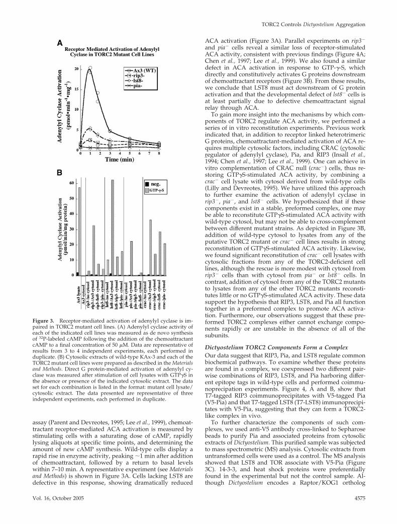
assay (Parent and Devreotes, 1995; Lee et al., 1999), chemoat-tractant receptor-mediated ACA activation is measured bystimulating cells with a saturating dose of cAMP, rapidlylysing aliquots at specific time points, and determining theamount of new cAMP synthesis. Wild-type cells display arapid rise in enzyme activity, peaking �1 min after additionof chemoattractant, followed by a return to basal levelswithin 7–10 min. A representative experiment (see Materialsand Methods) is shown in Figure 3A. Cells lacking LST8 aredefective in this response, showing dramatically reduced
ACA activation (Figure 3A). Parallel experiments on rip3�
and pia� cells reveal a similar loss of receptor-stimulatedACA activity, consistent with previous findings (Figure 4A;Chen et al., 1997; Lee et al., 1999). We also found a similardefect in ACA activation in response to GTP-�-S, whichdirectly and constitutively activates G proteins downstreamof chemoattractant receptors (Figure 3B). From these results,we conclude that LST8 must act downstream of G proteinactivation and that the developmental defect of lst8� cells isat least partially due to defective chemoattractant signalrelay through ACA.
To gain more insight into the mechanisms by which com-ponents of TORC2 regulate ACA activity, we performed aseries of in vitro reconstitution experiments. Previous workindicated that, in addition to receptor linked heterotrimericG proteins, chemoattractant-mediated activation of ACA re-quires multiple cytosolic factors, including CRAC (cytosolicregulator of adenylyl cyclase), Pia, and RIP3 (Insall et al.,1994; Chen et al., 1997; Lee et al., 1999). One can achieve invitro complementation of CRAC null (crac�) cells, thus re-storing GTP�S-stimulated ACA activity, by combining acrac� cell lysate with cytosol derived from wild-type cells(Lilly and Devreotes, 1995). We have utilized this approachto further examine the activation of adenylyl cyclase inrip3�, pia�, and lst8� cells. We hypothesized that if thesecomponents exist in a stable, preformed complex, one maybe able to reconstitute GTP�S-stimulated ACA activity withwild-type cytosol, but may not be able to cross-complementbetween different mutant strains. As depicted in Figure 3B,addition of wild-type cytosol to lysates from any of theputative TORC2 mutant or crac� cell lines results in strongreconstitution of GTP�S-stimulated ACA activity. Likewise,we found significant reconstitution of crac� cell lysates withcytosolic fractions from any of the TORC2-deficient celllines, although the rescue is more modest with cytosol fromrip3� cells than with cytosol from pia� or lst8� cells. Incontrast, addition of cytosol from any of the TORC2 mutantsto lysates from any of the other TORC2 mutants reconsti-tutes little or no GTP�S-stimulated ACA activity. These datasupport the hypothesis that RIP3, LST8, and Pia all functiontogether in a preformed complex to promote ACA activa-tion. Furthermore, our observations suggest that these pre-formed TORC2 complexes either cannot exchange compo-nents rapidly or are unstable in the absence of all of thesubunits.
Dictyostelium TORC2 Components Form a ComplexOur data suggest that RIP3, Pia, and LST8 regulate commonbiochemical pathways. To examine whether these proteinsare found in a complex, we coexpressed two different pair-wise combinations of RIP3, LST8, and Pia harboring differ-ent epitope tags in wild-type cells and performed coimmu-noprecipation experiments. Figure 4, A and B, show thatT7-tagged RIP3 coimmunoprecipitates with V5-tagged Pia(V5-Pia) and that T7-tagged LST8 (T7-LST8) immunoprecipi-tates with V5-Pia, suggesting that they can form a TORC2-like complex in vivo.
To further characterize the components of such com-plexes, we used anti-V5 antibody cross-linked to Sepharosebeads to purify Pia and associated proteins from cytosolicextracts of Dictyostelium. This purified sample was subjectedto mass spectrometric (MS) analysis. Cytosolic extracts fromuntransformed cells were used as a control. The MS analysisshowed that LST8 and TOR associate with V5-Pia (Figure3C). 14-3-3, and heat shock proteins were preferentiallyfound in the experimental but not the control sample. Al-though Dictyostelium encodes a Raptor/KOG1 ortholog
Figure 3. Receptor-mediated activation of adenylyl cyclase is im-paired in TORC2 mutant cell lines. (A) Adenylyl cyclase activity ofeach of the indicated cell lines was measured as de novo synthesisof 32P-labeled cAMP following the addition of the chemoattractantcAMP to a final concentration of 50 �M. Data are representative ofresults from 3 to 4 independent experiments, each performed induplicate. (B) Cytosolic extracts of wild-type KAx-3 and each of theTORC2 mutant cell lines were prepared as described in the Materialsand Methods. Direct G protein-mediated activation of adenylyl cy-clase was measured after stimulation of cell lysates with GTP�S inthe absence or presence of the indicated cytosolic extract. The dataset for each combination is listed in the format: mutant cell lysate/cytosolic extract. The data presented are representative of threeindependent experiments, each performed in duplicate.
TORC2 Controls Dictyostelium Aggregation
Vol. 16, October 2005 4575

(GenBank accession no. DDB0191024), it is not present inthese immunoprecipitates, suggesting that Dictyosteliumforms a separate TORC1. Interestingly, we did not observeRIP3 in the complex, although it coimmunoprecipitates withPia. These differences between the coimmunopreciptationdata and the MS analysis could be due to instability of thecomplex and differential loss of some components duringthe purification and/or differences in the efficiency of pep-tide ionization in the MS, leading to different efficiencies ofpeptide recovery in the analysis, although this is unlikelygiven the dynamic range and sensitivity of the MS. Never-theless, these findings indicate that, similar to the yeast andmammalian counterparts, Dictyostelium LST8, RIP3, and Piaform one or more TORC2 complexes (Loewith et al., 2002;Jacinto et al., 2004). We did not identify any orthologues toyeast AVO2 and no clear ortholog is found in the Dictyoste-lium genome database.
Dictyostelium TORC2 Regulates Cell Movement duringChemotaxisAn aggregation-deficient developmental phenotype canarise from defects in either ACA activation or chemotaxis,which are functionally independent (Comer et al., 2005).Previous studies suggested that pia� cells exhibit chemotaxisdefects (Chen et al., 1997) and demonstrated that rip3� cellshave reduced cell polarity and a lower chemotaxis index(Lee et al., 1999). To assess the ability of the lst8� cells tochemotax, we examined their chemotaxis toward a micropi-pette filled with cAMP. We quantified their behavior usingDIAS computer software (Wessels et al., 1988, 1998) andcompared this strain to wild-type cells and pia� and rip3�
cells, whose chemotaxis parameters had not been previouslymeasured. We found that, although the lst8� cells can mi-
grate toward the micropipette, they do so with reducedspeed and directionality and with significant loss in polaritycompared with wild-type cells (Figure 5, A and B; Table 1).rip3� cells showed similar defects, consistent with previousfindings (Lee et al., 1999). The chemotaxis defects of pia�
cells were not as strong as those for lst8� and rip3� cells. Tofurther characterize the chemotaxis defects of rip3�, pia�,and lst8� cells, we analyzed the mutants using the under-agarose assay (Laevsky and Knecht, 2001; Comer et al.,2005), in which cells migrate under a layer of agarose in agradient of chemoattractant. Under these conditions, thelst8� cells, as well as the rip3� and pia� cells, do not migrateas far toward the cAMP source as wild-type cells and, mostnotably, do not organize into streams, a process that requiresACA activation (Kriebel et al., 2003; unpublished data).These data are consistent with the notion that LST8, RIP3,and Pia function together in a complex that regulates bothACA activation/signal relay and chemotaxis.
Proper chemotaxis requires the concerted regulation ofanterior F-actin extension and posterior myosin II contrac-tion (Ridley et al., 2003). We therefore examined whetherdeficiencies in LST8, RIP3, or Pia result in defective chemoat-tractant-mediated F-actin polymerization or myosin II as-sembly. The lst8� cells exhibit a nominal but reproduciblereduction in the extent of chemoattractant-stimulated F-ac-tin polymerization in both the first (�5-s) and second(�45-s) peaks of activation, which correlate with the initialcringe response and the subsequent pseudopod extension,respectively, observed after uniform chemoattractant stim-ulation (Hall et al., 1988; Figure 5C). The rip3� and pia� cellsexperience a similar modest decrease in the first peak, butthe level of the second F-actin peak is indistinguishable fromthat of wild-type cells. We do not think that any change in
Figure 4. TORC2 complex. (A and B) Thecoimmunoprecipitation of V5-tagged Piawith T7-tagged RIP3 (A) or T7-tagged LST8(B). Cells were cotransformed with expres-sion vectors expressing V5-Pia and T7-RIP3or V5-Pia and T7-LST8 or expression on T7-RIP3 or only T7-LST8. The immunoprecipita-tion was carried out using anti-V5 antibody(see Materials and Methods) and the Westernblot was probed (IB) with either anti-T7 oranti-V5 antibody as shown. TCL shows theprotein in total cell lysates. (C) Partial list ofpeptides identified in the mass spectrometryanalysis of the V5-Pia-containing complex.Proteins found in common with the controlsample are not shown.
S. Lee et al.
Molecular Biology of the Cell4576

the first F-actin peak that is observed is at all responsible forthe null polarity and chemotaxis defects. All three mutantstrains exhibit moderately altered patterns of chemoattrac-
tant-mediated myosin II assembly. In wild-type cells, thelevels of myosin II associated with the cytoskeleton undergoan initial slight decrease 5–10 s after uniform chemoattrac-
Figure 5. Chemotaxis properties of TORC2 mutant strains. (A) Chemotaxis images of wild-type (KAx-3) and lst8� cells using DIC optics. Imagesshown are taken at the time the micropipette containing the chemoattractant is inserted (0 time) and at 15 min. See Materials and Methods for details.(B) Images of chemotaxing cells obtained using DIAS computer software (see Materials and Methods). The overlapping images are taken at 1-minintervals. (C) Chemoattractant stimulated F-actin polymerization and myosin II assembly as described in the Materials and Methods.
Table 1. DIAS analysis
RIP3
K680E,R681E
Strain KAx-3 lst8 null pia null rip3 null KAx-3 rip3 null rip3 nullSpeed (um/min) 9.96 � 0.70 5.39 � 0.38 7.41 � 0.25 5.36 � 1.97 10.2 � 2.12 9.37 � 0.18 6.70 � 0.82Dir ch (deg) 24.1 � 4.06 40.4 � 6.62 41.1 � 0.56 46.8 � 9.84 16.0 � 3.10 30.9 � 5.64 32.2 � 4.09Roundness 52.4 � 3.04 75.8 � 3.46 66.3 � 1.29 62.2 � 3.62 54.3 � 3.93 60.5 � 5.78 71.0 � 2.48Directionality 0.78 � 0.01 0.57 � 0.10 0.48 � 0.01 0.53 � 0.12 0.84 � 0.02 0.74 � 0.04 0.57 � 0.04
DIAS analysis of chemotaxis. Numbers are mean values � SD. Speed indicates speed of cell’s centroid movement along the total path.Direction change is a relative measure of the number and frequency of turns the cell makes. Larger numbers indicate more turns and lessefficient chemotaxis. Directionality is a measure of the linearity of the pathway. Cells moving in a straight line to the needle have adirectionality of 1.00. Roundness is an indication of the polarity of the cells. Larger numbers indicate the cells are more round (less polarized).
TORC2 Controls Dictyostelium Aggregation
Vol. 16, October 2005 4577

Figure 6.
S. Lee et al.
Molecular Biology of the Cell4578

tant stimulation and then increase about twofold, peaking at�30 s, before decreasing to basal levels (Steimle et al., 2001;Park et al., 2004). The rip3� cells exhibit the strongest phe-notype, with a significantly reduced peak of myosin II as-sembly upon chemoattractant stimulation (Figure 5C). Theessentially wild-type F-actin response and moderate defectsin myosin II assembly suggest that these cells do not have afundamental inability to undergo cytoskeletal changes inresponse to chemoattractant. However, all three null strainssimultaneously extend random pseudopodia from multiplepoints around the entire periphery of the cell, in contrast towild-type cells, which predominantly extend pseudopodiain the direction of the chemoattractant gradient. The poordirectionality of chemotaxing of the rip3�, pia�, and lst8�
cells supports the hypothesis that at least part of the loss ofdirectionality results from more random movement inherentin cells that form multiple lateral pseudopodia. Our data,however, do not exclude the possibility that these cells showa defect in gradient interpretation or spatial-temporal sig-naling to the cytoskeleton. This possibility is strongest forlst8� cells as some of these cells exhibit directionality defectsthat are stronger than those exhibited by the other strainsand for other apolar mutants such as myoII� or pakB/pakC�/� cells (Wessels et al., 1988; Lee et al., 2004).
Ras Is Involved in the Regulation of RIP3RIP3 was identified in a yeast two-hybrid screen using con-stitutively active human H-Ras as bait and was found tointeract with GTP-bound forms of Dictyostelium RasG andhuman H-Ras (Lee et al., 1999). Sequence comparison iden-tified a putative Ras-binding domain (RBD) in RIP3 (Figure6A). The Arg residue at position 681 is conserved in many ofthese domains, suggesting it might be important for medi-ating interactions between the RBD and Ras-GTP. We exam-ined this by mutating this Arg and the preceding Lys to Glu(K680E, R681E) and found that the RIP3 RBD carrying thesesubstitutions no longer interacted with RasG-GTP in a two-hybrid assay (Figure 6B). Expression of wild-type RIP3 com-plemented the developmental and chemotaxis defects ofrip3� cells (Figure 6C; Table 1). The chemotaxis parameterswere slightly reduced compared with wild-type cells, butthey were similar to wild-type cells overexpressing RIP3,suggesting any changes were due to an overexpression ofRIP3 protein (Table 1; Figure 5A). In contrast, whenRIP3K680E,R681E was expressed in rip3� cells, it only partiallycomplemented the developmental and chemotaxis defects.rip3� cells expressing RIP3K680E,R681E form aggregates athigher cell densities where cell-cell contacts can assist inaggregate formation but the organisms exhibit developmen-tal defects and are delayed in morphogenesis. The cellscannot aggregate at lower cell densities where chemotaxisplays an increasingly important role. Similar effects havebeen noted for several signaling mutants, including akt/pkbnull cells (Meili et al., 1999). Further, rip3� cells expressing
RIP3K680E,R681E exhibit only a small improvement in chemo-taxis speed, and the directionality of movement, an indicatorof chemotaxis index, is similar to that of rip3� cells. Theseresults suggest that a functional RBD is important for fullRIP3 function.
Dictyostelium TORC2 Acts Downstream of Ras and PI3KNumerous studies have shown that PI3 kinases (PI3Ks) playmultiple roles in chemoattractant signaling in Dictyosteliumand mammalian leukocytes (Funamoto et al., 2001; Stephenset al., 2002; Merlot and Firtel, 2003; Van Haastert and Dev-reotes, 2004). Cells lacking PI3K function exhibit chemotaxisdefects similar to those of the rip3�, pia�, and lst8� cells(Funamoto et al., 2001, 2002; Sasaki et al., 2004). In Dictyoste-lium, PI3K1 and PI3K2 translocate from the cytosol to theleading edge membrane during chemotaxis and removal ofthe membrane-targeting domain impairs chemoattractantsignaling through PI3K (Funamoto et al., 2002). Ras-medi-ated activation of PI3K is thought to preferentially promotelocalized activation of the chemotactic machinery, thussharply amplifying chemoattractant signaling at the leadingedge of chemotaxing cells (Sasaki et al., 2004). To gain insightinto the mechanism by which TORC2 regulates chemotaxisand signal relay, we studied the impact of TORC2 on PI3Ksignaling pathways. We first assessed the extent of chemoat-tractant-stimulated Ras activation in the TORC2 mutantsusing a pulldown assay in which GTP-bound Ras is isolatedthrough its binding to the RBD of human Raf1 and thenquantified using a Western blot assay (Sasaki et al., 2004).This assay examines the activation of RasG, RasD, and RasBwith the majority of the Ras-GTP bound to the Raf1-RBD inaggregation-competent cells attributed to RasG (Sasaki et al.,2004). As depicted in Figure 7A, all strains exhibited kineticsand extent of Ras activation similar to those of wild-typecells. We next assessed the role of the TORC2 componentsRIP3, Pia, and LST8 in the cellular distribution of PI3K. Wetransformed wild-type, rip3�, pia�, and lst8� cells with GFP-N-PI3K1, which is necessary and sufficient for PI3K1 corticallocalization, and studied its distribution in chemotaxingcells. As previously reported, we observed that the majorityof the cortically localized GFP-N-PI3K1 is found at the lead-ing edge of wild-type chemotaxing cells. Because of im-proved imaging technology, we now also find that a smallerfraction of GFP-N-PI3K1 localizes at the cell posterior (Fig-ure 8). Both localizations are sites of enriched F-actin poly-merization, consistent with our demonstration that en-hanced GFP-N-PI3K cortical localization is dependent onnew F-actin (Sasaki et al., 2004). As expected, wild-type cellshave a ruffled leading edge with numerous protrusions. Incontrast, rip3�, pia�, and lst8� cells have broader GFP-N-PI3K1-containing domains with multiple lateral pseudopo-dia and broader posteriors, illustrating their loss-of-polarityphenotype (Figures 5, A and B, and 8; data for pia� cells arenot published). Nevertheless, we find that GFP-N-PI3K1 istargeted to the cortex in the mutants. Furthermore, we ob-served that the mutant cell lines have normal kinetics ofGFP-N-PI3K1 translocation to the cortex in response to uni-form chemoattractant stimulation (unpublished data). Thesedata demonstrate that the TORC2 components are not re-quired for Ras activation or for the spatial targeting of PI3K.
We next evaluated whether the components of TORC2regulate PI3K activation, using translocation of the CRACand PhdA PH domain to the plasma membrane as markersfor the accumulation of PI(3,4,5)P3, the major product ofPI3K. Earlier studies demonstrated that chemoattractant-mediated translocation of CRAC and PhdA to the plasmamembrane is a G protein-, PI3K-, and PH domain-dependent
Figure 6 (facing page). Analysis of RIP3. (A) Sequence compari-son of RIP3 Ras-binding domain. RIP3 (AAD43567, D. discoideum);AVO1 (NP_014563, S. cerevisiae); RMIL/v-Raf1 (EAL24023, Homosapiens); 1340152/A-Raf1 (AAH07514, H. sapiens), h-KRAF(AAP03432, H. sapiens); RGSE-1h/RGS14 (043566, H. sapiens); AAD/CG5248-PL (NP_732776, Drosophila melanogaster); STEF (NP_036008,Mus musculus); STL (NP_524647, D. melanogaster). (B) Two-hybridinteraction of wild-type RIP3 and RIP3K680E,R681E with constitutivelyactive Dictyostelium RasG (RasGQ61L). (C) Development of rip3� cellsexpressing RIP3 or RIP3K680E,R681E plated on nonnutrient agar athigh (3 � 106/cm2) and low (0.75 � 106/cm2) density.
TORC2 Controls Dictyostelium Aggregation
Vol. 16, October 2005 4579

event (Parent et al., 1998; Funamoto et al., 2001, 2002). Figure7, B–D, depict the plasma membrane translocation ofPHCRAC-GFP in rip3�, pia�, and lst8� cells following a uni-form, saturating dose of chemoattractant, as judged by bothfluorescence microscopy and Western blot analysis of mem-brane preparations (data for PhdA not published). All threemutant cell lines retain the capacity to generate membranebinding sites for PHCRAC-GFP and PhdA, displaying normalkinetics and an extent of translocation similar to that ofwild-type cells. From these data, we infer that RIP3, Pia, andLST8 are not directly required to activate PI3K.
To gain further insight into the impact of TORC2 on PI3Ksignaling pathways, we examined activation of the PI3K-regulated effector PKB in rip3�, pia�, and lst8� cells bydirectly measuring the chemoattractant-mediated activationof PKB. Surprisingly, we found that in rip3� and pia� cellsexhibit a significant impairment in PKB activation comparedwith wild-type cells (Figure 7D). PKB activation was lessimpaired in lst8� cells, although the activation was stillreproducibly lower than that of wild-type cells (Figure 7D).Combined with our observation that CRAC and PhdA trans-location/PI3K activation is not impaired in the TORC2 mu-tants, these data are consistent with a model in whichTORC2 acts either downstream or parallel to PI3K in thechemoattractant-mediated activation of PKB.
Dictyostelium cells express two distinct PKB-related ki-nases, PKB (the ortholog of mammalian PKB) and PKBR1.PKBR1 is required for morphogenesis during the multicel-lular stages of Dictyostelium development (Meili et al., 2000).It harbors highly conserved kinase and C-terminal hydro-phobic domains that include conserved sites of phosphory-lation equivalent to T308 and S473 in human PKB. However,in contrast to PKB, PKBR1 lacks a true PH domain (it has anN-terminal, PH-like domain) and localizes constitutively tothe plasma membrane via an N-terminal myristoylationmodification. PKBR1 is activated by chemoattractants in aPI3K-independent manner, presumably because PKBR1 isconstitutively associated with the plasma membrane anddoes not require de novo PtdIns(3,4,5)P3 synthesis for itsmembrane localization. pkbr1� cells exhibit very weak che-motaxis phenotypes but a double PKB/PKBR1 null strainhas strong growth and chemotaxis defects, indicating thatboth kinases control chemotaxis pathways. These pheno-types are more severe than those of rip3�, pia�, and lst8�
cells and significantly more severe than those of pkb� cells.We therefore examined the extent of chemoattractant-in-duced PKBR1 phosphorylation in rip3�, pia�, and lst8� cells.Remarkably, as with PKB, PKBR1 phosphorylation is im-paired in rip3� and pia� cells and less affected in lst8� cells(Figure 6E), further indicating that the defect in PKB phos-phorylation is not a result of improper targeting to theplasma membrane or defects in PI3K activation. Rather,these results are consistent with the Dictyostelium TORC2regulating the phosphorylation of PKB and PKBR1. Al-though the effects on cell movement are strongest in lst8�
cells, these cells exhibit the weakest effect on PKB andPKBR1 activation. These observations suggest that differentcomponents of the TORC2 complex have different effects onTOR activity in different pathway. Our data do not excludethe possibility of multiple TORC2 complexes with differentor overlapping functions or activities.
DISCUSSION
The Composition of Dictyostelium TORC2We demonstrate here that Dictyostelium cells express aTORC2 that is essential for multicellular development, but
not growth. As in S. cerevisiae, Dictyostelium Pia, RIP3, andLST8, the orthologues of yeast AVO3, AVO1, and LST8,form a complex in vivo. Our MS analyses of Pia-containing
Figure 7. Regulation of the PI3K pathway in TORC2 mutants. (A)Ras activation in wild-type and lst8�, rip3�, and pia� cells is shown.(B and C) Translocation of PH-CRAC-GFP to the cortex examinedbiochemically (B) or by fluorescence microscopy (C). (D and E)Chemoattactant-mediated activation (kinase assay) of Akt/PKB (D)and PKBR1 (E) in wild-type and lst8�, rip3�, and pia� cells. Activa-tion of PKBR1 in pi3k1/2 null cells is also shown (E). Histone 2B(H2B) is used as a substrate. Upper lanes show the kinase assay.Lower lanes show a Western blot of Akt/PKB or PKBR1 protein inthe immunoprecipitate. See Materials and Methods for details.
S. Lee et al.
Molecular Biology of the Cell4580

complexes identified TOR and LST8 but not Raptor, which ispresent in the Dictyostelium genome and is a known compo-nent of rapamycin-sensitive TORC1 in other systems (Lor-berg and Hall, 2004). Although RIP3 was not observed in theMS analysis of Pia-containing complexes, coimmunoprecipi-tation experiments determined it is associated with Pia invivo. Although rapamycin inhibits growth in Dictyostelium,presumably through TORC1, our studies indicate thatTORC2 is rapamycin-insensitive, because the signalingpathways that are genetically controlled by TORC2 arenot inhibited by the drug. This is consistent with thefindings in yeast and mammalian cells, although we havenot formally proven the existence of an independentTORC1 biochemically (Loewith et al., 2002; Jacinto et al.,2004; Sarbassov dos et al., 2004). As cells lacking Pia, RIP3,or LST8 do not have growth defects, it is clear that arapamycin-sensitive TOR complex that does not containPia, RIP3, or LST8 controls growth in Dictyostelium (Lor-berg and Hall, 2004). Furthermore, our attempts to createa cell line lacking the single TOR gene found in theDictyostelium genome (http://dictybase.org) were unsuc-cessful, consistent with our rapamycin results and show-ing that TOR is required for growth.
Our attempts to purify Dictyostelium TORC2 suggest thatthe complex is not very stable, and we obtained the bestcoimmunoprecipitation results using chemical cross-linking.Our biochemical findings, combined with the common phe-notypes of rip3�, pia�, and lst8� cells, provide strong indi-cations of a Dictyostelium TORC2 that minimally containsTOR, LST8, RIP3, and Pia. Intriguingly, our MS analysesrevealed that 14-3-3 and several heat-shock proteins areassociated with Pia, the latter of which we assume functionas molecular chaperones, possibly to help form or stabilizeTORC2. 14-3-3 may further stabilize the complex or TOR bybridging phosphoserine residues.
TORC2 Integrates Signaling Pathways RegulatingAggregationPrevious studies and those presented here show that rip3�,pia�, and lst8� null strains all share common phenotypes:the inability to activate adenylyl cyclase in response to che-moattractant signaling; and a severe loss of cell polarization,directionality in movement, and chemotaxis speed (Chen etal., 1997; Lee et al., 1999). Using reconstitution of these nullcell lines, we provide strong evidence that Pia, RIP3, and
LST8 function together in a preformed complex to regulateACA. The mechanism by which this occurs remains to bedetermined. Our reconstitution experiments demonstratethat TORC2 is formed in the absence of CRAC, a PI3Keffector required for the activation of adenylyl cyclase. It waspreviously established that ACA activity can be reconsti-tuted in lysates derived from cells lacking both Pia andCRAC only when both proteins are added back (Chen et al.,1997), suggesting that ACA activation requires an inputfrom both TORC2 and CRAC.
We also demonstrate that rip3�, pia�, and lst8� cells ex-hibit a reduction in chemoattractant-mediated activation ofAkt/PKB and PKBR1, a PKB-related kinase that is alsoactivated by phosphorylation of a conserved site in theactivation loop and C-terminal hydrophobic residues corre-sponding to T308 and S473, respectively, in human PKB(Alessi et al., 1996, 1997; Williams et al., 2000). It was recentlyshown that TORC2 directly phosphorylates PKB on S473 inDrosophila Kc167 cells as well as in a variety of human celllines (Sarbassov et al., 2005). Our data strongly suggest thatTORC2 acts in a similar manner in Dictyostelium, in whichthe reduced chemoattractant-mediated phosphorylation ofPKB and PKBR1 could due to the specific loss of phosphor-ylation in the C-terminal domain. Consistent with thismodel, we find that although Akt/PKB activation in Dictyo-stelium is PI3K-dependent (Meili et al., 1999), as it is inmammalian cells, the PI3K pathway and the activation ofRas, which lies upstream of PI3K, is not detectably affectedin lst8�, pia�, and rip3� cells. A complete loss of both Akt/PKB and PKBR1 in Dictyostelium results in chemotaxis phe-notypes significantly more severe than those of rip3�, pia�,and lst8� cells (Meili et al., 1999, 2000). It is currently difficultto determine if the observed partial loss of PKB and PKBR1phosphorylation is responsible for the chemotaxis and po-larity defects of the rip3�, pia�, and lst8� cells, as we expectthe TORC2 pathway to control other chemoattractant sig-nals. Furthermore, we do not know whether TORC2 activityis constitutive or stimulated in response to chemoattractant.Although we expect the latter, presently we have no way ofdirectly assaying this.
A commonality of the yeast, mammalian cell, and Dictyo-stelium TORC2 pathways is a loss of cell polarity (Schmidt etal., 1996; Chen et al., 1997; Lee et al., 1999; Jacinto et al., 2004;Sarbassov dos et al., 2004). Dictyostelium TORC2 mutantsproduce multiple pseudopodia simultaneously along the
Figure 8. PI3K localization in chemotaxingcells. Fluorescent images of GFP-N-PI3K1, areporter for the localization of PI3K, aretaken from real-time digital recordings oflst8�, rip3�, and wild-type chemotaxing cells(Funamoto et al., 2001; Sasaki et al., 2004). Themicropipette is located to the left of the cellsoff the field of view.
TORC2 Controls Dictyostelium Aggregation
Vol. 16, October 2005 4581
cell’s cortex when placed in a chemoattractant gradient. Wefind that lst8�, rip3�, and pia� cells have reduced chemoat-tractant-mediated F-actin polymerization and myosin II as-sembly, with the strongest defect in myosin II assemblyobserved in rip3� cells. The reduced myosin II assemblymay be due to the reduction in Akt/PKB activity, which isrequired for maximal myosin II assembly in Dictyostelium(Chung et al., 2001). In yeast and mammalian cells, TORC2functions, at least in part, by controlling protein kinase Cand/or Rho GEF activity, although a direct link betweenthese signaling pathways, TORC2, and polarity has yet to beestablished (Schmidt et al., 1997; Jacinto and Hall, 2003;Jacinto et al., 2004; Lorberg and Hall, 2004). In Dictyostelium,loss of cell polarity during chemotaxis is at least partiallydue to reduced Akt/PKB and PKBR1 activation.
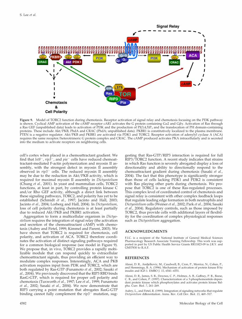
Aggregation to form a multicellular organism in Dictyo-stelium requires the integration of signal relay (the activationand secretion of the chemoattractant cAMP) with chemo-taxis (Aubry and Firtel, 1999; Kimmel and Parent, 2003). Wehave shown that TORC2 is required for chemotaxis, cellpolarity, and activation of ACA. TORC2 therefore coordi-nates the activation of distinct signaling pathways requiredfor a common biological response (see model in Figure 9).We propose that, in vivo, TORC2 provides a rapidly mobi-lizable module that can respond quickly to extracellularchemoattractant signals, thus providing an efficient way tomodulate complex responses. Interestingly, ACA and PKBactivation requires input from PI3K and TORC2, which areboth regulated by Ras-GTP (Funamoto et al., 2002; Sasaki etal., 2004). We previously discovered that the RIP3 RBD bindsRasG-GTP, which is required for proper cell polarity andchemotaxis (Tuxworth et al., 1997; Lee et al., 1999; Funamotoet al., 2002; Sasaki et al., 2004). We now demonstrate thatRIP3 carrying a point mutation that abrogates RasG-GTPbinding cannot fully complement the rip3� mutation, sug-
gesting that Ras-GTP/RIP3 interaction is required for fullRIP3/TORC2 function. A recent study indicates that strainsin which Ras function is severely abrogated display a loss ofdirectionality and ability to directionally respond to thechemoattractant gradient during chemotaxis (Sasaki et al.,2004). The fact that this phenotype is significantly strongerthan those of cells lacking PI3K1 and PI3K2 is consistentwith Ras playing other parts during chemotaxis. We pro-pose that TORC2 is one of these Ras-regulated processes.This complex level of coordinated control of chemotaxis andsignal relay is consistent with other complex feedback loopsthat regulate leading edge formation in both neutrophils andDictyostelium cells (Weiner et al., 2002; Park et al., 2004; Sasakiet al., 2004). Regulatory controls, such as those imposed byTORC2, thus provide cells with additional layers of flexibil-ity for the coordination of complex physiological responsessuch as Dictyostelium aggregation.
ACKNOWLEDGMENTS
F.I.C. is a recipient of the National Institute of General Medical SciencesPharmacology Research Associate Training Fellowship. This work was sup-ported in part by US Public Health Service Grants RR11823-09 to J.R.Y. andGM37830 to R.A.F.
REFERENCES
Alessi, D. R., Andjelkovic, M., Caudwell, B., Cron, P., Morrice, N., Cohen, P.,and Hemmings, B. A. (1996). Mechanism of activation of protein kinase B byinsulin and IGF-1. EMBO J. 15, 6541–6551.
Alessi, D. R., James, S. R., Downes, C. P., Holmes, A. B., Gaffney, P. R., Reese,C. B., and Cohen, P. (1997). Characterization of a 3-phosphoinositide-depen-dent protein kinase which phosphorylates and activates protein kinase Bal-pha. Curr. Biol. 7, 261–269.
Aubry, L., and Firtel, R. (1999). Integration of signaling networks that regulateDictyostelium differentiation. Annu. Rev. Cell Dev. Biol. 15, 469–517.
Figure 9. Model of TORC2 function during chemotaxis. Receptor activation of signal relay and chemotaxis focusing on the PI3K pathwayis shown. Cyclical AMP activation of the cAMP receptor cAR1 activates the G protein containing G�2 and G��. Activation of Ras througha Ras GEF (unpublished data) leads to activation of PI3K and the production of PI(3,4,5)P3 and the translocation of PH domain-containingproteins. These include Akt/PKB, PhdA and CRAC (PhdA; unpublished data). PKBR1 is constitutively localized to the plasma membrane.PTEN is a negative regulator. Akt/PKB and PKBR1 are activated via PDK1 and TORC2. Receptor activation of adenylyl cyclase A (ACA)requires the same receptor/heterotrimeric G protein complex and CRAC. The cAMP produced activates PKA intracellularly and is secretedinto the medium to activate receptors on neighboring cells.
S. Lee et al.
Molecular Biology of the Cell4582
Chen, M. Y., Long, Y., and Devreotes, P. N. (1997). A novel cytosolic regulator,pianissimo, is required for chemoattractant receptor and G protein-mediatedactivation of the 12 transmembrane domain adenylyl cyclase in Dictyostelium.Genes Dev. 11, 3218–3231.
Chung, C. Y., Potikyan, G., and Firtel, R. A. (2001). Control of cell polarity andchemotaxis by Akt/PKB and PI3 kinase through the regulation of PAKa. Mol.Cell 7, 937–947.
Comer, F. I., Lippincott, C. K., Masbad, J. J., and Parent, C. A. (2005). ThePI3K-mediated activation of CRAC independently regulates adenylyl cyclaseactivation and chemotaxis. Curr. Biol. 15, 134–139.
Funamoto, S., Meili, R., Lee, S., Parry, L., and Firtel, R. A. (2002). Spatial andtemporal regulation of 3-phosphoinositides by PI 3-kinase and PTEN medi-ates chemotaxis. Cell 109, 611–623.
Funamoto,S.,Milan,K.,Meili,R.,andFirtel,R.A.(2001).Roleofphosphatidylino-sitol 3� kinase and a downstream pleckstrin homology domain-containingprotein in controlling chemotaxis in Dictyostelium. J. Cell Biol. 153, 795–809.
Hall, A. L., Schlein, A., and Condeelis, J. (1988). Relationship of pseudopodextension to chemotactic hormone-induced actin polymerization in amoeboidcells. J. Cell. Biochem. 37, 285–299.
Hay, N., and Sonenberg, N. (2004). Upstream and downstream of mTOR.Genes Dev. 18, 1926–1945.
Insall, R., Kuspa, A., Lilly, P. J., Shaulsky, G., Levin, L. R., Loomis, W. F., andDevreotes, P. (1994). CRAC, a cytosolic protein containing a pleckstrin ho-mology domain, is required for receptor and G protein-mediated activation ofadenylyl cyclase in Dictyostelium. J. Cell Biol. 126, 1537–1545.
Insall, R. H., Borleis, J., and Devreotes, P. N. (1996). The aimless RasGEF isrequired for processing of chemotactic signals through G-protein-coupledreceptors in Dictyostelium. Curr. Biol. 6, 719–729.
Jacinto, E., and Hall, M. N. (2003). Tor signalling in bugs, brain and brawn.Nat. Rev. Mol. Cell. Biol. 4, 117–126.
Jacinto, E., Loewith, R., Schmidt, A., Lin, S., Ruegg, M. A., Hall, A., and Hall,M. N. (2004). Mammalian TOR complex 2 controls the actin cytoskeleton andis rapamycin insensitive. Nat. Cell Biol. 6, 1122–1128.
Kimmel, A. R., and Parent, C. A. (2003). Dictyostelium discoideum cAMPchemotaxis pathway. In: http://stke.sciencemag.org/cgi/cm/CMP_7918.
Kriebel, P. W., Barr, V. A., and Parent, C. A. (2003). Adenylyl cyclase local-ization regulates streaming during chemotaxis. Cell 112, 549–560.
Laevsky, G., and Knecht, D. A. (2001). Under-agarose folate chemotaxis ofDictyostelium discoideum amoebae in permissive and mechanically inhibitedconditions. Biotechniques 31, 1140–1149.
Lee, S., Parent, C. A., Insall, R., and Firtel, R. A. (1999). A novel Ras-interactingprotein required for chemotaxis and cyclic adenosine monophosphate signalrelay in Dictyostelium. Mol. Biol. Cell 10, 2829–2845.
Lee, S., Rivero, F., Park, K. C., Huang, E., Funamoto, S., and Firtel, R. A. (2004).Dictyostelium PAKc is required for proper chemotaxis. Mol. Biol. Cell 15,5456–5469.
Lilly, P. J., and Devreotes, P. N. (1995). Chemoattractant and GTP gammaS-mediated stimulation of adenylyl cyclase in Dictyostelium requires translo-cation of CRAC to membranes. J. Cell Biol. 129, 1659–1665.
Loewith, R., Jacinto, E., Wullschleger, S., Lorberg, A., Crespo, J. L., Bonenfant,D., Oppliger, W., Jenoe, P., and Hall, M. N. (2002). Two TOR complexes, onlyone of which is rapamycin sensitive, have distinct roles in cell growth control.Mol. Cell 10, 457–468.
Lorberg, A., and Hall, M. N. (2004). TOR: the first 10 years. Curr. Top.Microbiol. Immunol. 279, 1–18.
Meili, R., Ellsworth, C., and Firtel, R. A. (2000). A novel Akt/PKB-relatedkinase is essential for morphogenesis in Dictyostelium. Curr. Biol. 10, 708–717.
Meili, R., Ellsworth, C., Lee, S., Reddy, T., Ma, H., and Firtel, R. (1999).Chemoattractant-mediated transient activation and membrane localization ofAkt/PKB is required for efficient chemotaxis to cAMP in Dictyostelium. EMBOJ. 18, 2092–2105.
Merlot, S., and Firtel, R. A. (2003). Leading the way: directional sensingthrough phosphatidylinositol 3-kinase and other signaling pathways. J. CellSci. 116, 3471–3478.
Parent, C., Blacklock, B., Froehlich, W., Murphy, D., and Devreotes, P. (1998).G protein signaling events are activated at the leading edge of chemotacticcells. Cell 95, 81–91.
Parent, C. A., and Devreotes, P. N. (1995). Isolation of inactive and G protein-resistant adenylyl cyclase mutants using random mutagenesis. J. Biol. Chem.270, 22693–22696.
Parent, C. A., and Devreotes, P. N. (1996). Molecular genetics of signaltransduction in Dictyostelium. Annu. Rev. Biochem. 65, 411–440.
Park, K. C., Rivero, F., Meili, R., Lee, S., Apone, F., and Firtel, R. A. (2004). Racregulation of chemotaxis and morphogenesis in Dictyostelium. EMBO J. 23,4177–4189.
Ridley, A. J., Schwartz, M. A., Burridge, K., Firtel, R. A., Ginsberg, M. H.,Borisy, G., Parsons, J. T., and Horwitz, A. R. (2003). Cell migration: integratingsignals from front to back. Science 302, 1704–1709.
Sarbassov, D. D., Guertin, D. A., Ali, S. M., and Sabatini, D. M. (2005).Phosphorylation and regulation of Akt/PKB by the rictor-mTOR complex.Science 307, 1098–1101.
Sarbassov dos, D., Ali, S. M., Kim, D. H., Guertin, D. A., Latek, R. R.,Erdjument-Bromage, H., Tempst, P., and Sabatini, D. M. (2004). Rictor, a novelbinding partner of mTOR, defines a rapamycin-insensitive and raptor-inde-pendent pathway that regulates the cytoskeleton. Curr. Biol. 14, 1296–1302.
Sasaki, A. T., Chun, C., Takeda, K., and Firtel, R. A. (2004). Localized Rassignaling at the leading edge regulates PI3K, cell polarity, and directional cellmovement. J. Cell Biol. 167, 505–518.
Schmidt,A.,Bickle,M.,Beck,T.,andHall,M.N.(1997).Theyeastphosphatidylino-sitol kinase homolog TOR2 activates RHO1 and RHO2 via the exchange factorROM2. Cell 88, 531–542.
Schmidt, A., Kunz, J., and Hall, M. N. (1996). TOR2 is required for organiza-tion of the actin cytoskeleton in yeast. Proc. Natl. Acad. Sci. USA 93, 13780–13785.
Steimle, P. A., Yumura, S., Cote, G. P., Medley, Q. G., Polyakov, M. V.,Leppert, B., and Egelhoff, T. T. (2001). Recruitment of a myosin heavy chainkinase to actin-rich protrusions in Dictyostelium. Curr. Biol. 11, 708–713.
Stephens, L., Ellson, C., and Hawkins, P. (2002). Roles of PI3Ks in leukocytechemotaxis and phagocytosis. Curr. Opin. Cell Biol. 14, 203–213.
Sutoh, K. (1993). A transformation vector for Dictyostelium discoideum with anew selectable marker bsr. Plasmid 30, 150–154.
Tuxworth, R. I., Cheetham, J. L., Machesky, L. M., Spiegelmann, G. B., Weeks,G., and Insall, R. H. (1997). Dictyostelium RasG is required for normal motilityand cytokinesis, but not growth. J. Cell Biol. 138, 605–614.
Van Haastert, P. J., and Devreotes, P. N. (2004). Chemotaxis: signalling theway forward. Nat. Rev. Mol. Cell. Biol. 5, 626–634.
Washburn, M. P., Wolters, D., and Yates, J. R., III (2001). Large-scale analysisof the yeast proteome by multidimensional protein identification technology.Nat. Biotechnol. 19, 242–247.
Weiner, O. D., Neilsen, P. O., Prestwich, G. D., Kirschner, M. W., Cantley,L. C., and Bourne, H. R. (2002). A PtdInsP(3)- and Rho GTPase-mediatedpositive feedback loop regulates neutrophil polarity. Nat. Cell Biol. 4, 509–513.
Wessels, D., Soll, D. R., Knecht, D., Loomis, W. F., De Lozanne, A., andSpudich, J. (1988). Cell motility and chemotaxis in Dictyostelium amebaelacking myosin heavy chain. Dev. Biol. 128, 164–177.
Wessels, D., Voss, E., Von Bergen, N., Burns, R., Stites, J., and Soll, D. R. (1998).A computer-assisted system for reconstructing and interpreting the dynamicthree-dimensional relationships of the outer surface, nucleus and pseudopodsof crawling cells. Cell Motil. Cytoskelet. 41, 225–246.
Williams, M. R., Arthur, J. S., Balendran, A., van der Kaay, J., Poli, V., Cohen,P., and Alessi, D. R. (2000). The role of 3-phosphoinositide-dependent proteinkinase 1 in activating AGC kinases defined in embryonic stem cells. Curr.Biol. 10, 439–448.
TORC2 Controls Dictyostelium Aggregation
Vol. 16, October 2005 4583