Embed Size (px)
Citation preview
Topography-driven PhotorefractiveKeratectomyResults of Corneal Interactive Programmed TopographicAblation Software
Giovanni Alessio, MD, Francesco Boscia, MD, Maria Gabriella La Tegola, MD, Carlo Sborgia, MD
Objective: This study evaluated the efficacy, predictability, stability, and safety of a software program(Corneal Interactive Programmed Topographic Ablation (CIPTA) LIGI, Taranto, Italy) which, by transferringprogrammed ablation from the corneal topography to a flying-spot excimer laser, provides customized laserablation.
Design: Noncomparative consecutive case series.Participants: Forty-two eyes of 34 subjects with a mean age of 33.9 (range, 20–54) had CIPTA at the
Cattedra di Ottica Fisiopatologica of Bari (Italy). Twenty-eight eyes were treated for hyperopic astigmatism and14 for myopic astigmatism. All the subjects had irregular astigmatism.
Operation: Topography was acquired by a corneal topography mapping system (Orbscan, Orbtek, Inc., SaltLake City, UT). These data were processed to obtain a customized altimetric ablation profile, which wastransferred to a flying-spot laser (Laserscan 2000, Lasersight, Orlando, FL).
Main Outcome Measures: Data on uncorrected (UCVA) and best-corrected visual acuity (BCVA), predict-ability, and stability of refraction and any complications were analyzed.
Results: Mean follow-up was 13.2 months. At the last postoperative examination, 26 eyes (92.8%) in thehyperopic group and 12 eyes (85.7%) in the myopic group had an UCVA superior to 20/40. Twelve hyperopiceyes (42.8%) and five myopic eyes (35.7%) had a UCVA of 20/20. All patients fell between 1 diopter of attemptedcorrection in the spherical equivalent. Only 1 (2.4%) of the 42 eyes, belonging to the hyperopic group, lost 1Snellen line of BCVA. We did not observe any decentration and/or haze after photorefractive keratectomytreatment or any irregularity in the flap-stroma interface in the three laser in situ keratomileusis operationsperformed in this study.
Conclusions: The combination of topographic data with computer-controlled flying-spot excimer laserablation is a suitable solution for correcting irregular astigmatism due to different causes. Ophthalmology 2000;107:1578–1587 © 2000 by the American Academy of Ophthalmology.
Excimer lasers have worked well in patients with sphericalerror or with regular astigmatism. However, the results havenot been so satisfactory in subjects with irregular or asym-metric astigmatism. The reason for this is that all excimerlasers, although using different algorithms, carry out iden-tical ablations in all patients with the same refractive error
and optical zone. Some of the theoretical advantages oftopography-driven photorefractive keratectomy (PRK) are abetter astigmatic correction, the possibility of correctingirregular astigmatism, and a smaller ablation volume com-pared with standard treatments, resulting in better visualperformance.
Various authors have studied the possibility of perform-ing topography-assisted excimer laser treatments. Seitz etal1 presented their data for topography-based flying-spotcorrection of irregular corneal astigmatism based on theZernike decomposition of topography height data.Wiesinger-Jendritza et al2 published their results with laserin situ keratomileusis (LASIK) assisted by corneal topog-raphy on 23 eyes of 22 patients for the treatment of irregularastigmatism, and reported a high percentage of undercor-rection and regression because of underestimation of thecorneal irregularity.
In January 1996 we developed a software program (Cor-neal Interactive Programmed Topographic Ablation[CIPTA] LIGI, Taranto, Italy) that couples a corneal alti-
Originally received: February 18, 1999.Accepted: April 11, 2000. Manuscript no. 99101.
From the Cattedra di Ottica Fisiopatologica, Department of Otorhinolar-yngology and Ophthalmology, University of Bari, Bari, Italy.
Presented in part at the annual meeting of the Association for Research inVision and Ophthalmology, Ft. Lauderdale, Florida, May 1999, and at theannual meetings of the American Academy of Ophthalmology, New Or-leans, Louisiana, November 1998, and Orlando, Florida, October 1999.
The authors have no proprietary interest in any of the materials used in thisstudy.
Reprint requests to Carlo Sborgia, MD, Department of Otorhinolaryngol-ogy and Ophthalmology, University of Bari, Piazza Giulio Cesare, 11 Bari,Italy.
1578 © 2000 by the American Academy of Ophthalmology ISSN 0161-6420/00/$–see front matterPublished by Elsevier Science Inc. PII S0161-6420(00)00224-4

metric topography mapping system (Orbscan, Orbtek, Inc.,Salt Lake City, UT) with a flying-spot laser (Laserscan2000, Lasersight, Orlando, FL).
The software is based on the following principles:
1. The ablation takes into account the true corneal shapeof the patient, acquired with the elevation map, notonly a mathematical model of it.
2. The volume of the ablation is defined by the intersec-tion of the three-dimensional shape of the cornea andthe best aspheric surface for refraction. Among all thepossible ablation patterns, the one that minimizes theablation volume, while at the same time respectingthe optical zone, is chosen. This is determined bychoosing the smallest perimeter that fully circum-scribes the entrance pupil in scotopic conditions.
3. The transition zone of the intersection between thetwo surfaces described has a constant slope in alldirections, thus minimizing the risk of regression. Forthis reason, the transition zone is larger in the merid-ian with the highest refractive change.
Patients and Methods
Patients
After having tested the program on polymethyl methacrylate andeye bank eyes, we started the clinical trials in October 1997. Wetreated 42 eyes of 34 subjects (22 men, 12 women) ranging in agefrom 20 to 54 (mean, 33.9; standard deviation [SD], 9) at theCattedra di Ottica Fisiopatologica, Department of Otorhinolaryn-gology and Ophthalmology of the University of Bari (Italy). Meanfollow-up was 13.2 months (range, 6–18; SD, 5.5). In this studywe have not considered the first 20 eyes treated by means ofCIPTA because these were considered part of the learning curve,which was required both for the software and the surgeons. Theconversion factor between ablation on polymethyl methacrylateand living human cornea had to be assessed. Because the softwareis interactive, we had to learn how to weight the relative impor-tance of centering the width of the optic and of the transition zone.
All the patients had irregular or asymmetric corneal astigma-tism. This was defined as such an irregular corneal surface that itcould not be refracted with any optical corrective device andrequired surface smoothing.3 Twenty-eight subjects were treatedfor hyperopic astigmatism; 13 subjects had a spherical equivalent(SE) between 0 and13 diopters (D) and 15 between13.12 and16 D. Fourteen subjects needed treatment for myopic astigma-tism; 5 subjects had an SE between 0 and23 D, 3 between23.12and 26 D, 6 between26.12 and210 D. The hyperopic groupincluded 28 eyes of 20 patients (11 men and 9 women). Nineteeneyes had native irregular astigmatism. Nine eyes had postsurgicalastigmatism: one postcataract surgery and eight post-PRK (fiveposthyperopic and three postmyopic PRK). The myopic groupincluded 14 eyes of 14 patients (11 men and 3 women). Two eyeshad native irregular astigmatism. Twelve eyes had postsurgicalastigmatism: seven postmyopic PRK (one central island and sixdecentered treatments), two post-LASIK (one button-hole and onedecentered treatment), and two surgical leukomas.
Subjects with clinical or topographic signs of keratoconus orevidence of corneal infection were excluded.
Patients gave informed consent to the experimental nature ofthe treatment.
Clinical ExaminationAll eyes had a comprehensive preoperative ophthalmic examina-tion including slit-lamp biomicroscopy, applanation tonometry,indirect ophthalmoscopy, and optic pachymetry by means of theOrbscan. In the two eyes with corneal leukomas, we performedacoustic pachymetry because the Orbscan lacks accuracy for thismeasurement when corneal scattering is high.
Uncorrected visual acuity (UCVA) and best spectacle-cor-rected visual acuity (BCVA) were tested preoperatively andmonthly during follow-up. Refraction was measured preopera-tively under cycloplegia and postoperatively using the fogging(high plus) technique. We evaluated subjective refraction becauseof the difficulty in assessing this objectively in irregular astigma-tisms. In our series the difference between refractive and cornealastigmatism never exceeded 1 D.
Vector analysis of astigmatism, according to Alpins’ method,4
was based on the refractive data. The following parameters used inthe “Results” section are briefly explained. Target-induced astig-matism (TIA) corresponds to the amount and direction of thedioptric force required to achieve the astigmatic goal from thepreoperative astigmatic state. Surgical-induced astigmatism (SIA)corresponds to the amount and direction of corneal steepeningneeded to achieve the operative result from the preoperative as-tigmatic state. Ideally, the TIA and SIA are the same in magnitudeand axis and are perpendicular to the axis of preoperative astig-matism, so as to correct the latter. The magnitude of error is adirect parameter for measuring how close in magnitude the SIA isto the TIA and is calculated by subtracting the vectorial TIA fromthe vectorial SIA. The angle of error is the angle between thevectors of the SIA and the TIA on a 180° vector diagram. Successcan be determined by assessing how close the magnitude and meanangle of error are to zero.
Subjective evaluation of quality of vision was assessed byasking the patients whether halos, glare, and monocular diplopiawere less, the same, or worse than before surgery with glasses orcontact lenses.
Corneal topography was acquired before surgery and monthlyafter the treatment.
Surgical TechniquePreliminary steps of the procedure included acquisition of thecorneal shape by means of the Orbscan and a knowledge of thepatient’s subjective refraction. To increase the accuracy of thetopography, three acquisitions are required differing,3 mm inheight in the central 5 mm. The pupil diameter was measuredunder scotopic conditions with the infrared camera of the laser eyetracker.
These data were processed by CIPTA to obtain a customizedaltimetric ablation profile, which was transferred, by means of aninterface, to the Laserscan 2000.
Orbscan uses a calibrated video and scanning slit-beam systemto measure three-dimensional locations of several thousand pointson the corneal surfaces, which are used to construct the truetopographic surface.
Laserscan 2000 works with a microspot of 800mm, a frequencyof 100 Hz, and an energy at the cornea of 0.7 to 1.2 mJ per pulse.
CIPTA features six interactive steps:
1. Centering of the refractive treatment. The treatment couldbe centered on the fixation, the corneal reflex of the fixationtarget of the Orbscan, when the original visual axis of thepatient was to be preserved (in hyperopic and/or nativeasymmetric astigmatism); on the pupil centrally, in decen-tered treatments; on the corneal apex, in extremely irregularcorneas to enable maximal sparing of tissue ablation; on the
Alessio et al z Topography-driven PRK
1579
pachymetric thinnest point, on the cursor; or on the mathe-matical coordinates. These options are available for futureapplications of the software to prepare the bed for lamellarkeratoplasty for example. In this study we used the first twooptions.
2. Definition of the minimal useful refractive diameter (innerlimit) and selection of the outer limit of the treatment.
3. Choice of treatment axis: this could be the visual axis or theaxis of the best aspheric surface for the patient.
4. Input of the desired refractive correction and/or the kerato-metric values.
5. The level of ablation could be superficial (PRK) or intras-tromal (LASIK). At this stage, the software constructs thenew ideal aspheric corneal surface.
6. The slope and minimum width of the transition zone may bechanged by the surgeon.
At the end of the interactive process, the ablation statistics andscheme appear. At this stage it is possible to measure the differ-ence in height, for each point, between the two surfaces: real(preoperative) and ideal (postoperative aspheric surface).
Finally, the software processes the ablation map, which istransferred to the flying-spot excimer laser by means of a grid ofCartesian coordinates.
The spot’s energy was calibrated on the SE of each treatment.All treatments were performed as a one-step procedure and aimedat emmetropia.
In PRK, 18% ethanol epithelial debridement (Ethanol 95°,CarloErba Reagenti, Milano, Italy) was performed in primarytreatments, whereas in retreatments, excimer laser epithelial de-bridement to a depth of 50mm was carried out. In post-LASIKretreatment, the flap was elevated by a spatula.
After surgery, a contact lens was applied, and topical 0.1%preservative-free gentamicin and indomethacin drops were pre-scribed four times daily until the epithelium healed. Fluoro-metholone drops (0.2%) were used four times a day for the firstmonth, three times a day for the second month, twice a day for thethird month, and once a day for the fourth month. Acetylcysteinedrops (5%) were used three times a day for 4 months (because ofthe potential role of collagenase inhibitors on corneal transparencyafter PRK Corbett MC, O’Brart DPS, Patmore A, et al [InvestOphthalmol Vis Sci 35(Suppl): 1723, 1994].
Results
Efficacy
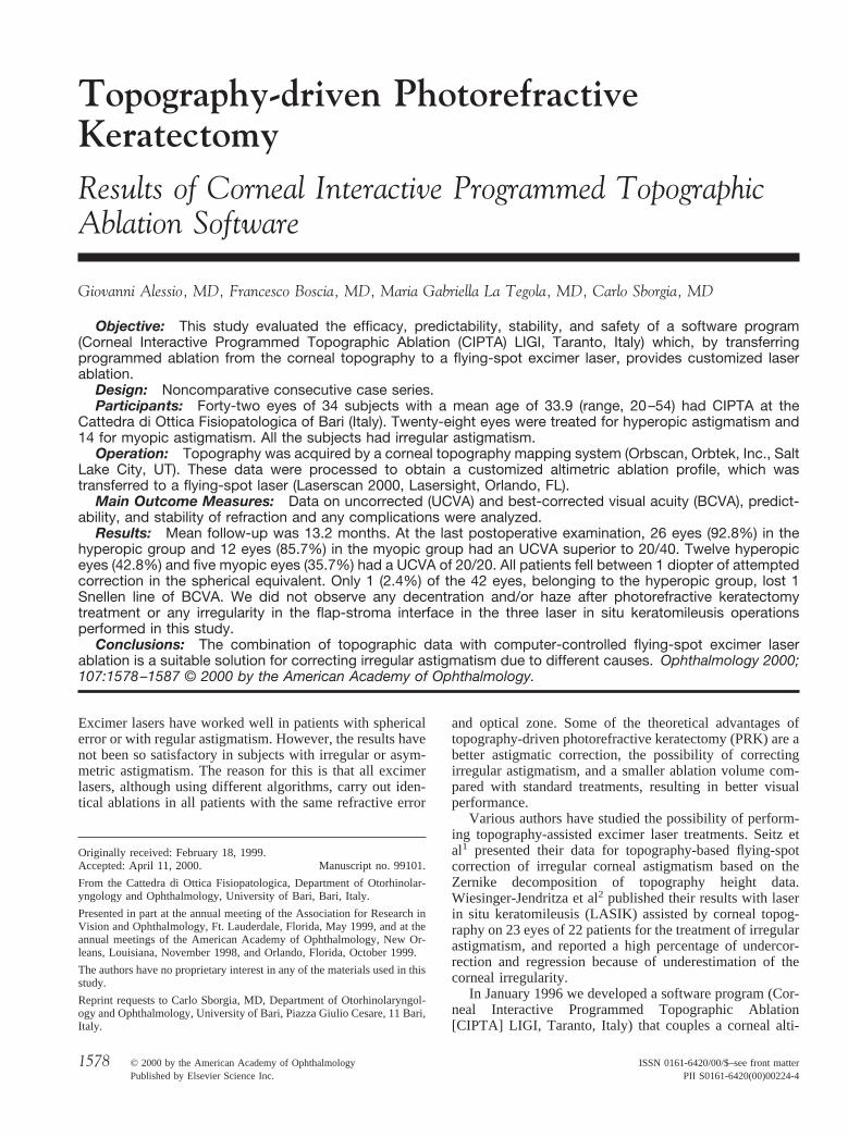
In the hyperopic group, mean UCVA improved from 0.54 loga-rithm of the minimum angle of resolution (LogMAR)5 (20/80)preoperatively (range, 1–0.1 LogMAR; SD, 0.2) to 0.09 LogMAR(20/22) (range, 0.5–0 LogMAR; SD, 0.12) at the last examination;in the myopic group, mean UCVA improved from 0.9 LogMAR(20/200) preoperatively (range, 1.3–0.3 LogMAR; SD, 0.4) to0.13 LogMAR (20/27) (range, 0.4–0 LogMAR; SD, 0.14) at thelast examination (Fig 1). In the hyperopic group, the percentage ofeyes with UCVA superior to 20/40 increased from 25% (7 eyes)preoperatively to 92.8% (26 eyes) postoperatively; in the myopicgroup, the percentage of eyes with UCVA superior to 20/40increased from 14.3% (2 eyes) to 85.7% (12 eyes) postoperatively.None of the eyes had a preoperative UCVA of 20/20, whereas aftertreatment 12 hyperopic eyes (42.8%) and 5 myopic eyes (35.7%)had a visual acuity of 20/20 (Table 1).
Predictability—Spherical Equivalent
In the hyperopic group, mean preoperative SE was13.12 D(range, 0–15 D; SD, 1.4); at the last postoperative examination,mean SE was10.2 D (range,21–11 D; SD, 0.5). In the myopicgroup, mean preoperative SE was24.58 D (range,20.375–28.375 D; SD, 2.8); at the last postoperative examination, meanSE was20.27 D (range,21–0.5; SD, 0.46). Table 2 showspreoperative and postoperative refractive data (mean6SD), up to18 months.
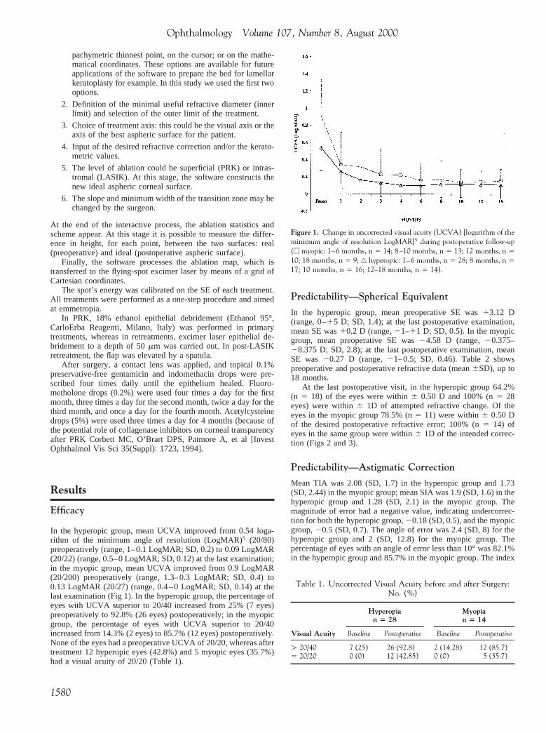
At the last postoperative visit, in the hyperopic group 64.2%(n 5 18) of the eyes were within6 0.50 D and 100% (n5 28eyes) were within6 1D of attempted refractive change. Of theeyes in the myopic group 78.5% (n5 11) were within6 0.50 Dof the desired postoperative refractive error; 100% (n5 14) ofeyes in the same group were within6 1D of the intended correc-tion (Figs 2 and 3).
Predictability—Astigmatic Correction
Mean TIA was 2.08 (SD, 1.7) in the hyperopic group and 1.73(SD, 2.44) in the myopic group; mean SIA was 1.9 (SD, 1.6) in thehyperopic group and 1.28 (SD, 2.1) in the myopic group. Themagnitude of error had a negative value, indicating undercorrec-tion for both the hyperopic group,20.18 (SD, 0.5), and the myopicgroup,20.5 (SD, 0.7). The angle of error was 2.4 (SD, 8) for thehyperopic group and 2 (SD, 12.8) for the myopic group. Thepercentage of eyes with an angle of error less than 10° was 82.1%in the hyperopic group and 85.7% in the myopic group. The index
Figure 1. Change in uncorrected visual acuity (UCVA) [logarithm of theminimum angle of resolution LogMAR]5 during postoperative follow-up(e myopic: 1–6 months, n 5 14; 8–10 months, n 5 13; 12 months, n 510; 18 months, n 5 9; ‚ hyperopic: 1–6 months, n 5 28; 8 months, n 517; 10 months, n 5 16; 12–18 months, n 5 14).
Table 1. Uncorrected Visual Acuity before and after Surgery:No. (%)
Hyperopian 5 28
Myopian 5 14
Visual Acuity Baseline Postoperative Baseline Postoperative
. 20/40 7 (25) 26 (92.8) 2 (14.28) 12 (85.7)5 20/20 0 (0) 12 (42.85) 0 (0) 5 (35.7)
Ophthalmology Volume 107, Number 8, August 2000
1580
of success (directly proportional to the difference vector andinverse to the TIA vector; ideal result5 zero, implying that thetargeted astigmatism correction has been achieved) was 0.15 in thehyperopic group and 0.26 in the myopic group (Table 3).
Safety
In the hyperopic group, mean BCVA passed from 0.1 LogMAR5
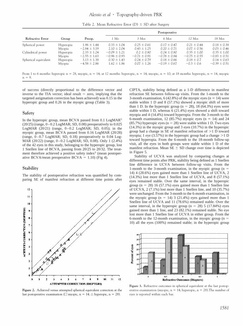
(20/25) (range, 0–0.2 LogMAR; SD, 0.08) preoperatively to 0.025LogMAR (20/21) (range, 0–0.2 LogMAR; SD, 0.05); in themyopic group, mean BCVA passed from 0.16 LogMAR (20/28)(range, 0–0.7 LogMAR; SD, 0.18) preoperatively to 0.04 Log-MAR (20/22) (range, 0–0.2 LogMAR; SD, 0.08). Only 1 (2.4%)of the 42 eyes in this study, belonging to the hyperopic group, lost1 Snellen line of BCVA, passing from 20/25 to 20/32. The treat-ment therefore achieved a positive safety index5 (mean postoper-ative BCVA/mean preoperative BCVA5 1.10) (Fig 4).
Stability
The stability of postoperative refraction was quantified by com-paring SE of manifest refraction at different time points after
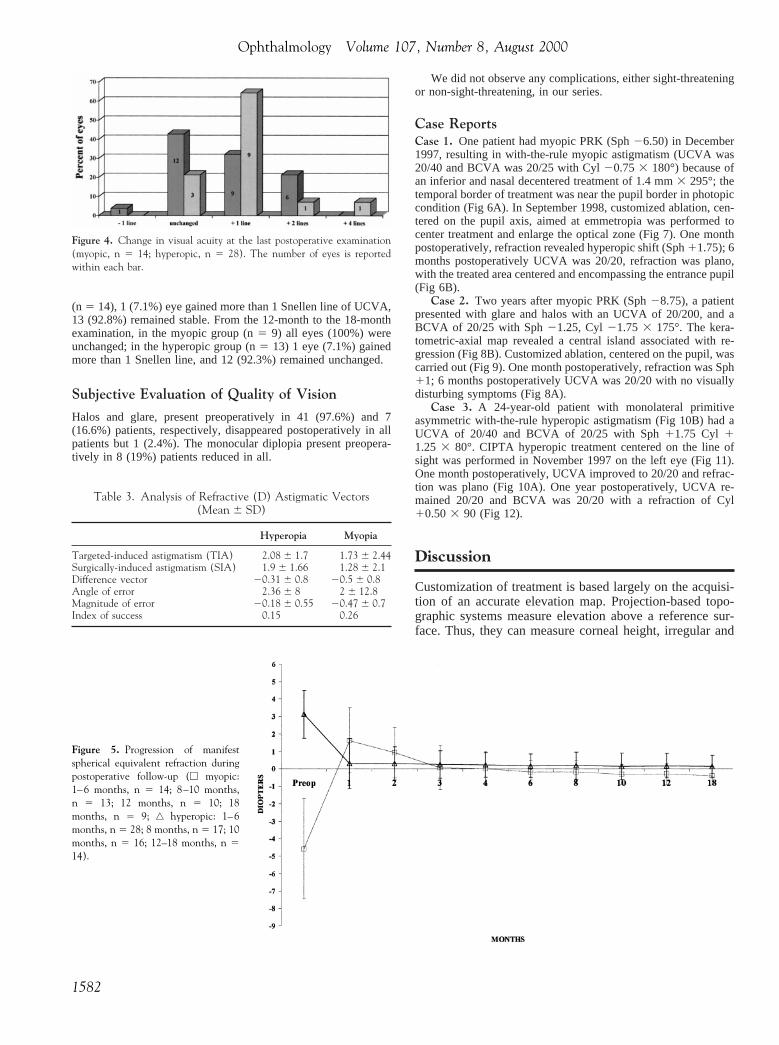
CIPTA, stability being defined as a 1-D difference in manifestrefraction SE between follow-up visits. From the 1-month to the3-month examination, 6 (42.8%) of the myopic eyes (n5 14) werestable within 1 D and 8 (57.1%) showed a myopic shift of morethan 1 D. In the hyperopic group (n5 28), 18 (64.3%) eyes werestable within 1 D, whereas 6 (21.4%) eyes showed a shift towardmyopia and 4 (14.4%) toward hyperopia. From the 3-month to the6-month examination, 12 (85.7%) myopic eyes (n5 14) and 24(85.7%) hyperopic eyes (n5 28) were stable within 1 D. Two eyes(14.3%) in the myopic group and 3 eyes (10.7%) in the hyperopicgroup had a change in SE of manifest refraction of.1 D towardmyopia; 1 eye (3.57%) in the hyperopic group had a change.1 Dtoward hyperopia. From the 6-month to the 18-month follow-upvisit, all the eyes in both groups were stable within 1 D of themanifest refraction. Mean SE6 SD change over time is depictedin Figure 5.
Stability of UCVA was analyzed by comparing changes atdifferent time points after PRK, stability being defined as 1 Snellenline difference in UCVA between follow-up visits. From the1-month to the 3-month examination, in the myopic group (n514) 4 (28.6%) eyes gained more than 1 Snellen line of UCVA, 2(14.3%) lost more than 1 Snellen line of UCVA, and 8 (57.1%)eyes remained stable. Over the same interval, in the hyperopicgroup (n5 28) 16 (57.1%) eyes gained more than 1 Snellen lineof UCVA, 2 (7.1%) lost more than 1 Snellen line, and 10 (35.7%)were unchanged. From the 3-month to the 6-month examination, inthe myopic group (n5 14) 3 (21.4%) eyes gained more than 1Snellen line of UCVA and 11 (78.6%) remained stable. Over thesame interval, in the hyperopic group (n5 28) 5 (17.84%) eyesgained more than 1 line, and 23 (82.1%) remained stable. No eyelost more than 1 Snellen line of UCVA in either group. From the6-month to the 12-month examination, in the myopic group (n510) all the eyes (100%) remained stable; in the hyperopic group
Figure 2. Achieved versus attempted spherical equivalent correction at thelast postoperative examination (e myopic, n 5 14; ‚ hyperopic, n 5 28).
Figure 3. Refractive outcomes in spherical equivalent at the last postop-erative examination (myopic, n 5 14; hyperopic, n 5 28).The number ofeyes is reported within each bar.
Table 2. Mean Refractive Error (D) 6 SD after Surgery
Refractive Error Group Preop.
Postoperative
1 Mo 3 Mos 6 Mos 12 Mos 18 Mos
Spherical power Hyperopia 1.96 6 1.44 0.33 6 1.04 0.25 6 0.61 0.17 6 0.47 0.21 6 0.44 0.18 6 0.38Myopia 22.84 6 3.19 2.10 6 2.04 0.43 6 1.25 0.20 6 0.71 0.07 6 0.56 0.03 6 0.46
Cylindrical power Hyperopia 2.33 6 1.24 20.09 6 1.21 0.2 6 0.80 0.24 6 0.80 0.35 6 1.00 0.35 6 1.00Myopia 22.55 6 1.63 20.96 6 0.93 20.71 6 0.91 20.78 6 0.84 20.75 6 0.55 20.83 6 0.51
Spherical equivalent Hyperopia 3.13 6 1.38 0.30 6 1.43 0.24 6 0.79 0.18 6 0.66 0.18 6 0.7 0.14 6 0.63Myopia 24.58 6 2.86 1.62 6 1.86 0.07 6 1.26 20.19 6 0.67 20.3 6 0.6 20.39 6 0.51
From 1 to 6 months: hyperopic n 5 28, myopic, n 5 14; at 12 months: hyperopic, n 5 14, myopic, n 5 10; at 18 months: hyperopic, n 5 14, myopicn 5 9.
Alessio et al z Topography-driven PRK
1581
(n 5 14), 1 (7.1%) eye gained more than 1 Snellen line of UCVA,13 (92.8%) remained stable. From the 12-month to the 18-monthexamination, in the myopic group (n5 9) all eyes (100%) wereunchanged; in the hyperopic group (n5 13) 1 eye (7.1%) gainedmore than 1 Snellen line, and 12 (92.3%) remained unchanged.
Subjective Evaluation of Quality of Vision
Halos and glare, present preoperatively in 41 (97.6%) and 7(16.6%) patients, respectively, disappeared postoperatively in allpatients but 1 (2.4%). The monocular diplopia present preopera-tively in 8 (19%) patients reduced in all.
We did not observe any complications, either sight-threateningor non-sight-threatening, in our series.
Case ReportsCase 1. One patient had myopic PRK (Sph26.50) in December1997, resulting in with-the-rule myopic astigmatism (UCVA was20/40 and BCVA was 20/25 with Cyl20.753 180°) because ofan inferior and nasal decentered treatment of 1.4 mm3 295°; thetemporal border of treatment was near the pupil border in photopiccondition (Fig 6A). In September 1998, customized ablation, cen-tered on the pupil axis, aimed at emmetropia was performed tocenter treatment and enlarge the optical zone (Fig 7). One monthpostoperatively, refraction revealed hyperopic shift (Sph11.75); 6months postoperatively UCVA was 20/20, refraction was plano,with the treated area centered and encompassing the entrance pupil(Fig 6B).
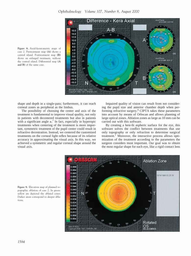
Case 2. Two years after myopic PRK (Sph28.75), a patientpresented with glare and halos with an UCVA of 20/200, and aBCVA of 20/25 with Sph21.25, Cyl21.75 3 175°. The kera-tometric-axial map revealed a central island associated with re-gression (Fig 8B). Customized ablation, centered on the pupil, wascarried out (Fig 9). One month postoperatively, refraction was Sph11; 6 months postoperatively UCVA was 20/20 with no visuallydisturbing symptoms (Fig 8A).
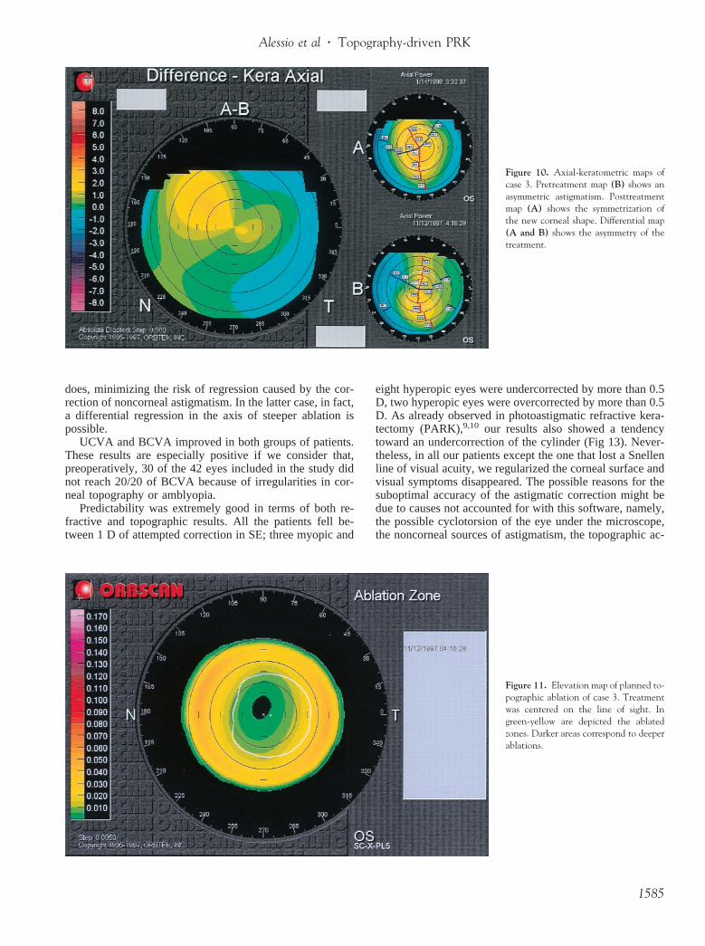
Case 3. A 24-year-old patient with monolateral primitiveasymmetric with-the-rule hyperopic astigmatism (Fig 10B) had aUCVA of 20/40 and BCVA of 20/25 with Sph11.75 Cyl 11.25 3 80°. CIPTA hyperopic treatment centered on the line ofsight was performed in November 1997 on the left eye (Fig 11).One month postoperatively, UCVA improved to 20/20 and refrac-tion was plano (Fig 10A). One year postoperatively, UCVA re-mained 20/20 and BCVA was 20/20 with a refraction of Cyl10.503 90 (Fig 12).
Discussion
Customization of treatment is based largely on the acquisi-tion of an accurate elevation map. Projection-based topo-graphic systems measure elevation above a reference sur-face. Thus, they can measure corneal height, irregular and
Figure 4. Change in visual acuity at the last postoperative examination(myopic, n 5 14; hyperopic, n 5 28). The number of eyes is reportedwithin each bar.
Table 3. Analysis of Refractive (D) Astigmatic Vectors(Mean 6 SD)
Hyperopia Myopia
Targeted-induced astigmatism (TIA) 2.08 6 1.7 1.73 6 2.44Surgically-induced astigmatism (SIA) 1.9 6 1.66 1.28 6 2.1Difference vector 20.31 6 0.8 20.5 6 0.8Angle of error 2.36 6 8 2 6 12.8Magnitude of error 20.18 6 0.55 20.47 6 0.7Index of success 0.15 0.26
Figure 5. Progression of manifestspherical equivalent refraction duringpostoperative follow-up (e myopic:1–6 months, n 5 14; 8–10 months,n 5 13; 12 months, n 5 10; 18months, n 5 9; ‚ hyperopic: 1–6months, n 5 28; 8 months, n 5 17; 10months, n 5 16; 12–18 months, n 514).
Ophthalmology Volume 107, Number 8, August 2000
1582

nonreflective surfaces, and the entire corneal surface withuniformly high accuracy in the center and periphery.Placido ring systems, on the contrary, derive height byintegrals: this can cause error; moreover, they do not pro-vide reliable elevation data for shapes that have nonlinearchanges in curvature, as occur after refractive surgery.6 Inaddition, the slit-beam videokeratograph acquires the realcorneal shape regardless of the cornea’s orientation relativeto the instrument, whereas Placido-based systems requirethe rings to be coaxial with the cornea. Also, the latterassumes the surface to be continuous and differential: ifthere is a break on the corneal surface, there will be a break
in the display produced and localized areas of irregularastigmatism may be ignored.
A limitation of the Orbscan topography is the long ac-quisition time (1.4 sec) that decreases the accuracy of theelevation map. To increase its precision, at least three mea-surements are carried out. These are assumed to be accuratewhen the difference in height in the central 5 mm amongthem is,3mm.
A flying-spot laser with a narrow beam with a Gaussianprofile is necessary to perform topography-assisted treat-ments. Thanks to its small spot size (800mm), Laserscan2000 can provide flexible, high-accuracy treatments of any
Figure 6. Elevation maps using a best fitsphere of 39.9 D of case 1. Preablationmap (A) shows a treatment decenteredinferonasally. Postablation map (B)shows a recentered treatment encom-passing the entrance pupil. Differentialmap (A and B) is similar to the plannedablation.
Figure 7. Elevation map of planned to-pographic ablation of case 1. In green-yellow are depicted the ablated zones.Darker areas correspond to deeper abla-tions.
Alessio et al z Topography-driven PRK
1583
shape and depth in a single-pass; furthermore, it can reachcorneal zones as peripheral as the limbus.
The possibility of choosing the center and axis of thetreatment is fundamental to improve visual quality, not onlyin patients with decentered treatments but also in patientswith a significant anglek.7 In fact, especially in hyperopictreatments when centering of the treatment is more impor-tant, symmetric treatment of the pupil center could result inrefractive decentration. Instead, we centered the customizedtreatments on the corneal light reflex because of its relativeaccuracy in approximating the visual axis. In this way, weachieved a symmetric and regular corneal shape around thevisual axis.
Impaired quality of vision can result from not consider-ing the pupil size and anterior chamber depth when per-forming refractive surgery.8 CIPTA takes these parametersinto account by means of Orbscan and allows planning oflarge optical zones. Ablation zones as large as 10 mm can becarried out with this software.
By creating a best-fit aspheric surface for the eye, thissoftware solves the conflict between treatments that useonly topography or only refraction to determine surgicaltreatment.1 Moreover, the interactive process allows opti-mization of the treatment according to the parameters thesurgeon considers most important. Our goal was to obtainthe most regular shape for each eye, like a rigid contact lens
Figure 8. Axial-keratometric maps ofcase 2. Pretreatment map (A) shows acentral island. Posttreatment map (B)shows an enlarged treatment, withoutthe central island. Differential map (Aand B) of the same case.
Figure 9. Elevation map of planned to-pographic ablation of case 2. In green-yellow are depicted the ablated zones.Darker areas correspond to deeper abla-tions.
Ophthalmology Volume 107, Number 8, August 2000
1584
does, minimizing the risk of regression caused by the cor-rection of noncorneal astigmatism. In the latter case, in fact,a differential regression in the axis of steeper ablation ispossible.
UCVA and BCVA improved in both groups of patients.These results are especially positive if we consider that,preoperatively, 30 of the 42 eyes included in the study didnot reach 20/20 of BCVA because of irregularities in cor-neal topography or amblyopia.
Predictability was extremely good in terms of both re-fractive and topographic results. All the patients fell be-tween 1 D of attempted correction in SE; three myopic and
eight hyperopic eyes were undercorrected by more than 0.5D, two hyperopic eyes were overcorrected by more than 0.5D. As already observed in photoastigmatic refractive kera-tectomy (PARK),9,10 our results also showed a tendencytoward an undercorrection of the cylinder (Fig 13). Never-theless, in all our patients except the one that lost a Snellenline of visual acuity, we regularized the corneal surface andvisual symptoms disappeared. The possible reasons for thesuboptimal accuracy of the astigmatic correction might bedue to causes not accounted for with this software, namely,the possible cyclotorsion of the eye under the microscope,the noncorneal sources of astigmatism, the topographic ac-
Figure 10. Axial-keratometric maps ofcase 3. Pretreatment map (B) shows anasymmetric astigmatism. Posttreatmentmap (A) shows the symmetrization ofthe new corneal shape. Differential map(A and B) shows the asymmetry of thetreatment.
Figure 11. Elevation map of planned to-pographic ablation of case 3. Treatmentwas centered on the line of sight. Ingreen-yellow are depicted the ablatedzones. Darker areas correspond to deeperablations.
Alessio et al z Topography-driven PRK
1585

quisition of the elevation maps, the eye-tracking system ofthe laser, and the surgeon’s experience in programming thecustomized ablation.
Although the follow-up is too short to draw definitiveconclusions, so far we have not observed any significantregression. This could be related to the large and homoge-neous ablation and transition zone diameters and to theconstant slope of the transition zone realized by CIPTA.
Our approach differs substantially from Seitz et al’sexperimental data,1 being based on a directly acquired ele-vation map as opposed to the Zernike decomposition oftopography height data.
Wiesinger-Jendritza et al’s data2 featured a high percent-age of undercorrection and regression caused by underesti-mation of corneal irregularity. A possible explanation is thattheir algorithms were based on corneal height values inte-grated by the axial radii of curvature and not on altimetricdata. Furthermore, the scanning slit laser used cannot pro-vide such flexible ablations as those generated by the flying-spot laser.
In contrast to the preceding methods, CIPTA can providepreoperative statistical information about the ablation vol-ume, area, and depth; also, because Orbscan can acquirepachymetric data, it can calculate the residual apex thick-ness.
Regularizing the corneal shape has the theoretical advan-tage of improving the quality of vision. Although in thisstudy we did not measure contrast sensitivity, all patientsbut one reported a subjective improvement in vision, with areduction of halos, glare, and monocular diplopia.
Although the use of customized ablation appears to havebeen effective in this population, there was no attempt tocompare this treatment with a control group having standardspherocylindrical treatment, which might have been effec-tive in some cases.
The ultimate goal of CIPTA is to create a better-than-normal corneal refractive surface. Another possible ap-proach to customized ablation aims to achieve an aberra-tion-free visual system. Although this presents theoreticaladvantages compared with CIPTA, the real influence of theother dioptric surfaces still has to be ascertained.
References
1. Seitz B, Langenbucher A, Kus MM, Harrer M. Experimentalcorrection of irregular corneal astigmatism using topography-based flying-spot-mode excimer laser photoablation. Am JOphthalmol 1998;125:252–6.
Figure 12. Axial-keratometric map ofcase 3, 1 year after corneal interactiveprogrammed topographic ablation,shows the symmetry of residual astigma-tism.
Figure 13. Surgically-induced astigmatism versus targeted induced astig-matism at the last postoperative examination (e myopic, n 5 14;‚ hyperopic, n 5 28).
Ophthalmology Volume 107, Number 8, August 2000
1586

2. Wiesinger-Jendritza B, Knorz MC, Hugger P, Liermann A.Laser in situ keratomileusis assisted by corneal topography. JCataract Refract Surg 1998;24:166–74.
3. Troutman RC, Buzard KA. Terminology and definitions, op-tical and surgical principles, tenets of refractive surgery. In:Troutman RC, Buzard KA, eds. Corneal Astigmatism: Etiol-ogy, Prevention and Management. St. Louis: Mosby YearBook, 1992; 5–22.
4. Alpins NA. A new method of analyzing vectors for changes inastigmatism. J Cataract Refract Surg 1993;19:524–33.
5. Koch DD, Kohnen T, Obstbaum SA, Rosen ES. Format forreporting refractive surgical data [editorial]. J Cataract RefractSurg 1998;24:285–7.
6. Schultze RL. Accuracy of corneal elevation with four cornealtopography systems. J Refract Surg 1998;14:100–4.
7. Buzard KA. Optical aspects of refractive surgery. In: ElanderR, Rich LF, Robin JB, eds. Principles and Practice of Refrac-tive Surgery. Philadelphia: Saunders, 1997; chap. 4.
8. Nixon WS. Pupil size in refractive surgery [letter]. J CataractRefract Surg 1997;23:1435–6.
9. Taylor HR, Kelly P, Alpins N. Excimer laser correction ofmyopic astigmatism. J Cataract Refract Surg 1994;20:243–51.
10. Lee JS, Oum BS, Lee BJ, Lee SH. Photorefractive keratec-tomy for astigmatism greater than22.00 diopters in eyes withlow, high, or extreme myopia. J Cataract Refract Surg 1998;24:1456–63.
Alessio et al z Topography-driven PRK
1587