Embed Size (px)
Citation preview

Uwe Ebeling 1
Helmuth Steinmetz2
Yanxiong Huang2
Thomas Kahn3
Received September 30, 1988; revision requested December 7, 1988; revision received February 7, 1989; accepted February 8, 1989.
This work was supported by grants from the Deutsche Forschungsgemeinschaft (SFB 200; TP Z2) .
' Department of Neurosurgery, lnselspital , CH-301 0 Bern, Switzerland. Address reprint requests to U. Ebeling.
2 Department of Neurology, University of Dusseldorf, Moorenstr. 5, D-4000 Dusseldorf 1, FRG.
3 Department of Diagnostic Radiology, University of Dusseldorf, Dusseldorf 1, FRG.
0195-6108/89/1005-0937 © American Society of Neuroradiology
Topography and Identification of the Inferior Precentral Sulcus in MR Imaging
937
Sagittal MR imaging was used to investigate cerebral sulci bordering the functionally important areas on the lateral suprasylvian surface. The aim of the study was to identify characteristic relationships of the inferior precentral sulcus to nearby sulci and gyri. MR findings in 20 healthy volunteers were compared with those in 62 intact postmortem hemispheres. MR techniques are described for the direct identification of the anterior ascending ramus of the sylvian fissure and the inferior precentral sulcus. These sulci, which border Broca's area and the primary motor area, can be reliably identified with sagittal MR. Four different types of sulcus topography were recognized. Most frequently, the inferior precentral sulcus is the sulcus posterior to the anterior ascending sylvian ramus (95% in the MR study, 87% in the anatomic study). Occasionally, an additional sulcus is interposed (5%, 10%), or an ascending ramus is absent (0%, 3%).
Identification of these landmarks is important for the exact preoperative localization of cortical lesions as well as for the intraoperative interpretation of individual sulcus patterns.
AJNR 10:937-942, September/October 1989; AJR 153: November 1989
With the advent of modern imaging techniques, the noninvasive visualization and identification of cortical structures became possible. Precise knowledge of the radiotopography of cerebral gyri and sulci is necessary for the localization of lesions in cortical areas and for decisions concerning surgical treatment. Moreover, the intraoperative recognition of cortical landmarks enables the neurosurgeon to preserve eloquent fields. In earlier studies, the characteristic configuration of the superior central and precentral sulcus was described [1-5]. Until now, a systematic investigation of the radioanatomy of the inferior motor cortex has been lacking. The main question to be answered by the present study is whether unique sulcal landmarks for the identification of the motor cortex can be recognized on the lateral hemispheric surface.
Anatomic Considerations
The precentral sulcus, which runs anterior and parallel to the central sulcus , is mostly divided into a superior and an inferior portion [6, 7] , the latter being of interest for the present study. The inferior frontal sulcus runs anterior and perpendicular to the inferior precentral sulcus (Fig . 1 ). The frequency of sulcal junctions was examined anatomically by Cunningham [9] in 50 hemispheres and later by Lang and Belz [1 0] in 1 00 hemispheres. A connection between the central sulcus and the sylvian fissure was found in 5% [9] and 19% [1 0] in these studies. A junction between the inferior precentral sulcus and the sylvian fissure existed in 15% [9] and 42% [1 0]. A more constant connection was present between the inferior precentral sulcus and the inferior frontal sulcus in 67.4% of Cunningham 's sample [9] and in 76% of Eberstaller's specimens [7].

938 EBELING ET AL. AJNR:1 0, September/October 1989
Central Sulcus
Fig. 1.- Schematic of typical sulcus pattern of lateral hemispheric aspect; sulcus terminology according to Gray's Anatomy [8] . . Ant_erior suprasylvian region is seen within circle. In this article, the antenor hm_bs of the sylvian fissure are termed anterior horizontal ramus and antenor ascending ramus (instead of anterior ramus and ascending ramus), which accords with Eberstaller's [7] and Cunningham's [9] nomenclature.
Anterior and parallel to the inferior precentral sulcus runs the anterior ascending ramus of the sylvian fissure (Fig. 1 ). It is defined by its continuity with the circular (periinsular) sulcus [7, 9], which forms the deep circumferential rim of the island of Reil. The ascending ramus thus "subopercularizes" the inferior frontal gyrus (F3). F3 is limited superiorly by the inferior frontal sulcus, posteriorly by the precentral sulcus, and inferiorly by the stem of the sylvian fissure.
Between the anterior ascending sylvian ramus and the inferior precentral sulcus lies the opercular portion of F3 (Broca area of the dominant hemisphere). Between the precentral and the central sulcus lies the precentral gyrus (primary motor area). The sulci bordering these important regions were studied to determine whether characteristic landmarks for the identification of the inferior precentral sulcus could be found radiologically and anatomically.
Materials and Methods
MR Study
Twenty healthy volunteers , 22-63 years old , were examined on a 0.35-T superconductive magnet. T1-weighted sagittal SE images were obtained from the whole brain. The technical factors were 500/ 40/4 (TRfTE/excitations), 256 256 matrix , and 5-mm-thick contiguous slices. Prior to the actual measurements, the position of the head was adjusted under the control of repeated, multiplanar, fastlocating MR scans until the interhemispheric fissure was aligned parallel to the sagittal imaging plane (orthomorphic condition). This method had the advantage that all sagittal images were obtained in reference to a common, constant, intracerebral plane (the interhemispheric plane). lnterindividual variations of cerebral radiotopography, caused by the adoption of more variable extracerebral references (as used in -ray CT), were thus e eluded. The stereotactic MR procedure used has been described elsewhere [4). The following sulci were evaluated for each brain hemisphere: central sulcus, inferior precentral sulcus, anterior ascending ramus of the sylvian fissure, inferior frontal sulcus. and eventual additional sulci of the lateral
suprasylvian region . Two different techniques were applied for the identification of the sulci:
1 . The medial central sulcus was identified at the superior hemispheric margin as the next sulcus anterior to the ascending marginal ramus of the cingulate sulcus (4, 7]. By using a fast cinematographic display mode,* the bottom of the central sulcus was traced dynamically in 5-mm lateral steps to its lowest point on the most lateral MR brain slice. The sulcus situated anterior to this lateral end of the central sulcus was identified as the inferior precentral sulcus.
2. The anterior ascending ramus of the sylvian fissure was identified in the depth of the sylvian cistern according to the criteria described above (continuity with the circular sulcus). It was traced laterally to the most lateral MR brain slice (Fig. 2). The next sulcus running perpendicular to and above the ascending ramus was identified as the inferior frontal sulcus. The topography of the lateral suprasylvian region was drawn for each hemisphere including the sulcal junctions and eventual sulci interposed between the ascending ramus and the inferior precentral sulcus.
Anatomic Study
Sixty-two unselected, intact hemispheres from the C. & 0 . Vogt brain collectiont were examined. The same sulci as described above were evaluated. The aforementioned criteria for identification of both the central sulcus and the anterior ascending sylvian ramus were tested. A drawing was made for each hemisphere.
Results
The radiologic recognition of the inferior precentral sulcus by identification procedure 1 and of the anterior ascending ramus of the sylvian fissure by identification procedure 2 (see Methods) was possible in all MR cases. With analogous procedures of sulcus identification in the anatomic study, the identifications were correct in all postmortem specimens. Two postmortem hemispheres lacked an anterior ascending ramus. According to the topographic characteristics of the lateral suprasylvian region , all brain hemispheres evaluated radiologically or anatomically could be grouped in four categories (Fig. 3, Table 1).
Type 1
This was the most frequent type, encountered in 90% of the hemispheres studied by MR and in 76% of the anatomic specimens. It was characterized by the juxtaposition of the anterior ascending sylvian ramus and the inferior precentral sulcus plus the presence of a junction between the inferior frontal and the inferior precentral sulcus (Figs. 2 and 3A).
Type 2
Type 2 was encountered in 5% of the hemispheres studied radiologically and in 11% of the anatomic specimens. Its characteristics corresponded to those of type 1 except for
• Mipron image processing workstation, Kontron Electronics, D-8057 Eching. FRG.
Brain Research Institute, University of Dusseldorf.
AJNR:1 0, September/October 1989 MR OF INFERIOR PRECENTRAL SULCUS 939
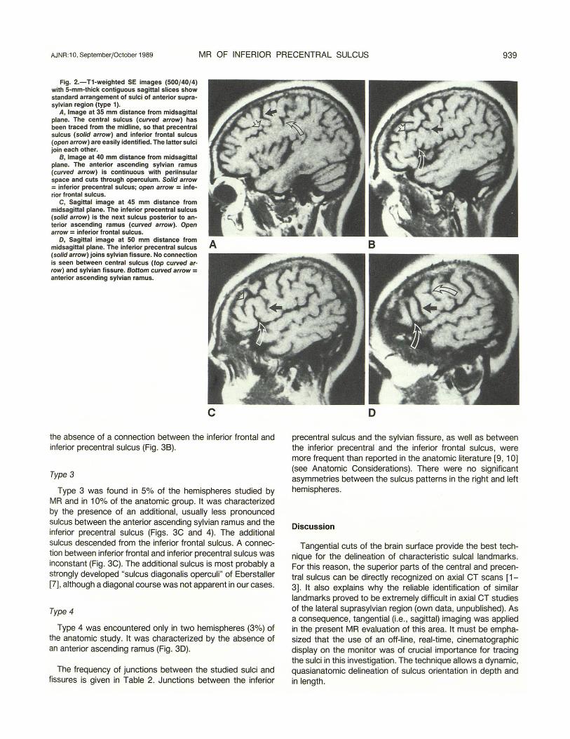
Fig. 2.- T1-weighted SE images (500/40/4) with 5-mm-thick contiguous sagittal slices show standard arrangement of sulci of anterior suprasylvian region (type 1 ).
A, Image at 35 mm distance from midsagittal plane. The central sulcus (curved arrow) has been traced from the midline, so that precentral sulcus (solid arrow) and inferior frontal sulcus (open arrow) are easily identified. The latter sulci join each other.
B, Image at 40 mm distance from midsagittal plane. The anterior ascending sylvian ramus (curved arrow) is continuous with periinsular space and cuts through operculum. Solid arrow = inferior precentral sulcus; open arrow = inferior frontal sulcus.
C, Sagittal image at 45 mm distance from midsagittal plane. The inferior precentral sulcus (solid arrow) is the next sulcus posterior to anterior ascending ramus (curved arrow). Open arrow = inferior frontal sulcus.
D, Sagittal image at 50 mm distance from midsagittal plane. The inferior precentral sulcus (solid arrow) joins sylvian fissure. No connection is seen between central sulcus (top curved arrow) and sylvian fissure. Bottom curved arrow = anterior ascending sylvian ramus.
A
c the absence of a connection between the inferior frontal and inferior precentral sulcus (Fig. 38).
Type 3
Type 3 was found in 5% of the hemispheres studied by MR and in 1 0% of the anatomic group. It was characterized by the presence of an additional, usually less pronounced sulcus between the anterior ascending sylvian ramus and the inferior precentral sulcus (Figs. 3C and 4). The additional sulcus descended from the inferior frontal sulcus. A connection between inferior frontal and inferior precentral sulcus was inconstant (Fig . 3C). The additional sulcus is most probably a strongly developed "sulcus diagonalis operculi" of Eberstaller [7] , although a diagonal course was not apparent in our cases.
Type4
Type 4 was encountered only in two hemispheres (3%) of the anatomic study. It was characterized by the absence of an anterior ascending ramus (Fig. 3D).
The frequency of junctions between the studied sulci and fissures is given in Table 2. Junctions between the inferior
B
D
precentral sulcus and the sylvian fissure , as well as between the inferior precentral and the inferior frontal sulcus, were more frequent than reported in the anatomic literature [9, 1 0] (see Anatomic Considerations). There were no significant asymmetries between the sulcus patterns in the right and left hemispheres.
Discussion
Tangential cuts of the brain surface provide the best technique for the delineation of characteristic sulcal landmarks. For this reason, the superior parts of the central and precentral sulcus can be directly recognized on axial CT scans [1-3] . It also explains why the reliable identification of similar landmarks proved to be extremely difficult in axial CT studies of the lateral suprasylvian region (own data, unpublished). As a consequence, tangential (i.e., sagittal) imaging was applied in the present MR evaluation of this area. It must be emphasized that the use of an off-line, real-time, cinematographic display on the monitor was of crucial importance for tracing the sulci in this investigation. The technique allows a dynamic, quasianatomic delineation of sulcus orientation in depth and in length.

940 EBELING ET AL. AJNR:10, September/October 1989
A B
c D
TABLE 1: Frequency of Topographic Types 1-4 in 40 Hemispheres Studied by MR and in 62 Hemispheres Studied Anatomically
Type MR Study Anatomic Study (n = 40) (n = 62)
1 36 (90%) 47 (76%) 2 2 (5%) 7 (11 %) 3 2 (5%) 6 (10%) 4 0 (0%) 2 (3%)
The criteria used for the identification of the suprasylvian landmarks proved to be valid. The anterior ascending sylvian ramus is a valuable landmark in this area. It cuts through the functionally most important posterior inferior frontal gyrus and can be reliably identified according to its continuity with the circular (periinsular) sulcus. The topography of the adjacent lateral suprasylvian region is highly constant. The inferior precentral sulcus is the next sulcus posterior to the ascending ramus in about 90% of cases (types 1 and 2). A junction between the inferior frontal and the inferior precentral sulcus is also very common. The recognition of this characteristic
Fig. 3.-The four types of anterior suprasylvian sulcus topography (for frequencies, see Table 1). CS = central sulcus, PS = inferior precentral sulcus, JFS = inferior frontal sulcus, SF= sylvian fissure (posterior horizontal ramus), ASR = anterior ascending sylvian ramus, AHR = anterior horizontal sylvian ramus; dotted lines = inconstant sulcus segments (see Table 2).
A, Type 1. The inferior precentral sulcus is the next sulcus posterior to anterior ascending ramus; inferior frontal sulcus joins precentral sulcus (see also Figs. 1 and 2).
8 , Type 2. The inferior precentral sulcus is the next sulcus posterior to anterior ascending ramus; no connection exists between inferior frontal and precentral sulcus.
C, Type 3. An additional branch (AB) descends from inferior frontal sulcus and is interposed between anterior ascending sylvian ramus and inferior precentral sulcus (see also Fig. 4).
D, Type 4. An anterior ascending sylvian ramus is lacking.
pattern (Figs. 2 and 3A) may be of help for the neurosurgeon when interpreting the suprasylvian surface topography, especially in small craniotomies. Two types of anatomic variation may occur. In 5-10%, an additional sulcus is interposed between the ascending ramus and the inferior precentral sulcus (type 3). In up to 3%, an ascending ramus is absent (type 4). Identification of the inferior precentral sulcus is therefore not always possible by evaluating the lateral suprasylvian surface alone. The relatively frequent junction between the inferior precentral sulcus and the sylvian fissure (Table 2) may serve as an additional topographic criterion. However, as can be seen from the discrepancy between the radiologic and anatomic data in Table 2, the frequency of a communication of the inferior precentral sulcus with the inferior frontal sulcus or with the sylvian fissure may be overestimated with our MR technique. Regarding the connection with the inferior frontal sulcus, this is most probably due to very small gyral bridges that sometimes link the posterior portions of the inferior and medial frontal convolution and that may be missed because of partial volume effects. This would also explain the difference between the radiologic and anatomic frequency of types 1 and 2 in our study (Table 1). Gyral connections of

AJNR:1 0, September/October 1989 MR OF INFERIOR PRECENTRAL SULCUS 941
Fig. 4.-MR study in a case of type 3 sulcus topography of the anterior suprasylvian region; T1-weighted SE image, 500/40/4, 5-mm-thick sagittal slice at 42.5 mm distance from midsagittal plane. Between the inferior precentral sulcus (solid arrow) and anterior ascending sylvian ramus (curved white arrow), an additional sulcus (open arrow) descends from the inferior frontal sulcus to cut into the opercular portion of inferior frontal convolution. Curved black arrow = central sulcus.
TABLE 2: Frequency of Junctions Between the Inferior Frontal Sulcus, Inferior Precentral Sulcus, Central Sulcus, and Sylvian Fissure (SF) in 40 Hemispheres Studied by MR and in 62 Hemispheres Studied Anatomically
Junction MR Study Anatomic Study (n = 40) (n = 62)
IFS- PS 38 (95%) 50 (81 %) PS- SF 35 (88%) 37 (60%) CS-SF 5 (13%) 9 (15%)
Note.-IFS = inferior frontal sulcus, PS = inferior precentral sulcus, SF = sylvian fissure, CS = central sulcus.
Fig. 5.-Schematic illustrates average distances (mm) between coronal suture (CoS), inferior central sulcus (CS), inferior precentral sulcus (PS), and anterior ascending sylvian ramus (ASR) (values for right hemisphere are from (10] and [11]).
Fig. 6.-lntraoperative identification of anterior suprasylvian cortical landmarks; surgeon 's view from a superior and lateral direction following right frontal craniotomy and resection of a low-grade astrocytoma in a patient with focal seizures (type 1 sulcus topography). Curved arrow = anterior ascending sylvian ramus; note sylvian veins entering ascending ramus. Black open arrow = inferior frontal sulcus. Tumor was located in posterior portions of inferior and medial frontal convolution. The posterior limit of resection is the inferior precentral sulcus (white open arrows). Correct identification of precentral gyrus was assured by electrical stimulation at points marked by tickets 6, 7, and 8 on the cortex. No paresis occurred postoperatively. 5
varying size ("Ubergangswindungen" [7]) were found to separate the inferior frontal and inferior precentral sulcus in 24% of the specimens studied by Eberstaller [7] , which is similar to our 19% in the anatomic sample compared with only 5% in the MR group (Table 2). In fact, the assignment of some of our MR cases to either type 1 or 2 was ambiguous. Likewise, gyral links between the most inferior precentral gyrus and F3 may be very small. Because this region is slightly folded into the sylvian fissure, the false impression of a connection between the inferior precentral sulcus and the sylvian fissure may appear on the most lateral MR slice in such cases. This explains the difference between the radiologic and anatomic data of Table 2 concerning the frequency of a junction between the sulci.
Additional topographic data may be helpful for the intraoperative localization of the inferior precentral sulcus. In previous anatomic and CT studies, the average sagittal distance between the inferior coronal suture and the central sulcus was 33 mm (range, 24-40 mm) [11-13]. Because the coronal suture is easily identified during craniotomy, it can be used as a rough surgical guide to the inferior motor cortex (Fig . 5).
The sagittal imaging technique provides a view of the lateral suprasylvian region that is very similar to that encountered intraoperatively (Fig. 6). Individual sulcus maps can be obtained preoperatively and thus serve for intraoperative identification of important sulci and their anatomic variations to preserve eloquent fields. The described technique of direct MR delineation of the suprasylvian sulci may also be of importance for the topographic analysis, especially of small ischemic lesions in anatomicofunctional studies on aphasia. Mass lesions, however, often deform cortical landmarks. In such cases, valuable preoperative information can be obtained from intraindividual comparison with the unaffected hemisphere. Provided that the MR examination was performed under an orthomorphic condition, the position of the
6

942 EBELING ET AL. AJNR:1 0, September/October 1989
inferior precentral sulcus or of the anterior ascending sylvian ramus as identified on the intact hemisphere can be roughly transferred by superimposition onto the contralateral side. Furthermore, if the identification of one sulcal landmark is possible in the vicinity of a lesion, the frontal operculum and precentral convolution can be localized indirectly with high reliability (Fig. 6). The average widths of these gyri provide further help for orientation in such cases. Lang and Belz [1 0] reported the following mean sagittal widths from 50 left and 50 right postmortem hemispheres: 11 .3 mm for the left opercular portion of F3 (range, 6-22 mm); 11 .9 mm for the right opercular portion of F3 (6-31 mm); 11 .9 mm for the left inferior precentral convolution (5-18 mm); and 17.7 mm for the right inferior precentral convolution (7 -21 mm) (Fig. 5).
Nevertheless, the boundaries of functional areas do not necessarily coincide with macroanatomic limits, which may be the case especially in speech function [14]. Electrical cortical stimulation is therefore of additional value in the surgical management of critical lesions of the lateral suprasylvian region [14] (Fig. 6).
ACKNOWLEDGMENTS
We thank Profs. Hopf and May (C. & 0 . Vogt Brain Research Institute, University of Dusseldorf, FAG) for allowing us to use the brains of the C. & 0 . Vogt brain collection .
REFERENCES
1. Ebeling U, Huber P, Reulen HJ . Localization of the precentral gyrus in the computed tomogram and its clinical application. J Neurol1986;233:73-76
2. Freund HJ, Hummelsheim H. Lesions of premotor cortex in man. Brain 1985;108:697-734
3. Kido OK, Le May M, Levonson AW, Benson WE. Computed tomographic localization of the precentral sulcus. Radiology 1980; 135 : 373-377
4. Steinmetz H, FOrst G, Freund HJ. Cerebral cortical localization: application and validation of the proportional grid system in MR imaging. J Comput Assist Tomogr 1989;13:10-19
5. Steinmetz H, FOrst G, Meyer BU. Craniocerebral topography within the international 10-20 system. Electroenceph Clin Neurophysiol 1989;72 : 499-506
6. Crosby EC, Humphrey T, Lauer EW. Correlative anatomy of the nervous system . New York: Macmillan, 1962
7. Eberstaller 0 . Das Stirnhirn. Ein Beitrag zur Anatomie der Oberflache des Gehirns. Wien-Leipzig: Urban & Schwarzenberg, 1890
8. Williams PL, Warwick R. Functional neuroanatomy of man. Neurology section from Gray's Anatomy. Edinburgh: Churchill Livingstone, 1975
9. Cunningham OJ. Contribution to the surface anatomy of the cerebral hemispheres. Dublin: Royal Irish Academy, 1892
10. Lang J, Belz J. Form und Masse der Gyri und Sulci an der Facies superolateralis und Facies inferior hemispherii. J Hirnforsch 1981;22: 517-533
11 . Ebeling U, Rikli D, Huber P, Reulen HJ. The coronal suture, a useful bony landmark in neurosurgery? Acta Neurochir 1987;89: 130-134
12. Horsley V. On the topographical relations of the cranium and the surface of the cerebrum. In: Cunningham CJ, ed. Contribution to the surface anatomy of the cerebral hemispheres. Dublin: Royal Irish Academy, 1892:306-355
13. Passet J. Ober einige Unterschiede des Grosshirns nach dem Geschlecht. Archiv fur Anthropologie (Braunschweig) 1882;14:89-141
14. Ojemann GA. Individual variability in cortical localization of language. J Neurosurg 1979;50 : 164-169