Embed Size (px)
Citation preview

Topic B – Part 2
Proteins (3Hrs)
IB Chemistry
Topic B – Biochem

B2 Proteins - 3 hours
B.2.1 Draw the general formula of 2-amino acids. (1)
B.2.2 Describe the characteristic properties of 2-amino acids (2)
B.2.3 Describe the condensation reaction of 2-amino acids to form polypeptides. (2)
B.2.4 Describe and explain the primary, secondary ('-helix and (-pleated sheets), tertiary and quaternary structure of proteins. (3)
B.2.5 Explain how proteins can be analyzed by chromatography and electrophoresis. (3)
B.2.6 List the major functions of proteins in the body. (1)
B2

B2.6 - Proteins Proteins
Proteins are macromolecules that are polymers of amino acids. They have two main roles
Structural Proteins; proteins go into making muscle tissue, connective tissue, and skin, hair, and nails, just to name a few.
Function Proteins; proteins are enzymes which catalyze biochemical reactions
Building up macromolecules requires energy and an enzyme lowers the amount of energy that is necessary.
Also as messengers known as hormones
3
B2
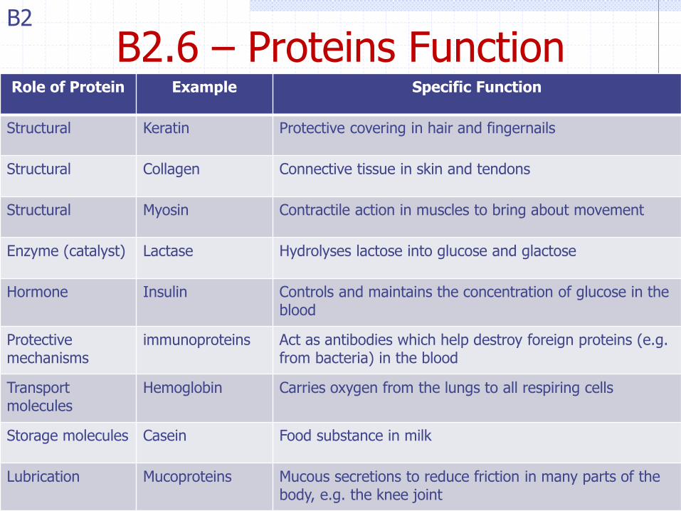
B2.6 – Proteins Function Role of Protein Example Specific Function
Structural Keratin Protective covering in hair and fingernails
Structural Collagen Connective tissue in skin and tendons
Structural Myosin Contractile action in muscles to bring about movement
Enzyme (catalyst) Lactase Hydrolyses lactose into glucose and glactose
Hormone Insulin Controls and maintains the concentration of glucose in the blood
Protective mechanisms
immunoproteins Act as antibodies which help destroy foreign proteins (e.g. from bacteria) in the blood
Transport molecules
Hemoglobin Carries oxygen from the lungs to all respiring cells
Storage molecules Casein Food substance in milk
Lubrication Mucoproteins Mucous secretions to reduce friction in many parts of the body, e.g. the knee joint
B2

B2.1 - Amino Acids and Proteins
Amino Acids are the building blocks of proteins
There are 20 amino acids that go into producing proteins.
Each have a carboxylic acid AND an amino group
COOH, loses an OH group and NH2 loses an H, forming a bond between two amino acids and water.
These amino acids are polymerized by a dehydration synthesis to form long chains of repeating amino acids called a protein. (taking water out)
The arrangement of the amino acids in the polymer determine the structure of the protein which confers to it is function or structural attributes.
Meaning the shape/order/structure of the amino acids in a protein determines it‟s function
5
B2


B2.1 - Essential Amino Acids
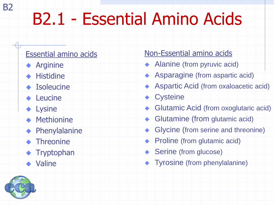
Of the 20 amino acids that make up proteins 10 of them can be synthesized by the human body
The other 10 amino acids must be acquired from food sources. These amino acids are known as essential amino acids
B2
B2.1 - Essential Amino Acids
Essential amino acids
Arginine
Histidine
Isoleucine
Leucine
Lysine
Methionine
Phenylalanine
Threonine
Tryptophan
Valine
Non-Essential amino acids
Alanine (from pyruvic acid)
Asparagine (from aspartic acid)
Aspartic Acid (from oxaloacetic acid)
Cysteine
Glutamic Acid (from oxoglutaric acid)
Glutamine (from glutamic acid)
Glycine (from serine and threonine)
Proline (from glutamic acid)
Serine (from glucose)
Tyrosine (from phenylalanine)
B2
B2.1 - Essential Amino Acids
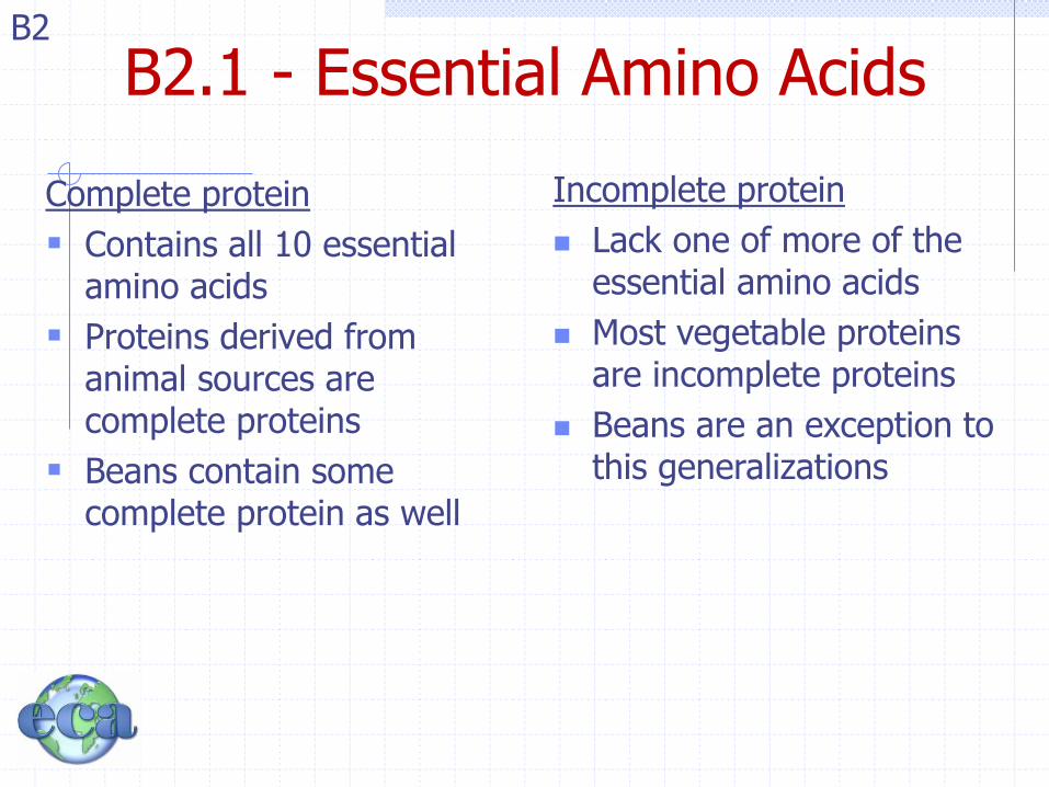
Complete protein
Contains all 10 essential amino acids
Proteins derived from animal sources are complete proteins
Beans contain some complete protein as well
Incomplete protein
Lack one of more of the essential amino acids
Most vegetable proteins are incomplete proteins
Beans are an exception to this generalizations
B2

B2.3 – Condensation, dehydration synthesis
With three-letter abbreviations.
These twenty amino acids that make up proteins, can be linked together through dehydration synthesis.
The carboxyl group of one amino acid bonds with the amino group of a second acid to yield a dipeptide and water. Proteins are long chains of amino acids linked by peptide bonds.
10
B2

B2.2 - Amino Acids are Amphoteric Amino acids are amphoteric. They are capable of behaving
as both an acid and a base, since they have both a proton
donor group and a proton acceptor group.
In neutral aqueous solutions the proton typically migrates
from the carboxyl group to the amino group, leaving an ion
with both a (+) and a (-) charge.
B2

B2.2 – (1) The Zwitterion Crystaline amino acids have relatively high melting or
decomposition points.
Soluble in polar solvents.
Amino acids exists as dipolar ions known as Zwitterions.
Internal acid-base reaction, a hydrogen ion is transferred fro the carboxylic acid to the amino group.
B2

B2.2 – (2) Buffer Action In aqueous solution the amino and carboxylic acid functional groups both ionize or dissociate
Carboxylic group releases hydrogen ions and hence acts as a Bonsted-Lowry acid
-COOH (aq) -COO- (aq) + H+ (aq)
The amino group can accept a hydrogen ion from solution and so acts as a Bronsted-Lowry base
-NH2 (aq) + H+ (aq) -NH3+ (aq)
In solutions of neutral (both dissociated), acidic (low pH, amino acid accepts hydrogen, +), basic (high pH, amino acid donates
hydrogen, -)
Consequently, amino acids regulate the pH of a system by “mopping up or donating H+ ions)
B2

B2.2 – (2) Buffer Action 2 B2

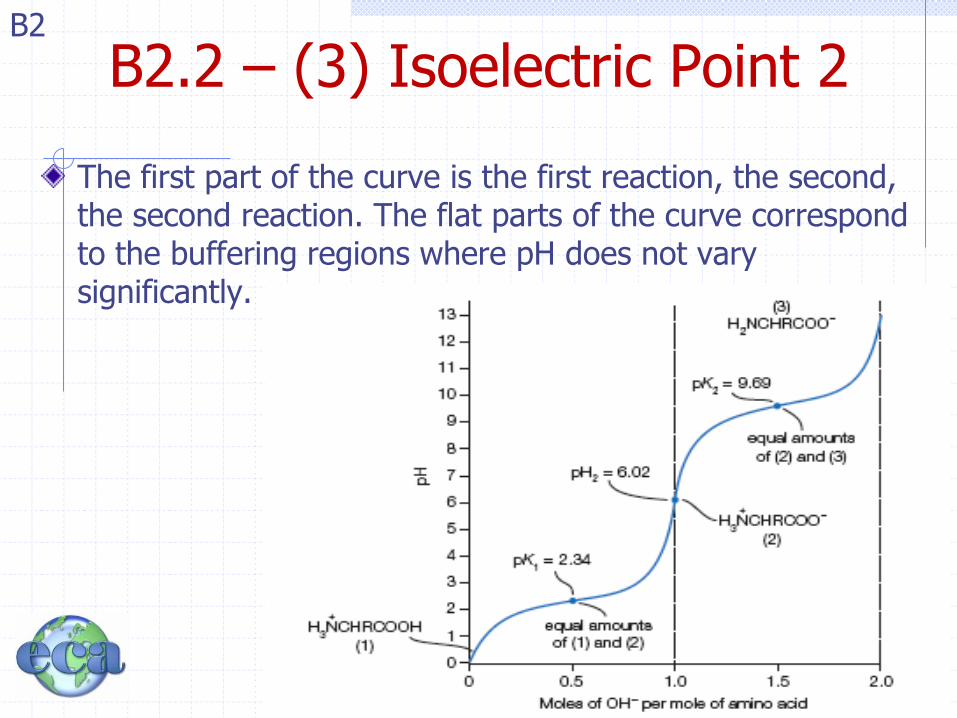
B2.2 – (3) Isoelectric Point Aminio acids such as alanine (where R is a methyl group) are dibasic when it‟s fully deprotonated. It can donate two protons H+ when in titration with a strong base.
H3N+CH(CH3)COOH(aq) + OH-(aq) H3N
+CH(CH3)COO-(aq) + H2O(aq)
H3N+CH(CH3)COO-(aq) + OH-(aq) H2NCH(CH3)COO-(aq) + H2O(aq)
The isoelectric effect is the basis for a separation technique known as electrophoresis.
B2
B2.2 – (3) Isoelectric Point 2
The first part of the curve is the first reaction, the second, the second reaction. The flat parts of the curve correspond to the buffering regions where pH does not vary significantly.
B2

B2.3 – Forming Polypeptides
Two amino acids can combined to form a simple dipeptide
B2
By convention, when drawing polypeptide chains the amino group is written on the left
N-terminus C-terminus

B2.3 – Forming Polypeptides
Chains of amino acids can form polypeptides (proteins) that are 1000‟s of AA‟s long
We will get into the folding and structure of proteins in B2.4 on Friday, please read ahead.
B2

B2.3 - Gly-Ala OR Ala-Gly B2
B2.3 – Amino Acid Residue
A peptide (protein) is NOT made up of amino acids, rather it is made up of amino acid leftovers (residue).
When amino acids form peptide bonds together they give off water, losing H or OH each. They are no longer the same structure
For this reason we use the term amino acid residue
B2

B2.3 – Reversible?
The process of forming a peptide bond can be reversed with the use of a strong acid in the presence of water.
B2

B2.4 – Protein Structure
There are four main contributing factors to the structure of proteins, primary, secondary, tertiary, and quaternary
B2

B2.4 – Primary Structure
The linear sequence of amino acids in the polypeptide chain
Change in a single amino acid (such as a mutation) can cause drastic differences in function
B2

B2.4 – Secondary Structure
• Secondary, refers to the regular and permanent arrangement of sections of the polypeptide chain, both are stabilized by hydrogen bonds
• α-helix (alpha, spiral)
• β-sheet (beta, sheets)
B2

B2.4 – Secondary/α-helix
• The coiled conformation of the polypeptide chain.
• Right-handed
• -R groups on outside and are perpendicular
• N (in peptide bond) bonded to O (of peptide carbonyl group, four residues down the chain.
B2
Triple helix – braided rope

B2.4 – Secondary/β-sheet • Formed by
polypeptides whose amino acid residues have small and compact side-chains.
• Composed of adjacent polypeptide chains (of the same protein) side-by-side and connected with hydrogen bonding
B2

27
Learning Check 1
Indicate the type of structure as
(1) primary (2) alpha helix
(3) beta pleated sheet (4) triple helix
A. Polypeptide chain held side by side by H bonds
B. Sequence of amino acids in a polypeptide chain
C. Corkscrew shape with H bonds between amino acids
D. Three peptide chains woven like a rope
B2

28
Solution 1
Indicate the type of structure as
(1) primary (2) alpha helix
(3) beta pleated sheet (4) triple helix
A. 3 Polypeptide chain held side by side by H bonds
B. 1 Sequence of amino acids in a polypeptide chain
C. 2 Corkscrew shape with H bonds between amino acids
D. 4 Three peptide chains woven like a rope
B2
B2.4 – Tertiary Structure
Overall 3D shape of a single protein.
Held by a specific shape by hydrogen bonds and van der Waala (intermolecular) and disulfide bonds, ionic bonds (intramolecular forces) involving side-chains.
Proteins can be classified as
Fibrous: long molecules arranged to form fibers
Globular: Folded into a compact and precise shape
B2

B2.4 – Tertiary / Fibrous
Fibrous: long molecules arranged to form fibers
Collogen is the most abundant fibrous protein in the human body
B2
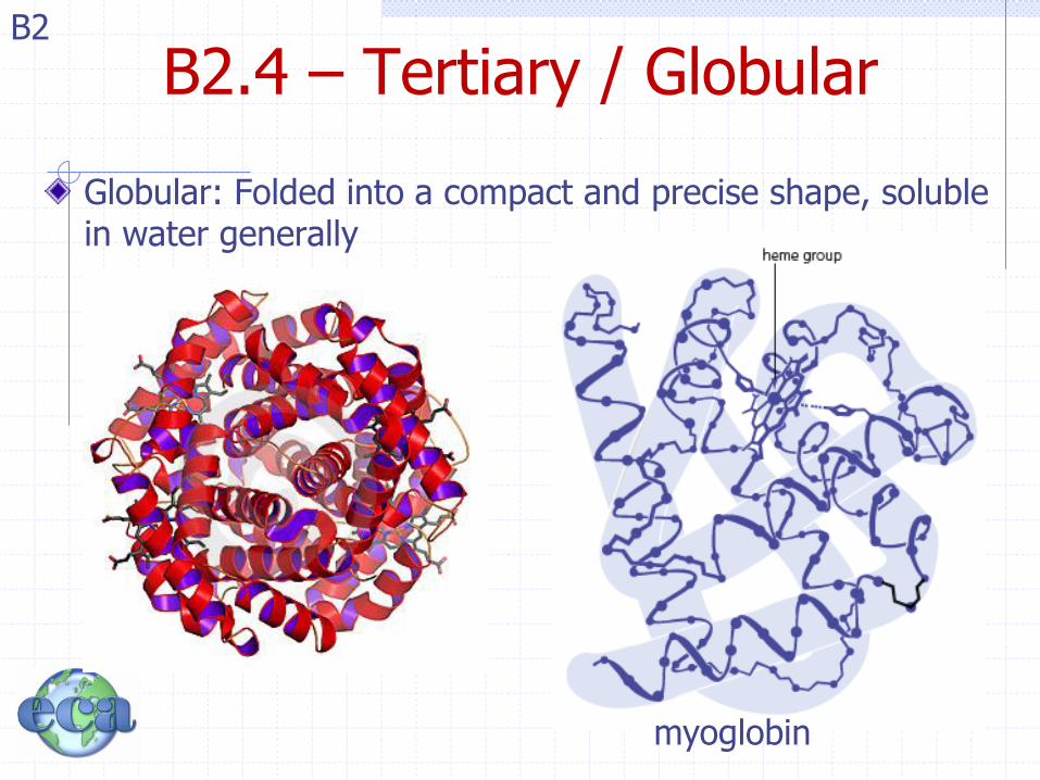
B2.4 – Tertiary / Globular
Globular: Folded into a compact and precise shape, soluble in water generally
B2
myoglobin

32
Globular and Fibrous Proteins
Globular proteins Fibrous proteins
“spherical” shape long, thin fibers
Insulin Hair
Hemoglobin Wool
Enzymes Skin
Antibodies Nails
B2

B2.4 – Tertiary/Di-sulfide Bridge
This also includes any covalent cross links
Disulfide bridges –S–S– can form between two cysteine amino acid side chains.
B2

B2.4 – Tertiary/Human Hair(1) B2
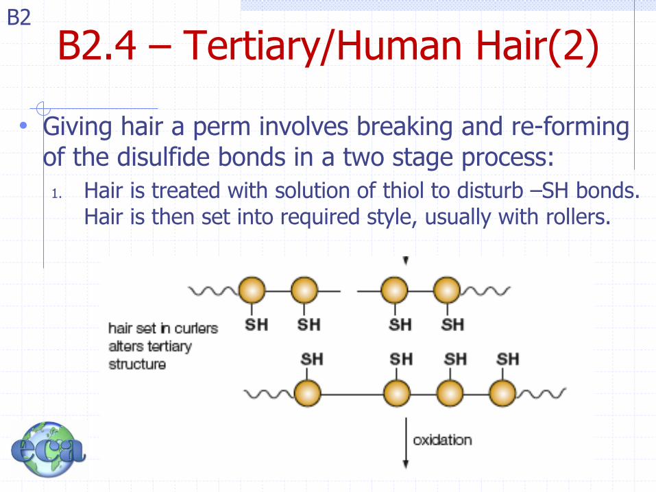
• The curled or straight nature of hair is due to disulfide bonds.
• Hair is made of keratin, which includes the amino acid cystine, which has a thiol (-SH group).
B2.4 – Tertiary/Human Hair(2) B2
• Giving hair a perm involves breaking and re-forming of the disulfide bonds in a two stage process:
1. Hair is treated with solution of thiol to disturb –SH bonds. Hair is then set into required style, usually with rollers.

B2.4 – Tertiary/Human Hair(3) B2
2. Then a formation of new disulfide bonds by oxidation of the thiol groups which set the new style “permanently” into position.

B2.4 – Tertiary Structure B2

38
Learning Check 2
Select the type of tertiary interaction as
(1) disulfide (2) ionic
(3) H bonds (4) hydrophobic
A. Leucine and valine
B. Two cysteines
C. Aspartic acid and lysine
D. Serine and threonine
B2

39
Solution 2
Select the type of tertiary interaction as
(1) disulfide (2) ionic
(3) H bonds (4) hydrophobic
A. 4 Leucine and valine
B. 1 Two cysteines
C. 2 Aspartic acid and lysine
D. 3 Serine and threonine

B2.4 – Quaternary Structure
• When a protein consists of two or more tightly bound polypeptide chains, the overall conformation or shape is referred to as the quaternary structure.
B2
Structure of Hemoglobin • 2 α-chains • 2 β-chains

41
Learning Check 3
Identify the level of protein structure
1. Primary 2. Secondary
3. Tertiary 4. Quaternary
A. Beta pleated sheet
B. Order of amino acids in a protein
C. A protein with two or more peptide chains
D. The shape of a globular protein
E. Disulfide bonds between R groups
B2

42
Solution 3
Identify the level of protein structure
1. Primary 2. Secondary
3. Tertiary 4. Quaternary
A. 2 Beta pleated sheet
B. 1 Order of amino acids in a protein
C. 4 A protein with two or more peptide
chains
D. 3 The shape of a globular protein
E. 3 Disulfide bonds between R groups
B2
B2.4 - Denaturation
Enzymes (proteins) function within a narrow range of pH and temperature values.
High or low values (pH or T) cause the protein to go through a physical change denature.
This involves loss of tertiary and quaternary structures, and is although often reversible since the primary and secondary structures remain the same, will change the function of the protein during this time.
B2

B2.4 – Protein Folding Disorders
The structure of a protein and it‟s ability to carry out it‟s biological function are so strongly correlated that even small structural defects can lead to a number of protein folding disorders.
Sickle cell anemia (single residue mutation)
Bovine spongiform encephalopathy (BSE) or “mad cow disease.”
Disorder leads to the buildup of insoluble protein in the brain
B2

B2.4 – Denaturing / Flip Creutzfeld-Jacob disease
(CJD) is the human equivilent of “mad cow disease”.
Researchers have found that normal prion protins in the brain consist of α-helicies,
but in CJD these proteins „flip‟ and unfold into a protein with β-sheets, now
insoluble, collect in the brain
B2

B2.5 – Analysis of Proteins The analysis of proteins is begun by determining it‟s amino
acid composition (not the primary sequence, the composition.)
First you must separate the amino acids from one another by breaking bonds through hydrolysis by using acid. This is the opposite of condensation used to form peptide bonds
There are two methods, chromatography and electrophoresis
B2
protein amino acids
Hydrolysis enzyme or heat/acid
condensation
H2O
H2O
B2.5 – Analysis / Chromatography Chromatography is a technique used to separate and
identify the components of a mixture, particularly when colored
Amino acids are colorless in solution, but take on color when treated with a locating reagent
Peptide chain must be hydrolyzed first as discussed in the previous slide
The solution is then “spotted” near the bottom of chromatographic paper and this position is known and marked as the origin
Paper is then suspended in a chromatographic tank containing a small volume of solvent ensuring that the “ origin” spot is above the solvent.
B2

Solvent rises up the paper by capillary action, passes through the origin and the amino acid distributes between two phases below, and move up the paper at different speeds and are spread according to their solubilities.
Stationary Phase (water in paper)
Mobile Phase (the solvent)
Paper is then removed from the tank and
developed by spraying with locating
reagent ninhydrin, making most of the
amino acids a purple color
Amino acids are then given an Rf value
B2.5 – Analysis / Chromatography(2) B2
Solvent front

A two dimensional (2D) resolution can then be achieved by adding second solvent and rotating the paper 90o
B2.5 – Analysis / Chromatography(3) B2

B2.5 – Analysis / Electrophoresis
Technique used to separate a mixture based on the movement of charged particles in an electric field
Amino acids carry different charges depending on side chains (R) and their pH so can be separated by these means
Gel electrophoresis, typically made by polyacrylamide
Amino acid mixture is placed in wells and an electric field is applied and AA‟s move toward electrodes at different rates
When complete, the amino acids can be detected by stain or made to fluroesce (glow) under UV light
B2

B2.5 – Analysis / Electrophoresis (2)
Gel Electrophoresis set up
B2

52
Learning Check 4
Identify the level of protein structure
1. Electrophoresis 2. Mobile Phase
3. Chromatography 4. Stationary Phase
A. The origin point moves with water
B. Technique used to separate a mixture based on the movement of charged particles in an electric field
C. The origin point moves with solvent
D. Technique used to separate and identify the components of a mixture
B2

53
Answers 4
Identify the level of protein structure
1. Electrophoresis 2. Stationary Phase
3. Chromatography 4. Mobile Phase
A. (2)The origin point moves with solvent
B. (1)Technique used to separate a mixture based on the movement of charged particles in an electric field
C. (4)The origin point moves with water
D. (3)Technique used to separate and identify the components of a mixture
B2

54
Learning Check 5
What are the products of the complete hydrolysis of Ala-Ser-Val?
B2

55
Solution 5
The products of the complete hydrolysis of Ala-Ser-Val are
alanine
serine
valine
B2

56
Learning Check 6
Tannic acid is used to form a scab on a burn. An egg becomes hard boiled when placed in hot water. What is similar about these two events?
B2

57
Solution 6
Acid and heat cause a denaturation of protein. They both break bonds in the secondary and tertiary structure of protein.
B2

B2.6 – Functions of Proteins
Proteins are crucial components for basic life processes
Responsible for transport throughout a cell or organism
Responsible for maintaining cellular structures
Responsible for basic metabolism
B2

B2.6 – Functional Protein Function
Functional proteins
Transport of oxygen throughout the blood and its storage in muscle cells
Completed by hemoglobin and myoglobin
B2

B2.6 – Structural Protein Function
Structural proteins
Elastin is included in the walls of arteries and veins as well as the brochioles (airways) in the lungs.
Collagen is the main structural protein of connective tissue in animals and the most abundant protein in mammals. It is the major component of cartilage, ligaments (bone to bone), and tendons (bone to muscle).
Muscle is composed of protein, example actin and myosin form the fibers
B2

B2.6 – Protein Function in the Body
This diagram is meant to aid in connecting your knowledge of chemistry with the use of chemical compounds in the body
B2

B2.6 – Pore Protein Function Pore proteins form channels that transport ions,
water, and other molecules through the cell membrane.
Many proteins have enzymatic activity such as catalyzing the digestion of protein, starch and lipids in the gut.
Digestive enzymes operate outside the cell
Intracellular enzymes control respiration, photosynthesis (in plants), and DNA repair and replication
B2

B2.6 – Immunoprotein Function
Immunoproteins (antibodies) are present in blood and identify and neutralize harmful bacteria and viruses.
Antibodies are produced by a group of white blood cells known as B cells. (HIV infects T cells which regulate the B cells of the immune system.)
B2
B2.6 – Hormone Function
Hormones are chemical messengers that carry a signal from a cell or group of cells to another via the blood stream.
All animals and plants produce a variety of hormones.
In general hormones control the function of their target cells.
Insulin is a protein-based hormone produced by mammals in the pancreas. Regulates blood glucose levels
B2